THE MECHANICAL BEHAVIOR OF DENTIN:
IMPORTANCE OF MICROSTRUCTURE, CHEMICAL
COMPOSITION AND AGING
Carolina Montoya Mesa Universidad Eafit
A dissertation submitted to the Universidad Eafit for the Degree of Doctor of Philosophy
2
Preface and Declaration
The work described in this dissertation was carried out at Universidad Eafit between January 2003 and February 2017.
I would like to thank my supervisor Dr Alex Ossa. for his guidance and invaluable discussions. Special thanks to Dr. Dwayne. Arola from the Materials Science and Engineering department at the University of Washington for the continuous support during this investigation. Also to Prof. Santiago Arango and Dr. Alejandro Peláez from the Dental Clinic of Universidad Cooperativa de Colombia for providing teeth for this study.
3
THE MECHANICAL BEHAVIOR OF DENTIN: IMPORTANCE
OF MICROSTRUCTURE, CHEMICAL COMPOSITION AND AGING
Carolina Montoya Mesa
Summary
Dental fracture is one of the three most common forms of failure of restored teeth and the most common cause of tooth loss or extraction in elderly patients. Previous investigations conducted on aging of hard tissues have identified that there is a considerable reduction in the mechanical properties (i.e. fracture toughness, fatigue and flexural resistance) of dentin with aging and that may predispose tooth fracture. These declines in properties have been attributed to microstructural and chemical composition changes over time. However, these aging processes have not been really quantified and related with the changes in mechanical properties. Accordingly, the aim of this work is to evaluate the aging process of coronal dentin in terms of the evolution of microstructure, changes in chemical composition and mechanical properties from selected age groups (young and old donors). The changes in these properties were evaluated in three different regions (outer, middle and inner) in order to identify spatial variations within the crown.
4
An extensive experimental study was carried out in chapter 3 to identify the changes in microstructure of dentin with aging by means of optical and electron microscopy; while changes in chemical composition were analyzed using Raman Spectroscopy to calculate the mineral-to-collagen ratio. Changes in mechanical properties were measured using Vickers micro-hardness.
Chapter 4 describes the importance of tubule density to the fracture toughness of dentin for young and old donor’s groups. An approach previously proposed to study the mechanical behavior of porous materials was used to model the fracture toughness of coronal dentin in terms of the tubule characteristics. Results were then compared with published results from previous studies.
The time-dependent deformation response of dentin was analyzed via spherical indentation experiments at different indentation loads in Chapter 5. From the experimental observations was proposed a simple model to describe the time dependent deformation behavior of dentin. This model was based on previously proposed theories for indentation of time dependent materials, showing that the effective strain rate of dentin depends on its chemical composition (i.e. mineral-to-collagen ratio) and microstructure (i.e. lumen area fraction). The descriptions of the model were compared with the experimental results showing good agreement. The same model was validated with experimental results of aged dentin, finding a low change in the deformation response of dentin with aging, as presented in chapter 6.
5
6
Products
As a result of this doctoral research the following products have been obtained:
Peer reviewed publications
Montoya, C., Arola, D., Ossa, E. A. (2017). Time Dependent Deformation Behavior of Dentin. Archives of Oral Biology, 76, 20-29.
Montoya, C., Arola, D., Ossa, E. A. (2016). Importance of tubule density to the fracture toughness of dentin. Archives of Oral Biology, 67, 9-14.
Montoya, C., Arango-Santander, S., Peláez-Vargas, A., Arola, D., Ossa, E. A. (2015). Effect of aging on the microstructure, hardness and chemical composition of dentin. Archives of oral biology, 60 (12), 1811-1820.
Oral conference presentations
Montoya, C., Arola, D., Ossa, E. A. Prediction of fracture toughness of human dentin. 6th International Conference on Mechanics of Biomaterials and Tissues, 2015, Hawaii, USA.
Montoya, C., Arola, D., Ossa, E. A. Time dependent behavior of human dentin. TMS 2015, Orlando, USA.
Montoya, C. Propiedades mecánicas de la dentina. XX Jornada de Investigación- Facultad de Odontología UCC, 2014, Medellín, Colombia.
7
Montoya, C., Ossa, E. A. Composición química y microestructura de la dentina de pacientes colombianos. VII Congreso Internacional de Materiales, 2013, Medellín, Colombia.
Conference proceedings publications
8
Contents
Preface and Declaration ... 2
Summary ... 3
Products ... 6
Peer reviewed publications... 6
Oral conference presentations ... 6
Conference proceedings publications... 7
Contents ... 8
List of Tables ... 14
List of Figures ... 15
Chapter 1 ... 20
Introduction ... 20
Chapter 2 ... 23
Review of previous research on the mechanical behavior and aging of human dentin . 23 2. 1 Introduction ... 23
2. 2 Tissues of the human body ... 23
2. 3 Microstructure and chemical composition of dentin ... 24
9
2.3. 1 Hardness and Elastic Modulus ... 27
2.3. 2 Flexure Strength ... 29
2.3. 3 Fracture Toughness ... 30
2.3. 4 Compressive behavior ... 31
2.3. 5 Fatigue strength ... 32
2.3. 6 Viscoelastic Properties ... 32
2. 5 Aging process of dentin ... 35
2. 6 Conclusions ... 38
2. 7 Tables ... 40
2. 8 Figures ... 44
Chapter 3 ... 45
Effect of aging on hardness, microstructure and chemical composition of dentin ... 45
3. 1 Introduction ... 45
3. 2 Experimental investigation ... 46
3. 3 Experimental Results ... 49
Microstructure ... 49
Hardness ... 51
Chemical composition ... 52
3. 4 Discussion ... 53
10
Hardness ... 55
Chemical composition ... 57
3. 5 Conclusions ... 59
3. 6 Tables ... 61
3. 7 Figures ... 62
Chapter 4 ... 73
Importance of Tubule Density to the Fracture Toughness of Dentin ... 73
4. 1 Introduction ... 73
4. 2 Experimental investigation ... 74
4. 3 Experimental results ... 75
4. 4 Discussion ... 76
4. 5 Conclusions ... 81
4. 6 Figures ... 82
Chapter 5 ... 86
Time dependent deformation behavior of human dentin ... 86
5. 1 Introduction ... 86
5. 2 Background ... 87
5. 3 Experimental investigation ... 89
Spherical indentation tests ... 90
11
Microstructural Analysis ... 91
5. 4 Results ... 91
Spherical indentation tests ... 91
Chemical Composition Analysis ... 93
Microstructural Analysis ... 94
5. 5 Time dependent deformation model for dentin ... 94
5. 6 Approximate calibration of the model ... 96
5. 7 Discussion ... 98
5. 8 Conclusion ... 101
5. 9 Tables ... 103
5. 10 Figures ... 106
Chapter 6 ... 117
Contributions of aging to the time dependent deformation of dentin ... 117
6. 1 Introduction ... 117
6. 2 Experimental investigation ... 118
Sample collection and preparation ... 118
Spherical Indentation Tests ... 118
Chemical composition analysis ... 118
Microstructural Analysis ... 119
12
6. 3 Experimental results ... 120
Spherical Indentation... 120
Chemical composition analysis ... 121
Microstructure ... 123
6. 4 Discussion ... 124
6. 5 Conclusions ... 129
6. 6 Tables ... 130
6. 7 Figures ... 131
Chapter 7 ... 138
Ethnic Background influence on the aging process of dentin: Preliminary Results .... 138
7. 1 Introduction ... 138
7. 2 Experimental investigation ... 140
7. 3 Experimental results ... 141
Microstructure ... 141
Spherical Indentation Tests ... 142
Chemical Composition ... 143
7. 4 Discussion ... 143
7. 5 Conclusions ... 149
7. 6 Tables ... 151
13
Chapter 8 ... 158
Conclusions ... 158
Chapter 2: Review of previous research on the mechanical behavior and aging of human dentin ... 158
Chapter 3: Effect of aging on hardness, microstructure and chemical composition of dentin ... 159
Chapter 4: Importance of Tubule Density to the Fracture Toughness of Dentin . 160
Chapter 5: Time dependent deformation behavior of human dentin ... 160
Chapter 6: Contributions of aging on the time dependent deformation of dentin 161
Chapter 7: Ethnic Background influence on the aging process of dentin ... 161
14
List of Tables
Table 2. 1. Results reported in the literature for the hardness of dentin. ... 40
Table 2. 2. Results reported in the literature for the Young Modulus of dentin. ... 41
Table 2. 3. Results reported in the literature for the flexural strength of dentin. ... 42
Table 2. 4. Results reported in the literature for the fracture toughness of dentin. ... 43
Table 3. 1. Results from the ANOVA (p-values) in comparing the microstructure of dentin from young and old donor teeth. Note the statistically significant differences for the occlusion ratio for the middle and outer dentin. ... 61
Table 5. 1. Indentation model parameters and c as a function of the power-law exponent n (reproduced from Bower et al. (1993)). ... 103
Table 5. 2. Dentin parameters obtained from the proposed model. ... 104
Table 5. 3. Parameters describing the basic power-law creep behavior for different hard tissues. ... 105
Table 6. 1. Dentin parameters obtained to describe the time dependent behavior of aged dentin. ... 130
15
List of Figures
1 6
Fi g ur e 3. 8. Distri b uti o n of mi n er al -t o-c oll a ge n r ati o of d e nti n fr o m y o u n g a n d ol d d o n or t e et h a c c or di n g t o d e pt h. ... 6 9
Fi g ur e 3. 9. C o m p aris o n of t h e h ar d n ess a n d c h e mi c al c o m p ositi o n distri b uti o ns i n a t o ot h as e vi d e nt fr o m l o n git u di n al s e cti o ni n g. a) h ar d n ess a n d b) c h e mi c al c o m p ositi o n ( mi n er al t o c oll a g e n r ati o) fr o m t h e t o ot h of a 1 8 ye ar ol d d o n or, c) h ar d n ess a n d d) c h e mi c al c o m p ositi o n fr o m t h e t o ot h of a 6 5 ye ar ol d d o n or. ... 7 0
Fi g ur e 3. 1 0. O c cl usi o n r ati o f or t h e t hr e e diff er e nt r e gi o ns of c or o n al d e nti n a n d a c o m p aris o n of r es ults f or t h e y o u n g a n d ol d d o n or t e et h. ... 7 1
Fi g ur e 3. 1 1. Vi c k ers h ar d n ess o bt ai n e d f or d e nti n of y o u n g a n d ol d d o n or t e et h a c c or di n g t o d e pt h. T h e a p pli e d l o a d i s p ar all el (//) a n d p er p e n di c ul ar ( ) t o t h e d e nti n al t u b ul es. C ol u m ns wit h si g nifi c a nt diff er e n c es ( p ≤ 0. 0 5) ar e gr o u p e d i n a li n e a n d m ar k e d wit h a cr oss ( +) a n d a ast eris k ( *). ... 7 2
Fi g ur e 4. 1. Mi cr o gr a p hs of t h e mi cr ostr u ct ur e f or t h e y o u n g a n d ol d d e nti n as a f u n cti o n of l o c ati o n. a-b) O ut er d e nti n; c -d) Mi d dl e d e nti n; e -f) I n n er d e nti n. N ot e t h e o blit er at e d d e nti n al t u b ul es i n mi cr o gr a p hs f or t h e mi d dl e a n d o u t er d e nti n of t h e ol d d o n or t e et h. ... 8 2
x
Fi g ur e 4. 2. L u m e n ar e a fr a cti o n ( ) f or t h e t hr e e diff er e nt r e gi o ns of c or o n al d e nti n a n d a c o m p aris o n of r es ults f or t h e y o u n g a n d ol d d o n or t e et h. ... 8 3
1 7
Fi g ur e 4. 4. Esti m at e d fr a ct ur e t o u g h n ess f or diff er e nt r e gi o ns of c or o n al d e nti n f or t h e
y o u n g a n d ol d d o n or t e et h. T h es e esti m at es ar e o bt ai n e d fr o m t h e l u m e n ar e a fr a cti o n x( )
m e as ur e m e nts a n d t h e us e of t h e B als hi n e q u ati o n ( B als hi n, 1 9 4 9 ). ... 8 5 Fi g ur e 5. 1. S c h e m ati c di a gr a m of a h alf -s pa c e u n d er i n d e nt ati o n b y a ri gi d s p h er e.
T h e v ari a bl es F, R, h a n d a r e pr es e nt t h e i n d e nt ati o n f or c e, i n d e nt or r a di us of c ur v at ur e, d e pt h of i n d e nt ati o n a n d t h e r a di us of p er m a n e nt i n d e nt ati o n, r es p e cti v el y. ... 1 0 6
Fi g ur e 5. 2. S c h e m ati c di a gr a m of a s e cti o n e d m ol ar wit h t h e e x p os e d d e nti n e m b e d d e d i n c ol d c ur e d e p o x y r e a d y f or t h e i n d e nt ati o n t est. T h e l ett ers D a n d E r ef er t o d e nti n a n d e n a m el, r es p e cti v el y. ... 1 0 7
Fi g ur e 5. 3. S el e ct e d i n d e nt ati o n d e pt h v ers us ti m e r es ults f or i n n er d e nti n at a p pli e d l o a ds of 1, 1 0 a n d 5 0 N e wt o ns. ... 1 0 8
Fi g ur e 5. 4. St e a d y st at e i n d e nt ati on r at e ( ) v ers us i n d e nt ati o n l o a d r es p o ns e f or t h e t hr e e r e gi o ns of d e nti n e v al u at e d. ... 1 0 9
Fi g ur e 5. 5. C o m p aris o n of t h e e x p eri m e nt al st e a d y -st at e eff e cti v e str ess a n d eff e cti v e
str ai n r at e of c or o n al d e nti n ( m ar k ers) wit h pr e di ct e d r es p o ns es (li n es). ... 1 1 0
Fi g ur e 5. 6. D e p e n d e n c e of t h e r ef er e n c e eff e cti v e str ai n r at e ( ) o n t h e mi n er al-t o
c oll a g e n r ati o ( ) wit hi n t h e t hr e e diff er e nt r e gi o ns of c or o n al d e nti n. ... 1 1 1
Fi g ur e 5. 7. Eff e ct of i n d e nt ati o n l o a d o n t h e eff e cti v e str ai n r at e f or diff er e nt r e gi o ns of c or o n al d e nti n. C ol u m ns wit h o ut si g nifi c a nt diff er e n c es ( p > 0. 0 5) ar e gr o u p e d wit h a li n e.
... 1 1 2 Fi g ur e 5. 8. Distri b uti o n of t h e mi n er al -t o-c oll a g e n r ati o of d e nti n a c c or di n g t o t h e
18
Figure 5. 9. Schematic diagram showing how changes in the degree of mineralization affect the response of the reference strain rate . ... 114 Figure 5. 10. Experimental results reported for the steady state creep rate for bone (Rimnac et al. 1993) and radicular dentin (Jantarat et al. 2002) and comparison with results of the current study. ... 115 Figure 5. 11. Schematic representation of how the dentinal tubules are distributed in dentin. a) and b) top views of an indentation test on the regions indicated... 116 Figure 6. 1. Selected indentation depth versus time results for different regions of dentin at a constant applied load of 60N. The results correspond from an incisor of a 70-year-old donor. ... 131
19
20
Chapter 1
Introduction
The effect of aging on the microstructure and mechanical properties of bone has been studied extensively due to its importance to the elderly and their quality of life (e.g. Currey et al., 1996; Zioupos et al., 1998; Zioupos et al., 1999; Wang et al., 2002; Wang et al., 2004; Nalla et al., 2004a; Ural and Vashishth 2006; Ural and Vashishth, 2007). However, the effect of aging on dental hard tissues (including dentin and enamel) has received rather limited attention. That is surprising when one considers the importance of human teeth to mastication and dietary intake.
21
Microstructural changes occurring in dentin with aging have been associated with obliteration of dentinal tubules and variations on chemical composition (Kinney et al., 2005; Rosen et al., 1999). However, the aging process of human dentin have not been really quantified and related with the changes in mechanical properties. Therefore, the aim of this doctoral work is to quantitatively evaluate the aging process of coronal dentin in terms of the evolution of microstructure, changes in chemical composition and mechanical properties from selected age groups (young and old donors). The changes in these properties were evaluated in three different regions (outer, middle and inner dentin) in order to identify spatial variations within the crown.
The overall hypothesis of this project is that aging of dentin and the decrease in mechanical properties is directly correlated with the change in the microstructure and chemical composition. To achieve this objective, the following aims were developed:
Specific Aim 1: Test the hypothesis that human dentin undergoes a significant change in the microstructure and obliteration of dentinal tubules with increasing patient age.
Specific Aim 2: Test the hypothesis that human dentin undergoes significant changes in its chemical composition with increasing patient age.
Specific Aim 3: Test the hypothesis that human dentin undergoes a significant change in hardness and in its distribution within the crown with increasing patient age.
Specific Aim 4: Test the hypothesis that the viscoelastic or time dependent deformation response of dentin change along the tooth and undergo significant changes with increasing patient age.
22
23
Chapter 2
Review of previous research on the mechanical behavior and aging of
human dentin
2. 1 Introduction
A brief description of the main literature on microstructure, chemical composition and mechanical behavior of dentin and its changes with aging is presented in this chapter. Detailed reviews can be found elsewhere (e. g. Pashley et al., 1989; Kinney et al., 2003; Zhang et al., 2014). Additionally, throughout the entire document additional information on previous research can be found where necessary to complement the discussion.
2. 2 Tissues of the human body
A tissue is a collection of cells having similar structure and function, usually having a common embryonic origin and working together to develop specialized activities (Gomez de Ferraris and Campos Munoz, 2009). According to the embryonic origin the tissues in the human body can be classified as (Henrikson and Kaye, 1986):
Epithelium: Responsible of cover and protect organs, constituting the inner lining of
the cavities, hollow organs and ducts of the body as well as form mucous and glands. Connective tissue: Protect, support and bind together different types of tissues and
24
Muscular tissue: Produces movement and according to the location and function can be
skeletal or striated muscle, smooth or non-striated muscle, and cardiac muscle. Nervous tissue: Initiates and transmits potentials that help coordinate activities.
These tissues, depending on their level of mineralization and therefore their hardness, may be classified as soft or hard tissues.
Soft tissues are responsible of connect, support, or surround other structures and organs of the body (i.e. ligaments, tendons and skin) (Derby and Akhta, 2015). While hard tissues are those that have been mineralized, and therefore serve as protective shield or structural support (Grumezescu, 2016). The hard tissues of humans are bone, tooth enamel, dentin, and cementum. Detailed reviews about each of these tissues and their chemical composition, formation and chemical properties can be found in: Kambic (1994), Rho et al. (1998), Meyes et al. (2008) and Lloyd et al. (2015).
2. 3 Microstructure and chemical composition of dentin
Dentin is a hard tissue that occupies the majority of the human tooth. By volume it consists of approximately 45% mineral material, 33% organic material (collagen type I) and 22% water (Nanci, 2012). Although this chemical composition is assumed to dentin, it has been widely reported in literature that these percentages change within the tooth from the pulp to the Dentin Enamel Junction (DEJ) and along radicular dentin (Ryou et al., 2011; Xu et al., 2009; Tesch et al., 2001).
25
appositional growth and deposition of secondary dentin (Gomez de Ferraris and Campos Munoz, 2009).
The microstructure of dentin is largely dominated by its tubules, which are responsible for housing the odontoblastic processes and maintain dentin vitality. The tubules extend from the pulp to the DEJ with a double “S” shape in coronal dentin and only one curvature in the dentin root (radicular dentin) (Nanci, 2012). These are called primary curvatures and are formed as the progressive stacking of dentinal tubules during the formation of dentin. As a result of this process there are a higher number of dentinal tubules near the pulp (40.000 tubules/mm2) and fewer near the DEJ (17.000 tubules/mm2) (Fehrenbach and Popowics, 2015).
Dentinal tubule diameters also vary along dentin, diameters of dentinal tubules range from approximately 1 to 3 μm, depending on patient age and its location, having larger diameters near the pulp (2.00m) and smaller near the DEJ (1.20m) (Ide Ingle et al., 2008). Additionally, dentinal tubules have collateral ramifications that contribute to dentinal permeability and sensitivity; this branching have been reported as more evident in root dentin than in the crown. This condition promotes bacterial penetration and therefore periodontal disease in elderly patients (Pashley and Pashley, 1991).
26
complete and is slowly deposited centripetally after the development of the tooth (Kinney et
al., 1996). This process of formation makes that PTD show three clearly distinguishable areas: a hypo-calcified region near the outer edge, a highly-mineralized zone that is in continuous formation and a hypo-mineralized area, which is continuously forming and mineralizing throughout life (Gomez de Ferraris and Campos Munoz, 2009).
The process of formation and mineralization of dentin, known as dentinogenesis takes place during the stage of apposition of the tooth; where enamel, cementum and dentin are secreted in layers by ameloblasts, cementoblasts, and odontoblasts respectively (Goldberg et al., 2011). During dentinogenesis, odontoblasts release a non-mineralized matrix of collagen fibers that are later mineralized in the maturation stage by the formation of hydroxyapatite crystals (Fehrenbach and Popowics, 2015). This process takes place as long as the tooth is alive and that is why dentin is considered a live tissue (Nanci, 2012).
Since dentin is not a uniform tissue due to its variation in chemical composition, dentinal tubule density and diameter of dentinal tubules, various types of dentin have been identified (Nanci, 2012; Gomez de Ferraris and Campos Munoz, 2009; Spangberg, 1989):
Primary dentin: This dentin forms the external shape of the tooth. Depending on its
location can be named as mantle dentin (first formed dentin) and circumpulpar dentin (forms the remainder tissue). This tissue is formed during odontogenesis and until the tooth enters occlusion.
27
Tertiary dentin: Also named reactionary or reparative dentin. This dentin is formed as
a result of external stimuli (i.e. caries or trauma) in order to protect the sensitive pulp. This dentin formation occurs irregularly depending on the aggressiveness of the stimulus.
Based on its differences in chemical composition and microstructure, dentin is considered a hierarchical biological composite (Ziskind et al., 2011) and its mechanical properties change along different regions of dentin and with patient age.
2. 4 Mechanical Properties of dentin
The mechanical properties of dentin have been studied using techniques commonly used for the characterization of ceramic materials. Some of the properties that have been determined are hardness, Young’s modulus, flexural strength, fracture toughness, fatigue strength, compressive strength and some viscoelastic properties. Some of the results obtained are described below:
2.3. 1 Hardness and Elastic Modulus
Hardness and elastic modulus of dentin have been determinated in different types and regions of dentin. Some of the techniques used include nanoindentation, Atomic Force Microscopy (AFM) and microindentation.
28
between 18 GPa and 22 GPa for intertubular dentin. The higher value obtained for peritubular dentin was attributed to a higher mineralization degree of this tissue when comparing with intertubular dentin (Kinney et al., 1996). By means of nanoindentation Ryou et al. (2012) found hardness values of 2.38 GPa and 1.31 GPa for peritubular and intertubular dentin, respectively; while the Young´s modulus had values of 29.8 GPa for peritubular dentin and 19.4 GPa for intertubular dentin. From the results, the authors found that intertubular dentin is almost isotropic and the anisotropic behavior of dentin is determined by the direction of dentinal tubules and the direction in which testing is performed.
An average hardness of 0.5 GPa has been reported using a Vickers microindentation techniques, with no significant dependence on indentation load or indentation time (Chuenarrom et al., 2009). Additionally, Gutiérrez-Salazar and Reyes-Gasga (2003) used Vickers hardness to determinate how the tooth hardness change from outer enamel surface to inner dentin layer finding that enamel’s hardness ranges from 2.65 GPa to 3.53 GPa and from 0.49 GPa to 0.58 GPa for dentin.
29
Dentin hardness has been widely studied and some of the differences obtained between authors can be attributed to the specimen preparation procedure, indentation load or type of tooth analyzed. Nonetheless, how dentin hardness changes spatially within the tooth and how this differences relates with the chemical composition and tubular characteristics (i.e. tubule density and diameter of dentinal tubules) have not been completely understood.
2.3. 2 Flexure Strength
Different types of bending tests have been performed to dentin in order to compare its properties with those obtained for restorative materials or adhesives used in restoration procedures. For example, Ryou et al. (2011) used specimens of two regions of coronal dentin to perform flexural test - the regions analyzed were named as “top” (region near DEJ) and “inner” dentin (region near pulp) -. All the samples showed a linear elastic behavior with a small zone of plastic deformation before failure. Further, it was found that the “top” dentin had the higher elastic modulus and strength when comparing with the inner regions. The flexure strength was found ranging from roughly 130 MPa to 180 MPa and the strain ranging from 0.008 mm/mm to 0.011 mm/mm. The fracture of the specimens was found to begin in the area subjected to tension stresses. Similar tests were performed by Plotino et al. (2007) on root dentin, in this particular case in order to evaluate the strength of different synthetic root materials like carbon and glass fiber composites, zirconia, gold, stainless steel and titanium.
The flexure modulus for root dentin showed values of 17.5 3.8 GPa and flexure strength of
212.9 41.9 MPa. A comparison of results for natural and synthetic materials showed similar
30
an average flexure strength of 164.4 ± 9.1 MPa. A summary of the flexure strength of dentin results is shown in the table 2.3.
The changes in flexure strength for the different regions of dentin have been attributed to the chemical composition and microstructure of each region, where the higher values of flexural strength were found for outer dentin and radicular dentin where a lower number of dentinal tubules were found.
2.3. 3 Fracture Toughness
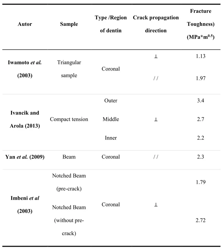
Various approaches have been used to measure the fracture toughness of dentin, including notched and compact tension specimens with different orientations of dentinal tubules with respect to the direction of crack propagation. Knowledge of the fracture toughness of dentin is important to establish the effects of a restoration procedures in the advent and propagation of cracks in dentin.
Triangular samples were used by Iwamoto et al. (2003) to study the fracture toughness of dentin. Samples were classified in three groups; i) those with load applied perpendicular to the plane of dentinal tubules, ii) parallel and aligned to the plane of dentinal tubules; and iii) the plane of crack propagation parallel and transverse to the plane of dentinal tubules. A scheme of the samples used is shown in Figure 2.1. From the results, they argued that the orientation of dentinal tubules with respect to the direction of the load had a statistically significant effect on fracture toughness of the tissue. Values of 1.13 MPa*m0.5 were found for samples with the load applied perpendicular to the plane of dentinal tubules and an average of
2.00 MPa*m0.5 was found when the load was applied parallel to the dentinal tubules with the
31
Another type of test that has been used to study the fracture toughness of dentin involves notched specimens in three-point bending and two directions of crack propagation, in plane and anti-plane, with respect to dentinal tubules. The results obtained were used to calculate the stress intensity factor, showing that this factor is higher when the load is applied anti-plane due to a higher proportion of peritubular dentin with a higher mineralization degree (Yan et al., 2009).
Compact tension specimens were used by Ivancik and Arola (2013) to examine how fracture toughness changes in outer, middle, and inner coronal dentin. They found that outer dentin required 50% greater stress to propagate the crack as compared with inner dentin. This behavior was attributed to differences in tubule density and diameter of dentinal tubules along the tooth.
Additional test performed to measure the fracture toughness of dentin were reported by Imbeni et al. (2003), Nalla et al. (2003a) and Wang (2005); who found similar results to those mentioned above. A summary of the reported results are shown in table 2.4
2.3. 4 Compressive behavior
Human teeth are subjected to compressive loads and friction during mastication of food, whereas dentin works under compression stresses only. This makes that the understanding of the compressive behavior of dentin become very important for this tissue. Several studies have found compressive strength values that range between 275 MPa and 300
MPa (Craig and Peyton, 1958) and elastic modulus 14 GPa (Kinney et al., 2003). The elastic
32
2.3. 5 Fatigue strength
The fatigue strength of dentin is of great importance as teeth are continuously subjected to cyclic stresses during mastication. Likewise, after a restoration process is common to find cracks that can propagate even with low stresses but applied continuously. A detailed review on the fatigue behavior of dentin can be found in Kruzic and Ritchie (2008).
Studies on the fatigue behavior of human dentin have distinguished that dentin exhibits the traditional S–N response (Arola et al., 2010; Arola and Reprogel, 2005; Nalla et al., 2003b). From these results, has been established that dentin exhibits an apparent endurance limit that ranges from approximately 20–50 MPa, and that it is dependent on the frequency of loading (Nalla et al., 2003b, Kruzic et al., 2003), the stress applied (Nalla et al., 2004b), and tubule orientation with respect to the loading direction (Arola and Reprogel, 2006).
The dependence of these factors can be explained by the variations in the microstructure of dentin (i.e. Tubule density and diameter) and changes in chemical composition (i.e. collagen content) along the tooth.
2.3. 6 Viscoelastic Properties
33
stresses to analyze the relaxation behavior of the tissue, finding that dentin showed a linear dependence on the logarithm of time. They found that the relaxation modulus of dentin showed a linear dependence on the logarithm of time. The mathematical model of viscoelasticity previously presented by Alfrey and Doty (1945) was further compared with their experimental results finding that the stress relaxation response of dentin followed a linear viscoelastic behavior. Despite of these results, it is not clear if the same behavior can be extrapolated to coronal dentin, which supports different loads and shows distinct microstructure. On the other hand, Jantarat et al. (2002) also used radicular dentin cylinders manufactured from incisors and canines. Compressive loads of 100 N, 300 N, 500 N and 700 N were applied and held for time periods of 90 min. Strain recovery was measured for 60 min after removing the load. A typical viscoelastic behavior was found for the creep measurements with a strain increase over time while the stress was kept constant. Statistical differences were found between stress and creep rate when comparing for the different loads applied. Similar test made on radicular dentin have been performed by Trengrove et al. (1995) and Jafarzadeh et al. (2004) finding some viscoelastic behavior for the tissue when a load in constant for a period of time.
Viscoelastic test on coronal dentin performed by Pashley et al. (2003) in order to study the stress relaxation of demineralized dentin under tension showed that the dentin matrix exhibits both stress–relaxation and creep behavior. However, the stress–relaxation and tensile creep were independent of the initial strain applied.
34
demineralized material is nearly elastic, and that hardness and elastic modulus do not change with different maximum loads. According to these results, it was established that collagen in dentin does not contribute to the elastic modulus but it does to dentin strength and toughness. On the other hand, Ryou et al. (2012) studied the viscoelastic behavior of peritubular and intertubular dentin. Indentations were made using loads of 400, 700 and 1000 μN with a corresponding dynamic load of 20 μN and frequencies varying from 2 to 100 Hz. Complex
modulus, loss moduli, storage moduli and tan showed an increase in magnitude with loading
frequency. The complex and storage moduli of peritubular dentin were significantly larger than for intertubular dentin. No significant differences in the loss modulus and tan between intertubular and peritubular dentin were found, implying similar viscoelastic behavior in the two types of dentin. Recently, Chuang et al. (2015) performed nanoindentation creep tests to study the viscoelastic properties of dentin after de- and re-mineralization processes finding that the demineralization process increase the primary and secondary creep regimes, while the remineralization reduces the primary creep of dentin without increasing viscoelasticity.
35
1.5 mm diameter tungsten-carbide ball to analyze the Hertzian response (i.e. contact modulus) of dentin with different loading rates and indentation directions as a function of dentinal tubules orientation (i.e. parallel and perpendicular), finding significant differences in the response of dentin with respect to loading rate but with no differences with tubules orientation.
2. 5 Aging process of dentin
Geriatric dentistry has attracted attention in recent years given the need to preserve and improve the quality of life of elder people. Additionally, maintaining the physiological function of the different organs in the aging population could help to reduce the burden on the existing medical systems as older individuals consume medical services (Sieck, 2003). The aging process begins since the day of birth and continues throughout life; however, the effects of the aging process are more evident in the third decade of life and are increasingly obvious after that (Vaughn, 2011).
The aging process of teeth includes wear of enamel, tooth loss due to alveolar resorption and periodontal disease and color change (darkening) (Nanci, 2012). On the other hand, in the case of dentin it has been found that the aging process can generate the following changes (Murray et al., 2002):
Increase in dentin thickness and decrease of the pulp chamber due to the deposition of
secondary dentin throughout life: An approximate rate of secondary deposition of 43 µm per year, or 0.119 μm per day have been estimated (Solheim et al., 1992). Reduction in the number of odontoblasts in the pulp chamber: Quantitative studies
36
Obliteration of dentinal tubules: The most obvious change in dentin with aging is the
microstructural change due to the obliteration of dentinal tubules with sclerotic or transparent dentin. This process changes the chemical composition of dentin increasing its mineral content (Nanci, 2012).
As can be seen, all these changes are related and have an effect not only on the chemical composition as mentioned earlier, but on the mechanical behavior, permeability and sensitivity of the tissue (Arola and Reprogel, 2005). A few studies have been published aiming at identify the changes in mechanical properties of dentin with aging. An extensive review of the aged dentin properties was published by Arola et al. (2009). Some of the changes found for the mechanical properties of dentin include:
A decrease in dentin tubule diameter due to deposition of secondary coronal dentin (Nanci, 2012). Nonetheless, these changes have not been quantified. This process of obliteration is known to begin at the root and move towards the crown of the tooth (Vasiliadis et al., 1983).
An increase in the mineral content of dentin has been found in aged dentin (Koester et
al., 2008a). Likewise, changes in crystallinity of the mineral material have been reported; intertubular dentin mineral crystallites are 7–19% smaller in aged than in young dentin, while the dentin mineral crystals deposited - obliterating - in dentinal tubules are chemically similar to the intertubular mineral (Porter et al., 2005). Increases in elastic modulus and hardness have been also found for radicular dentin, being more significant in the cervical portion of the root (Xu et al., 2014).
Increases in the elastic modulus of 5% and 9% in the hardness of the outer layer of
37
dentin. On the other hand, Zheng et al. (2005) analyzed the changes in hardness and
Young’s modulus of dentin with aging and reported that dentin does not undergo a significant change in hardness or Young’s modulus with age in the middle and inner dentin. However, they found an increase of 16% in hardness and around 5% in Young’s modulus within outer dentin.
There is a decrease in flexural strength of dentin of almost 20 MPa per decade of life
that begins shortly after reaching adulthood (30 years) (Arola et al., 2009).
A reduction in fatigue strength of dentin was found in aged dentin at higher levels of
stress. However, at low stress levels transparent dentin appears to have the same behavior than young dentin (Kinney et al., 2005).
There is reduction in the fatigue crack growth resistance of dentin with increasing age.
The average rate of fatigue crack growth in old dentin is greater by a factor of 102 in comparison to young dentin (Arola et al., 1999).
A reduction of 75% in the energy required to fracture dentin between young (age≤30)
and old patients (age>55) was also reported (Arola et al., 2009).
Ryou et al. (2015) found a difference in the damping behavior of dentin with aging and
38 2. 6 Conclusions
After reviewing the studies available on the mechanical properties of dentin and its aging process, the follow conclusions can be drawn to support the development of this doctoral project:
1. It has been widely reported in literature that aging causes a change in dentin microstructure, product of the obliteration of dentinal tubules. Most of these studies have been performed on root dentin, where aging begins, and the results obtained have been analyzed qualitatively using microscopy technics. However, these changes have not been quantified and there is no information available on how the obliteration of dentinal tubules changes spatially in coronal dentin. Obtaining this information for coronal dentin is important as the crown is responsible to support most of the stresses during mastication. Identifying these quantitative changes is essential to find relations between the decrease in mechanical properties due to the aging process.
2. It has been reported in literature that aging causes a change in the chemical composition of dentin. These changes have been related with an increase in mineral content due to the obliteration of dentinal tubules. However, the literature review shows no studies aimed at determine and quantify the changes in chemical composition of aged coronal dentin (spatial variations). These changes might be related with an increase in mineralization, crosslinking of collagen or changes in crystallinity.
39
dentinal tubules density), but not with chemical composition changes that occur in the tooth. For this correlation, old dentin can include other additional changes that occur with aging such as obliteration of dentinal tubules. Due to differences in microstructural features of dentin it is expected to find a correlation between hardness and age.
40 2. 7 Tables
Table 2. 1. Results reported in the literature for the hardness of dentin.
Author Technique Region /Type of
dentin
Hardness GPa)
Kinney et al. (1996) AFM
PTD1 2.40
ITD2 0.51
Ryou et al. (2012) Nanoindentation
PTD 2.38
ITD 1.31
Chuenarrom et al. (2009)
Microindentation/
Vickers
Bulk dentin 0.50
Gutiérrez-Salazar and Reyes-Gasga (2003)
Microindenttion/
Vickers
Outer 0.49
Inner 0.58
Angke et al. (2003) Berkovich indenter.
Primary dentin/
Outer 0.91
Primary dentin/
Inner 0.52
1 PTD: Peritubular dentin
41
Table 2. 2. Results reported in the literature for the Young Modulus of dentin.
Author Technique Region /Type of
dentin
Young modulus
(GPa)
Kinney et al. (1996) AFM
PTD 25.0
ITD 20.0
Ryou et al. (2012) Nanoindentation
PTD 29.8
ITD 19.4
Angke et al. (2003) Berkovich indenter.
Primary dentin/
Outer 16.9
Primary dentin/
42
Table 2. 3. Results reported in the literature for the flexural strength of dentin.
Author Region /Type of dentin Flexural Strength (MPa)
Ryou et al. (2011)
Outer 180.0
Inner 130.0
Staninec et al. (2008) Coronal 164.4
43
Table 2. 4. Results reported in the literature for the fracture toughness of dentin.
Autor Sample Type /Region
of dentin
Crack propagation direction
Fracture Toughness) (MPa*m0.5)
Iwamoto et al. (2003)
Triangular
sample Coronal
⊥ 1.13
/ / 1.97
Ivancik and
Arola (2013) Compact tension
Outer
⊥
3.4
Middle 2.7
Inner 2.2
Yan et al. (2009) Beam Coronal / / 2.3
Imbeni et al (2003)
Notched Beam (pre-crack)
Coronal ⊥
1.79
Notched Beam (without
pre-crack)
44 2. 8 Figures
45
Chapter 3
Effect of aging on hardness, microstructure and chemical composition of
dentin
3. 1 Introduction
Within the field of dentistry, the importance of aging has become of greater interest in recent years due to its impact on the practice of restorative dentistry. Indeed, the tooth undergoes certain changes with age, including wear of enamel, the formation of transparent dentin, a decrease in the number of odontoblasts and an increase in dentin thickness as well as a production of reactionary dentin (Nanci, 2012). The changes in dentin microstructure produce variations in its mechanical properties, which are important for the introduction of restorative treatments and the greater potential for tooth fractures.
46
Changes in mechanical properties of dentin with aging have largely been attributed to the increase in mineralization due to filling of the dentinal tubules. However, it remains unclear whether these changes can be attributed to the mineral occupying the dentinal tubules, a complimentary change of the mineral of the intertubular dentin, or crosslinking of collagen by non-enzymatic processes (Miura et al., 2014). In fact, little information is available on the relationship between the changes in microstructure of dentin with age and spatial variations in chemical composition. Thus, the aim of this chapter is to identify the changes in microstructure, chemical composition and hardness of dentin with aging from selected age groups of Colombian patients.
3. 2 Experimental investigation
Human third molars were obtained from selected patients after written consent and following all the protocols required by the Dental Clinic at Universidad Cooperativa de Colombia (UCC). Exclusion criteria included presence of caries and previous restorations. The teeth were obtained from donors residing in Medellín, Colombia, and were divided into two age groups, namely a “young” group with donors between 18 and 25 years of age (N=12), and an “old” group with donors between 47 and 65 years of age (N=8). There were an equal number of male and female samples in both groups. Immediately after extraction, all the specimens were kept in Hank’s Balanced Salt Solution (HBSS) at 2°C to avoid dehydration and loss of mineral (Habelitz et al., 2001). In addition, the specimens were tested within two weeks of extraction to limit the loss of mineral and organic materials.
47
indentation analysis and microscopic evaluations, the specimens were embedded in cold-cured epoxy resin, following similar procedures used by other researchers (Park et al., 2008; Rivera et al, 2013; Brauer et al., 2011). The exposed dentin in the resin mount was polished using silicon carbide abrasive paper with successive smaller particle sizes until reaching 1200 grit. Further polishing by means of standard red felt polishing cloth wheels was then performed using diamond particle suspensions of 3 m in size. After polishing, all samples were ultrasonically cleaned in an HBSS bath for 30 min before microscopic observation in order to eliminate particles of the diamond particle suspension or tissue resulting from the polishing process. The polished specimens were then kept in a HBSS bath solution prior to testing.
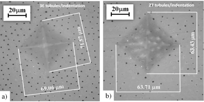
Vickers testing was used to study the variation in hardness as a function of dentin depth. Microindentation was performed using a micro-hardness tester (Wilson Instruments, Model 402 MVD, Norward, MA, USA) with a Vickers diamond indenter. Ten indentations were made on each surface, starting at the DEJ. Grinding and polishing was then performed to
remove approximately 500 m of material, after which another 10 indentations were
performed. This procedure was repeated until the pulpal surface was reached. Indentations were made using an indentation load of 1.96 N and dwell times of 10 sec. These testing conditions generate an indentation large enough so that the hardness corresponds to the overall dentin hardness, which includes hardness of intertubular and peritubular dentin. Indentations were carefully made with a distance of at least 10 diagonals in length from each other in order to avoid any deformation from neighboring indentations.
48
where F is the indentation load and d is the indentation diagonal.
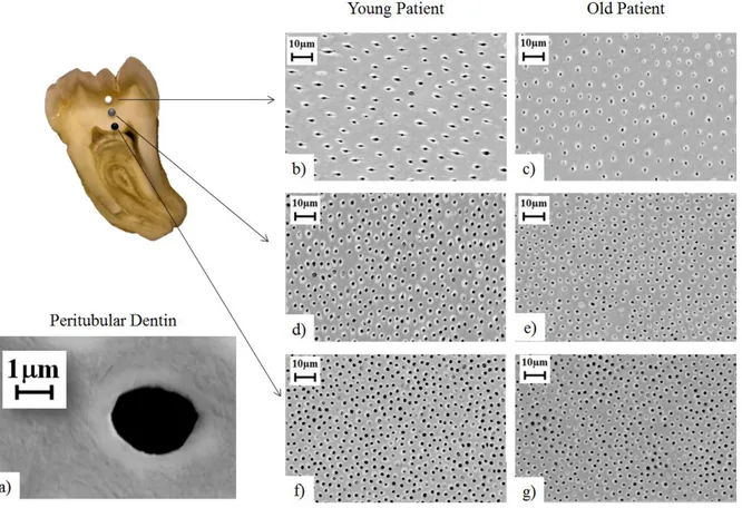
The same specimens were used to evaluate the dentin microstructure using an optical microscope (Axiovert 40 MAT, Carl Zeiss Microscopy, NY). Tubule density, diameter of the tubule lumens and diameter of the peritubular dentin were measured and calculated at each depth. A determination of such parameters was carried across the sample surface. Seven images, each with constant area, were randomly selected from every polished surface. In each image, the amount of tubules was calculated and expressed as tubules/mm2. The tubule diameter and peritubular dentin diameter were also obtained. Values from the seven images were averaged to obtain information from each depth.
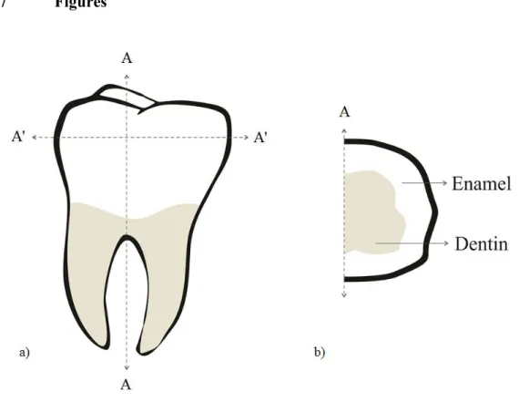
The results obtained for hardness and microstructure at each depth were normalized to the dentin thickness and then classified as outer (normalized depth between 0.76 to 1.00), middle (between 0.36 to 0.75) or inner (depth between 0.00 to 0.35) dentin. Differences in hardness between the outer, middle and inner dentin, as well as between young and old patients were evaluated using a two-way analysis of variance (ANOVA), defining significance of results by p-value ≤ 0.05, a Tukey post-hoc analysis was then performed.
49
were obtained over the spectral region of 400 to 1100 cm-1.The Raman spectrometer had a
laser diode with a wavelength of 785 nm and a spot diameter of approximately 1.1 µm. The mineral-to-collagen ratio was calculated from the ratio of area under the 4PO4 peak at 589 cm-1, which is associated with the phosphate bending of hydroxyapatite, and the area under the amide III peak at 1254 cm-1, which is associated with movements of the peptide bond present in collagen (Goodyear et al., 2009; Kazanci et al., 2006). These bands were selected for analysis as they are reportedly less susceptible to orientation effects and the polarization direction of the incident light (Kazanci et al., 2006).
Differences in the mineral-to-collagen ratio between the outer, middle and inner dentin, as well as between young and old patients were evaluated using a two-way analysis of variance (ANOVA), defining significance of results by p-value ≤ 0.05; a Tukey post-hoc analysis was then performed. Representative maps of hardness and mineral-to-collagen ratio were also obtained from selected teeth to convey the spatial variations present.
3. 3 Experimental Results
Microstructure
50
obliterated, with greater number in the middle and outer dentin. Micrographs of tubules for young and old donor teeth are shown in Figure 3.3.
A quantitative comparison of the microstructural characteristics of dentin from the young and old groups is shown in Figure 3.4. Specifically, Figure 3.4(a) shows the tubule density in different regions of coronal dentin. A reduction in the tubule density was observed with increasing proximity to the DEJ in both age groups. For the young donor group, the
average tubule density in the outer and inner regions was 25,000 tubules/mm2 and
35,000 tubules/mm2, respectively. A similar distribution was found for the old donor group (i.e. decrease in tubule density approaching the DEJ), but there was some difference in the tubule count. The most noticeable change was found in the outer dentin with lower tubule density (10% less) in comparison to that of young donors.
51 Hardness
The influence of indentation load on hardness was studied in order to identify the proper load for measuring the hardness of dentin. Results of that evaluation are shown in Figure 3.5. The measured hardness values decrease from approximately 1.5 GPa at 0.23 N indentation load to 0.7 GPa at 1.96 N load. A plateau in hardness is observed for loads of 1.96 N and greater. Consequently, all further hardness measures were conducted with a load of 1.96 N. The average hardness for the three regions of dentin are shown in Figure 3.6 for both the young and old donor teeth. These values correspond to hardness measured with load applied parallel to the dentinal tubules, as shown in Figs 3.5b and 3.5c.
52 Chemical composition
The distribution of mineral-to-collagen ratio as a function of distance across the coronal dentin for young and old donor teeth is shown in Figure 3.8. The dentin in both age groups showed a similar behavior, with increasing mineral-to-collagen ratio approaching the DEJ. A higher ratio indicates a lower proportion of organic material. The differences found among the areas evaluated in the young donors group were not statistically significant (p>0.05), as opposed to the old donors group where they were statistically significant (p≤0.05).
When comparing the mineral-to-collagen ratios between the young and old patients, nearly a 4% difference was found nearest the pulp within the inner dentin. However, the difference in mineral-to-collagen ratio was approximately 40% in the middle dentin and 70% in the outer dentin. In comparing results for the mineral-to-collagen ratio between young and old donor teeth, the differences were statistically significant (p≤0.05).
Comparisons of hardness and mineral-to-collagen ratio distributions in representative teeth from each age group after longitudinal sectioning can be seen in Figure 3.9. The results correspond to a young donor (18 years of age) and an old donor (65 years of age). For the young donor tooth there was an increase in hardness from the pulp up to the DEJ (Figs. 3.9a). As for the chemical composition of the young dentin (Fig. 3.9b), there was an increase in the mineral-to-collagen ratio with increasing proximity to the DEJ. In comparing Figures 3.9a and 3.9b it is seen that lower hardness values were obtained in regions of lower mineral-to-collagen ratio (inner dentin). Conversely, where a higher proportion of mineral was present, the hardness was greater.
53
tooth shows a distorted pulp chamber due to deposition of secondary dentin within. This process can lead to complete pulp obliteration over sufficient time (Tronstad, 2003).
3. 4 Discussion
Microstructure
Consistent with the findings of earlier studies, there were differences in tubule density and dentinal tubule diameter as conveyed from the measures within the three regions of dentin evaluated (Fig. 3.2). The results obtained for tubule density in the young patients group are
consistent with those of Marshall et al. (1997), who reported values of 20,000 tubules/mm2
(outer) to 43,000 tubules/mm2 (inner).
For the dentin of old donor teeth only full or partially opened tubules were considered in the tubule density measurements. Obliterated tubules were excluded from the measurements to assess how dentinal tubule density changes with aging as caused by the filling process. Clearly a reduction in the number of open tubules contributes to changes in dentin permeability and potentially other aspects of its physical behavior. For example, the changes with aging could be important to dentinal sensitivity and the resistance to tooth fracture.
54
was some difference in the measures for diameter in the outer dentin, with 9% lower values in the old dentin. Undoubtedly this difference is attributed to the onset of sclerosis.
In general, the measures of peritubular cuff diameter were approximately twice the diameter of the tubule lumens. The formation of peritubular dentin occurs after the mineralization of intertubular dentin has completed (Gomez de Ferraris and Campos Munoz, 2009). Deposition of dentin is continued by odontoblasts following a circular pattern surrounding the dentinal tubules. During this process, and as the odontoblasts produce several layers of dentin, the pulpal chamber reduces its size, odontoblasts move to the interior of the pulp, and odontoblastic processes remain inside the dentinal tubules (Gomez de Ferraris and Campos Munoz, 2009).
To identify the change in number of obliterated dentinal tubules relative to the total number of open tubules for each region of dentin, the occlusion ratio was calculated according to:
A higher occlusion ratio indicates a higher fraction of obliterated dentinal tubules. Measured estimates for the occlusion ratio in the three regions of dentin evaluated are shown in Figure 3.10. As expected, the occlusion ratio in young donor teeth is approximately zero in all three regions. Indeed, the visible changes in microstructure of dentin with aging begin in
the third decade of life (Gomez de Ferraris and Campos Munoz, 2009; Nazari et al., 2009;
55
correspond to an average of 1170 obliterated tubules/mm2 in outer dentin and 120 obliterated
tubules/mm2 in inner dentin. Thus, in the tooth crown filling of the lumens begins at the ends of the tubules.
In comparing the results obtained for microstructure between young and old patients (e.g. tubule density, diameter, and peritubular dentin diameter) there was no significant difference (p > 0.05). However, there was a significant difference in the occlusion ratio between the young and old groups, and between the outer and inner dentin (p ≤ 0.05) as shown in Table 3.1.
Hardness
56
found by other authors, where values between 0.25 GPa to 0.8 GPa have been reported (Pashley et al., 1985). It is important to note that previous studies have generally involved data derived from the teeth of patients living in North America. Differences in the microstructure and mechanical behavior of dentin have been noted in a previous comparison of donor groups from North and South America (Ivancik et al., 2014). That may be relevant here.
An increase in hardness occurred with increasing proximity of the DEJ for both age groups. The lower hardness of inner dentin is at least partly associated with the higher number (and area) of dentinal tubules (Pashley et al., 1985). The area occupied by dentinal tubules in the inner dentin is approximately 20% higher than the outer dentin (Pashley et al., 1985; Ivancik and Arola, 2013). This behavior might be attributed to differences in chemical composition and variations between the amount of organic and inorganic material within dentin. However, according to the results obtained for the mineral-to-collagen ratio, these differences were not statistically significant in the young patients group.
57
dentin from old donor teeth did exhibit significantly greater hardness. The discrepancy between the earlier studies and present results may be explained by the use of nano-indentation techniques, and differences in the representation of peritubular or intertubular dentin with single indents. A load-dependency may also contribute to unique responses for the intertubular and peritubular components. More work is needed in this area.
Chemical composition
An increase in mineral-to-collagen ratio was found with increasing proximity to the DEJ for both young and old donors, but no significant differences were found along the tooth. These results contradict the findings by Ryou et al (2011), who found a reduction in the mineral-to-collagen ratio from the pulp to the DEJ in an evaluation of coronal dentin from US donors using FTIR. Ryou et al (2011) used the bands associated with the ʋ3PO4 peak and the Amide I peak to find the mineral-to-collagen ratio of dentin, contrary to the bands used in this study. Authors like Kazanci et al. (2006) have argued that it is important to consider the effect of orientation and polarization of different bands for Raman Spectroscopy analysis. They have also found that Amide III band (used in this study) is less susceptible to polarization effects than other amide bands. However, Märten et al (2010) measured the volume fraction of mineral within the coronal dentin using Small-angle X-ray scattering (SAXS) and found that the mineral volume fraction in dentin is uniform and only found statistically significant differences near the DEJ.
58
DEJ. A correlation made by Pashley et al. (1985) between the change in hardness and tubule density demonstrated that the decrease in hardness of dentin towards the pulp can be accounted for by the decreased hardness of the intertubular dentin and that correlation with tubule density may be coincidental.
It is important to note that most of the results found in the literature were obtained from studies using teeth of mostly anglo-saxons living in the USA. Therefore, the differences in dentin features from Colombian donors (i.e. the variation in the mineral-to-collagen ratio along the tooth) might be attributed to individual characteristics, such as oral health and nutritional status, among others. In addition, several authors have suggested that a relation
between ethnicity and some dental features, such as tooth size (Merz et al., 1991; Bishara et
al., 1989), enamel thickness (Hall et al., 2007) and tooth formation rate (Olze et al., 2007), might exist. Merz et al. (1991) found that dental arches in black patients are significantly wider and deeper than the ones in white patients; while Hall et al. (2007) found thicker enamel on the distal aspect of the black donor tooth. Differences between tooth dimensions of permanent teeth in three populations: Egyptians, Mexicans, and the North Americans were found by Bishara et al. (1989). However, only a few studies on dentin characteristics and how they might be determined by ethnicity have been carried out (Bajaj et al., 2008).
The results obtained for the mineral-to-collagen ratio are in agreement with those found for hardness distribution, where higher hardness values were associated with areas where there was a higher amount of mineral.
59
been reported that the total obliteration of tubules for coronal dentin might occur near the age of 70 (Tronstad, 2003).
It is important to note that the results shown in the hardness maps (Figs 3.9a and 3.9c) were obtained after longitudinal sectioning of the tooth (section A-A in Fig. 3.1a). Consequently, the indentation load was applied perpendicular to the dentinal tubules. A comparison of hardness measurements from the parallel and perpendicular loading orientation is shown in Figure 3.11. In comparing the results for these two directions, significant differences in hardness were found between the middle and inner dentin (p ≤0.05) of young donor teeth. For the old dentin there were no significant differences (p > 0.05). The unique behavior of the inner and middle dentin may be attributed to a greater number of tubules in these regions, which yields a higher proportion of voids and lower hardness. For old dentin there is a consistent increase in hardness approaching the DEJ with both orientations of indentation. In this case, the increase is more significant than the one discussed earlier (load applied parallel to dentinal tubules) due to the presence of large areas of peritubular dentin within the indentation as a result of obliteration of the dentinal tubules.
3. 5 Conclusions
According to the results obtained, the following conclusions were drawn:
1. The tubule density in the dentin of young and old donor teeth ranged from approximately 22,000 to 35,000 tubules/mm2. The tubule diameter ranged from approximately 1.2 m to 1.8 m. There was a significant decrease in tubule density from the pulp to the DEJ in both age groups studied (p ≤ 0.05).
60
largest proportion of obliterated dentinal tubules was near the DEJ and decreased towards the pulp.
3. A significant decrease in hardness was found with increasing distance from the DEJ
for both young and old donor teeth (p ≤ 0.05). A larger hardness was found in the old dentin when compared to young dentin. There were significant differences (p ≤ 0.05) in the hardness in the outer and middle dentin between the young and old donor teeth.
4. The mineral-to-collagen ratio was found to increase with proximity to the DEJ for
both young and old patients. This behavior is opposed to previous results reported in the literature for dentin from US donors, which in turn raises questions whether the chemical composition of dentin and its percentages of organic material and collagen might be affected by ethnicity.
5. The outer dentin exhibited the highest mineral-to-collagen ratio, while lower values were found in the areas near the pulp. A greater mineral-to-collagen ratio was found for the old dentin, with the highest values found in the outer dentin.
61 3. 6 Tables
Table 3. 1. Results from the ANOVA (p-values) in comparing the microstructure of dentin from young and old donor teeth. Note the statistically significant differences for the occlusion ratio for the middle and outer dentin.
Young vs. Old
Region Tubule
Density
Occlusion Ratio
Tubule Diameter
Peritubular Diameter
Outer 0.1425 0.0002 0.2123 0.6466
Middle 0.4333 0.0001 0.7358 0.4379
62 3. 7 Figures
63
64
65
66
67
68
Figure 3. 7. Indentation of dentin for determination of Vickers hardness. The indentation locations are within the outer dentin of a young (a) and old (b) donor tooth.
69
70
71
72
Figure 3. 11. Vickers hardness obtained for dentin of young and old donor teeth according to