Organ-focused mutual information for nonrigid multimodal registration
of liver CT and Gd-EOB-DTPA-enhanced MRI
Laura Fernandez-de-Manuel Gert Wollny Jan Kybic Daniel Jiménez-Carretero Jose M. Tellado
Enrique Ramon Manuel Deseo Andres Santos Javier Pascau Maria J. Ledesma-Carbayo
A B S T R A C T
Accurate detection of liver lesions is of great importance in hepatic surgery planning. Recent studies have shown that the detection rate of liver lesions is significantly higher in gadoxetic acid-enhanced magnetic resonance imaging (Gd-EOB-DTPA-enhanced MRI) than in contrast-enhanced portal-phase computed tomography (CT); however, the latter remains essential because of its high specificity, good performance in estimating liver volumes and better vessel visibility. To characterize liver lesions using both the above image modalities, we propose a multimodal nonrigid registration framework using organ-focused mutual information (OF-MI). This proposal tries to improve mutual information (MI) based registration by adding spatial information, benefiting from the availability of expert liver segmentation in clinical protocols. The incorporation of an additional information channel containing liver segmentation information was stud-ied. A dataset of real clinical images and simulated images was used in the validation process. A Gd-EOB-DTPA-enhanced MRI simulation framework is presented. To evaluate results, warping index errors were calculated for the simulated data, and landmark-based and surface-based errors were calculated for the real data. An improvement of the registration accuracy for OF-MI as compared with MI was found for both simulated and real datasets. Statistical significance of the difference was tested and confirmed in the simulated dataset (p < 0.01).
1. Introduction
The advances made in recent decades in the understanding of liver anatomy and physiology (Couinaud, 1999), together with the improvement of medical imaging techniques (Handels and Ehr-hardt, 2009; Radtke et al., 2007) and the progressive safety of sur-gical instrumentation, allow surgeons to design complex liver resections more accurately and effectively without jeopardizing patient safety. Further, preoperative planning has become an essential task before undertaking liver surgery. It requires the
mapping of hepatic vasculature, spatial localization of tumors and their relation with other tumors or vascular structures, and the estimation of remnant liver volume in order to determine the suitability of a patient for surgery and to decide the procedure. Accurate detection of individual liver lesions is of great importance because their number, location, and relationships determine both resectability (the probability of performing a resection safely) and radicality (the probability of a potential cure by achieving an R0 resection) (Solbiati et al., 1999).
sensitivity and specificity for detecting hepatic metastases and is in most cases more convenient than MRI for evaluating the extrahe-patic abdomen (Oliva and Saini, 2004) and for estimating remnant liver volumes because of its higher spatial resolution, better vessel visibility, and wide availability and acquisition speed. However, the lesion detection rate has been found to be significantly higher with Gd-EOB-DTPA-enhanced MRI as compared with CT, especially for small lesions (Donati et al., 2010; Hammerstingl et al., 2008) (Fig. 1). Gd-EOB-DTPA (gadoxetic acid, Primovist® in Europe, Eo-vist® in the US, by Bayer Healthcare Pharmaceuticals) is an organ-specific contrast medium for hepatic MRI, in use since 2005. In de-layed Tl-weighted MRI, it produces strong signal enhancement in normal liver parenchyma and absence of signal for focal liver lesions with absence of hepatocellular activity. Consequently, detection of liver metastases and other secondary malignant liver tumors is im-proved. However, vessels are not opacified.
Considering the advantages and disadvantages of the different imaging modalities and contrast media, several researchers have pointed out the importance of combining MRI and CT for the detec-tion and localizadetec-tion of hepatic lesions and their reladetec-tion with ves-sels for therapy planning (Bluemke et al., 2000; Kong et al., 2008; Lange et al., 2005a). In this work, we propose a nonrigid registra-tion framework for aligning contrast-enhanced portal-phase CT and delayed Tl-weighted Gd-EOB-DTPA-enhanced MRI into a common coordinate system. As far as we know, this problem has never been tackled before. To use all available data and improve the registration robustness and accuracy, we propose the use of an organ-focused mutual information (OF-MI) registration criterion.
I.I. State of the art
To date, commercial systems for planning hepatic surgery mostly align images rigidly. However, registering soft tissues with rigid registration may result in errors as high as 19-20 mm (Archip et al., 2007; Lee et al., 2005), due to deformations that may be caused by liver movements because of respiration, variations of po-sition, and corporal mass changes over time. To improve detection and characterization in terms of volume and relation with vascula-ture of primary liver cancers (for example hepatocellular
carci-Fig. 1. Contrast-enhanced portal-phase CT image and delayed Tl-weighted gadoxetic acid MRI from a patient after right hepatectomy. Arrows show metastases within the different modalities. Each row represents different slice positions. The figure shows how metastases clearly identified in MRI are hardly visible in CT. Better vessel visibility is observed in the CT.
noma), secondary tumors (for example, liver metastases secondary to colorectal cancer) and other liver diseases, an accu-rate multimodal nonrigid image registration algorithm is clearly required.
Some studies have presented methods for liver monomodal im-age registration (Carrillo et al., 2000; Lange et al., 2005b). Other proposed techniques have focused on compensating multimodal image differences in the location and motion of the liver in relation to other organs by using rigid approaches (Van Dalen et al., 2004) or nonrigid methods based on finite elements, B-splines or demons (Archip et al., 2007). Different similitude criteria have been also ap-plied, such as voxel similarity or surface based criteria (Lee et al., 2005). However, to the best of our knowledge, the performance in terms of correspondences between internal liver structures, le-sions, and vascular landmarks such as vessel bifurcations has not been evaluated. Additionally, CT/Gd-EOB-DTPA-enhanced MRI registration methods have not been proposed before.
Voxel intensity measures have been shown to be robust mea-sures of image similarity. There are several possible image metrics that are used in voxel similarity-based image registration (Crum et al., 2004; Hill et al., 2001; Maintz and Viergever, 1998; Rueckert and Schnabel, 2011; Zitova and Flusser, 2003): correlation coeffi-cient, sum of squared differences, or mutual information (MI). MI (Maes et al., 1997; Mattes et al., 2003, 2001; Pluim et al., 2003; Wells et al., 1996) is one of the more successful medical image sim-ilarity measures. However, extending the maximization of MI to nonrigid image registration and applying it to extensive areas of body images is still an active field of research. Moreover, the most important drawback of MI is that, due to the absence of spatial information, intensity relationships in one region can occasionally mislead the algorithm in another region where the intensity rela-tionships are completely different (e.g., problems with spatially varying intensity inhomogeneity in MRI (Loeckx et al., 2010) or li-ver vessel misalignments in contrast-enhanced CT and delayed Tl-weighted Gd-EOB-DTPA-enhanced MRI (Fig. 2)).
Some studies have focused on improving registration accuracy by considering the use of additional image gradient information (Pluim et al., 2000a,b), neighbor pixel information (Heinrich et al., 2012b; Kybic and Vnucko, 2012; Rueckert et al., 2000), tex-tural information (Heinrich et al., 2012a), or different approaches to weighted MI (Park et al., 2010; Rodriguez-Carranza and Loew, 1999; Van Dalen et al., 2004). One approach to weighted MI is the regularization of MI with the use of weights based on overlaps (Rodriguez-Carranza and Loew, 1999) without including spatial information. Other weighted MI approaches increase histogram contributions of certain pixels (Park et al., 2010) or restrict the reg-istration to certain regions (Van Dalen et al., 2004). Nevertheless, the main problems with the application of the last method are the lack of information on the borders of the regions and neighbor-ing structures, and havneighbor-ing too few samples to obtain a good entro-py estimation. These problems may be less significant in rigid scenarios. However, they are more relevant when nonrigid trans-formations are required, hence, the weighted MI performance strongly depends on the concrete registration problem.
Recent approaches add spatial context to mutual information, either by studying different spatial encoding schemes (Zhuang et al., 2011) or by searching for the correspondence of a priori learned set of image patches (Yi and Soatto, 2011). In Hermosillo et al. (2002) the formulation of a locally computed similarity mea-sure is presented and in Rogelj et al. (2003) a variant to obtain pointwise similarity metric is described.
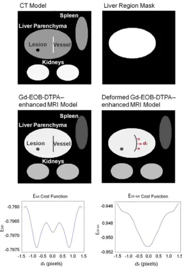
ap-CT Model Liver Region Mask
Gd-EOB-DTPA-enhancedMRI Model
Deformed Gd-EOB-DTPA-enhancedMRI Model
EMI Cost Function EOF-MI Cost Function
-1.0 -0.5 0.0 0.5 1.0 dx (pixels)
-0.946
0.948
-0.950
-0.952
-1.5 -1.0 -0.5 0.0 0.5 1.0 1.5 dx (pixels)
Fig. 2. Cost functions for a synthetic 2D model as a function of horizontal transformation. A local nonrigid deformation is applied with maximum amplitude dx from —1.5 pixels to 1.5 pixels in the middle of the vessel region. The £Mi cost function shows multiple local minima and a global minimum shifted away from 0 where the correct solution should be. The £0F-MI cost function shows a clean global minimum in the correct location.
plied to analyze local tissue contrast changes in brain MRI nonrigid registration. The RMI was defined as:
RMI(T,fi,X) = H(T) + H(K) + H(X) - H(T,R,X) (1)
with X expressing the spatial position of cubic overlapping subre-gions in the reference image R and the test image T. In Loeckx et al. (2010) a conditional mutual information (cMI) was defined and calculated between two images T, R, given a certain spatial dis-tribution X Besides the intensity dimensions, a third spatial channel was incorporated into the histogram definition, expressing the spa-tial location of every joint intensity pair:
cMI(T,K|X) = H(T|X) + H(K|X) - H{T,R\X) (2)
In Russakoff et al. (2004), a regional MI was described, introduc-ing neighborhood regions of pixels into a multidimensional histo-gram. In this work, each pixel co-occurrence was represented by more than one entry in the joint histogram depending on its neigh-bor pixel co-occurrences. However, one should notice that as de-scribed in Russakoff et al. (2004), the main problem in increasing the dimensionality of joint histograms is the need of a higher num-ber of samples to obtain a reasonable estimate of entropy distribu-tion. Therefore, these methods require a large number of image voxels hence increasing the computational complexity enor-mously. In Russakoff et al. (2004) a method to make the problem more tractable is proposed, taking advantage of the fact that the
entropy of a discrete distribution is invariant to rotations and translations and making the simplifying assumption that high-dimensional distributions are approximately normally distributed. In Studholme et al. (1996) preliminary rigid registration results demonstrated the possibility of extending the histogram with a small number of unconnected regions of similar intensity ranges (e.g. air or fat tissue). As the reference image R and the regions L were inherently registered, they proposed an extension I of the mutual information for more than two variables defined by:
1(7", R; I) = H{R, I) + H(T) - H(T, R, L) (3)
Their preliminary results motivated us to use the same extension of the mutual information for more than two variables. However, they proposed the use of regions calculated by using only intensity ranges. Unconnected regions containing the same range of intensi-ties were considered as different, which does not allow separating intensity relationships based on anatomical reasons nor having re-gions with more than one range of intensities.
1.2. Our contribution
As already pointed out above, delayed Tl-weighted Gd-EOB-DTPA-enhanced MRI causes strong signal enhancement for normal liver parenchyma. For this reason, the relationship between inten-sities in CT and MRI images is very different inside and outside the liver. Hence, using the classical formulation of MI, intensity rela-tionships in a region can occasionally mislead the algorithm in an-other region where the relationships are completely different; especially in nonrigid registrations.
Liver volume estimation from preoperative CT is a routine man-datory process to determine the suitability of a patient for surgery and to make the final clinical decision before extensive hepatecto-mies (Fernandez-de-Manuel et al., 2011; Heimann et al., 2009). To estimate remnant liver volumes, both manual and automatic seg-mentation tools are applied in the daily clinical routine. Conse-quently, liver segmentation from CT images are available in most hospitals for patients considered for liver surgery. For that reason, incorporating the liver segmentation information into the registra-tion process is very feasible.
We discarded the approaches presented in Loeckx et al. (2010), Russakoff et al. (2004) and Studholme et al. (2006) because those techniques cannot take advantage of the available liver segmentations.
In this work, we propose the use of an organ-focused mutual information (OF-MI) criterion. We extend the joint histogram with an additional information channel using an extension of the mu-tual information for more than two variables similar to the one proposed in Studholme et al. (1996) but with a different probabil-ity distribution estimation. Studholme et al. (1996) used regions segmented based on intensity ranges, however, they suggested to exploit a higher level of anatomical knowledge. Therefore, we take advantage of an anatomical segmentation resulting in regions with varying intensities. We consider that using anatomical regions actually allows taking maximum advantage of the definition pro-posed in Studholme et al. (1996). Additionally, we have extended its implementation to nonrigid multimodal registration. Conse-quently, our work's main contributions are related to the experi-mental novelty and the obtained results in a clinical application that benefits from the proposed approach.
synthetically generated data as well as its application to relevant clinical liver datasets (CT and Gd-EOB-DTPA-enhanced MRI), and a comparison of the registration performance using OF-MI com-pared with MI as registration criterion.
The proposed criterion takes into account an organ (liver) seg-mentation based on the semi-automatic method described in Fer-nandez-de-Manuel et al. (2009) and Jiménez-Carretero et al. (2011).
The algorithm has been validated and compared with the stan-dard MI on a simulated 3D dataset with 63 image pairs, using a de-layed Tl-weighted Gd-EOB-DTPA-enhanced MRI simulation framework. Additionally a dataset of seven real subjects referred to surgery with one contrast-enhanced portal-phase CT and one delayed Tl-weighted Gd-EOB-DTPA-enhanced MRI each have been used.
2. Methods
In Sections 2.1 and 2.2, we will introduce the general frame-work and MI, describing briefly the equations presented in previ-ous work (Kybic and Unser, 2003; Thévenaz and Unser, 2000). We will then explain our contribution in Section 2.3.
2.1. Problem definition and registration framework
The intensity-based nonrigid registration algorithm used ex-tends the previous B-spline method of Kybic and Unser (2003). The algorithm determines a set of B-spline coefficients that de-scribe a nonrigid transformation that maximizes an image similar-ity measure. The transformation model is defined as a linear combination of B-spline basis functions located on a uniform grid. B-spline functions have been widely used to represent deforma-tions (Kybic and Unser, 2003; Ledesma-Carbayo et al., 2005; Oguro et al., 2009; Rueckert et al., 1999; Schnabel et al., 2001), motivated by their compact support, computational simplicity, good approx-imation properties, and implicit smoothness. We also use B-spline functions for representing continuous images derived from a set of samples (Kybic and Unser, 2003; Thévenaz and Unser, 2000). Moreover, B-spline basis functions are used as Parzen windows (Thévenaz and Unser, 2000) in the similarity criteria, as described later.
The input images are given as two N-dimensional discrete sig-nals: the test image T and the reference image R with intensities /t(i) and /r(i), respectively, where i e J c Zw, and / is an
N-dimen-sional discrete interval representing the set of all voxel coordinates in the image. For convenience in our formulation we use a contin-uous representation/tc(x) of the discrete test image/t(i) as follows:
/¡F(x) = ] T a,/?m(x-i)
ielacz" ( 4 ) /t(i)=/t c(i) V i e / c ZN
where f¡m represents an N-dimensional tensor product of centered
B-splines of degree m (Kybic, 2001), a, are the B-spline coefficients that represent the original test image given by its samples/t(i), and
la is the set of nodes used to represent the image.
Let g(x) be a deformation function that finds the spatial corre-spondence between coordinates in the test and reference images. The deformation is represented using splines:
g ( x ) = x + ] T c,/?n(x/h-j) (5)
Je'bCZ"
described by a finite number of parameters c = {c¡},j e ¡b c ZN; where ¡b is an N-dimensional discrete interval representing the set
of parameter indexes, h is the knot spacing on a regular grid over
the image, and f¡„ represents an N-dimensional tensor product of centered B-splines of degree n.
The warped test image W is defined as /w(x) =/tc(g(x)). We de-fine the solution to our registration problem as the result of the minimization g = arg mingeG £(g), where G is the space of all admis-sible deformation functions g and £ is the criterion. For the pro-posed application, we consider the criterion:
E(g)=Ed(W,R) + yEI(g) (6)
where E¿ is an Mi-based image dissimilarity criterion and £r is a reg-ularization term with weight y used to prevent discontinuities and to guarantee overall smoothness. For this particular problem, we use a discrete approximation to the norm of the Laplacian of the continuous deformation as £r (Kybic, 2001).
To minimize the criterion E with respect to a finite number of parameters c we use a gradient descent optimizer with quadratic step size estimation, as recommended in Kybic and Unser (2003). The optimization uses a multiresolution approach for the image model. The multiresolution methodology used creates a pyramid of subsampled images optimal in the L2 sense, taking advantage of the spline representation (Unser et al., 1993). The problem is solved by starting at the coarser level of the pyramid (the most subsampled image) and proceeding to the finest level.
2.2. Mutual information
The joint intensity probability distribution is estimated by means of Parzen windows because of their good properties, such as computational efficiency (Unser, 1999).
Following Thévenaz and Unser (2000), the contribution to the joint histogram of a single pair of pixels with intensities (fw,fr) is
tributed over several discrete bins (t, r) with t and r belonging to dis-crete sets of intensities associated with the test and reference images, with ranges from 0 to nbinsT - 1 and nbinsR - 1, respectively.
Intensities [fw,fr) can take values in a continuum in the ranges (fw
mm, fw max) and (fr mm, fr max), respectively. Using B-spline functions
of degree m-i and m2, the discrete joint probability of co-occurring intensities in the overlap of the two images fw and/r is expressed as:
p{t,r;c)=^Y. A»,(t " T(i;c)) • (¡mi(r - p(i)) (7)
and the discrete marginal probability distributions for the warped test and the reference images, respectively, are:
PTÍt;C)=T¡- ] T /¡m i(t-T(i;C))
(8)
P * (r) = 7 n E A "2(r- P ( i ) ) |Jc
Wz«
where ¡c is a discrete set of samples ¡c c /. T and p are the test and
reference images after scaling the continuous interval (0, nbinsT - 1)
and (0, nbinsR - 1), respectively:
^m^) ~ (/wvl)CJ — Jwmin) ' r c
/w max — Jwmin ÍQ\
P(i) = (fr(i)-/r m í n)- " M " 1
J r max Jrmin
The MI-based dissimilarity criterion £MI can be defined from the above probabilities as a function of the deformation parameters c:
^
=
^^
=
-W
{t
'
r
'
e)
^^h
(10)
We can also express Mi-based dissimilarity criterion £Mi be-tween a warped image W and a reference image R in terms of the marginal and joint entropies:
Ed = Em(W,R) = H(W,R) - H(W) - H(K) (11)
For the optimization algorithm, partial derivatives of the £Mi with respect to Cj are needed:
9EM\
9clk
E
8EMi 8f[(x)
dfw(i) dxk x=g(i)
3g*(i)
dciM
(12)
where k is the dimension of the N-dimensional Cj. For further details on the derivatives calculation, we refer the reader to Thevenaz and Unser (2000).
2.3. Organ-focused mutual information
We present here an OF-MI criterion that allows including prob-abilities of voxels belonging to the object or the background.
We introduce an additional information channel consisting of an image L containing for every voxel its probability Pc,(i) of belonging to the background Q0 and the object (liver) £?i, Pa¡ (i)
sat-isfying E v A , (i) = i vi, i = o, l.
Based on (3) (Studholme et al., 1996) we define OF-MI-based dissimilarity criterion £0F-MI between a warped image Wand a pair (P,L) consisting of a reference image R and the probability image L in terms of the marginal and joint entropies:
Ed = Eo?-m{W,R,L) = H{W,R,L) - H(W) - H{R,L) (13)
The joint probability histogram is extended with a third dimen-sion of size 2, with / the coordinate representing inside (/ = 1) and outside (/ = 0) the liver region. Using B-spline functions of degree mx and m2, we define a 3D discrete joint probability distribution:
p(t,r,i;c)=-L Y.P^-fSm(t-r(i;c))-flmi(r-p(i)) (14)
1 c |i e lcc ZN
The marginal organ-focused joint intensity probability distribu-tion for the reference image is:
|Jc
Wz«
(15)
The marginal intensity probability distribution for the test image is given in (8).
The dissimilarity criterion £0F-MI is defined from the above probabilities as follows:
p(t,r, i;C) £OF-MI (W,R,L)
-EEEp(
t-
r-
i;
c)-
1°g
Vt Vr VI PT(t;c)-Pitt(>",i) (16)
The partial derivatives of the £0F-MI with respect to Cj are: <9(EOF-MI) _ •sr-.d(E0f_MI) dfi(x)
dc),k
E
<9L(i) dxkdgk(J) dci,k
(17)
(For further detail on the derivatives calculation, we refer the reader to the Appendix A).
From (13) and (16) the expressions for the marginal and joint entropies remain as follows:
H(W, R, L)
= - E E E p f t r>';c) • 1O
SP(Í> r.';c) Vt Vr VIH(W) = - 5 > ( t ; C ) . l 0 g pr( t ; C ) (18)
H(P.,L) = - ^ ^ pf f i( r , í ) . l o g pf f i( r , í )
2.3.1. Estimation of the region probabilities ?Qt(i)
The probability image L containing Pc,(i),/ = 0,1 is computed from the hepatic masks (liver segmentation) obtained for the
sur-gery planning procedure. Uncertainty of the segmentations could be taken into account by smoothing the mask edges using a Gauss-ian filtering; however, experiments revealed that this does not bring any benefit for this application. Therefore, in this work, the voxel probability of belonging to a region will be either 0 or 1; as each voxel contributes only to one region.
Considering a binary image/Q(x) with dimensions identical to /r(x) that represents the clinical segmented liver in the reference
image, we define Pc,(i) as follows:
i V i ) •• f
Q(x)\xM if I = 1
l - /f l( x ) |f c I if 1 = 0
(19)
In this work the initial liver binary images /Q(x) have been cre-ated semi-automatically by a liver segmentation application based on active contours previously described in Fernandez-de-Manuel et al. (2009) and Jiménez-Carretero et al. (2011).
Notice that with our proposed OF-MI we are not neglecting any area of the image, as all the regions are represented in the 3D joint intensity probability distributions and optimized together.
2.3.2. MI versus OF-MI synthetic examples
To illustrate theoretically the behavior of the proposed ap-proach compared with MI and its shortcomings related to its assumption of equal statistical relationships over the whole do-main of the images, two 2D basic synthetic images were created representing a contrast-enhanced portal-phase CT model and a de-layed Tl-weighted Gd-EOB-DTPA-enhanced MRI model. Both models contained the liver, the kidneys, the spleen, and an intrahe-patic lesion and vessel. They represented a realistic distribution of intensities and were initially registered completely. We used the CT liver segmentation (liver region mask in Fig. 2). We then applied a mild horizontal nonrigid deformation with maximum amplitude dx around the center of the hepatic vessel to the Gd-EOB-DTPA-enhanced MRI model and evaluated the dependency of the crite-rion on dx, with dx ranging from -1.5 pixels to 1.5 pixels (Fig. 2).
The £MI exhibits multiple local minima and a global minimum far from the correct location. This happens because the intensity relationships between CT and MRI pixels in extrahepatic organs (spleen) mislead the algorithm into considering that the hepatic vessel in MRI should be aligned with the liver tissue in CT. On the other hand, the E0F-MI cost function shows a clean global min-imum at the correct location, as the intensity relationships inside and outside the liver are considered separately.
3. Validation methodology
In this section, we will first describe the simulated and real im-age datasets. Then, we will illustrate the definition of the quantita-tive measures used to validate the experiments. After that, the registration parameters for both, the MI and the OF-MI criteria are given, followed by the validation results.
3.1. Data
3.1.1. Simulated images dataset
The gray values of the simulated MRI images were calculated by a nonlinear intensity transformation based on intensity distribu-tions of contrast-enhanced portal-phase CT and delayed Tl-weighted Gd-EOB-DTPA-enhanced MRI (Fig. 3a and b). By direct observation of these distributions, we assigned the interval of intensities approximating those in MRI to the interval of intensities in the CT for each relevant organ or structure, differentiating be-tween liver structures and background. Therefore, different trans-formations were applied to different CT regions, assigning tissue-dependent signal intensity to each region (liver and liver back-ground) (Fig. 3c and d). In order to apply these intensity transfor-mations, CT regions were calculated by segmenting the liver in the CT. Segmentations were made by an independent person blind to those used during the registration process in order to guarantee the independence of the simulation step with respect to the registrations.
To simulate the partial volume effect in MRI (Tohka et al., 2004), a 3D low-pass Gaussian filter with a standard deviation for the Gaussian kernel of [1,1,3] was applied after the intensity transformation.
Gd-EOB-DTPA-enhanced MRI images commonly show better edge enhancement than CT images. To simulate this particular fea-ture, borders were first calculated by morphological edge detection and added by summation to the existing image.
The MRI signal is corrupted by an additive noise process (Kwan et al., 1999). As noise distributions in MRI images are nearly white Gaussian for signal-to-noise ratios (SNR) greater than 2 (Gudbjarts-son and Patz, 1995), we added independent realizations of white Gaussian noise to our simulated dataset. We measured the amount of added noise as a signal-to-noise ratio according to Bushberg et al. (2002):
SNR = A/an (20)
where A is the mean image pixel intensity, and o„ is the standard deviation of the Gaussian noise. Different levels of Gaussian noise were added to the simulated data ranging from SNR 3 to SNR 7.
Additionally, known transformations were applied to the origi-nal CT images to generate reference images for the registration experiments. The transformations were modeled using a closed-form function that defines the spatial dependence of the declosed-forma- deforma-tion, mimicking nonrigid organ movements due to respiration and volume changes along time caused by differences in patient weight, organ disposition, or liver volume growth due to illness evolution or portal vein embolization. In our transformation model we assume that the deformation is 0 in the center of the body and maximum at a liver distance, as the soft-tissue motion of the liver is highly influenced by the motion of both the diaphragm and the ribcage (Villard et al., 2011). According to some sources, local liver
CT intensity values MRI (Gadaxetc Add T1W delayed) intensity values
1600
1400
1200
> 1000
800
600
LIVER LIVER LIVER KIDNEY SPLEEN PANCREAS PARENCHYMA M I X VESSEL
LIVER LIVER LIVER KIDNEY SPLEEN PANCREAS PARENCHYMA MTX VESSEL
Liver intensity transformation Background intensity transformation
1600
1400
1200
1000
800
600
400
200
I
-100 -50 50 100 150
Initial Intensity
200 250 300
1600
50 100 150
Initial Intensity
300
Fig. 3. Intensity distribution analysis in contrast-enhanced portal-phase CT (a) and delayed Tl-weighted Gd-EOB-DTPA-enhanced MRI (b) for abdominal organs and
deformation due to respiration can range from 10 to 26 mm in amplitude between the extremes of the respiratory cycle (Blackall et al., 2005; Clifford et al., 2002; Rohlfing et al., 2001). Moreover, we approximate the deformation as a continuous movement. Therefore, we represent the dependency of the deformation on the distance to the center of the image by using a sinusoidal func-tion that allows us to have a symmetric deformafunc-tion all around the contour of the body with maximum value in the center of the liver. The simulated deformation is defined by t(i) = {tk(!k)}k=li 2, 3. where i e / c Z3 and / is the 3-dimensional discrete interval representing the set of all voxel coordinates in the image. t(i) is applied voxel by voxel as defined by the equation:
t
k(i
k) = i
k-m
k. sin (,
Xc-""''fe.f\ (21)
where xc are the voxel coordinates with minimum deformation, xw are the voxel coordinates with maximum deformation and m is the maximum deformation. xc represents the center of the image and xw represents the points at 1/3 of the extreme of the image that comprise the liver in all the models. Therefore, these parameters de-pend on the image size sim = {simk}k=li 2, 3 as follows:
Xc = Vc • Sim ( 2 2 )
Xw — Pw ' Sim
where pc = 0.5 and pw = 1/3. Considering the literature references about local liver deformation (Blackall et al., 2005; Clifford et al., 2002; Rohlfing et al., 2001), we decided to apply to each of our 63 models a maximum deformation value that varies randomly be-tween 4 mm and 28 mm at liver level. mk represents the magnitude
of the maximum deformation for each dimension and takes random values in the range [4,28] (mm).
An example of a synthetic delayed Tl-weighted Gd-EOB-DTPA-enhanced MRI can be seen and compared with a real MRI image in Fig. 4 as well as an example of a simulated deformation.
3.Í.2. Real images dataset
A dataset consisting of seven clinical subjects with a wide vari-ety of pathological scenarios was used. Each case has one contrast-enhanced portal-phase CT and one delayed Tl-weighted Gd-EOB-DTPA-enhanced MRI from a retrospective clinical dataset. The contrast-enhanced portal-phase helical CTs were performed
Fig. 4. (a) Original CT, (b) CT with simulated deformation, (c) synthetic gadoxetic
acid MRI, (d) real gadoxetic acid MRI.
with a 16-MDCT scanner (Brilliance 16; Philips Medical Systems, Eindhoven, The Netherlands) in all cases. The scanning parameters were 120 kVp, 250-300 mA s, 2-mm slice thickness with an over-lap of 1 mm (pitch, 0.9), and a single-breath-hold helical acquisi-tion. The images were obtained in the craniocaudal direcacquisi-tion. Hepatic portal-phase scanning began 70 s after injection of 120 ml of a nonionic iodinated contrast agent (Ioversol, Optiray Ultraject 300; Covidien). For delayed Tl-weighted Gd-EOB-DTPA-enhanced MRI, 20-min delayed hepatobiliary phase images were obtained with a Tl-weighted 3D turbo-field-echo sequence (Tl high-resolution isotropic volume examination, THRIVE; Philips Medical Systems, Eindhoven, The Netherlands) (3.4/1.8; flip angle 10°; matrix size, 336 x 206; bandwidth, 995.7 Hz/pixel) with a 2-mm section thickness, no intersection gap, and a field of view of 32-38 cm. Details regarding clinical information are summarized in Table 1.
MRI images were manually aligned onto the corresponding CT by a point-based rigid registration.
The images were then cropped in axes X, V, and Z to restrict the subsequent image-processing steps to the complete body region at liver level. The images were then resampled to pixel size [1,1,1] mm, guaranteeing isotropy. CT was used as the reference image, and MRI as the test image.
3.2. Error measures
3.2.1. Measures on the simulated dataset
Registration of simulated CT and MRI datasets was evaluated in terms of geometric error by comparing the resulting transforma-tion with the applied analytical one on a voxel-by-voxel basis using the warping index (WI) (Thevenaz et al., 1998). The WI cal-culation was restricted to the liver region:
where g* is the true deformation, R represents the set of all voxel coordinates inside the liver, and |-| the Euclidean distance.
3.2.2. Measures on the real dataset
To establish an independent validation procedure, radiologists annotated all real images manually, defining a set of 10 intrinsic anatomical hepatic landmarks for each pair of images. Registration results were evaluated in terms of the mean distance error be-tween corresponding anatomical landmarks before and after regis-tration for each subject (landmark-based mean errors). Landmarks were located in vessel intersections, hepatic fissures and ligaments, and small lesions.
Because of the high dependency of error results on the accuracy of the landmark selections, additional error criteria were also con-sidered. For this purpose, the liver was manually segmented in the CT and MRI images. Segmentations were made by an independent expert blind to those used during the registration process in order to guarantee the independence of the registrations with respect to the validation. Comparing liver segmentation before and after reg-istration allows calculation of surface-based mean errors (ME):
M E
= ¿ E
d
> (
24
)
lJl ieS
Table 1
Real dataset clinical description ("Roman numerals represent Couinaud hepatic segments (Couinaud, 1999)).
Subject Clinical information Image dates CT-visible lesions MRI-visible lesions
Hepatectomy Metastases Chemotherapy Metastases Chemotherapy
Metastases
Hepatectomy Metastases Metastases Cholecystectomy
Metastases Sectorectomy Metastases
MRI 26 days post-CT
CT 45 days post-MRI
MRI 4 months post-CT
MRI 1 month post-CT
1 in hepatic duct
21 days post-CT
10 months post-CT
12 days post-CT
1 in IV 1 in VIII 0
3 in VIII 1 in IV 1 in II
1 in VII
1 in hepatic duct 1 in IVb"
1 in II/III 1 in IVa/VIII 1 in VI 1 in I 1 in VIII
1 in VIII 1 in VII/VIII 1 in II
0
1 in IV 1 in II
3.3. Parameter optimizations
To investigate the best parameter combination, we tested the performance of the algorithm with one real training subject. Com-mon parameters in the intensity-based nonrigid registration algo-rithm are:
• degrees of the B-spline functions: m, n, m,, and m2, • knot spacing of the transformation grid: h, • weight for the regularization term: y,
• number of bins for the intensity probability distributions: nbimT,
nWnsR>
• number of multiresolution levels: after some initial experi-ments, this was fixed at 3.
3.3.1. Choosing the B-spline degrees
The B-spline degrees for the image, m, and deformation model, n, were chosen as cubic because previous studies have shown that these perform better than linear and quadratic splines (Kybic and Unser, 2003). However, for the B-spline joint probability distribu-tion model (m-i, m2), the chosen degree was quadratic. By using quadratic B-splines, we ensure derivability of the joint probability distributions and avoid an increase in histogram dispersion.
3.3.2. Choosing the node spacing and the regularization weight The main criterion for choosing the knot spacing h in (5) and the regularization weight y in (6) is the estimated intrinsic resolu-tion (smoothness) of the deformaresolu-tion to be recovered. To estimate these optimum parameters, multiple registrations were run using Ml and OF-MI with different values for the node spacings in the three dimensions hk e {12,14,16,18,20}, (fe = 1,2,3) given in mm
and the weight y e {0,0.001,0.002 0.01} for the regularization term. Surface-based ME values after applying registrations based on OF-MI and Ml are shown in Figs. 5 and 6. The optimum param-eters are hk = 18 mm Vfe and y = 0.001 for both Ml and OF-MI.
3.3.3. Choosing the number of bins in the histogram
A low number of bins reduces the noise level in Ml and helps avoid trapping the optimization in a local minimum (Kim et al., 1997) while increasing the approximation error (Thevenaz and Un-ser, 1996). In this work we fixed the number of bins to 32 x 32 for all resolution levels.
ME after registration using OF-MI
02 0.004 0.006 0.008 Regularization weight
0.01 Knot spacing (mm)
Fig. 5. Surface-based ME values after applying registrations based on OF-MI for
different knot spacings hk Vic and regularization weights y in the real training subject.
ME after registration using Ml
0.002 0.004 0.006 0.008 0.01 *> «not spacing (mm) Regularization weight
Fig. 6. Surface-based ME values after applying registrations based on MI for
3.4. Results
3.4.1. Results with simulated dataset
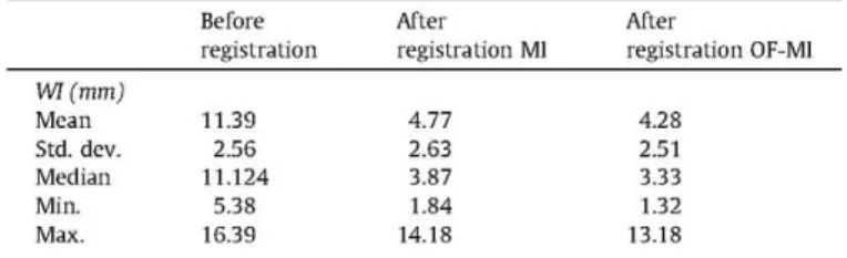
Warping index (WI) results for the 63-subject dataset are shown in Table 2. First, we can observe an important improvement for the nonrigid registrations compared with the initial values for both MI and OF-MI criteria.
Based on the Kolmogorov-Smirnov test, we cannot assume nor-mality for the pair-wise difference of the WI values distribution. Therefore, we apply the Wilcoxon matched-pairs signed-rank test to study the significant difference of the registration error mea-sures. A significant reduction in WI values using registrations based on OF-MI compared with those based on MI (p < 0.01) was confirmed.
Both methods are affected by noise, but OF-MI presents a more robust behavior with respect to SNR changes (Fig. 7).
3.4.2. Results with real dataset
Visual inspection of the results shows important qualitative improvements after applying nonrigid registration to the images, especially when using OF-MI. Specific structures inside the liver are registered better when using OF-MI, which facilitates accurate localization of lesions from the MRI into the CT for surgery plan-ning. Fig. 9 shows the fusion of CT and MRI before and after regis-tration with MI and OF-MI for one of the subjects. Even when most of the organ surfaces are visually well registered with both criteria; the registration with OF-MI is better in some critical areas affecting the inner liver vessels (see Figs. 8 and 10). Fig. 9 shows hepatic sur-faces registration improvements with OF-MI with respect to the initial scenario and with respect to the use of MI in the nonrigid registration. In Fig. 10 a detailed view of the fitting of a subset of the vascular branches is given. As can be seen from Fig. 10(b and e) versus (c and f) in comparison to the use of MI, the use of OF-MI results in a considerable better alignment between these vessels.
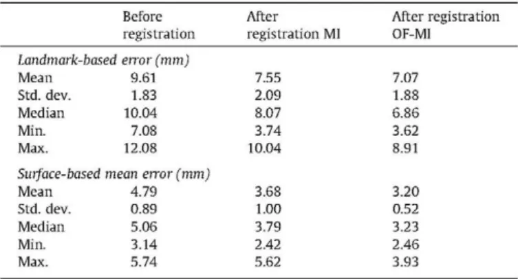
Numerical registration results are summarized in Table 3. Bet-ter results are obtained with respect to all criBet-teria with OF-MI than with MI, with maximum improvements of 1.27 mm for the land-mark-based mean geometric errors (subject 5) and 2.39 mm for the surface-based mean errors (subject 5).
In order to see the effect on the registration results of segmen-tation accuracy when calculating the masks for the OFMI criteria, we have performed independent registration experiments on all seven data sets checking the effect of inter-subject segmentation errors within the real context. Therefore, when using masks for the OFMI criteria segmented by an additional non-expert user, we observed a landmark-based mean error of 7.42 mm, on average for all seven data sets, also smaller than that obtained using MI (7.55 mm). As the inter-subject variability of the segmentation accuracy using the semi-automatic method described in Fernan-dez-de-Manuel et al. (2009) and Jiménez-Carretero et al. (2011) is 1.35 mm on average, we can conclude that for segmentation er-rors due to inter-subject variability and smaller than 1.35 mm, the OFMI registration method gives consistent and robust results, bet-ter to those obtained with standard MI.
Additionally, Fig. 11 shows the effect on the registration results of artificial segmentation errors in the mask for the OFMI criteria. We have applied morphological dilation and erosion on the initial segmentation of one real subject using a sphere of different radius from 1 to 4 voxels as structuring element, and we have studied the effect on the final results. Assuming that a segmentation obtained by applying the semi-automatic method described in Fernandez-de-Manuel et al. (2009) and Jiménez-Carretero et al. (2011) is the proper one, we express the errors using the surface-based mean er-rors (ME) (mm) (24) between the original segmentation and the di-lated/eroded versions. We can see that with mean errors in the masks used in the OFMI criteria smaller than 3 mm, the final reg-istration results are always better than those of the standard MI. Additionally, we have found that the OF-MI metric is less sensitive to errors resulting from erosions than from dilations of the initial mask.
Table 2
Warping Index results with simulated dataset (y = 0.001 and hk = 18 mm Vfc).
Wl(mm)
Mean Std. dev. Median Min. Max.
Before registration
11.39 2.56 11.124
5.38 16.39
After registration MI
4.77 2.63 3.87 1.84 14.18
After
registration OF-MI
4.28 2.51 3.33 1.32 13.18
12 10
&
i
5
5 4 2
\
Before registration
Registration with OF-MI
Registration with Ml
A
XSNR
Fig. 7. Warping Index results after applying registrations with MI and OF-MI to a
simulated dataset with SNR ranging from 1 to 8 (registration parameters: y = 0.001 and hk = 18 mm Vfc.
4. Discussion
We have described a nonrigid registration framework that takes advantage of available expert liver segmentations in clinical proto-cols to ensure good alignment of the inner structures of the liver. The validation of the proposed registration method shows that the OF-MI metric improves the results obtained with the classical formulation of MI. Maximum improvements were as high as 1.27 mm for the mean landmark-based geometric errors (subject 5) reaching up to 6 mm for some particular landmarks. The com-parison of Fig. 10(b and e) versus Fig. 10(c and f) illustrates the contribution of OF-MI, that provides a substantially better align-ment of vessels. The significance of these improvealign-ments should be considered in the context of the target application: liver surgery planning. In this scenario, the definition of the surgical approach may depend on the accuracy of the registration in certain areas in-side the liver and therefore any improvement in the registration results affecting those areas will facilitate these decisions. Even when most of the organ surface is visually well registered with MI or OF-MI, and the numerical difference between the methods is not large; the registration with OF-MI becomes significant in some critical areas affecting the liver parenchyma and inner vascu-lar structure.
Fig. 8. (a) Contrast-enhanced portal-phase CT subject 1 (fixed image); (b) Gd-EOB-DTPA-enhanced MRI before registration (first column) and fusion with CT (overlay fusion
second column and multiplication fusion third column); (c) GdEOBDTPAenhanced MRI after registration with MI (first column) and fusion with CT (overlay fusion -second column - and multiplication fusion - third column); (d) Gd-EOB-DTPA-enhanced MRI after registration with OF-MI (first column) and fusion with CT (overlay fusion - second column and multiplication fusion - third column). Arrows number 1 show improvements in body contour fitting after registration. Arrows number 2 show that the fitting of liver boundaries is performed better when using OF-MI. Arrows number 3 and 4 show that the fitting of several liver vessels is performed better when using OF-MI.
Fig. 9. CT liver segmentation in red (a, e, f, g, h) and MRI liver segmentation in green: before registration (b and f), after registration with MI (c and g) and after registration
with OF-MI (d and h) in subject 7. Comparison of liver segmentation before and after registration (f, g, h). Note the better alignment of the liver boundaries when using OF-MI (h) as compared to the classical formulation of MI (g) (see white arrows). (For interpretation of the references to color in this figure legend, the reader is referred to the web version of this article.)
ablation of focal liver tumors assisted by the fusion of preoperative CT/MRI and intraoperative ultrasound (Jung et al., 2012), the target selection and calculation of the lesions ablative volume from the pre-procedure images must accurately be blended with the real-time ultrasound in order to place the thermal electrode on the se-lected targets. Millimetric displacement and tracking inaccuracy
Fig. 10. Contrast-enhanced portal-phase CT of subject 7 (sagittal and transversal plains), Gd-EOB-DTPA-enhanced MRI (coronal plain), CT liver segmentation (red structure),
and vessel segmentations from the CT (orange), as well as from the MRI (purple, green, pink), before registration (a and d), after registration optimizing classical MI (b and e), and after registration by optimizing OF-MI (c and f). Upper row: complete vascular branchs, lower row: detail of one particular vascular branch. Note the better alignment of the vascular branches when optimizing OF-MI (c and f) as compared with MI (b and e) (see white arrows). (For interpretation of the references to color in this figure legend, the reader is referred to the web version of this article.)
Table 3
Results with real dataset. (y = 0.001 and hk = 18 mm Vk).
Before registration
Landmark-based error (mm)
Mean Std. dev. Median Min. Max.
Surface-based
Mean Std. dev. Median Min. Max.
9.61 1.83 10.04 7.08 12.08
mean error (mm)
4.79 0.89 5.06 3.14 5.74
After registration MI
7.55 2.09 8.07 3.74 10.04
3.68 1.00 3.79 2.42 5.62
After registration OF-MI
7.07 1.88 6.86 3.62 8.91
3.20 0.52 3.23 2.46 3.93
multiple lesions (Gold et al., 2008), or undergoing repeated liver resections. In these patients, major hepatic surgery with ample oncological margins larger than 10 mm cannot be performed and the safest approach is a parenchymal-sparing liver resection that requires a precise study of the oncological margin of each lesion and lesion-to-vascular topography. In (Casciola et al., 2011), the authors point out the necessity of using both a contrast enhanced CT and a liver gadoxetic acid-enhanced MRI for robot-assisted parenchymal-sparing liver surgery in order to evaluate, during the preoperative work-up, the technical feasibility of a liver resec-tion and the viability of a minimally invasive approach. Different cases where the tumor was in contact with a main portal branch or with a hepatic vein were studied in Casciola et al. (2011), describing the different surgery strategies depending on the level of contact between the lesion and the vessel. The proper
identifica-14
12
1 0
8
6
-4
2
^ — ^ — Before Registration
Registration with M l
— — Registration with OF-MI
fc •>> T. V ' v "V •% »•
f> & <f> <& & & <£• ^ cp
<y <y <y7 \$>> >$' v§>- <v? <y- <y Radius of the structuring element. In brackets, associated ME (mm). Dilations-:- | ->Erosions
Fig. 11. Landmark-based mean error before and after registration using MI and
OF-MI for a real subject. Effect in the OF-OF-MI results of realistic amounts of segmentation errors when calculating the masks for the OFMI criteria (ME with respect to the proper segmentation in brackets).
tion and fitting of the vessels among imaging modalities facilitates the accurate delimitation of the adjacency of tumors and veins and the proper detection of vascular invasion determining whether the patient is unresectable or eligible for surgery.
modify the results, making the applicability of the proposed meth-od in clinical environment more realistic and reliable.
Finally, this work can also be useful in other therapeutic appli-cations. Scenarios such as radiotherapy treatment planning using multimodal imaging (Kessler et al., 1991; Tan et al., 2010; Thor-warth et al., 2013) could also benefit from the advantages of the inclusion of additional regional information using OF-MI. Consider-ing that segmentations are normally available for the dosimetry planning (Acosta et al., 2010; Bazalova and Graves, 2011; Lu et al., 2011), their use for the registration could imply benefits without affecting the clinical protocols.
5. Conclusions
In this work we have proposed a multimodal nonrigid registra-tion framework to characterize liver lesions using simultaneously contrast-enhanced portal-phase CT and delayed Tl-weighted Gd-EOB-DTPA-enhanced MRI using OF-MI, and we have compared it with the classical formulation of MI. We took advantage of actual liver segmentation available in standard clinical protocols and we used them in the criterion. This solution allows the statistical dependence between the two modalities to differ inside and out-side the organ of interest.
We have shown important improvements in all considered val-idation criteria after applying nonrigid registration to simulated and real multimodal liver studies, in comparison with unaligned images. The improvement was in general better when using OF-MI than with OF-MI. We tested and confirmed the statistical signifi-cance of the improvement in the simulated data (p < 0.01). Specific structures inside the liver are registered better when using OF-MI, facilitating more accurate localization of lesions from the MRI into the CT for surgery planning. In addition, OF-MI presents more ro-bust behavior with respect to SNR changes and more stable results with smaller dispersion than MI.
Acknowledgments
This work was supported in part by Spain's Ministry of Science and Innovation through the Project TEC2010-21619-C04-03/01, CDTI-CENIT (AMIT), INNPACTO (PRECISION & XIORT) IPT-300000-2010-3, Instituto de Salud Carlos III (PI09/91058, PI09/91065, PI09/90568 and PI09/90987), Comunidad de Madrid (ARTEMIS S2009/DPI-1802) and the European Regional Develop-ment Funds (FEDER). Jan Kybic was supported by Czech Science Foundation project P202/11/0111.
Appendix A
A?. Derivatives of OF-MI
The partial derivatives of the £0F-MI with respect to Cj (17) are:
<9(EoF-Ml)_v-<9(E0F-Ml) df[(X) Cfo(i)
do
where
<9(£oF-Ml)
= vd ( £ o F - M i ) ¿>/tc(x) ¿ - <9/„(i) ' dxk
x=g(i) do IX
9fw(i)
y y y dp(t,r,l;c)
losp(t,r,l; c)
Vt Vr VI 9fw(i) PÁt;c)
(A.1)
(A.2)
Differentiating the joint probability distribution p(t,r,/;c) with respect to the warped image at i in (A.2) can be expressed as:
g p ( t , r , i ; c ) 1 nbinsT-\ dpm^)
171 7 1 7 ^ W • Pm2(r-p(i,c)) — — —
\'c\ Jw max Jwmin UL> | = Í - T ( Í ¡ C )
(A.3)
The explicit expression for the derivative of the B-spline func-tion is:
9 I ^ = /Jmi_, (f + 1 ¡2) - ft^ (f - 1 ¡2) (A.4) The partial derivatives of ff (4) are calculated as a tensor product (Kybic, 2001):
X^k
(A.5)
Finally, the derivative of the deformation function is calculated from (5):
dgk(i)
do. •),k = A , ( i / h - j ) (A.6)
where k is the dimension of the JV-dimensional Cj deformation parameters.
References
Acosta, O., Dowling, J., Cazoulat, G., Simon, A., Salvado, O., De Crevoisier, R., Haigron, P., 2010. Atlas based segmentation and mapping of organs at risk from planning ct for the development of voxel-wise predictive models of toxicity in prostate radiotherapy, prostate cancer imaging. In: Computer-Aided Diagnosis, Prognosis, and Intervention. Springer, pp. 4 2 - 5 1 .
Archip, N., Tatli, S., Morrison, P., Jolesz, R, Warfield, S.K., Silverman, S., 2007. Non-rigid registration of pre-procedural MR images with intra-procedural unenhanced CT images for improved targeting of tumors during liver radiofrequency ablations. Medical Image Computing and Computer-Assisted Intervention-MICCAI, pp. 969-977.
Bazalova, M., Graves, E.E., 2011. The importance of tissue segmentation for dose calculations for kilovoltage radiation therapy. Medical Physics 38, 3039. Blackall, J.M., Penney, G.P., King, A.P., Hawkes, D.J., 2005. Alignment of sparse
freehand 3-D ultrasound with preoperative images of the liver using models of respiratory motion and deformation. IEEE Transactions on Medical Imaging 24, 1405-1416.
Bluemke, D.A., Paulson, E.K., Choti, M.A, DeSena, S., Clavien, P.A., 2000. Detection of hepatic lesions in candidates for surgery: comparison of ferumoxides-enhanced MR imaging and dual-phase helical CT. American Journal of Roentgenology 175, 1653-1658.
Bushberg, J.T., Bushberg, J.T., SeibertJr.JA, E.M.L., Boone, J.M., 2002. The Essential Physics of Medical Imaging. Williams & Wilkins.
Carrillo, A., Duerk, J.L., Lewin, J.S., Wilson, D.L, 2000. Semiautomatic 3-D image registration as applied to interventional MRI liver cancer treatment. IEEE Transactions on Medical Imaging 19,175-185.
Casciola, L., Patriti, A., Ceccarelli, G., Bartoli, A., Ceribelli, C, Spaziani, A., 2011. Robot-assisted parenchymal-sparing liver surgery including lesions located in the posterosuperior segments. Surgical Endoscopy 25, 3815-3824.
Clifford, M.A., Banovac, F., Levy, E., Cleary, K., 2002. Assessment of hepatic motion secondary to respiration for computer assisted interventions. Computer Aided Surgery 7, 291-299.
Couinaud, C, 1999. Liver anatomy: portal (and suprahepatic) or biliary segmentation. Digestive Surgery 16, 459-467.
Crum, W.R, Hartkens, T., Hill, D., 2004. Non-rigid image registration: theory and practice. British Journal of Radiology 77, S140.
Dokmak, S., Agostini, J.Jacquin, A., Cauchy, F., Farges, O., Belghiti.J., 2012. High risk of biliary fistula after isolated segment VIII liver resection. World Journal of Surgery 36, 2692-2698.
Donati, O.F., Hany, T.F., Reiner, C.S., von Schulthess, G.K., Marincek, B., Seifert, B., Weishaupt, D., 2010. Value of retrospective fusion of PET and MR images in detection of hepatic metastases: comparison with 18F-FDG PET/CT and Gd-EOB-DTPA-enhanced MRI. Journal of Nuclear Medicine 51, 692-699.
Fernandez-de-Manuel, L., Rubio, J.L, Ledesma-Carbayo, M.J., Pascau, J., Tellado, J.M., Ramón, E., Deseo, M., Santos, A., 2009.3D Liver Segmentation in Preoperative CT Images using a Level-Sets Active Surface Method, Annual International Conference of the IEEE Engineering in Medicine and Biology Society. EMBS, Minneapolis, pp. 3625-3628.
Fernandez-de-Manuel, L, Ledesma-Carbayo, M.J., Jiménez-Carretero, D., Pascau, J., Rubio-Guivernau, J.L., Tellado, J.M., Ramon, E., Deseo, M., Santos, A., 2011. Liver Segmentation and Volume Estimation from Preoperative CT Images in Hepatic Surgical Planning: Application of a Semiautomatic Method Based on 3D Level Sets. Theory and Applications of CT Images. InTech, pp. 79-94.
Gudbjartsson, H., Patz, S., 1995. The Rician distribution of noisy MRI data. Magnetic Resonance in Medicine 34, 910-914.
Hammerstingl, R., Huppertz, A., Breuer, J., Balzer, T., Blakeborough, A, Carter, R., Fuste, LC, Heinz-Peer, G., Judmaier, W , Laniado, M., 2008. Diagnostic efficacy of gadoxetic acid (Primovist)-enhanced MRI and spiral CT for a therapeutic strategy: comparison with intraoperative and histopathologic findings in focal liver lesions. European Radiology 18, 457-467.
Handels, H., Ehrhardt, J., 2009. Medical image computing for computer-supported diagnostics and therapy advances and perspectives. Methods of Information in Medicine 48,11-17.
Heimann, T., van Ginneken, B., Styner, M.A., Arzhaeva, Y., Aurich, V., Bauer, C, Beck, A., Becker, C, Beichel, R., Bekes, G., Bello, F., Binnig, G., Bischof, H., Bornik, A, Cashman, P.M.M., Chi, Y., Cordova, A., Dawant, B.M., Fidrich, M., Furst, J.D., Furukawa, D., Grenacher, L, Hornegger, J., Kainmuller, D., Kitney, R.I., Kobatake, H., Lamecker, H., Lange, T., Lee, J., Lennon, B., Li, R, Li, S., Meinzer, H.P., Nemeth, G., Raicu, D.S., Rau, A.M., van Rikxoort, E.M., Rousson, M., Rusko, L., Saddi, K.A., Schmidt, G., Seghers, D., Shimizu, A., Slagmolen, P., Sorantin, E., Soza, G., Susomboon, R., Waite, J.M., Wimmer, A., Wolf, I., 2009. Comparison and evaluation of methods for liver segmentation from CT datasets. IEEE Transactions on Medical Imaging 28,1251-1265.
Heinrich, M., Jenkinson, M., Brady, M., Schnabel, J., 2012a. Textural mutual information based on cluster trees for multimodal deformable registration. IEEE International Symposium on Biomedical Imaging, ISBI. IEEE, Barcelona, pp. 1471-1474.
Heinrich, M.P., Jenkinson, M., Bhushan, M., Matin, T., Gleeson, F.V., Brady, S.M., Schnabel, J.A., 2012b. MIND: modality independent neighbourhood descriptor for multi-modal deformable registration. Medical Image Analysis 16, 1423-1435.
Hermosillo, G., Chefd'Hotel, C, Faugeras, O.D., 2002. Variational methods for multimodal image matching. International Journal of Computer Vision 50,329-343.
Hill, D.L.G., Batchelor, P.G., Holden, M., Hawkes, D.J., 2001. Medical image registration. Physics in Medicine and Biology 46, R1-R45.
Jiménez-Carretero, D., Fernandez-de-Manuel, L, Pascau, J., Tellado, J.M., Ramon, E., Deseo, M., Santos, A., Ledesma-Carbayo, M.J., 2011. Optimal multiresolution 3D level-set method for liver segmentation incorporating local curvature constraints. In: Annual International Conference of the IEEE Engineering in Medicine and Biology Society, EMBS, Boston, Massachusetts, USA, pp. 3419-3422.
Jung, E.M., Friedrich, C, Hoffstetter, P., Dendl, LM., Klebl, F., Agha, A., Wiggermann, P., Stroszcynski, C, Schreyer, A.G., 2012. Volume navigation with contrast enhanced ultrasound and image fusion for percutaneous interventions: first results. PLoS One 7, e33956.
Kessler, M.L, Pitluck, S., Petti, P., Castro, J.R., 1991. Integration of multimodality imaging data for radiotherapy treatment planning. International Journal of Radiation Oncology* Biology* Physics 21, 1653-1667.
Kim, B., Boes, J.L, Frey, K.A., Meyer, C.R., 1997. Mutual information for automated unwarping of rat brain autoradiographs. Neurolmage 5, 31-40.
Kong, G., Jackson, C, Koh, D., Lewington, V., Sharma, B., Brown, G., Cunningham, D., Cook, G.J.R., 2008. The use of 18F-FDG PET/CT in colorectal liver metastases-comparison with CT and liver MRI. European Journal of Nuclear Medicine and Molecular Imaging 35,1323-1329.
Krücker, J., Xu, S., Venkatesan, A., Locklin, J.K., Amalou, H., Glossop, N., Wood, B.J., 2011. Clinical utility of real-time fusion guidance for biopsy and ablation. Journal of Vascular and Interventional Radiology 22, 515-524.
Kwan, R.K.S., Evans, A.C., Pike, G.B., 1999. MRI simulation-based evaluation of image-processing and classification methods. IEEE Transactions on Medical Imaging 18,1085-1097.
Kybic, J., 2001. Elastic Image Registration using Parametric Deformation Models. Ecole Polytechnique Fedérale de Lausanne, Lausanne.
Kybic, J., Unser, M., 2003. Fast parametric elastic image registration. IEEE Transactions on Image Processing 12,1427-1442.
Kybic, J., Vnucko, I., 2012. Approximate all nearest neighbor search for high dimensional entropy estimation for image registration. Signal Processing 92, 1302-1316.
Lange, T., Wenckebach, T., Lamecker, H., Seebass, M., Hünerbein, M., Eulenstein, S., Gebauer, B., Schlag, P., 2005a. Registration of different phases of contrast enhanced CT/MRI data for computer assisted liver surgery planning: evaluation of state of the art methods. The International Journal of Medical Robotics and Computer Assisted Surgery 1, 6-20.
Lange, T., Wenckebach, T., Lamecker, H., Seebass, M., Hunerbein, M., Eulenstein, S., Schlag, P.M., 2005b. Registration of portal and hepatic venous phase of MR/CT data for computer-assisted liver surgery planning. CARS: Computer Assisted Radiology and Surgery 1281, 768-772.
Ledesma-Carbayo, M.J., Kybic, J., Deseo, M., Santos, A., Suhling, M., Hunziker, P., Unser, M., 2005. Spatio-temporal nonrigid registration for ultrasound cardiac motion estimation. IEEE Transactions on Medical Imaging 24,1113-1126. Lee, W.C.C., Tublin, M.E., Chapman, B.E., 2005. Registration of MR and CT images of
the liver: comparison of voxel similarity and surface based registration algorithms. Computer Methods and Programs in Biomedicine 78,101-114. Loeckx, D., Slagmolen, P., Maes, F., Vandermeulen, D., Suetens, P., 2010. Nonrigid
image registration using conditional mutual information. IEEE Transactions on Medical Imaging 29,19-29.
Lu, C, Chelikani, S., Papademetris, X., Knisely, J.P., Milosevic, M.F., Chen, Z., Jaffray, DA, Staib, L.H., Duncan, J.S., 2011. An integrated approach to segmentation and
nonrigid registration for application in image-guided pelvic radiotherapy. Medical Image Analysis 15, 772-785.
Maes, F., Collignon, A., Vandermeulen, D., Marchal, G., Suetens, P., 1997. Multimodality image registration by maximization of mutual information. IEEE Transactions on Medical Imaging 16,187-198.
Maintz, J., Viergever, M.A., 1998. A survey of medical image registration. Medical Image Analysis 2 , 1 - 3 6 .
Mattes, D., Haynor, D.R, Vesselle, H., Lewellyn, T.K., Eubank, W., 2001. Nonrigid multimodality image registration. In: SPIE, pp. 1609-1620.
Mattes, D., Haynor, D.R., Vesselle, H., Lewellen, T.K., Eubank, W., 2003. PET-CT image registration in the chest using free-form deformations. IEEE Transactions on Medical Imaging 22,120-128.
Oguro, S., Tokuda, J., Elhawary, H., Haker, S., Kikinis, R., Tempany, C, Hata, N., 2009. MRI signal intensity based B-Spline nonrigid registration for pre-and intraoperative imaging during prostate brachytherapy. Journal of Magnetic Resonance Imaging 30, 1052-1058.
Oliva, M.R., Saini, S., 2004. Liver cancer imaging: role of CT, MRI, US and PET. Cancer Imaging 4, S42.
Oudkerk, M., Torres, C.G., Song, B., Konig, M., Grimm, J., Fernandez-Cuadrado, J., Op de Beeck, B., Marquardt, M., van Dijk, P., de Groot, J.C., 2002. Characterization of liver lesions with Mangafodipir Trisodium-enhanced MR imaging: multicenter study comparing MR and dual-phase spiral CT1. Radiology 223, 517. Park, S.B., Rhee, F.C., Monroe, J.L, Sohn, J.W., 2010. Spatially weighted mutual
information image registration for image guided radiation therapy. Medical Physics 37, 4590-4601.
Pluim, J., Maintz, J., Viergever, M., 2000a. Image registration by maximization of combined mutual information and gradient information. In: Medical Image Computing and Computer-Assisted Intervention - MICCAI. Springer, pp. 103-129.
Pluim, J., Maintz, J., Viergever, M., 2000b. Image registration by maximization of combined mutual information and gradient information. IEEE Transactions on Medical Imaging 19, 809.
Pluim, J.P.W., Maintz, J.B.A., Viergever, M.A., 2003. Mutual-information-based registration of medical images: a survey. IEEE Transactions on Medical Imaging 22, 986-1004.
Radtke, A, Nadalin, S., Sotiropoulos, G.C., Molmenti, E.P., Schroeder, T., Valentin-Gamazo, C, Lang, H., Bockhorn, M., Peitgen, H.O., Broelsch, CE., Malago, M., 2007. Computer-assisted operative planning in adult living donor liver transplantation: a new way to resolve the dilemma of the middle hepatic vein. World Journal of Surgery 31,175-185.
Rodríguez-Carranza, CE., Loew, M.H., 1999. Global optimization of weighted mutual information for multi-modality image registration. In: Proc. SPIE, pp. 89-96.
Rogelj, P., Kovacic, S., Gee, J.C., 2003. Point similarity measures for non-rigid registration of multi-modal data. Computer Vision and Image Understanding 92,112-140.
Rohlfing, T., Maurer Jr. C.R., O'Dell, W.G., Zhong, J., 2001. Modeling liver motion and deformation during the respiratory cycle using intensity-based free-form registration of gated MR images. In: Proc. Visualization, Display, and Image-guided Procedures, pp. 337-348.
Rueckert, D., Schnabel, J.A., 2011. Medical image registration. Biomedical Image Processing, 131-154.
Rueckert, D., Sonoda, LI., Hayes, C, Hill, D.L.G., Leach, M.O., Hawkes, D.J., 1999. Nonrigid registration using free-form deformations: application to breast MR images. IEEE Transactions on Medical Imaging 18, 712-721.
Rueckert, D., Clarkson, M., Hill, D., Hawkes, D., 2000. Non-rigid registration using higher-order mutual information. In: Proc SPIE, pp. 438-447.
Russakoff, D., Tomasi, C, Rohlfing, T., Maurer, C, 2004. Image similarity using mutual information of regions. Computer Vision-ECCV, pp. 596-607.
Schnabel, J., Rueckert, D., Quist, M., Blackall, J., Castellano-Smith, A., Hartkens, T., Penney, G., Hall, W., Liu, H., Truwit, C, 2001. A generic framework for non-rigid registration based on non-uniform multi-level free-form deformations. In: Proc. International Conference on Medical Image Computing and Computer-Assisted Intervention MICCAI. Springer, pp. 573-581.
Solbiati, L, Cova, L, Ierace, T., Marelli, P., Dellanoce, M., 1999. Liver cancer imaging: the need for accurate detection of intrahepatic disease spread. Journal of Computer Assisted Tomography 23, S29-S37.
Studholme, C, Hill, D., Hawkes, D., 1996. Incorporating connected region labelling into automatic image registration using mutual information. Workshop on Mathematical Methods in Biomedical Image Analysis, pp. 23-31.
Studholme, C, Drapaca, C, Iordanova, B., Cardenas, V., 2006. Deformation-based mapping of volume change from serial brain MRI in the presence of local tissue contrast change. IEEE Transactions on Medical Imaging 25, 626-639. Tan, J., Lim Joon, D., Fitt, G., Wada, M., Lim Joon, M., Mercuri, A., Marr, M., Chao, M.,
Khoo, V., 2010. The utility of multimodality imaging with CT and MRI in defining rectal tumour volumes for radiotherapy treatment planning: a pilot study. Journal of Medical Imaging and Radiation Oncology 54, 562-568. Thévenaz, P., Unser, M., 1996. A pyramid approach to sub-pixel image fusion based
on mutual information. In: Int. Conference on Image Processing, vol. 261. IEEE, pp. 265-268.
Thévenaz, P., Unser, M., 2000. Optimization of mutual information for multiresolution image registration. IEEE Transactions on Image Processing 9, 2083-2099.
Thorwarth, D., Müller, A.-C, Pfannenberg, C, Beyer, T., 2013. Combined PET/MR Imaging Using 68Ga-DOTATOC for Radiotherapy Treatment Planning in Meningioma Patients, Theranostics, Gallium-68, and Other Radionuclides. Springer, pp. 425-439.
Tohka, J., Zijdenbos, A., Evans, A., 2004. Fast and robust parameter estimation for statistical partial volume models in brain MRI. Neurolmage 23, 84-97. Unser, M., 1999. Splines: a perfect fit for signal and image processing. IEEE Signal
Processing Magazine 16, 22-38.
Unser, M., Aldroubi, A., Eden, M., 1993. The L2-polynomial spline pyramid. IEEE Transactions on Pattern Analysis and Machine Intelligence 15, 364-379. Van Dalen, J., Vogel, W., Huisman, H., Oyen, W., Jager, G., Karssemeijer, N., 2004.
Accuracy of rigid CT-FDG-PET image registration of the liver. Physics in Medicine and Biology 49, 5393.
Villard, P.-R, Boshier, P., Bello, R, Gould, D., 2011. Virtual Reality Simulation of Liver Biopsy with a Respiratory Component, Liver Biopsy. InTech, pp. 315-334.
Weinmann, H.J., Ebert, W., Misselwitz, B., Schmitt-Willich, H., 2003. Tissue-specific MR contrast agents. European Journal of Radiology 46, 33-44.
Wells III, W.M., Viola, P., Atsumi, H., Nakajima, S., Kikinis, R., 1996. Multi-modal volume registration by maximization of mutual information. Medical Image Analysis 1, 35-51.
Yi, Z., Soatto, S., 2011. Multimodal registration via spatial-context mutual information. In: Lecture Notes in Computer Science, IPMI. Springer, pp. 424-435.
Zhuang, X., Arridge, S., Hawkes, D.J., Ourselin, S., 2011. A nonrigid registration framework using spatially encoded mutual information and free-form deformations. IEEE Transactions on Medical Imaging 30,1819-1828.