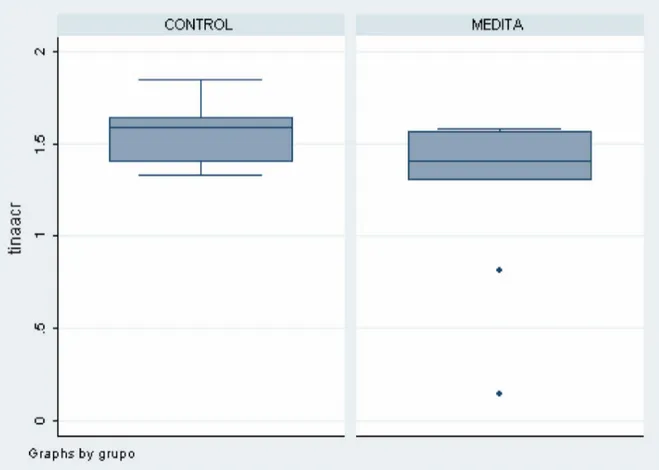
Brain Changes in Long Term Zen Meditators Using Proton Magnetic Resonance Spectroscopy and Diffusion Tensor Imaging: A Controlled Study
Texto completo
Figure


Documento similar
The expansionary monetary policy measures have had a negative impact on net interest margins both via the reduction in interest rates and –less powerfully- the flattening of the
Normal Achilles tendon on both short (A) and long (B) axis ultrasound and magnetic resonance imaging in axial STIR (C), sagittal T1 (D), and STIR (E) sequences.. A very useful
Spectrum of lesions observed by computed tomography and magnetic resonance imaging scans in young athletes that participated in the 2018 Youth Olympic Games in Buenos
2D and 3D biometric parameters were mea- sured from slice-to-volume reconstructed images, including 3D measurements of supratentorial brain tissue, lateral ventricles,
3p orbitals, as shown in Figure 8 for 100 K and Figures S1 and S2 for 300 K structures. As mentioned above, the EXSCI approach has been employed to reduce the
The intra-reader reliability of bone related and soft-tissue results, the patterns of BML associated with tendon enthesopathy (ICC total = 0.79, range across the locations 0.44 to 1)
We used structural magnetic resonance imaging (MRI) to esti- mate global brain volumes and performed a voxel-based morphometry and lesion symptom mapping analysis in order to
It has previously been reported that low cerebral NIRS values in preterm infants during the first days of life are asso- ciated with higher grades of intraventricular hemorrhage and