Inflamatory infiltrate in skeletal muscle injected with Bothrops asper venom
6
0
0
Texto completo
(2) 210. REVISTA D E BIOLOGIA TROPICAL. expressed in percentage , taking as 1 00% the CK content of the contralateral gastrocnemius. In samples obtained 6 and 48 hr after enven omation, the pellet was resuspended in 5 .0 mI of 0.5 M NaOH and allowed to stand overnight at room temperature . The solution was then centrifuged and the protein content ("non collagen protein") of the supernatant was determined according to Lowry et al. ( 1 95 1 ). Non-collagen protein of envenomated gas trocnemius was expressed in percentage , taking as 1 00% the values of the contralateral muscle . Electrophoresis of muscle proteins: The pellet resultant from centrifugation of muscle homogenate was resuspended in 250 J..Ll of disti lled water and an aliquot was analyzed by slab SDS-polyacrylamide gel electrophoresis (SDS PAGE), using 1 2% acrylamide (Laemmli, 1 970). SDS-PAGE was performed in samples obtained 6 hr and 48 hr after envenomation . Studies on the inflammatory infiltrate: The technique of Maskrey et al. ( 1 977) was used with sorne modifications. Groups of four mice (20-22 g) were injected i ro . in the right gastrocnemius with 1 00 J..Lg of venom . At different time intervals (6 hr, 24 hr, 48 hr and 72 hr) mice were killed and envenomated muscle Was removed and chopped with scissors in 2 .0 mI of phosphate-buffered saline . The tissue suspension was then incubated , with continuous agitation , for 30 min at 37 C. The suspension was flltered through bolting silk and inflammatory cells were counted in a hemocy tometer. Just before counting, the cell suspension was treated with a solution (3% acetic acid) that lyses erythrocytes whlle leaving leucocytes and macrophages intact ; this was done in order to count only inflammatory cells and not erythrocytes present in the tissue due to hemorrhage . Then , the suspension Was centrifuged in a cytocentrifuge and smears were prepared and stained with Wright in order to identify the inflammatory cells. Cells were classified as macrophages, polymorphonuclear leucocytes and lymphocytes. Histology and uItrastructure: Groups of four mice (20-22 g) were injected as described above . At different time intervals, animals were killed and pieces of envenomated muscle were obtained . Samples for histology were processed routinely . Other samples were processed for. electron microscopy and embedded in Spurr resin (Arroyo and Gutiérrez, 1 98 1 ). Thin sec tions were stained with uranyl aceta te and lead citrate and examined in a Hitachi HU- 1 2A electron microscope of the Electron Microscopy Unít , Universidad de Costa Rica . Control mke were injected with 0 . 1 mI of phosphate-buffer saline solution (pH 7 .2). RESULTS Quantitation of inflammatory inflltrate: Control gastrocnemius muscle contained 85 ,000 ± 7 ,000 leucocytes. There was a signifi cant increase (P< 0.005 ) in muscle obtained 6 hr after envenomation ( 1 67 ,500 ± 1 2 ,500). By 24 hr , a further increase was observed, reaching values of 8 1 0,000 ± 1 25 ,000 inflam matory cells per gastrocnemius. The most prominent elevation in inflammatory inflltrate occured at 48 hr (2 ,400,000 ± 760,000); by 72 hr the inflltrate increased only slightly when compared to values at 48 hr (Fig. 1 ). Cell types present in infiltrate : Examination of inflammatory cells by light microscopy allowed us to classify them in three groups: Macrophages, Iymphocytes and polymorphonu clear leucocytes. Among the latter, the vast majority of cells were neutrophlls. In control muscle, 48 ± 3% of the cells were macrophages, 44 ± 5% were polymorphonuclear leucocytes and 9 ± 1 % were lymphocytes. Infiltrate from samples obtained 6 and 24 hr after enven omation presented a high proportion of polymorphonuc1ear leucocytes (Table 1 ). By 48 hr there was a change in this pattern ; the relative number of macrophages increased and that of polymorphonuc1ear leucocytes decreased . This trend remained in samples obtained at 72 hr, when macrophages were the predominant cell type in the inflltrate. The percentage of lymphocytes in inflammatory inflltrate did not change , their values ranging from 2 ± 3% to 7 ± 1 .3% at all time periods (Table 1 ). Determi ation of creatine kinase in muscle: CK content of injected gastrocnemius decreased to values f 35 ± 3% 3 hr after envenomation . Thereafter here was no significant change in CK content (32 ± 6% at 6 hr; 3 1 ± 6% 24 h r ; a n d 3 0 ± 5 a t 48 hr)..
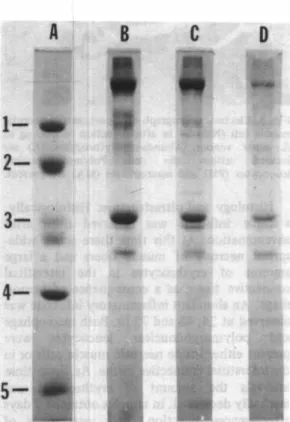
(3) GUTIERREZ et al.: Muscle injected with Bothrops venom. 211. TABLE 1 Distribution ofinflammatory cell types in muscle injected with Bothrops asper venom ... Envenomated Envenomated muscle Envenomated muscle Envenomated muscle muscle (24 hr) (48 hr) (72 hr) (6 hr). Cell types. Control muscle. Macrophage. 48 1 3%. Polymorphonuclear 45 ± 2% Leucocyte Lymphocyte. 7 ± 1%. 2 3 1 3%. 14 1 1%. Ú 1 8%. 5 3 1 2%. 72 ± 5 %. 84 1 2%. 60 1 9%. 40 1 3%. 7 1 1%. 7 1 1%. 5. ±. 1%. 2. ¡. ±. 0.3%. Results are expressed in relative terms (percenta¡;e; mean inflammatory cells present in the sample.. *. ;. ,. ... �. �. i U>O !. ... -1/. .. . j. .. . . ... . . .... . .. ±. SEM : n. A. -=. 4), taking as 1 00% the total number of. B. e. D. 1' -. _ _ __. .. TlMI (",1. Fig.. l . Time-course of inflammatory infiltrate in mousc gastrocnemius injected with 1 00 ¡.¡g of Bothrops asper venom. Each value rcpresents the mean ± SEM (n =4).. Cha nge in myofibrillar proteins: Degradation of myofibrillar proteins was almost negligible in musc1e samples obtained 6 hr after venom injection. At this time, non-collagen proteins had decreased to 95 ± 2 % when compared to control gastrocnemius. Electrophoretic analysis of musc1e proteins indicated that none of the major myofibrillar proteins had been extensively degraded (Fig. 2). There was evidence of protein degradation in musc1e obtained 48 hr after envenomation. In these samples , non-collagen protein decreased to a value of 79 ± 6%when compared to control gastrocnemius. Moreover, there was a decrease in the intensity of several bands in SDS-PAGE of musc1e proteins ( Fig. 2 ). 8 0th of these observations indicate that a process of. Fig. 2. SDS-PAGE of myofibrillar proteins obtained from mouse gastrocnemius. Lane A : Molecular weight markers; ( 1 ) phosphorylase b (94K); (2) albumin (67 K ) ; (3) ovalbumin '(43 K); (4) carbonic anhydrase ( 30 K); and P) Trypsin inhibitor (20 K). Lane B : Proteins of cortrol muscle. Lane C : Proteins of muscle obtained 6 after injection of 1 00 ¡.¡g of B. asper venom. Lane D : Proteins of muscle obtained 48 hr after venom i ection.. myofibrillar protein degradation was taking place..
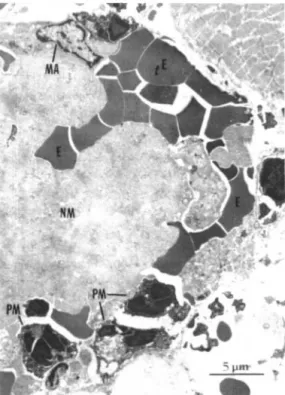
(4) 212. REVISTA D E BIOLOGIA TROPICAL. macrophages or neutrophils. Sorne of these cells were performing phagocytosis of material within necrotic muscle cells (Fig. 3 ). Myo fibrillar material, as well as mitochondria and vesicles, were located inside phagocytic vacuoles in macrophages and neutrophils. DISCUSSION. •. Fig. 3. EIectron micrograph of a portion of a nccrotic. muscIe cell (NM) 24 hr after injection of 1 00 ¡lg of asper venom. Abundant erythrocytes (E) are Iocated within the cell. Polymorphonuclear leucocytes (PM) and macrophages (MA) are presento. B.. Histology and ultrastructure: Histologically , a scarce inftltrate was observed 6 hr after envenomation. At this time there was a wide spread necrosis of muscle fibers and a large amount of erythrocytes in the interstitial connective tissue as a consequence of hemor rhage. An abundant inflammatory inftltrate was observed at 24 , 48 and 72 hr. 80th macrophage and polymorphonuclear leucocytes were present either inside necrotic muscle cells or in the interstitial connective tissue. At these time intervals the amount of erythrocytes was markedly decreased . In samples obtained 7 days after venom injection there were areas of muscle regeneration as well as areas of granula tion tissue ; however, there were sorne portions in which necrotic muscle cells had not been cleared out . In these areas the infiltrate was scarce and necrotic cells contained pale-stained myofibrillar material . Muscle obtained from control mice had a normal histological pattern with no evidence ofmyonecrosis or hemorrhage , and without an increase in the cellular infiltrate. U1trastructurally , most inflammatory celis were. Our results indicate that there is an abundant inflammatory infiltrate in skeletal muscle injected with B. asper venom. Inflammatory cells arrive at affected muscle several hr after myonecrosis develops. It has been described that B. asper venom induces drastic myone crosis rapidly after envenomation (Gutiérrez el al. , 1 980 a ; Gutiérrez el al. , 1 984). lnflamma tory cells begin to increase in number in necrotic muscle six hr after injection , reaching their peak by 48 hr. As in other cases of inflam matory responses, polymorphnuclear leuco cytes were more abundant in the early time intervals, whereas macrophages peaked at later time periods (Movat, 1 979). This pattern of inflammatory infiltrate composition is partially different from the ones observed in other examples of myonecrosis. For instance , in muscle damage induced by the local anesthetic bupivacaine and by plasmocid, the predominant inflammatory cells are macrophages (Nonaka el al. , 1 98 3 , 1 984). In regard to snake venoms, an abundant phagocy tic cell infiltrate after myonecrosis induced by the venoms of Micrurus nigrocinctus (Gutiérrez el al. , 1 980 b) and Oxyranus sculellatus (Harris and Maltin , 1 982), as well as by the toxins notexin and taipoxin (pluskal el al. , 1 97 8 ; Harris and Maltin, 1 982) has been described . In the case of myonecrosis by notexin, Harris and Johnson ( I 978) observed that between 3 and 6 hr after injection, necrotic fibers had been invaded by polymorphonuclear leucocytes, although Pluskal et al. ( I 978) suggested the possibility that different cell types might migra te to necrotic tissue at different times. One common finding in all of these studies is that inflammato� infiltrate starts to accumulate several hr a�ter the insult, despite the fact that myonecrosis is evident soon after injection of the myotox,c agents. Sorne ar�as of necrotic musc1e were almost devoid of ipflammatory cells even one week after envenpmation. In these areas, necrotic material ha4 not been removed. Since B. asper.
(5) GUTIERREZ er al. :. Muscle injected with Bothrops venom. venom drastically affects the microvasculature , it may be that damage to blood vessels is partially responsible for this lack of infiltrate in sorne areas. In myonecrosis induced by toxins that affect muscle cells without inducing hemorrhage , necrotic tissue is cleared by phagocytosis in the first 2 to 3 days (Harris et al. , 1 975 ; Harris and Maltin, 1 982). Our observations may have implications on the issue of muscle regeneration, since clearing of necrotic tissue by phagocytosis is a requirement for regeneration to proceed . Ishiura et al. ( I 984) have addressed the problem of myofibrillar degradation by muscle proteases in plasmocid-induced myonecrosis. They proposed a "two-step" mechanism in myofibrillar degradation. During the first 1 2 hr there is a limited hydrolysis of specific components, especially alpha-actinin, although the ultrastructural organization of myo filaments is severely affected. This early phase is responsible for onIy a minor myofibrillar degradation. After 1 2 hr, a macrophage-rich infiltrate accumulates in necrotic muscle , and Iysosomal cathepsins derived from these inflam matory cells are responsible for a drastic degra dation of myofibrillar proteins. Our results with B. asper venom agree with the "two-step" model , since electrophoretic studies showed that there was little, if any , myofibrillar degradation before 6 hr, a1though myofilaments were structurally affected at this period (Gutié rrez et al 1 984). One the other hand, a drop in non-collagen proteins was observed by 48 hr, together with a decrease in the intensity of most myofibrillar proteins in electrophoresis . This protein degradation corre lates with the presence of an abundant inflarnmatory infiltrate after 24 hr. Thus, the initial disorgani zation and clumping of myofilaments is not related to a generalized protein degradation, but probably to a limited alteration of structurally relevant components which regulate the assembly of myobrils. Therefore, their disturbance results in a loss of myofibrillar organization, without widespread protein hydrolysis. Later, the arrival of neutrophils and macrophages results in a more significant protein degradation . On the basis of the abundant inflltrate observed, it is interesting to study the possibil ity that enzymes released from leucocytes or macrophages may cause further damage to muscle cells. lt has been demonstrated that . •. 213. lysosomal enzymes of polymorphonuclear leucocytes, such as acid proteinases, colla genases and elastases, are responsible for tissue damage in inflammatory responses (Movat , 1 979). However, in our case, there was not a decrease in CK contents of muscle after 6 hr. This implies that muscle necrosis was a rapid phenomenon, after which no further muscle cell death occurred . Thus, inflarnmatory cells played a role in degradation and clearing of necrotic cells, but not in the development of further muscle cell damage . RESUMEN Se estudió los cambios y la composición del infiltrado inflamatorio que se desarrolla en el músculo gastronemio de ratones a consecuencia de inoculaciones de veneno de serpiente "tercio pelo" (Bothrops asper). El veneno produjo una severa y rápida mionecrosis, de acuerdo al aná lisis histológico y a la drástica disminución de los niveles musculares de la enzima creatina qui nasa (CK). Se observó un escaso infiltrado infla matorio a las 6 hr , pero hubo un aumento evi dente a las 24 , 48 , y 72 hr. A las 6 y 24 hr el in flltrado presentó un predominio de leucocitos polimorfonucleares neutrófilos, en tanto que a las 48 y 72 hr se observó un aumento en el por centaje de macrófagos. Histológicamente , las cé lulas inflamatorias se observaron en el interior de las fibras musculares necróticas , as í como en el espacio intersticial ; sin embargo , algunas áreas necróticas no contenían células inflamato rias. En un intento por correlacionar la presen cia de células inflamatorias con la degradación de las prote ínas miofibrilares, se observó muy poca degradación proteica a las 6 hr. Por otra parte, a las 48 hr se dio una disminución de las prote ínas "no colágenas" del músculo, así co mo una disminución en algunos componentes miofibrilares, de acuerdo al análisis electroforéti co. Las enzimas proteolíticas presentes en célu las inflamatorias pueden jugar un papel impor tante en la degradación de las prote ínas miofi brilares luego de mionecrosis inducida por el veneno de B. aspero ACKNOWLEDGEMENTS We thCUlk Gustavo Rojas , Oiga Arroyo , Bru no Lomolite, Alfredo Vargas and Javier Núñez for their valuable asistance, as well as Hilda.
(6) REVISTA D E BIOLOGIA TROPICAL. 214. Herrera and Rocío Monge . This work was supported by Vicerrectoría de Investigación, Universidad de Costa Rica, project No. 74 1 -8469 . Fernando Chaves received partial support from the Consejo Nacional de Investigaciones Cient íficas y Tecnológicas (CONICIT) of Costa Rica. REFERENCES Arroyo, O. & J.M. Gutiérrrez. 1 98 1 . Estudio ultraes tructural de la mionecrosis inducida en ratón por el veneno de terciopelo (Bothrops asper) de Costa Rica. Toxicon 1 9 : 17 3-782. Bolaños, R. 1 982. Las serpientes venenosas de Centro américa y el problema del ofidismo. Primera parte. Aspectos zoológicos, epidemiológicos y biomédi cos. Revista Costarricense de Ciencias Médicas 3 : 1 65-1 84. Gutiérrez, J .M. & F. Chaves. 1 980. Efectos proteolí ticos, hemorrágico y mionecrótico de los venenos de serpientes costarricenses de los géneros Bothrops, Crotalus y Lachesis. Toxicon 1 8 : 3 1 5· 321. Gutiérrez, J.M. , O . Arroyo & R . Bolaños. 1 980a. Mio necrosis, hemorragia y edema inducidos por el ve neno de Bothrops asper en ratón blanco. Toxicon 1 8 : 603-6 1 0. Gutiérrez, J .M., F. Chaves, E. Rojas & R. Bolaños R. 1 9 8Gb. Efectos locales inducidos por el veneno de la serpiente coral Micrurus nigrocinctus en ratón blanco. Toxicon 1 8 : 63 3-6 39.. Harris, J.B., M.A. Johnson & E. Karlsson. 1 975. Patho logical responses of fat skeletal muscles to a single subcutaneous injection of a toxin isolated from the venom of the Australian tiger snake, Notechis scu tatus scutatus. Clin. Exp. Pharmacol. Physiol. 2: 3 83-404. Homma, M. & A.T. Tu. 1 97 1 . Morphology of local tissue damage in experimental snake envenomation. Br. J. Exp. Pathol. 5 2 : 5 3 8-542. lshiura, S., 1 . Nonaka, T. Fujita & H . Sugita. 1 983. Effect of cycloheximide administration on bupivacaine-induced acute muscle degradation. J . Biochem. 9 4 : 1 63 1 -1 636. lshiura, S., 1 . Nonaka, H. Nakase, A. Tada & H. Sugita. 1 984. Two-step mechanism of myofibrillar protein degradation in acute plasmocid induced muscle necrosis. Biochim. Biophys, Act. 798: 3 33-342. Laemmli, U.K. 1 970. Cleavage of structural protein during the assembly of the head of bacteriophage T4. Nature 2 2 7 : 680-685 . Lowry , O.H . , N.J. Rosebrough, A.L. Farr & R.J. Randall. 1 95 1 . Protein measurement with the Folin-phenol reagent. J. Biol. Chem. 1 93 : 265·268. Maskrey, P. , M.G. Pluskal, J . B. Harris & R.J .T. Pennington. 1 977. Studies on incrcased acid hydrolase activities in denervated muscle. J . Neurochm. 2 8 : 403-409. Movat, H.Z. 1 979. The acute inflarnmatory reaction, p. 1 - 1 6 1 . In H.Z. Movat (ed). Inflammation. Immunity and Hypersensitivity. Cellular and Molecular Mechanisms. Harper & Row . Hagerstown.. Gutiérrez, 1.M., L. Cerdas, O. Arroyo, E. Rojas, B. Lo monte & J.A. Gené. 1 982. Patogénesis y neutra lización de los efectos locales inducidos por el ve neno de la serpiente "terciopelo" (Bothrops asper). Acta Médica Costarricense 25 : 255-262.. Nonaka, l . , A. Takagi, S. I shiura. H. Nakase & H . Sugita. 1 983. Pathophysiology of muscle fiber necrosis induced by bupivacaine hidrocholoride (Marcaine). Acta Neuropathol. 60: 1 6 7-1 74.. Gutiérrez, J.M., c.L. Ownby & G.V. Odell. 1 984. Pathogenesis of myonecrosis induced by crude venom and a myotoxin of Bothrops aspero Exp. Molec. Pathol. 40 : 367·379.. Ownby, c.L. 1 982. Pathology of rattlesnake cnveno mation, p. 1 63-209. In A.T. Tu (ed). Rattlesnake Venoms. Their Actions and Treatment. Man:el Dekker, New York.. Harris, J . B. & M.A. Johnson. 1 978. Further observa tions on the pathological responses of rat skeletal muscle to toxins isolated from the venom of the Australian tiger snake, Notechis scutatus scutatus. Clin. Exp Pharmacol. Physiol. 5 : 5 87-600.. Pluskal, M. G. , J .B. Harris, R.L Pennington, R.J . & D. Eaker. 1 978. Sorne biochemical responses 01' rat skeletal muscle to a single subcutaneous injection of a toxin (notexin) isolated from the venom 01' the Australian tiger snake Notechis scutatus scutatus. Clin. Exp. Pharmacol. Physiol. 5 : 1 3 1 141.. Harris, LB. & C.A. Maltin. 1 982. Myotoxic activity of the crude venom and the principal neurotoxin, taipoxin, of the Australian taipan, Oxyuranus scutellatus. Br. J. Pharmacol. 76: 6 1 ·75.. Tu, A.T. 1 977 Venoms: Chem istry and Mole¡;u lar Biology. Nqw York : John Wiley..
(7)
Figure
Documento similar
People with Parkinson’s have different motor symptoms related to movement, the most common of which are tremor, muscle rigidity and slowness of movement.. In addition, there are
Likewise, in this pharmaceutical group, there is a conveniently match between the highly research activity in the removal of these drugs in water, as well as this pharmaceutical
There are other factors in the development of cancer such as genetic, environmental, as well as the role of oxidative stress and free radicals in response to damage caused by
However, there were some gaps in the knowledge of this pathway; for instance, on the mechanism of regulation of PHR1 (master regulator of Pi starvation
Castellón de la Plana is a medium-sized Spanish coastal city with about 170,000 inhabitants, located in east Spain. All the areas identified in Castellón de la Plana in ARRU
Upon this basis, we investigate the potential adaptation of performance predictors defined in other areas of IR (mainly query performance in ad hoc retrieval), as well as the
Changes in serum IL-15 levels at 4 and 8 weeks as compared with baseline values were directly associated with changes in body weight, BMI, fat-free mass, and muscle mass
There is no synchronization in the activity of these neurons: there are several epochs in which neuron a 6 remains silent whereas neuron b 6 is active; However, tran-
