The Reprimo Gene Family: A Novel Gene Lineage in Gastric Cancer with Tumor Suppressive Properties
15
0
0
Texto completo
(2) Int. J. Mol. Sci. 2018, 19, 1862. 2 of 15. the gastrointestinal tract as well as in multiple other organs. Expression of RPRM is mainly induced by p53 after DNA damage, though expression of this gene has also been associated to expression of p73, another member of the p53 gene family [3]. Upregulation of RPRM results in cell cycle arrest at the G2/M checkpoint [4]. Epigenetic silencing of RPRM, mainly by DNA methylation of its promoter region, occurs at early stages of human cancer. Assessment of this biochemical DNA modification in J. Mol. Sci. 2018, 19, x 2 of 14 body fluidsInt.has opened an interesting opportunity for translational applications of this family of genes, as cancer biomarkers. RPRM and RPRM-Like (RPRML), the RPRM gene appears to be involved in tumor suppression in of the gastrointestinal tract asthe wellstructure, as in multiplegenomic other organs. Expression of RPRM is mainly of these Here,tumors we describe and discuss location and homologies induced by p53 after DNA damage, though expression of this gene has also been associated poorly characterized genes. In addition, we explore the role in development as well astofunctional expression of p73, another member of the p53 gene family [3]. Upregulation of RPRM results in cell diversification of the RPRM family. In gastric cancer as well as in other human neoplasms, this family cycle arrest at the G2/M checkpoint [4]. Epigenetic silencing of RPRM, mainly by DNA methylation of single exon and intronless genes hadstages led to discovery of novelofclinical applications of its promoter region, occurs at early of the human cancer. Assessment this biochemical DNA such as modification in body fluids hasdiagnosis opened an interesting opportunity for translational of non-invasive biomarkers for early and disease monitoring and theapplications development of new this family of genes, as cancer biomarkers. drugs for cancer therapies. Here, we describe and discuss the structure, genomic location and homologies of these poorly characterized genes. In addition, we explore the role in development as well as functional 2. Structure and Genomic Location of the RPRM Gene Family diversification of the RPRM family. In gastric cancer as well as in other human neoplasms, this family of single exonfamily and intronless genes had led to members: the discoveryRPRM, of novelRPRML clinical applications suchWhile as The RPRM gene is composed of three and RPRM3. RPRM non-invasive biomarkers for early diagnosis and disease monitoring and the development of new and RPRML are expressed in most of the vertebrate lineages, RPRM3 is only found in cartilaginous fish drugs for cancer therapies.. (e.g., sharks and rays), bony fish (e.g., zebrafish) and coelacanths [1]. In humans, RPRM and RPRML 2. Structure Location of the RPRM Family are both single exonand andGenomic intronless genes, which isGene an uncommon type of gene representing roughly 3% of the human genome [5]. RPRM and RPRML locatedRPRM, on the minus strand ofWhile chromosomes The RPRM gene family is composed of threeare members: RPRML and RPRM3. and RPRML are expressed most 1.47 of thekbvertebrate lineages, is only found in 2q23.3 andRPRM 17q21.32, respectively. RPRMinspans of genomic DNARPRM3 and encodes a 109-amino acid cartilaginous fish (e.g., sharks and rays), bony fish (e.g., zebrafish) and coelacanths [1]. In humans, protein with an estimated molecular weight of 11,774 Da. Similarly, RPRML spans 1.09 kb of genomic RPRM and RPRML are both single exon and intronless genes, which is an uncommon type of gene DNA encoding a 120-amino acid protein an estimated molecular weight Da. RPRM3 representing roughly 3% of the humanwith genome [5]. RPRM and RPRML are located of on12,312 the minus strand exon of chromosomes 2q23.3 and 17q21.32, respectively. RPRM spans 1.47minus kb of genomic DNA and is also a single gene and intronless gene, that is located on the strand of chromosome a 109-amino acid protein1.794 with an molecular weight 11,774 Da.aSimilarly, RPRML 23q32.2 inencodes zebrafish, RPRM3 spans kbestimated of genomic DNA andofencodes 103-amino acid protein spans 1.09 kb of genomic DNA encoding a 120-amino acid protein with an estimated molecular with an estimated molecular weight of 11,323 Da. weight of 12,312 Da. RPRM3 is also a single exon gene and intronless gene, that is located on the RPRM is a highly proteinin which hasRPRM3 two N-glycosylation sites at DNA amino acids 7 and minus strand ofglycosylated chromosome 23q32.2 zebrafish, spans 1.794 kb of genomic and encodes a 103-amino acid protein with estimated weight of 11,323 Da. 18, a potential serine-phosphorylation siteanat residuemolecular 98, a predicted sumoylation site at position 82 RPRM is a highly glycosylated protein which has two N-glycosylation sites at amino1)acids and and a potential transmembrane domain covering amino acids 56 to 76 (Figure [6].7 Furthermore, 18, a potential serine-phosphorylation site at residue 98, a predicted sumoylation site at position 82 RPRML has predicted N-glycosylation sites at amino acids 2 and 27, a predicted sumoylation site at and a potential transmembrane domain covering amino acids 56 to 76 (Figure 1) [6]. Furthermore, position 93RPRML and ahas transmembrane site covering amino acids 6727,toa 87 (Figure 1). As other predicted N-glycosylation sites at amino acids 2 and predicted sumoylation site atintronless position 93 as and a transmembrane siteRPRM covering amino acids 67 to 87 (Figure 1). As other intronless gene families, such JUN and FOX, the gene family is often implicated in cancer through their gene families, such as JUN and FOX, the RPRM gene family is often implicated in cancer through overrepresentation in cell growth and proliferation [7]. Functional analyses have suggested that RPRM their overrepresentation in cell growth and proliferation [7]. Functional analyses have suggested that is a transcriptional target for p53 and as a cell cycle arrest protein at the G2/M checkpoint, operating RPRM is a transcriptional target for p53 and as a cell cycle arrest protein at the G2/M checkpoint, through inhibition of the nuclear of the Cdc2/cyclin B1 complex [4].[4]. operating through inhibition translocation of the nuclear translocation of the Cdc2/cyclin B1 complex. Figure 1. Cont..
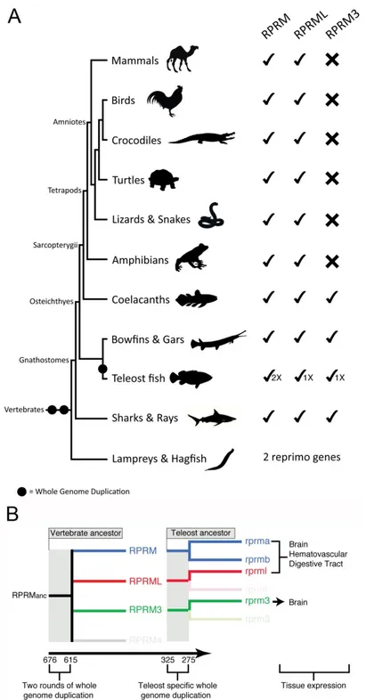
(3) Int. J. Mol. Sci. 2018, 19, 1862 Int. J. Mol. Sci. 2018, 19, x. 3 of 15 3 of 14. Figure 1. Domain structure and potential post-translational modification sites of human RPRM and. Figure 1. Domain structure and potential post-translational modification sites of human RPRM and RPRML proteins. Schematic representation shows the RPRM and RPRML putative N-glycosylation RPRML proteins. Schematic representation shows the RPRM and RPRML putative (green hexagons), serine-phosphorylation (blue circle) and sumoylation (purple triangle) N-glycosylation sites. The N-terminal, transmembrane and C-terminal domains are and represented by colored(purple boxes (turquoise, (green hexagons), serine-phosphorylation (blue circle) sumoylation triangle) sites. yellow, and red, respectively). * (asterisk) indicates positions which have aby single, fully boxes conserved The N-terminal, transmembrane andAn C-terminal domains are represented colored (turquoise, residue. A: (colon) indicates conservation between groups of strongly similar properties—scoring >0.5 yellow, and red, respectively). An * (asterisk) indicates positions which have a single, fully conserved in the Gonnet PAM 250 matrix. A. (period) indicates conservation between groups of weakly similar residue. A: properties—scoring (colon) indicates0.5conservation between groups of strongly similar properties—scoring in the Gonnet PAM 250 matrix. Post-translational modification sites were >0.5 in the Gonnet 250 matrix. A. (period) indicates conservation between groups of weakly predicted PAM using *NetNGlyc (http://www.cbs.dtu.dk/services/NetNGlyc/), **NetPhos (http://www.cbs.dtu.dk/services/NetPhos/) andPAM ***SumoPlot (http://www.abgent.com/sumoplot). similar properties—scoring 0.5 in the Gonnet 250 matrix. Post-translational modification sites were predicted using *NetNGlyc (http://www.cbs.dtu.dk/services/NetNGlyc/), **NetPhos 3. Evolution of the RPRM Gene Family (http://www.cbs.dtu.dk/services/NetPhos/) and ***SumoPlot (http://www.abgent.com/sumoplot).. Based on current sequenced genome data, the evolutionary history of the RPRM gene family traces back to the last common ancestor of vertebrates, which lived between 676 and 615 million of 3. Evolutionyears of the RPRM Family ago [1]. At thatGene time the ancestor of vertebrates presumably had only a single RPRM gene in its genome, which likely performed many of the physiological functions that the current gene family Based performs on current sequenced genome data, the evolutionary history of the RPRM gene family today. Through time, this ancestral gene diversified as a consequence of the two rounds of traces back whole to the last of vertebrates, which lived between and 615 million genomecommon duplicationsancestor (WGDs), which occurred early in the evolutionary history676 of vertebrates the single gene present the genome of the vertebratehad ancestor rise to RPRM four of years ago[8,9]. [1].Thus, At that time copy the ancestor ofinvertebrates presumably onlygave a single gene RPRM genes [1], threeperformed of which were retained actual vertebrates (Figure 2A). Thethat presence coin its genome, which likely many ofinthe physiological functions the of current gene duplicated genes that are found on the chromosomes where the RPRM genes are located in actual family performs today. Through time, this ancestral gene diversified as a consequence of the two species, and the fact that the genomic regions in which the RPRM genes are located derived from a rounds of whole genome duplications (WGDs), which this occurred early single linkage group in the chordate ancestor support hypothesis [10].in the evolutionary history of diversification vertebrates the in RPRM family vertebrates [8,9].During Thus,the the single copyofgene present the gene genome ofwas thedifferentially vertebrate retained, ancestorasgave rise not all RPRM genes are present in all main vertebrate groups (Figure 2A). Although it was previously to four RPRM genes [1], three of which were retained in actual vertebrates (Figure 2A). The presence of claimed that the RPRM gene was not retained in birds [1], searches in the most recent version of bird co-duplicated genes that are found on the chromosomes where the RPRM genes are located in actual genomes revealed the presence of the RPRM gene in this subgroup of amniotes. Thus, all groups of species, andjawed the fact that the genomic regions in which the RPRM genes are located derived from vertebrates (gnathostomes) have retained this gene (Figure 2A). The RPRML gene has also been identified allchordate main groups of gnathostomes, whereas RPRM3 was only retained in four a single linkage group ininthe ancestor support this hypothesis [10]. related vertebrate groups: cartilaginous fish (e.g., rays), holostean fish as not Duringdistantly the diversification of vertebrates the RPRM genesharks, familyskates, was and differentially retained, (e.g., bowfish, gars), teleost fish (e.g., zebrafish) and coelacanths (Figure 2A). In cyclostomes, the all RPRM genes are present in all main vertebrate groups (Figure 2A). Although it was previously group that includes lampreys and hagfish, two RPRM genes has been identified (Figure 2A). claimed thatHowever, the RPRM gene was not retained in failed birdsin[1], searches in theRPRM mostsequences recent version phylogenetic reconstructions have defining orthology. of this of bird group arethe recovered sisteroftothe eachRPRM other, which be subgroup due to features their genomes (e.g., genomes revealed presence genemight in this of of amniotes. Thus, allGC groups of bias) that differ substantially from all other vertebrate genomes [11–13], making it difficult to recover jawed vertebrates (gnathostomes) have retained this gene (Figure 2A). The RPRML gene has also been the true evolutionary history using phylogenetic approaches. identified in allMore main groups of gnathostomes, whereas RPRM3 was only retained in four distantly recently in vertebrate history, the RPRM gene family further expanded as a consequence related vertebrate groups: cartilaginous fish (e.g., sharks, skates, in and holostean fish (e.g., bowfish, of the teleost-specific genome duplication (TSGD) that occurred the rays), ancestor of this group between 325 and 275 million of years ago [14,15] (Figure 2A). Although the TSGD potentially doubled the includes gars), teleost fish (e.g., zebrafish) and coelacanths (Figure 2A). In cyclostomes, the group that number of all RPRM genes in the common ancestor of teleosts, only the rprm gene retained duplicated lampreys and hagfish, two RPRM genes has been identified (Figure 2A). However, phylogenetic copies, termed rprma and rprmb (Figure 2A).. reconstructions have failed in defining orthology. RPRM sequences of this group are recovered sister to each other, which might be due to features of their genomes (e.g., GC bias) that differ substantially from all other vertebrate genomes [11–13], making it difficult to recover the true evolutionary history using phylogenetic approaches. More recently in vertebrate history, the RPRM gene family further expanded as a consequence of the teleost-specific genome duplication (TSGD) that occurred in the ancestor of this group between 325 and 275 million of years ago [14,15] (Figure 2A). Although the TSGD potentially doubled the number of all RPRM genes in the common ancestor of teleosts, only the rprm gene retained duplicated copies, termed rprma and rprmb (Figure 2A)..
(4) Int. J. Mol. Sci. 2018, 19, 1862 Int. J. Mol. Sci. 2018, 19, x. 4 of 15. 4 of 14. Figure2.2.Evolution Evolution and diversification vertebrates. (A)(A) During thethe diversification Figure diversificationofofRPRM RPRMgenes genesinin vertebrates. During diversification of vertebrates RPRM genes were differentially retained, as not all RPRM genes are present all main of vertebrates RPRM genes were differentially retained, as not all RPRM genes are in present in all groups of vertebrates. Thus, RPRM and RPRML genes were retained in all main groups of jawed of main groups of vertebrates. Thus, RPRM and RPRML genes were retained in all main groups vertebrates (gnathostomes), whereas RPRM3 was only retained in four distantly related groups: jawed vertebrates (gnathostomes), whereas RPRM3 was only retained in four distantly related groups: cartilaginous fish (e.g., sharks, skates, and rays), holostean fish (e.g., bowfish and gars), teleost fish fish cartilaginous fish (e.g., sharks, skates, and rays), holostean fish (e.g., bowfish and gars), teleost (e.g.,zebrafish) zebrafish) and and coelacanths. coelacanths. In lampreys and hagfish, twotwo RPRM genes (e.g., Inthe thegroup groupthat thatincludes includes lampreys and hagfish, RPRM genes have been identified, however, phylogenetic and synteny analyses have failed in defining orthology. have been identified, however, phylogenetic and synteny analyses have failed in defining orthology. (B) Schematic representation of the evolution of the RPRM gene family in teleost fish. On the left, the (B) Schematic representation of the evolution of the RPRM gene family in teleost fish. On the left, RPRM gene family diversified as a product of the two rounds of whole-genome duplication occurred the RPRM gene family diversified as a product of the two rounds of whole-genome duplication in the vertebrate ancestor, as well as, a teleost-specific whole genome duplication. On the left, the occurred in the vertebrate ancestor, as well as, a teleost-specific whole genome duplication. On the left, expression territories of the three RPRM gene lineages present in teleost fish. the expression territories of the three RPRM gene lineages present in teleost fish..
(5) Int. J. Mol. Sci. 2018, 19, 1862. 5 of 15. 4. Developmental Expression Patterns of RPRM Gene Family The use of animal models is the primary step to understand the process of human carcinogenesis and the development of new drugs for cancer therapies [16]. In this scenario, Zebrafish combines the complexity of a whole vertebrate animal with the easy-to-use and high-throughput characteristics of in vitro models [17,18]. Despite differences with teleosts, most of the genetic pathways that regulate development are similar between zebrafish and human [19]. In zebrafish embryos—both duplicated copies of rprm (rprma and rprmb) are expressed in a tissue-specific manner in the gastrointestinal tract, brain and blood vessels. Strikingly, expression of RPRM protein in adult human was also detected in the same organs [2]. Expression of rprm3, a gene that is lost in all tetrapods, is restricted to the central nervous system (CNS) in zebrafish embryos and larvae [2]. Our recent observations also indicate that the three rprm genes (rprma, rprmb and rprml) genes are expressed in the developing digestive tract in embryonic fish [2]. Importantly, in humans, RPRM is a gene which expression is lost in many human gastrointestinal malignancies and serves as a potential biomarker for non-invasive detection of gastric cancer (reviewed in the following section). The RPRM protein is located in epithelial cells lining the intestinal crypts and gastric glands and smooth muscle cells (SMCs) from the Muscularis propria [2], As in humans, the digestive tube of the zebrafish is organized in concentric layers of SMCs [20]. However, the role of RPRM genes during the differentiation process of gastrointestinal epithelial cells, which occurs during the migration of these cells from the base of gastric glands and intestinal crypts, remains to be determined. In this scenario, the zebrafish model organism offers an interesting alternative to dissect this role in developmental physiology. Figueroa et al. [2] also show that, in zebrafish and humans, the RPRM and RPRML genes are expressed in vascular tissues. In humans, RPRM is expressed in endothelial and vascular smooth muscle cells (VSMCs). The expression of RPRM and RPRML in blood vessels suggests a potential involvement of RPRM during angiogenesis or vasculogenesis. Future studies require molecular manipulation of the RPRM gene family to unveil the role of RPRM genes in the formation and dynamics of the blood vessels. As we recently reported, in embryonic zebrafish larvae, rprma, rprmb and rprml are expressed distinctly in the developing brain [21]. Some of the structures that express these genes include the forebrain, telencephalon and the olfactory epithelium (OE), among other sensory organs. For example, the expression of rprma is largely restricted to the peripheral nervous system at the OE, while rprmb transcripts are located in most posterior neuronal territories such as the optic tectum and trigeminal ganglia. In contrast to rprma and rprmb, expression of rprml is not detectable in the retina. In adult zebrafish and humans, the RPRM messenger and protein are both located in brain tissues. The human RPRM is expressed in grey and white matter neurons and glial cells from the brain cortex, a tissue that displays low mitotic rates during adulthood [2,21,22]. These findings suggest that the RPRM genes may play a key role in the regulation of cell proliferation in brain development and/or regeneration during adulthood [21]. Models that compare brain-specific gene expression profiles between wild-type animals and those with loss-of-function of rprm and rprml will help to define the expression and function of the RPRM gene family in the processes that contribute to brain development. 5. Functional Diversification of the RPRM Gene Family Due to the three WGDs, the diversification of the RPRM gene family opens new opportunities for physiological innovation within this lineage. In the literature it has been difficult to probe the causal link between the WGDs and biological innovation [23], although for some well-studied gene families, this causal association has been demonstrated [24–26]. As mentioned above, expression profile analyses of the RPRM genes demonstrate that they exhibit unique—although partially overlapping—expression patterns during embryonic and larval vascular development [2]. On one hand, rprma, rprmb and rprml are all expressed in the digestive tube, blood vessels and brain; whereas rprm3 possesses a unique expression profile restricted only to the brain. Importantly, the expression patterns of.
(6) Int. J. Mol. Sci. 2018, 19, 1862. 6 of 15. rprma and rprmb transcripts in the zebrafish resembled expression profiles of the RPRM protein in humans. This evidence suggests that the developmental expression pattern for the RPRM gene family is the same in fish and mammals [1]. Furthermore, the compartmentalization in humans of RPRM genes in partially overlapping territories seems to agree with the pattern described for teleost fish (Figure 2B) [27]. Future studies should elucidate the functional role of the RPRM gene family during the physiological processes such as gut, vascular and neuronal development across the vertebrate subphylum. 6. Role of the RPRM Gene Family in Human Carcinogenesis In order to uncover the RPRM and RPRML tissue expression patterns in human samples, we have assessed in silico RNAseq data from the Genotype Tissue Expression (GTEx) database [28]. As shown in Figure 3A, RPRM has a variable expression across different tissues, whereas RPRML is expressed at very low levels in most tissues, except for brain compartments where expression levels are the highest. However, low transcript levels are expected for intronless genes, which generally express at lower levels than intron-containing genes despite having important biological roles [29]. Transcriptome studies in several vertebrate species reveal that RPRM genes are mainly expressed in the central nervous system. In accordance with this observation, our previous studies have shown that the transcript and protein for RPRM are expressed in the zebrafish and human brain [2]. In the case of cancer tissues, as shown in Figure 3B, both genes display down regulated expression in tumor tissues in comparison with non-tumor adjacent mucosa, including gastric cancer [30]. At experimental and clinical levels, only the RPRM gene has been examined in terms of biological functions and significance in human cancer. In gastric cancer cells, restoring the expression of RPRM by transfecting exogenous cDNA results in reduced colony formation and anchorage-independent growth [3,31]. Correspondingly, mouse xenografts models of gastric cancer cells deficient in RPRM expression have demonstrated enhanced tumor formation and volume [31,32]. In other tumors, such as breast cancer, pituitary tumors and renal cell carcinoma cell lines, overexpression of RPRM suppresses cell proliferation, cell migration, clonogenic capacity and invasiveness [33–35]. Furthermore, the role of RPRM in cell-cycle has also been explored. Ohki et al. [4] overexpressed RPRM through adenoviral infection in cells with wild-type (HeLa, Lovo, MCF7) and mutated (DLD1 and Saos2) p53, observing cell-cycle arrest in G2/M phase, independently of p53 mutational status. However, conflicting results have been reported in gastric cancer and pituitary cell lines where RPRM overexpression results in a significant increase in the sub-G1 population with minimal changes in S and G2/M populations [31,34]. Ectopic expression of RPRM cDNA in gastric cancer cell lines after exposure to DNA-damaging agents, such as 5-fluorouracil or cisplatin, results in an apoptotic phenotype 24 h after treatment [31]. In other types of tumors, with both wild-type and mutated p53 gene, overexpression of RPRM induces an apoptotic phenotype after 4 days of adenoviral infection, suggesting that RPRM may also repress cell growth by induction of apoptosis [4]. Taken together, these results suggest that RPRM has tumor suppressive properties not only in gastric cancer but also in other tumors. Clinical studies have shown that the loss of RPRM expression is as common event in gastric cancer [3,31,32,36] as in other tumors of the gastrointestinal tract including Barrett’s-associated esophageal adenocarcinoma, pancreatic and colorectal carcinoma [37–41]. Loss of expression of RPRM has been also reported in non-digestive tumors including breast, renal cell carcinoma, adrenocortical and pituitary tumors [33–35,42,43]. Conversely, enhanced RPRM expression has been described in metastatic brain tumors [44]..
(7) Int. J. Mol. Sci. 2018, 19, 1862 Int. J. Mol. Sci. 2018, 19, x. 7 of 15 7 of 14. Figure 3. RNA expression of RPRM and RPRML across different tissues. (A) Tissue-specific expression Figure 3. RNA expression of RPRM and RPRML across different tissues. (A) Tissue-specific expression profile from 570 human donors available in the Genotype-Tissue Expression (GTEx) database [28]. profile from 570 human donors available in the Genotype-Tissue Expression (GTEx) database [28]. Data is expressed as log10 of Transcripts Per Kilobase Million (TPM). (B) Expression levels of human Data is expressed as log10 of Transcripts Per Kilobase Million (TPM). (B) Expression levels of human tumor and matched normal tissue samples from Broad Institute TCGA Genome Data Analysis Center tumor and matched normal tissue samples from Broad Institute TCGA Genome Data Analysis [0]. Data is expressed as log2 of RSEM (RNA-Seq by Expectation Maximization). Center [30]. Data is expressed as log2 of RSEM (RNA-Seq by Expectation Maximization)..
(8) Int. J. Mol. Sci. 2018, 19, 1862. 8 of 15. RPRM is located within a CpG-enriched region of the genome. In these regions, a significant proportion of cytosines contain a methyl group in the fifth carbon when they are immediately preceded by a guanine (CpG sites). Although unevenly distributed across the genome, CpG sites generally cluster near gene promoters (CpG islands), thus controlling local chromatin structure and transcription factor binding [45]. In normal cells, a few CpG islands are usually methylated (DNA methylation), maintaining genomic stability and controlling expression of tissue-specific, imprinted and housekeeping genes [46]. In contrast, an aberrant pattern of DNA methylation has been observed in some cancers characterized by a genome wide low methylation state that promotes transcriptional activation of oncogenes, genomic instability, and loss of imprinting, while some CpG islands, particularly those located in the promoter regions of tumor suppressor genes, show a local hypermethylated state that may result in gene silencing [47]. This is one of the most common epigenetic alterations found in human cancers. In the case of RPRM, bisulfite sequence experiments to evaluate the density of methylated CpG sites of the promoter region have correlated positively with the levels of the transcriptional expression of the gene [3]. Consequently, restoring the expression of RPRM by the use of demethylating agents, such as 5-aza-cytidine has confirmed that the expression of RPRM gene is regulated by DNA methylation in gastric cancer cells [3,32,48]. In other neoplasm similar results have been obtain confirming the role of DNA methylation as the main mechanisms of regulation of RPRM gene expression [35,49]. In addition, DNA methylation of RPRM has been associated with a compact chromatin structure and further increasing transcriptional silencing of the gene [49]. Interestingly, an in agreement with the enhanced RPRM expression in metastatic brain tumors [44], bisulfite sequence studies in pituitary tumors have shown that loss of RPRM is not due to hypermethylation of the promoter region [34]. This observation raises the possibility that other mechanisms, genetic and/or epigenetic (i.e., microRNAs), might contribute to RPRM gene regulation. As previously mentioned, RPRM has been proposed as a transcriptional target for p53. In gastric cancer cells expressing wild-type p53, a significant down-regulation of RPRM has been described. Conversely, RPRM-induced changes were not seen in p53-deficient NCI-N87 cells. [50]. Analogous findings have been described by in silico analysis of the TCGA data, where a negative correlation between Survivin and RPRM expression was identified exclusively in patients with wild-type p53 protein status [50]. Based on a positive co-expression between RPRM and p73 proteins in a large cohort of tumor samples, the possibility that other members of the p53 gene family participate in the regulation of RPRM has been raised [3]. Since cytoplasmic overexpression of RPRM inhibit nuclear translocation of the Cdc2-Cyclin/B1 complex inducing cell cycle arrest at the G2/M stage [4] further binding and or co-immunoprecipation experiments should contribute to clarify the role of p73 in the regulation of the expression of RPRM (Figure 4). The clinical significance of the loss of RPRM expression in gastric cancer was first explored by Luo et al. [36]. This study analyzed RPRM protein expression along with tumor suppressor S100A2 (S100 calcium binding protein A2) in a cohort of 100 consecutive gastric cancer cases identifying loss of RPRM expression in up to 65% of cases. Interestingly, Luo et al. [36] found that there exists a positive relationship between the expressions of both genes. Furthermore, loss of RPRM expression was significantly associated with depth of tumor invasion, lymphatic vascular invasion and lymph node metastasis. These clinical findings have been confirmed by Saavedra et al. [3] showing also that loss of RPRM expression is particularly associated with the progression from stage I to stages III-IV (Japanese classification of gastric carcinoma) [51] in a cohort of Hispanic/Amerindian cases from Latin America, one of the highest regions in gastric cancer incidence worldwide [52]. Although none of the earlier studies were able to show that loss of expression of RPRM could influence overall survival in gastric cancer, our group has recently found that loss of RPRM expression does confer a worse prognosis only when accompanied with overexpression of Survivin, a well establish oncogene in gastric cancer [50,53]. Taken together, data suggest that RPRM requires a genetic background including other cancer-related genes, such as S100A2 and/or Survivin to drive the gastric carcinogenesis process..
(9) Int. J. Mol. Sci. 2018, 19, 1862. Int. J. Mol. Sci. 2018, 19, x. 9 of 15. 9 of 14. Figure Figure4.4.Schematic Schematicmodel modelofofRPRM-mediated RPRM-mediatedcell cellcycle cycleand andG2 G2arrest arrestmechanisms. mechanisms.RPRM RPRMhas hasbeen been identified 7/FoxA1 identifiedasasa atranscriptional transcriptionaltarget targetfor: for:(1) (1)p53 p53[4]; [4];(2)(2)histone histonedeacetylase deacetylase 7/FoxA1(HDAC7/FoxA1) (HDAC7/FoxA1) ininan anestrogen estrogenmediated mediatedmechanism mechanism[49]; [49];and and(3) (3)for forepigenetic epigeneticsilencing silencingby byhypermethylation hypermethylationofofits its promoter promoterregion region[54]. [54].AApotential potentialregulation regulationby byp73 p73itithas hasalso alsobeen beenproposed proposed[3]. [3].RPRM RPRMexpression expression results resultsinininhibited inhibiteddephosphorylation dephosphorylationofofCdc2, Cdc2,suppressing suppressingthe theactivation activationofofthe theCdc2-Cyclin Cdc2-CyclinB1 B1 complex. complex. Thus, Thus, inducing inducing cell cell cycle cycle arrest arrest at at G2 G2 suggesting suggesting aa potential potential role rolefor forRPRM RPRMas asaatumor tumor suppressor suppressorgene gene[4]. [4].The Thebalance balancetowards towardscell cellcycle cyclearrest arrestororproliferation proliferationcan canbe beshifted shiftedby bymultiple multiple antagonistic antagonisticeffectors, effectors,amongst amongstthem themRPRM. RPRM.Straight Straightlines lineswith witharrowheads arrowheadsindicate indicateactivation. activation.Lines Lines with no arrowhead indicate inhibition. Curved arrow on the bottom left indicates dephosphorylation with no arrowhead indicate inhibition. Curved arrow on the bottom left indicates dephosphorylation ofofCdc2/Cyclin onon thethe bottom right indicates nuclear translocation of Cdc2/CyclinB1 B1complex. complex.Curved Curvedthick thickarrow arrow bottom right indicates nuclear translocation dephosphorylated Cdc2/Cyclin B1 at G2/M checkpoint. of dephosphorylated Cdc2/Cyclin B1the at the G2/M checkpoint.. Interestingly,itithas hasrecently recentlybeen beenshown shownthat thatupregulation upregulationofofendogenous endogenousRPRM RPRMexpression expressionby by Interestingly, theuse useofofCRISPR/dCas9 CRISPR/dCas9 (Clustered (ClusteredRegularly RegularlyInterspaced InterspacedShort ShortPalindromic PalindromicRepeats Repeatsand andassociated associated the deadCas9) Cas9)system, system,aaplatform platformthat thatutilizes utilizesaacatalytically catalyticallydeactivated deactivatedCas9 Cas9(dCas9) (dCas9)linked linkedtotoeffector effector dead domainsforfor gene expression regulation (i.e., Synergistic Activation Mediator (SAM) complex) domains gene expression regulation (i.e., Synergistic Activation Mediator (SAM) complex) reduced reduced cell proliferation andapoptosis increasedinapoptosis in gastric cancer cells [55]. has Thisbeen finding has been cell proliferation and increased gastric cancer cells [55]. This finding expanded to expanded to other genes with tumor suppressor properties embedded in a CpG-enriched region other genes with tumor suppressor properties embedded in a CpG-enriched region of the genome suchof the genome as Maspin METTL3 [55,56]. newnot toolonly willfor bethe useful not only for as Maspin or such METTL3 [55,56].orThe use of this newThe tooluse willofbethis useful understanding the understanding of the epigenetic modifications in an endogenous biological context but also for of the epigenetic modifications in an endogenous biological context but also for the potential cancer the potential cancer therapies based on these findings. therapies based on these findings. Studieshave haveshown shownthat thatacross acrossthe thegastric gastricprecancerous precancerouscascade, cascade,RPRM RPRMbecomes becomesincreasingly increasingly Studies hypermethylated, in association with loss of protein expression. These findings are particularly hypermethylated, in association with loss of protein expression. These findings are particularly related related with the from transition frommetaplasia intestinal tometaplasia to dysplasia gastric cancer [57]. with the transition intestinal dysplasia and/or gastricand/or cancer [57]. Consequently, Consequently, differenceslevels in methylation been paired found between paired tumor adjacent and nonno differences inno methylation have beenlevels foundhave between tumor and non-tumor tumor adjacent cells [3]. Taken together, loss of expression and/or methylation of RPRM could be cells [3]. Taken together, loss of expression and/or methylation of RPRM could be proposed as a late proposed a late precancerous event in the gastric precancerous Methylation of region the RPRM promoter event in theasgastric cascade. Methylationcascade. of the RPRM promoter is associated region is associated with the infection of Helicobacter pylori particularly to cytotoxin-associated gene with the infection of Helicobacter pylori particularly to cytotoxin-associated gene A (CagA) strains [58]. A (CagA) strains [58]. Accordingly, methylation of has RPRM has been proposed as a Accordingly, methylation of RPRM promoter region beenpromoter proposedregion as a tissue biomarker for the tissue biomarker for the evaluation of H. pylori eradication [59]. evaluation of H. pylori eradication [59]. Accordingly, with this line of evidence, follow-up studies examining DNA methylation levels of RPRM on the longitudinal progression of the gastric precancerous lesions after H. pylori eradication,.
(10) Int. J. Mol. Sci. 2018, 19, 1862. 10 of 15. Accordingly, with this line of evidence, follow-up studies examining DNA methylation levels of RPRM on the longitudinal progression of the gastric precancerous lesions after H. pylori eradication, have revealed an increasing level of the DNA methylation six-years prior the progression of gastric lesions [60]. Interestingly, these changes were independent of the effect of the duration of H. pylori infection and other clinical parameters [60]. Since RPRML is also located within a CpG-enriched region of the genome, as has been well documented in the case of RPRM [3,35,48,49] and also correlates with transcriptional silencing [3,32], the evaluation of RPRML methylation may yield similar findings to that of RPRM. To assess this issue, an exploratory in silico analysis of RNAseq expression data from paired gastric adenocarcinomas and non-tumor adjacent mucosa from The Cancer Genome Atlas (TCGA) database [0] is in progress. In addition, in vitro gain/loss of function experiments focused on evaluation of tumorigenic or tumor suppressive effects may provide insights on the role of RPRML in cancer. 7. Methylated RPRM Cell-Free DNA as a Potential Non-Invasive Biomarker in Gastric Cancer The discovery of methylated cell-free DNA in body fluids has expanded the translational applications of DNA methylation of cancer-related genes [61,62]. Due to its relatively stable nature and availability, DNA methylation can be easily detected in serum, plasma and a variety of body fluids. Thus, the assessment of DNA methylation through cell-free DNA or liquid biopsy approaches has been proposed as a candidate for the diagnosis and management of cancer [63]. Our group was the first to propose the assessment of methylated cell-free DNA of the RPRM promoter gene region for non-invasive detection of gastric cancer with a sensitivity of 95.35% [95% CI: 84.19–99.43%] and specificity of 90.32% [95% CI: 74.25–97.96%] [48]. These values achieved the highest OR (OR = 191.33 [95% CI = 30.01, 1220.01]) in a comprehensive meta-analyses undertaken by Sapari el al. [64] after consolidation of 132 case-controls studies for potential biomarkers based on methylated DNA in gastric cancer. This approach has subsequently been extended to the detection of precancerous lesions [32,57,65]. Therefore, clinical trials addressing the role of RPRM as a non-invasive biomarker in gastric cancer should be performed. 8. Unanswered Questions and the RPRM Gene Family There are many biological questions still to be answered regarding RPRM gene family. Protein structure and the biological relevance of post-translational modifications such as glycosylation, phosphorylation and sumoylation should be assessed (Figure 1). Furthermore, the regulation of expression and the translational applications of the RPRM gene family in cancer medicine need to be further elucidated, along with a potential role in other pathologies [4]. Originally described as a p53-dependent cell cycle arrest mediator (Figure 4), yet the functions and mechanisms by which RPRM acts still remain unknown. Evolutionary studies, developmental genetics and molecular biology approaches will prove useful tools in determining the origin and function of this single exon and intronless RPRM gene family [7,66]. The evaluation of different and complementary lines of evidence such as (1) selective pressures; (2) phenotypical variations as a consequence of loss- or gain-of-function; and (3) pre- and post-transcriptional and translational variations may deliver a better understanding of this gene family. From an evolutionary perspective, comparative genomics and phylogenetic analyses may provide evidence of co-evolution and potential relationships between RPRM and other gene families, and thus uncover new signaling partners. Questions regarding a “de novo” origin or retrotranscription of mRNA followed by insertion of the resulting DNA copy into the genome should be addressed to clarify the origin and evolution of the RPRM single exon gene family. Using molecular and genetic approaches, which rely on DNA sequence or RNA/protein expression alterations, it may be possible to establish the pathophysiological role of the RPRM genes in the regulation of cellular functions. Mutation of RPRM family genes will be useful to identify phenotypic defects generated by RPRM loss-of-function as well as to dissect the role of RPRM in developmental processes..
(11) Int. J. Mol. Sci. 2018, 19, 1862. 11 of 15. The function and role of the RPRM genes in gastric carcinogenesis should be expanded to Int. J. Mol. Sci. 2018, 19, x 11 of 14 incorporate RPRML (Figure 5); as to date, only the human RPRM gene has been examined. Importantly, the unique expression pattern of rprml suggests that this is a functional gene and will hopefully initiate hopefully initiate studies into its presence and function in human gastric physiology and cancer studiestissues. into its presence and function in human gastric physiology and cancer tissues.. Figure 5. Unanswered questions in RPRM gene family. RPRM is as a p53-induced protein which. Figure 5. Unanswered questions in RPRM gene family. RPRM is as a p53-induced protein which induces cell cycle arrest at the G2/M checkpoint [4], through an unknown mechanism. Recently, induces cell cycle arrest at the G2/M checkpoint [4], through an unknown mechanism. Recently, RPRM genes have been shown to be expressed during brain, gut and blood vessel development [2]. RPRM genes have been shown to be expressed during brain, gut and blood vessel development [2]. Additional functions for RPRM include its role as a potential tumor suppressor gene (TSG), and a Additional functions for the RPRM includeof its as a potential suppressor gene non(TSG), biomarker—through assessment the role methylation status of tumor its promoter region—for and a biomarker—through the assessment of the methylation status of its promoter region—for invasive detection of gastric cancer and other tumors [48,62]. Much like RPRM, RPRML is also non-invasive detection of gastricdevelopment cancer and[2], other tumors Much like RPRM, RPRML expressed during embryonic but its role in [48,62]. physiological processes has never been is also expressed during embryonic development [2], but its role in physiological processes has never investigated. been investigated. Although the ectopic expression of RPRM induces cell cycle arrest and apoptosis in vitro [4,34,55], unanswered questions still remain as to how transcriptional silencing of RPRM predisposes Although the ectopic expression of RPRM induces cell cycle arrest and apoptosis in vitro [4,34,55], tissues to gastric cancer development. In multiple tumors, DNA methylation is the most common unanswered questions still remain as to how transcriptional silencing of loss-of-expression RPRM predisposes epigenetic mechanism of loss-of-expression of RPRM [62]. In fact, by tissues DNA to gastricmethylation cancer development. In multiple tumors, DNA methylation is the most common epigenetic of the RPRM promoter region it is an indicator of cancer progression and the continuous mechanism loss-of-expression of RPRM [62]. In can fact,restore loss-of-expression by DNA use ofof demethylating agents such 5-aza -cytidine RPRM gene function andmethylation dampen of tumor region progression this scenario, more progression specific approaches to re-activate RPRM of the indicators RPRM promoter it is [3]. an In indicator of cancer and the continuous use of expression, for example through CRISPR/dCas9-based artificial transcription factors, may open the demethylating agents such 5-aza -cytidine can restore RPRM gene function and dampen indicators door to new translational opportunities in cancer therapeutics of tumor progression [3]. In this scenario, more specific approaches to re-activate RPRM expression,. for example through CRISPR/dCas9-based artificial transcription factors, may open the door to new 9. Concluding Remarks translational opportunities in cancer therapeutics. The role of the RPRM gene family in vertebrate physiology and disease is still a bourgeoning field. A comprehensive characterization of the genetic interactions, signaling pathways, protein 9. Concluding Remarks modifications and regulatory mechanisms of the RPRM gene family may shed light on its role in both. The role of theand RPRM gene family in vertebrate physiology bourgeoning physiological oncological processes. RPRM is known to play aand role disease in tumorsisofstill the a stomach and field. A comprehensive characterization of the genetic interactions, signaling pathways, protein has translational applications in the monitoring and treatment of disease. Whether other members of modifications regulatory mechanisms of the properties RPRM gene family may shed on pathologies, its role in both this geneand family also have tumor suppressor or play relevant roles light in other remainsand a key question inprocesses. need of further research. In this thisinfamily of single genesand physiological oncological RPRM is known to scenario, play a role tumors of theexon stomach could lead applications to the discovery novel biomarkers and therapeutic targets, together with the has translational in theofmonitoring and treatment of disease. Whether other members development of new drugs and clinical applications. The coming years should bring forth active of this gene family also have tumor suppressor properties or play relevant roles in other pathologies, investigation to help defineresearch. and utilize RPRM gene family. remains a key question inunderstand, need of further Inthe this scenario, this family of single exon genes could lead to the discovery of novel biomarkers and therapeutic targets, together with the development Acknowledgments: BMBF-CONICYT 20140027, CRP-ICGEB CH15-01 and CONICYT-ANILLO ACT1402 (JDA), of newIMII drugs and clinical applications. The coming years(GIO), should bring forthand active investigation to P09/016-F (GIO), Fondecyt grants 1160627 (JCO), 1180241 1151411 (AHC) CONICYT-FONDAP 15130011 (AHC and GIO). help understand, define and utilize the RPRM gene family. Conflicts of Interest: The authors declare no conflict of interest.. Acknowledgments: BMBF-CONICYT 20140027, CRP-ICGEB CH15-01 and CONICYT-ANILLO ACT1402 (JDA), IMII P09/016-F (GIO), Fondecyt grants 1160627 (JCO), 1180241 (GIO), 1151411 (AHC) and CONICYT-FONDAP 15130011 (AHC and GIO). Conflicts of Interest: The authors declare no conflict of interest..
(12) Int. J. Mol. Sci. 2018, 19, 1862. 12 of 15. References 1.. 2.. 3.. 4.. 5. 6.. 7. 8. 9. 10.. 11. 12. 13.. 14. 15. 16.. 17. 18. 19. 20. 21.. Wichmann, I.A.; Zavala, K.; Hoffmann, F.G.; Vandewege, M.W.; Corvalan, A.H.; Amigo, J.D.; Owen, G.I.; Opazo, J.C. Evolutionary history of the reprimo tumor suppressor gene family in vertebrates with a description of a new reprimo gene lineage. Gene 2016, 591, 245–254. [CrossRef] [PubMed] Figueroa, R.J.; Carrasco-Avino, G.; Wichmann, I.A.; Lange, M.; Owen, G.I.; Siekmann, A.F.; Corvalan, A.H.; Opazo, J.C.; Amigo, J.D. Reprimo tissue-specific expression pattern is conserved between zebrafish and human. PLoS ONE 2017, 12, e0178274. [CrossRef] [PubMed] Saavedra, K.; Valbuena, J.; Olivares, W.; Marchant, M.J.; Rodriguez, A.; Torres-Estay, V.; Carrasco-Avino, G.; Guzman, L.; Aguayo, F.; Roa, J.C.; et al. Loss of expression of reprimo, a p53-induced cell cycle arrest gene, correlates with invasive stage of tumor progression and p73 expression in gastric cancer. PLoS ONE 2015, 10, e0125834. [CrossRef] [PubMed] Ohki, R.; Nemoto, J.; Murasawa, H.; Oda, E.; Inazawa, J.; Tanaka, N.; Taniguchi, T. Reprimo, a new candidate mediator of the p53-mediated cell cycle arrest at the G2 phase. J. Biol. Chem. 2000, 275, 22627–22630. [CrossRef] [PubMed] Louhichi, A.; Fourati, A.; Rebai, A. Igd: A resource for intronless genes in the human genome. Gene 2011, 488, 35–40. [CrossRef] [PubMed] Huret, J.L.; Ahmad, M.; Arsaban, M.; Bernheim, A.; Cigna, J.; Desangles, F.; Guignard, J.C.; Jacquemot-Perbal, M.C.; Labarussias, M.; Leberre, V.; et al. Atlas of genetics and cytogenetics in oncology and haematology in 2013. Nucleic Acids Res. 2013, 41, D920–D924. [CrossRef] [PubMed] Grzybowska, E.A. Human intronless genes: Functional groups, associated diseases, evolution, and mRNA processing in absence of splicing. Biochem. Biophys. Res. Commun. 2012, 424, 1–6. [CrossRef] [PubMed] Holland, P.W.; Garcia-Fernandez, J.; Williams, N.A.; Sidow, A. Gene duplications and the origins of vertebrate development. Development 1994, 1994, 125–133. Dehal, P.; Boore, J.L. Two rounds of whole genome duplication in the ancestral vertebrate. PLoS Boil. 2005, 3, e314. [CrossRef] [PubMed] Putnam, N.H.; Butts, T.; Ferrier, D.E.; Furlong, R.F.; Hellsten, U.; Kawashima, T.; Robinson-Rechavi, M.; Shoguchi, E.; Terry, A.; Yu, J.K.; et al. The amphioxus genome and the evolution of the chordate karyotype. Nature 2008, 453, 1064–1071. [CrossRef] [PubMed] Qiu, H.; Hildebrand, F.; Kuraku, S.; Meyer, A. Unresolved orthology and peculiar coding sequence properties of lamprey genes: The KCNA gene family as test case. BMC Genom. 2011, 12, 325. [CrossRef] [PubMed] Kuraku, S. Impact of asymmetric gene repertoire between cyclostomes and gnathostomes. Semin. Cell Dev. Boil. 2013, 24, 119–127. [CrossRef] [PubMed] Smith, J.J.; Kuraku, S.; Holt, C.; Sauka-Spengler, T.; Jiang, N.; Campbell, M.S.; Yandell, M.D.; Manousaki, T.; Meyer, A.; Bloom, O.E.; et al. Sequencing of the sea lamprey (Petromyzon marinus) genome provides insights into vertebrate evolution. Nat. Genet. 2013, 45, 415–421. [CrossRef] [PubMed] Meyer, A.; Van de Peer, Y. From 2R to 3R: Evidence for a fish-specific genome duplication (FSGD). BioEssays 2005, 27, 937–945. [CrossRef] [PubMed] Kasahara, M. The 2R hypothesis: An update. Curr. Opin. Immunol. 2007, 19, 547–552. [CrossRef] [PubMed] Gutierrez-Lovera, C.; Vazquez-Rios, A.J.; Guerra-Varela, J.; Sánchez, L.; de la Fuente, M. The potential of zebrafish as a model organism for improving the translation of genetic anticancer nanomedicines. Gene 2017, 8, 349. [CrossRef] [PubMed] Vogel, G. Genomics: Sanger will sequence zebrafish genome. Science 2000, 290, 1671. [CrossRef] [PubMed] Staton, C.A.; Reed, M.W.; Brown, N.J. A critical analysis of current in vitro and in vivo angiogenesis assays. Int. J. Exp. Pathol. 2009, 90, 195–221. [CrossRef] [PubMed] MacRae, C.A.; Peterson, R.T. Zebrafish as tools for drug discovery. Nat. Rev. Drug Discov. 2015, 14, 721–731. [CrossRef] [PubMed] Wallace, K.N.; Akhter, S.; Smith, E.M.; Lorent, K.; Pack, M. Intestinal growth and differentiation in zebrafish. Mech. Dev. 2005, 122, 157–173. [CrossRef] [PubMed] Stanic, K.; Quiroz, A.; Wichmann, I.; Corvalan, A.H.; Owen, G.I.; Opazo, J.C.; Lemus, C.; Concha, M.; Amigo, J.D. Expression of RPRM/rprm in the olfactory system of embryonic zebrafish (Danio rerio). Front. Neuroanat. 2018. [CrossRef] [PubMed].
(13) Int. J. Mol. Sci. 2018, 19, 1862. 22. 23. 24. 25. 26. 27. 28. 29.. 30. 31.. 32. 33.. 34.. 35.. 36. 37.. 38.. 39. 40.. 41.. 42.. 13 of 15. Ming, G.L.; Song, H. Adult neurogenesis in the mammalian brain: Significant answers and significant questions. Neuron 2011, 70, 687–702. [CrossRef] [PubMed] Van de Peer, Y.; Maere, S.; Meyer, A. The evolutionary significance of ancient genome duplications. Nat. Rev. Genet. 2009, 10, 725–732. [CrossRef] [PubMed] Hoffmann, F.G.; Opazo, J.C.; Storz, J.F. Whole-genome duplications spurred the functional diversification of the globin gene superfamily in vertebrates. Mol. Biol. Evol. 2012, 29, 303–312. [CrossRef] [PubMed] Hoffmann, F.G.; Opazo, J.C.; Storz, J.F. Differential loss and retention of cytoglobin, myoglobin, and globin-e during the radiation of vertebrates. Genome Biol. Evol. 2011, 3, 588–600. [CrossRef] [PubMed] Storz, J.F.; Opazo, J.C.; Hoffmann, F.G. Gene duplication, genome duplication, and the functional diversification of vertebrate globins. Mol. Phylogenet. Evol. 2013, 66, 469–478. [CrossRef] [PubMed] Braasch, I.; Gehrke, A.R.; Smith, J.J. The spotted gar genome illuminates vertebrate evolution and facilitates human-teleost comparisons. Nat. Genet. 2016, 48, 427–437. [CrossRef] [PubMed] GTEx Consortium, T. The genotype-tissue expression (GTEx) project. Nat. Genet. 2013, 45, 580–585. Shabalina, S.A.; Ogurtsov, A.Y.; Spiridonov, A.N.; Novichkov, P.S.; Spiridonov, N.A.; Koonin, E.V. Distinct patterns of expression and evolution of intronless and intron-containing mammalian genes. Mol. Biol. Evol. 2010, 27, 1745–1749. [CrossRef] [PubMed] Broad Institute TCGA Genome Data Analysis Center. Analysis-Ready Standardized TCGA Data from Broad GDAC Firehose 2016_01_28 Run; Broad Institute of MIT and Harvard: Cambridge, MA, USA, 2016. Ooki, A.; Yamashita, K.; Yamaguchi, K.; Mondal, A.; Nishimiya, H.; Watanabe, M. DNA damage-inducible gene, reprimo functions as a tumor-suppressor and is suppressed by promoter methylation in gastric cancer. Mol. Cancer Res. 2013, 11, 1362–1374. [CrossRef] [PubMed] Lai, J.; Wang, H.; Luo, Q.; Huang, S.; Lin, S.; Zheng, Y.; Chen, Q. The relationship between DNA methylation and reprimo gene expression in gastric cancer cells. Oncotarget 2017, 8, 108610–108623. [CrossRef] [PubMed] Morris, M.R.; Ricketts, C.; Gentle, D.; Abdulrahman, M.; Clarke, N.; Brown, M.; Kishida, T.; Yao, M.; Latif, F.; Maher, E.R. Identification of candidate tumour suppressor genes frequently methylated in renal cell carcinoma. Oncogene 2010, 29, 2104–2117. [PubMed] Xu, M.; Knox, A.J.; Michaelis, K.A.; Kiseljak-Vassiliades, K.; Kleinschmidt-DeMasters, B.K.; Lillehei, K.O.; Wierman, M.E. Reprimo (RPRM) is a novel tumor suppressor in pituitary tumors and regulates survival, proliferation, and tumorigenicity. Endocrinology 2012, 153, 2963–2973. [CrossRef] [PubMed] Buchegger, K.; Ili, C.; Riquelme, I.; Letelier, P.; Corvalan, A.H.; Brebi, P.; Huang, T.H.; Roa, J.C. Reprimo as a modulator of cell migration and invasion in the mda-mb-231 breast cancer cell line. Biol. Res. 2016, 49, 5. [CrossRef] [PubMed] Luo, J.; Zhu, Y.; Yang, G.; Gong, L.; Wang, B.; Liu, H. Loss of reprimo and s100a2 expression in human gastric adenocarcinoma. Diagn. Cytopathol. 2011, 39, 752–757. [CrossRef] [PubMed] Hamilton, J.P.; Sato, F.; Jin, Z.; Greenwald, B.D.; Ito, T.; Mori, Y.; Paun, B.C.; Kan, T.; Cheng, Y.; Wang, S.; et al. Reprimo methylation is a potential biomarker of barrett’s-associated esophageal neoplastic progression. Clin. Cancer Res. 2006, 12, 6637–6642. [PubMed] Nakazato, T.; Suzuki, Y.; Tanaka, R.; Abe, N.; Masaki, T.; Mori, T.; Ohkura, Y.; Sugiyama, M. Effect of reprimo down-regulation on malignant transformation of intraductal papillary mucinous neoplasm. Pancreas 2018, 47, 291–295. [CrossRef] [PubMed] Sato, N.; Fukushima, N.; Hruban, R.H.; Goggins, M. CPG island methylation profile of pancreatic intraepithelial neoplasia. Mod. Pathol. 2008, 21, 238–244. [PubMed] Chang, W.L.; Jackson, C.; Riel, S.; Cooper, H.S.; Devarajan, K.; Hensley, H.H.; Zhou, Y.; Vanderveer, L.A.; Nguyen, M.T.; Clapper, M.L. Differential preventive activity of sulindac and atorvastatin in Apc+/Min-FCCC mice with or without colorectal adenomas. Gut 2017. [CrossRef] [PubMed] Beasley, W.D.; Beynon, J.; Jenkins, G.J.; Parry, J.M. Reprimo 824 G>C and p53R2 4696 C>G single nucleotide polymorphisms and colorectal cancer: A case-control disease association study. Int. J. Colorectal Dis. 2008, 23, 375–381. [CrossRef] [PubMed] Buchegger, K.; Riquelme, I.; Viscarra, T.; Ili, C.; Brebi, P.; Huang, T.H.; Roa, J.C. Reprimo, a potential p53-dependent tumor suppressor gene, is frequently hypermethylated in estrogen receptor alpha-positive breast cancer. Int. J. Mol. Sci. 2017, 18, 1525. [CrossRef] [PubMed].
(14) Int. J. Mol. Sci. 2018, 19, 1862. 43.. 44.. 45. 46. 47.. 48.. 49.. 50.. 51. 52. 53. 54.. 55.. 56.. 57. 58.. 59.. 60.. 61.. 62.. 14 of 15. Soon, P.S.; Gill, A.J.; Benn, D.E.; Clarkson, A.; Robinson, B.G.; McDonald, K.L.; Sidhu, S.B. Microarray gene expression and immunohistochemistry analyses of adrenocortical tumors identify IGF2 and Ki-67 as useful in differentiating carcinomas from adenomas. Endocr. Relat. Cancer 2009, 16, 573–583. [CrossRef] [PubMed] Zohrabian, V.M.; Nandu, H.; Gulati, N.; Khitrov, G.; Zhao, C.; Mohan, A.; Demattia, J.; Braun, A.; Das, K.; Murali, R.; et al. Gene expression profiling of metastatic brain cancer. Oncol. Rep. 2007, 18, 321–328. [CrossRef] [PubMed] Bernstein, B.E.; Meissner, A.; Lander, E.S. The mammalian epigenome. Cell 2007, 128, 669–681. [CrossRef] [PubMed] Berdasco, M.; Esteller, M. Aberrant epigenetic landscape in cancer: How cellular identity goes awry. Dev. Cell 2010, 19, 698–711. [CrossRef] [PubMed] Esteller, M.; Hamilton, S.R.; Burger, P.C.; Baylin, S.B.; Herman, J.G. Inactivation of the DNA repair gene o6-methylguanine-DNA methyltransferase by promoter hypermethylation is a common event in primary human neoplasia. Cancer Res. 1999, 59, 793–797. [PubMed] Bernal, C.; Aguayo, F.R.; Villarroel, C.; Vargas, M.; Diaz, I.; Ossandón, F.J.; Santibáñez, E.; Palma, M.; Aravena, E.; Barrientos, C.; et al. Reprimo as a potential biomarker for early detection in gastric cancer. Clin. Cancer Res. 2008, 14, 6264–6269. [CrossRef] [PubMed] Malik, S.; Jiang, S.; Garee, J.P.; Verdin, E.; Lee, A.V.; O’Malley, B.W.; Zhang, M.; Belaguli, N.S.; Oesterreich, S. Histone deacetylase 7 and foxa1 in estrogen-mediated repression of rprm. Mol. Cell. Biol. 2010, 30, 399–412. [CrossRef] [PubMed] Cerda-Opazo, P.; Valenzuela-Valderrama, M.; Wichmann, I.; Rodriguez, A.; Contreras-Reyes, D.; Fernandez, E.; Carrasco-Aviño, G.; Corvalan, A.H.; Quest, A. Inverse expression of survivin and reprimo correlates with poor patient prognosis in gastric cancer. Oncotarget 2018, 9, 12853–12867. [CrossRef] [PubMed] The Japanese Research Society for Gastric Cancer. Japanese classification of gastric carcinoma: 3rd English edition. Gastric Cancer 2011, 14, 101–112. Torre, L.A.; Bray, F.; Siegel, R.L.; Ferlay, J.; Lortet-Tieulent, J.; Jemal, A. Global cancer statistics, 2012. CA Cancer J. Clin. 2015, 65, 87–108. [CrossRef] [PubMed] Liu, J.L.; Gao, W.; Kang, Q.M.; Zhang, X.J.; Yang, S.G. Prognostic value of survivin in patients with gastric cancer: A systematic review with meta-analysis. PLoS ONE 2013, 8, e71930. [CrossRef] [PubMed] Perri, F.; Longo, F.; Giuliano, M.; Sabbatino, F.; Favia, G.; Ionna, F.; Addeo, R.; Della Vittoria Scarpati, G.; Di Lorenzo, G.; Pisconti, S. Epigenetic control of gene expression: Potential implications for cancer treatment. Crit. Rev. Oncol. Hematol. 2017, 111, 166–172. [CrossRef] [PubMed] Garcia-Bloj, B.; Moses, C.; Sgro, A.; Plani-Lam, J.; Arooj, M.; Duffy, C.; Thiruvengadam, S.; Sorolla, A.; Rashwan, R.; Mancera, R.L.; et al. Waking up dormant tumor suppressor genes with zinc fingers, tales and the crispr/dcas9 system. Oncotarget 2016, 7, 60535–60554. [CrossRef] [PubMed] Chen, M.; Wei, L.; Law, C.T.; Tsang, F.H.; Shen, J.; Cheng, C.L.; Tsang, L.H.; Ho, D.W.; Chiu, D.K.; Lee, J.M.; et al. RNA N6-methyladenosine methyltransferase-like 3 promotes liver cancer progression through YTHDF2 dependent post-transcriptional silencing of SOCS2. Hepatology 2017. [CrossRef] Liu, L.; Yang, X. Implication of reprimo and hMLH1 gene methylation in early diagnosis of gastric carcinoma. Int. J. Clin. Exp. Pathol. 2015, 8, 14977–14982. [PubMed] Schneider, B.G.; Peng, D.F.; Camargo, M.C.; Piazuelo, M.B.; Sicinschi, L.A.; Mera, R.; Romero-Gallo, J.; Delgado, A.G.; Bravo, L.E.; Wilson, K.T.; et al. Promoter DNA hypermethylation in gastric biopsies from subjects at high and low risk for gastric cancer. Int. J. Cancer 2010, 127, 2588–2597. [CrossRef] [PubMed] Maeda, M.; Yamashita, S.; Shimazu, T.; Iida, N.; Takeshima, H.; Nakajima, T.; Oda, I.; Nanjo, S.; Kusano, C.; Mori, A.; et al. Novel epigenetic markers for gastric cancer risk stratification in individuals after helicobacter pylori eradication. Gastric Cancer 2018. [CrossRef] [PubMed] Schneider, B.G.; Mera, R.; Piazuelo, M.B.; Bravo, J.C.; Zabaleta, J.; Delgado, A.G.; Bravo, L.E.; Wilson, K.T.; El-Rifai, W.; Peek, R.M., Jr.; et al. DNA methylation predicts progression of human gastric lesions. Cancer Epidemiol. Biomark. Prev. 2015, 24, 1607–1613. [CrossRef] [PubMed] Duffy, M.J.; Napieralski, R.; Martens, J.W.; Span, P.N.; Spyratos, F.; Sweep, F.C.; Brunner, N.; Foekens, J.A.; Schmitt, M. Methylated genes as new cancer biomarkers. Eur. J. Cancer 2009, 45, 335–346. [CrossRef] [PubMed] Padmanabhan, N.; Ushijima, T.; Tan, P. How to stomach an epigenetic insult: The gastric cancer epigenome. Nature reviews. Gastroenterol. Hepatol. 2017, 14, 467–478..
(15) Int. J. Mol. Sci. 2018, 19, 1862. 63.. 64. 65.. 66.. 15 of 15. Leygo, C.; Williams, M.; Jin, H.C.; Chan, M.W.Y.; Chu, W.K.; Grusch, M.; Cheng, Y.Y. DNA methylation as a noninvasive epigenetic biomarker for the detection of cancer. Dis. Markers 2017, 2017, 3726595. [CrossRef] [PubMed] Sapari, N.S.; Loh, M.; Vaithilingam, A.; Soong, R. Clinical potential of DNA methylation in gastric cancer: A meta-analysis. PLoS ONE 2012, 7, e36275. [CrossRef] [PubMed] Wen, J.; Zheng, T.; Hu, K.; Zhu, C.; Guo, L.; Ye, G. Promoter methylation of tumor-related genes as a potential biomarker using blood samples for gastric cancer detection. Oncotarget 2017, 8, 77783–77793. [CrossRef] [PubMed] Jorquera, R.; Ortiz, R.; Ossandon, F.; Cardenas, J.P.; Sepulveda, R.; Gonzalez, C.; Holmes, D.S. SinEx DB: A database for single exon coding sequences in mammalian genomes. Database 2016. [CrossRef] [PubMed] © 2018 by the authors. Licensee MDPI, Basel, Switzerland. This article is an open access article distributed under the terms and conditions of the Creative Commons Attribution (CC BY) license (http://creativecommons.org/licenses/by/4.0/)..
(16)
Figure

![Figure 3. RNA expression of RPRM and RPRML across different tissues. (A) Tissue-specific expression profile from 570 human donors available in the Genotype-Tissue Expression (GTEx) database [28]](https://thumb-us.123doks.com/thumbv2/123dok_es/7318028.451326/7.892.140.757.135.1002/expression-different-specific-expression-available-genotype-expression-database.webp)
![Figure 4. Schematic model of RPRM-mediated cell cycle and G2 arrest mechanisms. RPRM has been identified as a transcriptional target for: (1) p53 [4]; (2) histone deacetylase 7/FoxA1 (HDAC7/FoxA1) in an estrogen mediated mechanism [49]; and (3) for epige](https://thumb-us.123doks.com/thumbv2/123dok_es/7318028.451326/9.892.164.725.122.549/schematic-mediated-mechanisms-identified-transcriptional-deacetylase-estrogen-mechanism.webp)
+2
![Figure 5. Unanswered questions in RPRM gene family. RPRM is as a p53-induced protein which induces cell cycle arrest at the G2/M checkpoint [4], through an unknown mechanism](https://thumb-us.123doks.com/thumbv2/123dok_es/7318028.451326/11.892.167.729.230.443/figure-unanswered-questions-induced-protein-induces-checkpoint-mechanism.webp)
Documento similar
As it is well known by now, the at the time Oxonian ethologist R. Dawkins introduced the concept of selfish gene back in 1976. This selfish gene was in fact in many cases a
For the development of those therapeutic actions, multifunctional protein nanoparticles appear as one of the most promising gene therapy vectors due to their
The DNA damage response gene atl-1 and egl-1, a cell death- related gene, are upregulated upon RNAi of s-adRP genes ..... RNAi of s-adRP genes induces the expression of
Keywords: Metal mining conflicts, political ecology, politics of scale, environmental justice movement, social multi-criteria evaluation, consultations, Latin
The following figures show the evolution along more than half a solar cycle of the AR faculae and network contrast dependence on both µ and the measured magnetic signal, B/µ,
What is perhaps most striking from a historical point of view is the university’s lengthy history as an exclusively male community.. The question of gender obviously has a major role
(hundreds of kHz). Resolution problems are directly related to the resulting accuracy of the computation, as it was seen in [18], so 32-bit floating point may not be appropriate
Even though the 1920s offered new employment opportunities in industries previously closed to women, often the women who took these jobs found themselves exploited.. No matter
