The lipid habitats of neurotransmitter receptors in brain
9
0
0
Texto completo
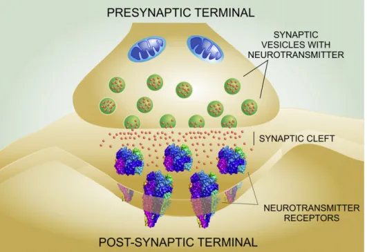
(2) M.V. Borroni et al. / Biochimica et Biophysica Acta 1858 (2016) 2662–2670. 1. Introduction The most complex machine in Nature, the human brain, is a chemically distinct organ: 50% of the brain's dry weight is composed of lipids, in contrast to 6–20% for other organs [114]. Among the principal components of the lipid dotation of the brain are cholesterol, sphingolipids (sphingomyelin, cerebrosides, sulfatides, gangliosides), and glycerophospholipids (phosphatidylcholine, phosphatidylethanolamine, phosphatidylinositols) [108]. Most brain lipids are considered to be structural in the sense that they form part of membranes and myelin, brain cells almost lacking triglycerides, the main energy reserve fat in other tissues. Neurons are highly specialized cells; their shape, composition and metabolism have been adapted in the course of phylogenetic evolution to receive, modify and transmit signals efficiently. These processes occur mostly at specific sites called synapses, where the signaling “output” neuron and the receiving “input” neuron converge. The most common modality of synaptic message is chemical. The molecules that convey the message are called neurotransmitters, small molecules encoding chemical signals. Neurotransmitters released by the presynaptic neuron bind to neurotransmitter receptors in the postsynaptic apparatus, generating either the activation or the inhibition of the target cell (Fig. 1). Neurotransmitter receptors can be classified into two main categories comprised by the superfamilies of pentameric ligand-gated ion channels (pLGIC) on the one hand and the G-protein coupled receptors (GPCR) on the other. The dichotomy is based on the mechanisms they employ to translate the signal: in the case of ligand-gated ion channels, binding of the neurotransmitter induces the opening of a pore that selectively allows certain ions to enter the postsynaptic cell. In the case of the GPCR, binding of neurotransmitters favors the coupling of the receptor with a G protein, triggering a signaling cascade, which takes the form of a metabolic process. Although they act through different mechanisms, both types of receptors are transmembrane proteins with a large proportion of their structure embedded in the lipid membrane bilayer, and both superfamilies share common motifs to bind cholesterol [7,33,34]. A recent review highlights the fact that both neuronal and glial cells are highly polarized, with morphologically distinguishable. 2663. “macrodomains” such as the oligodendroglial myelin sheath or the soma and the axon in neurons [5]. Synapses also constitute highly polarized subcellular structures, and they possess lipid compositions distinct from the rest of the cell membranes [5,44]. Such chemical specialization strongly suggests that neurotransmitter receptors require specific lipid habitats for correct functioning. The purpose of this review is to discuss the physicochemical nature of the lipid environment of paradigmatic neurotransmitter receptors and to evaluate their functional implications with a view to current possibilities of pharmacological modulation of these habitats and, consequently, of receptor function. We consider only the physiological status, not venturing into the pathophysiological alterations of lipid metabolism or lipid-protein interactions in disease. A special focus of this review is the interaction of neurotransmitter receptors with cholesterol. We have recently hypothesized that this relationship appeared early in phylogenetic evolution [12] and was preceded by the appearance of ancient forms of sterols like the hopanoids in bacterial homologs of pLGIC [13]. 2. Cholesterol and neurotransmitter receptors The brain is not only the organ of the economy with uniquely high lipid content; it is also the tissue with the highest concentration of cholesterol. The high membrane/volume ratio of neurons partly explains the high level of this lipid, which is abundant in synaptic membranes [44]. Although structurally simple, cholesterol deeply affects the properties of the plasma membrane where neurotransmitter receptors reside [32]. Cholesterol concentration alters the physical properties of the bulk membrane and also promotes the formation of distinct membrane domains with a higher degree of order than the bulk lipid membrane, termed liquid-ordered (Lo) domains [111]. These domains enriched in cholesterol and sphingolipids are transient, and thicker than the rest of the membrane, offering a platform with unique biophysical characteristics. The most important consequence of these transient lipid supramolecular assemblies is that they spatially and temporally confine signaling processes. Cholesterol is also capable of direct interactions with neurotransmitter receptors through consensus binding sequences like the so-called cholesterol recognition/interaction amino acid consensus motif (CRAC) or its. Fig. 1. Schematic depiction of a generic chemical synapse. Neurotransmitters are stored in synaptic vesicles in the presynaptic terminal. Upon activation, neurotransmitters are released into the synaptic cleft, where they diffuse to bind and activate the neurotransmitter receptors in the postsynaptic membrane..
(3) 2664. M.V. Borroni et al. / Biochimica et Biophysica Acta 1858 (2016) 2662–2670. inverted signature, CARC [7,32,33]. Thus, neurotransmitter receptors can be influenced by cholesterol through three not mutually exclusive mechanisms: i) changes in bulk membrane fluidity that may influence the ability of receptors to undergo the conformational changes required to exert their function; ii) association/dissociation with/from cholesterol- and sphingolipid-enriched Lo membrane domains and iii) specific interactions with cholesterol that can occur within or outside cholesterol-rich domains, via cholesterol binding sites. Whereas modulation of the membrane's physical properties by cholesterol appears to be a general mechanism affecting a broad range of membrane proteins and cell functions, direct binding of cholesterol to sites on the transmembrane regions of a protein and association with membrane domains are expected to be more specific and protein-dependent. We will next describe how these mechanisms modulate the signaling of structurally different neurotransmitter receptors to highlight the importance of cholesterol in correct neurotransmission. Cholesterol depletion is one of the most common strategies to address these phenomena. Table 1 provides a summary of the effects of cholesterol depletion on neurotransmitter receptors. 3. Lipid domains in synapses Biological membranes exhibit varying degrees of lateral heterogeneity at the micron- and sub-micron (nanometer) levels. This is a clear indication of heterogeneity in lipid domain size. In fact, the idea of a subtype of the latter (“ultranano-“) domains has recently been operationally defined by the inability of Förster resonance energy transfer (FRET) techniques to detect them, due to their small size (b 5 nm) [81]. These ultranano-domains, as well as the better characterized nano-domains, exhibit a lipid composition rich in saturated fatty acids, sphingomyelins and cholesterol, and this chemical composition is reflected in their physicochemical state, a characteristic liquid-ordered (Lo) state, different from that of the bulk membrane bilayer lipid, which is in a liquid-disordered (Ld) state [46,58,94,103]. Their protein composition is also, albeit less characteristically, distinct: Lo domains are predominantly enriched in glycosylphosphatidylinositol (GPI)anchored proteins, as well as in proteins involved in signal transduction and intracellular trafficking (trimeric and small GTPases, Src family kinases) [4]. Moreover, palmitoylation serves as a signal for Table 1 Effects of cholesterol depletion on neurotransmitter receptors. Receptor. Effect. Reference. nAChR. Gain of function Retention of nAChR protein in Golgi apparatus Reduction in receptor cell surface expression Acceleration of receptor endocytosis Increase in the size of receptor nanoclusters Reduction in agonist effectiveness Enhance the potentiating effect of diazepam Increase the potentiation by pregnanolone and alfaxalone Augment susceptibility to temperature, proteolytic degradation and pH-induced denaturalization Decrease ‘compactness’ of protein Reduction of the confinement of receptors in large (μm sized) domains. Reduction of open channel probability Increment in the rate constant of entry into desensitized state Redistribution of NMDA Glu/N2B subunit from lipid ordered to lipid disordered membrane fraction. [99]; [18] [82]. GABAA. 5HT-1A. NMDA. [82]; [18]; [20] [20] [53] [72]; [106,107] [72] [106,107] [100]. [79] [87] [35]; [57] [35]; [57] [35]. the anchoring of several neurotransmitter receptors in Lo domains [98]. It should be emphasized that the existence of Lo domains has been clearly demonstrated experimentally in artificial membranes, but their presence in cell membranes is still an often controversial issue. It is important, therefore, to clearly specify the experimental conditions under which their occurrence has been claimed to play a functional role in cellular systems. Finally, lipid domains in biological membranes not only exhibit size heterogeneity, but we can surmise that they may also display shape heterogeneities, and a varying span of lifetimes. In the case of the nervous system, lipid domains have been described to be involved in neuronal cell adhesion, axon guidance, synaptic transmission, growth factor-mediated signal transduction, vesicular trafficking, and membrane-associated proteolysis [4,5,21,109,113]. This makes Lo domains important for neural development and neuronal function at large. Their contribution to neuronal excitability stems from the observation of their participation in the clustering and regulation of neurotransmitter receptors on the one hand, and in the exocytic process of neurotransmitter release on the other [17,22,24,69,72,103]. Due to the essential structural role of cholesterol in the formation of lipid domains, we discuss these aspects in more detail in the following sections of the review, addressing the specific roles of cholesterol on liquid-ordered lipid platforms inhabited by different types of neurotransmitter receptors. 4. Nicotinic acetylcholine receptor (nAChR) and cholesterol The most extensively studied neurotransmitter receptor in terms of lipid-protein interactions is the nicotinic acetylcholine receptor (nAChR), a cation channel and prototypic member of the pentameric ligand-gated ion channel (pLGIC) superfamily of receptors [8,9,12]. Its privileged position arises from the early availability of a nAChR source with high yield and purity, the electric organ of Torpedo electric fish. Moreover, long before the monoclonal antibodies and GFP technology boosted the labeling of proteins, α-bungarotoxin, a very specific quasi-irreversible antagonist of the nAChR, allowed the radiochemicaland fluorescent-labeling of the receptor, opening the door for its cellular and sub-cellular localization, isolation, purification and extensive biochemical and physicochemical characterization. The nAChR is a transmembrane protein composed of five subunits organized around a central pore, the ion channel proper [23,51]. Each nAChR subunit contains four transmembrane segments, 20–30 amino acids in length, named M1–M4. We have proposed that the interface between the protein and lipid moieties, comprising both the lipid-exposed transmembrane portions of the nAChR protein and the nAChR-vicinal lipid, behaves as an identifiable and evolutionarily conserved domain that regulates receptor function [1,8,9,12]. Using native membrane fractions enriched in the postsynaptic membrane of Torpedo electrocytes, nAChR- associated lipids were shown to be immobilized with respect to the bulk membrane lipid [68]. Sterols were identified as part of these immobilized lipids. Two cholesterol pools have been described in nAChR-rich membranes from Torpedo: an easily extractable fraction that influences the bulk fluidity of the membrane and a tightly bound nAChR-associated fraction [61]. Consensus motifs for cholesterol binding are present in the transmembrane domain of all nAChR subunits, rendering a total of 15 cholesterol molecules per receptor [7]. In addition, in silico modeling studies led to the conclusion that in the total absence of cholesterol the nAChR structure would collapse [20]. This model postulates the occurrence of cholesterol sites deep inside the transmembrane region of the nAChR, a premise on which there is still no conclusive experimental evidence [20]. We surmise that the cholesterol sites lie at the lipid belt region surrounding the nAChR macromolecule, in direct contact with the membrane-facing surfaces of the transmembrane segments [7,34]. Independently of the exact topographical loci, the significance of cholesterol for nAChR function has been extensively documented in.
(4) M.V. Borroni et al. / Biochimica et Biophysica Acta 1858 (2016) 2662–2670. experiments using purified nAChR from Torpedo reconstituted into membranes of various compositions, pointing to the clear influence this lipid –in addition to some glycerophospholipids (see reviews in [8,9,12]) – has on the ability of the receptor to undergo the conformational transitions relevant to channel activation and desensitization. In living cells, acute cholesterol depletion has been reported to result in a transient ion channel gain-of-function in the cell line CHO-K1/A5 cells and in Torpedo nAChR expressed in Xenopus oocytes, whereas cholesterol enrichment has the opposite effect [18,99]. Subsequent studies provided a rational for these observations: Acute cholesterol depletion results in the marked acceleration of the rate of endocytosis of the receptor: The nAChR molecules remaining at the surface exhibit a moderate increase in the lifetime of their channel open state, which we interpreted as a compensatory mechanism for the temporary loss of cell-surface receptors [18].. 2665. In CHO-K1/A5 cells and in resting C2C12 myotubes, cell-surface muscle type nAChRs are organized in micron- and submicron-sized clusters, respectively. These receptor aggregates do not appear to be preferentially associated with cholesterol-rich domains, as suggested by early experiments in which membrane patches rich in nAChR were isolated from muscle cells and their cholesterol composition subjected to biochemical analysis [101]. More recent attempts to explore the possible association of the nAChR with lipid domains using wide-field fluorescence microscopy and a cholesterol-sensitive probe did not provide conclusive results [49,101]. In both cases the sensitivity and the spatial resolution of the techniques employed, respectively, did not suffice to ascertain the association of nAChRs with a given type of lipid domain. In the case of the homomeric neuronal α7nAChR expressed in somatic spines of chicken ciliary neurons, the receptor protein has been shown by wide-field fluorescence microscopy to. Fig. 2. Upper panel. CHO-K1/A5 cells co-stained with anti-nAChR Alexa647 (mAb210) and di-4-ANEPPDHQ, a probe used for studying differences in membrane order in living cells [49,75]. Regions of the cell-surface membrane on which the analysis was carried out are highlighted with a white rectangle. nAChR clusters associated with areas having GP values lower than the mean GP of the cell membrane (more disordered domains/domains with lower Chol content) are outlined in white; nAChR clusters with higher-than-the-mean GP are outlined in red (more ordered domains/domains with higher Chol content). Scale bar: 10 μm in images with a white rectangle, and 1 μm in magnified images. From ref. [49]. Middle and lower panels. Schematic representation of the distribution of nAChR molecules in liquid-ordered (Lo) and liquid-disordered (Ld) domains rich and poor in cholesterol, respectively, at the cell surface. The experimentally determined high GP areas in the upper panel are assumed to correspond to the thicker, cholesterol-rich domains, and vice-versa..
(5) 2666. M.V. Borroni et al. / Biochimica et Biophysica Acta 1858 (2016) 2662–2670. colocalize with GM1, a ganglioside reportedly associated with lipid Lo domains [22]. Although the topographical relationship between nAChR and a given type of lipid domain will require further experimentation, there is clear experimental evidence indicating that the size, stability and organization of nAChR clusters depend on cholesterol levels and actin cytoskeletal integrity [10,11,18,49]. Hence, the association of surface nAChRs with cholesterol-rich domains may confer these receptors stability and an adequate environment to perform their function (Fig. 2). In addition, trafficking of nAChRs to the plasmalemma as well as trafficking from the cell surface to the cell interior are both cholesteroldependent processes. In the exocytic pathway, receptors associate with cholesterol-rich domains as early as in the endoplasmic reticulum (ER) and the Golgi apparatus [67]. Moreover, cholesterol depletion retains nAChRs in the Golgi complex, resulting in a decrease in cellsurface nAChR levels [82]. Once nAChRs reach the cell surface, their stability is also highly dependent on cholesterol levels. Acute cholesterol depletion reduces the number of receptor domains by accelerating the rate of endocytosis and shifting the pathway to an Arf6-dependent process [19]. In summary, cholesterol depletion leads to nAChR conformational changes that alter its stability and its long-range dynamic association with other nAChR nanoclusters, accelerates its endocytosis, and transiently affects the channel kinetics of those receptors remaining at the surface. Cholesterol content at the plasmalemma may thus homeostatically modulate nAChR dynamics, cell-surface organization and lifetime of receptor nanodomains, and fine tune the ion permeation process. 5. GABAA receptor and cholesterol Early studies on the influence of cholesterol on the γ-aminobutyric acid receptor (GABAA-R) function arose from the discovery of the potent modulators of this pLGIC, the neurosteroids (steroids synthesized de novo in the brain from cholesterol). Since neurosteroids and cholesterol are structurally related and cholesterol is present in substantial amounts in the neuronal membranes, possible interaction of cholesterol with the GABAA-R through the neurosteroid site was early suggested [16]. However, although an inverse relationship between membrane cholesterol levels and neurosteroid efficacy was observed, the lack of competition between epicholesterol and neurosteroids, and the absolute requirement for GABAA-R potentiating activity of a 3-OH in the α configuration (in contrast cholesterol has this group in the β conformation) led to this possibility being discarded [40,92]. Thus, a specific allosteric site for cholesterol on the GABAA receptor was proposed rather than a less specific fluidity-related effect in supporting the action of neurosteroids on the GABAA-R [106,107]. More recent molecular dynamics studies postulate the binding of cholesterol at the GABAA-R subunit interfaces, deeply buried in between transmembrane helices. Cholesterol in these sites is mobile and instances of spontaneous unbinding and rebinding were observed in the modeling exercise [43]. Whether this flexibility represents the possibility of exchange with structurally-related modulators or with other membrane lipids remains to be explored. These sites may also provide the structural basis for a mechanism localizing the GABAA-R in particular lipid domains with different functional characteristics. Interestingly, following solubilization with Triton X-100 of rat forebrain tissue, different GABAA-R subunit combinations have been associated with different cell membrane compartments [63,72]. Subunits resembling extrasynaptic GABAA-Rs were found in detergent-resistant membranes (the putative biochemical correlate of cholesterol and sphingolipid-rich domains or lipid “rafts”) and assemblies corresponding to synaptic receptors showed only negligible association with this membrane fraction [63]. Furthermore, in WSS-1 cells, cholesterol depletion reduced agonist effectiveness and increased GABAA-R potentiation by benzodiazepines [72]. Moreover, in cultured hippocampal neurons, a U-shaped relationship between GABA affinity for the GABAA-R and. membrane cholesterol content was observed, suggesting that this lipid is required to maintain optimal agonist potency [106,107]. 6. 5-HT-1A receptor and cholesterol Serotonin-1A receptor (5-HT-1AR) is a transmembrane protein that spans the membrane through seven transmembrane domains. It belongs to the G-protein coupled receptor (GPCR) superfamily together with the adrenergic receptors and muscarinic acetylcholine receptors [47]. Modulation of the 5-HT-1AR by cholesterol occurs at different levels. At the protein level, receptor modeling studies suggest that the 5-HT-1AR is more compact in the presence of tightly bound cholesterol; such condensed conformation could contribute to receptor stability [79]. Indeed, in the absence of cholesterol, the 5-HT-1AR is more susceptible to temperature, proteolytic degradation and pH-induced denaturation [100]. Importantly, the presence of CRAC and CARC cholesterol-consensus motifs in the sequence of various GPCRs including the 5-HT-1AR were identified [7]. The number and location of these sequences differ among receptors, and this may confer different sensitivity to cholesterol action [15,54, 55,77,102]. Membrane cholesterol content and availability differentially modulate the specific binding of agonist and antagonist to serotonin receptors present in isolated hippocampal membranes [78,91]. Whereas cholesterol sequestration by the antibiotic nystatin inhibits the binding of antagonists to 5-HT-1AR receptors, the specific agonist binding is not altered [78,91]. Replenishment of membrane cholesterol is reported to be associated with the recovery of ligand binding activity [87–89]. Moreover, different 5-HT receptors respond differently to modification in cholesterol levels. Cholesterol depletion decreases binding of both agonist and antagonist radioligands to 5-HT-7R [105]. Since all the treatments change membrane fluidity to similar extents, these effects cannot be attributed solely to the physical state of the membrane. However, when the same experiments were carried out on whole cells instead of isolated membranes, the opposite effect was observed, suggesting that cell integrity is required to define the effect of cholesterol on this receptor. At the membrane level, cholesterol depletion reduces the confinement of 5-HT-1ARs in large (μm-sized) domains in the plasmalemma [90], a process attributed to destabilization of the cortical actin cytoskeleton that may occur under cholesterol depletion conditions [36,60], as observed by Borroni et al. [18] with the nAChR. Furthermore, the 5-HT-1AR was found in detergent-resistant membranes in different cell types, an effect which depended, in turn, on receptor palmitoylation [97]. As occurs with agonist binding capacity, preferential segregation of 5-HT-1AR into the cholesterol-poor liquid-disordered (Ld) phase was observed in giant unilamellar model membranes [39]. Thus, the localization of 5-HT-1AR in cholesterol-rich domains is more likely to be a cell-dependent event and not an intrinsic physicochemical property of the 5-HT-1AR protein, opening the possibility of receptor regulation through protein post-transcriptional modification (e.g. palmitoylation, see above) or selective recruitment of receptors by distinct membrane domains. 7. N-methyl D-aspartic acid receptors (NMDAR) and cholesterol NMDARs are a variety of glutamate receptors that mediate excitatory synaptic transmission in the CNS, and are so named because NMDA binds exclusively to these and not to other forms of glutamate receptors. Structurally, they are heterotetramers formed by two GluN1 and two GluN2 subunits surrounding a cation channel permeable to calcium [14,26,62]. NMDARs play a key role in synaptic plasticity and are associated with various forms of learning and memory. They are also the target of anesthetics like ketamine and psychoactive hallucinogenic drugs like phencyclidine..
(6) M.V. Borroni et al. / Biochimica et Biophysica Acta 1858 (2016) 2662–2670. Membrane cholesterol has been reported to modulate conformational transitions of the NMDAR present in cerebellar granule cells in culture: acute and chronic cholesterol depletion diminish NMDAR response by reducing ion channel open probability and incrementing the on-rate of desensitization. Cholesterol replenishment reverses NMDAR activity to control values [57]. However, to our knowledge, cholesterol binding sites on NMDARs have not been identified as yet. In contrast, association of NMDAR with cholesterol-rich domains has been extensively described over the past decade. Analysis of detergent-resistant membranes from the synaptosomal fraction of rat forebrains suggests that as much as 60% of NMDARs are located in cholesterol-enriched lipid domains [17], which by virtue of their particular physical properties might dynamically regulate NMDAR subunit composition and trafficking. In support of this possibility, changes in cholesterol content were shown to inhibit NMDA-stimulated influx of calcium in hippocampal cells in culture [35]. Cholesterol diminution redistributed the NMDAR GluN2B subunit from Lo to Ld membrane fractions [35]. 8. Regulation of neurotransmitters receptors by glycosphingolipids Sphingolipids are a family of structurally heterogeneous lipids comprising thousands of biochemically distinct molecules [28]. They tend to segregate laterally within membranes that are enriched in cholesterol, thus favoring the formation of Lo domains [27]. Moreover, as is also the case with cholesterol, sphingolipids can directly interact with neurotransmitter receptors. The latter display a specific sphingolipid-binding domain in their extracellular regions for neutral, cationic, and/or acid sphingolipids [32,33]. When receptors interact with sphingolipids through these binding domains they can undergo strong conformational changes, affecting ligand binding properties, signaling pathways and receptor's trafficking. Indeed, reduction in sphingomyelin and GSL levels upon treatment with metabolic inhibitors resulted in decreased serotonin binding to 5-HT-7 receptors in transfected HeLa cells [105]. Sphingolipids (especially sphingomyelin) have been shown to have a chaperone effect on e.g. the nAChR biosynthetic pathway, affecting the efficiency of the assembly process and subsequent receptor trafficking to the cell surface [6]. It is highly feasible that the physical contact between sphingomyelin and the nAChR results from electrostatic interactions between the protein and the charged sphingolipid [32]. If we keep in mind that both types of molecules are enriched in Lo lipid phases it is not surprising that the crosstalk between cholesterol, sphingolipids, and neurotransmitter receptors is operative in the fine regulation of receptor function.. 10. Ubiquity, mechanisms and implications of neurotransmitter receptor palmitoylation Palmitoylation is the post-translational modification of proteins with palmitic acid (a 16-carbon saturated fatty acid) by covalent attachment to cysteine, serine or threonine residues. To date, 23 palmitoyl-acyl transferases (PATs) have been described in mammalian systems. They contain a genetically conserved DHHC cysteine-rich domain which catalyzes the palmitoylation of multiple targets. Each enzyme palmitoylates different substrates and localizes to different subcellular compartments [73]. Moreover, several PATs have been linked to diverse neurological diseases, such as Parkinson's disease, mental retardation, Huntington's disease, and schizophrenia, highlighting the importance of this lipid modification in the regulation of neurotransmission [45,66,70,96]. De-palmitoylation, on the other hand, is catalyzed by palmitoyl-protein thioesterases, such as palmitoyl-protein thioesterase-1 (PPT1), acyl-protein thioesterase-1 (APT1) and acyl-protein thioesterase-2 (APT2). In contrast to the diversity of PATs, evidence suggests APT1 and APT2 are the major APTs that mediate the de-palmitoylation of diverse cellular substrates [64]. One obvious consequence of palmitoylation is that this posttranslational modification enhances the hydrophobicity of the target proteins and alters their biophysical properties. Importantly, this posttranslational modification is a reversible process, and as such has strong consequences on membrane organization, localization, trafficking and function in the target protein [83]. In the next section we will focus on the effects of palmitoylation on neurotransmitter receptors, also summarized in Table 2. 10.1. Glutamate receptors Glutamate receptors mediate fast excitatory synaptic transmission in the central nervous system. These receptors are tetrameric cation Table 2 Effects of palmitoylation on neurotransmitter receptors. Receptor type. Receptor subtype. Effect. Reference. Glutamate. AMPA. Prevents receptor degradation Regulates receptor stability Retains receptor in Golgi apparatus Inhibits interaction with protein 4.1 N Enhances endocytosis Increases phosphorylation levels by Src-family tyrosine kinases Accumulates NR2A and NR2B in Golgi apparatus Reduces receptor cell-surface expression Promotes association with protein Glu 4.1 N Stabilizes receptor surface expression Reduces receptor internalization Inhibits phosphorylation levels of GLuK2 by PKC Regulates ligand binding site formation at the ER Regulates receptor trafficking to the cell surface Regulates receptor cluster formation at synaptic sites Regulates receptor trafficking and postsynaptic accumulation Retains receptor in membrane microdomains Supports receptor oligomerization Supports receptor G-protein coupling Supports receptor effector signaling. [115] [115] [115]; [41] [41]; [65] [41]; [65] [42]. NMDA. KAINATE. 9. Lipid post-translational modification of neurotransmitter receptors At variance with receptor association with cholesterol or with lipid domains, which are essentially determined by noncovalent interactions [34], lipid post-translational modification of the target protein involves the addition of fatty acids to particular amino acids, a mechanism which carries important changes in the biological signaling properties of the modified protein. Lipid modifications of proteins include: isoprenylation, myristoylation, glycophosphatidyl inositol acylation and palmitoylation [48,116]. These modifications produce profound changes in the biophysical properties of the targeted protein and are able to affect membrane location and lipid domain association of neurotransmitter receptors. Attachment and detachment of the lipid is catalyzed by different sets of enzymes and the whole process is highly regulated. Since palmitoylation is by far and large the most important of these posttranslational modifications and directly affects neurotransmitter receptor's preference for a certain lipid habitat, we will discuss this acylation as a paradigmatic case in the next section.. 2667. Nicotinic. α7 α4. GABA. Serotonin. GABAA. 5-HT1A. 5-HT1B 5-HT4 5-HT7. Regulates isomerization of the receptor from the inactive form to the active form Regulates isomerization of the receptor from the inactive form to the active form. [42] [42] [25] [25] [25] [25] [30]; [2] [3] [95] [52]; [31] [97] [97] [97] [97] [85]; [59] [85]; [59].
(7) 2668. M.V. Borroni et al. / Biochimica et Biophysica Acta 1858 (2016) 2662–2670. channels consisting of four distinct subtypes: AMPA, NMDA, kainite, and δ receptors, and they are localized in neuronal and non-neuronal cells. Functional receptors result from the assembly of four subunits within the same functional receptor class: GluA1–GluA4 for AMPA receptors, GluK1–GluK5 for kainate receptors, GluN1, GluN2A–GluN2D, GluN3A, and GluN3B for the NMDA receptors and GluD1-GluD2 for the δ receptors. All glutamate receptors undergo palmitoylation, except for the δ receptors, with different consequences on receptor biology as discussed below for the different subtypes of glutamate receptors and subunits thereof, thus pointing to a general regulatory mechanism to tune glutamatergic signaling [110]. 10.2. AMPA receptors All AMPA-R subunits are palmitoylated at two distinct sites, one within the pore domain (C585 in GluA1), and the other one in the C-tail juxtamembrane region (C811 in GluA1) [41]. The enzymatic acylation of the AMPA-R precludes receptor degradation, and consequently receptor stability is acquired by this process in the ER, where AMPA receptor subunits are palmitoylated in their M2 hairpin domain [115]. Conversely, de-palmitoylation at this domain must occur when the AMPA-R reaches the Golgi apparatus in order to allow receptors to continue their exocytic trafficking to the cell surface. If AMPA-Rs remain palmitoylated, they are retained in the Golgi apparatus, where they accumulate. Thus, palmitoylation possibly acts as a “quality-control” mechanism at the Golgi level in order to prevent forward trafficking of misfolded AMPA-Rs [41,115]. Palmitoylation at the other site (GluA1 C-tail C811) inhibits the GluA1-4.1N protein interaction. This interaction is necessary for stabilizing AMPA-Rs at the cell surface. Consequently, palmitoylation at this site enhances endocytosis of AMPA-Rs [41,65]. It is thus clear that AMPA-R palmitoylation is a highly regulated, activitydependent process. Indeed, in neuronal cultures glutamate stimulation induces de-palmitoylation of AMPA-Rs whereas blockade of neuronal activity by tetrodotoxin (TTX) increases it [115]. Therefore, palmitoylation/depalmitoylation cycles of AMPA-Rs provide an important regulatory mechanism for synaptic plasticity of nothing less than the major excitatory neurotransmitter receptor in brain. 10.3. NMDA receptors Similar to what is observed with AMPA-Rs, palmitoylation of NMDARs has a prominent role in the trafficking of these receptors to the neuronal plasma membrane, their residence time there, and the endocytic process sequestering them from the cell surface. Palmitoylation occurs at two cysteine clusters in the carboxylterminus of GluN2A and GluN2B subunits [42,50]. This lipid modification differentially regulates retention in the Golgi apparatus and surface stability of NMDARs. Palmitoylation in cluster I results in increased levels of phosphorylation by Src-family tyrosine kinases, which leads in turn to decreased rates of constitutive receptor internalization [42]. In contrast, palmitoylation of cluster II causes accumulation of both NR2A- and NR2B-containing receptors in the Golgi apparatus and reduces NMDAR surface expression. 10.4. Kainate receptors The GluK2 subunit of kainate receptors has been shown to be palmitoylated at two distal sites within the C-terminal tail, the cysteines 858 and 871, with consequences in terms of receptor stability [25,71,84]. In contrast to what occurs with AMPA receptors, palmitoylation of GluK2 promotes association of kainate receptors with protein 4.1N and this association stabilizes surface receptors due to a reduced constitutive internalization. Moreover, when C871 is palmitoylated, it inhibits phosphorylation of GluK2 by PKC, a modification that antagonizes GluK2-4.1N interaction [25]. The data support the. idea that palmitoylation plays an important role in anchoring the receptor's C-terminal domain at the plasma membrane [25,76]. 10.5. nAChRs All muscle nAChR subunits as well as neuronal α4, β2 and α7 nAChR subunits have been found to be palmitoylated [2,3,30,74]. In the case of the homomeric α7 nAChR, palmitoylation is directly involved in regulating the formation of the ligand binding site during nAChR assembly in the ER [2,30]. In addition, cysteine 273 in the M1-M2 loop of the α4 nAChR was found to be a palmitoylation site and to have a prominent role in receptor trafficking to the cell surface [3]. In all cases, palmitoylation occurs during subunit assembly in the ER and regulates the formation of ligand binding sites. Although palmitoylation was demonstrated for many of the muscle and neuronal nAChR subunits, little is known about the role of this chemical postranslational modification in regulating the assembly, trafficking, surface organization and/or ion permeation function of nAChRs. 10.6. 5-HT receptors The serotonin receptors 5-HT 1A , 5-HT1B , 5-HT4 and 5-HT 7 are targets of palmitoylation. Interestingly, the consequences of this modification are different for each type of receptor [37]. All of them are palmitoylated at Cys residues located in the C-terminal receptor domain, although it was suggested that 5-HT7 may have additional acylation site(s) outside this region [38]. Palmitoylation of the 5-HT 7 and 5HT 4 receptor is reversible, agonist-dependent and is directly involved in the isomerization of the receptors from the inactive to the active form [59,85]. Moreover, receptor stimulation increases the turnover rate for palmitoylated 5-HT4 receptors [85]. In the case of 5-HT7 receptor, which can signal through Gs- or G12proteins, palmitoylation may act as a molecular switch selecting the signaling pathway to be activated [59]. For 5-HT 1A receptors, in contrast, palmitoylation is irreversible and insensitive to agonist stimulation [56,80,112]. Moreover, it was demonstrated that palmitoylation does not directly modulate oligomerization of 5-HT 1A , but rather serves to direct and retain 5-HT 1A receptor in defined membrane microdomains, supporting receptor oligomerization, G-protein coupling and effector signaling. [97]. 10.7. GABAA receptors γ-Aminobutyric acid type A receptors (GABAA) are predominantly composed of α, β, and γ2 subunits [93,104]. The major intracellular domain of the GABAA receptor γ2 subunit is rich in cysteine residues, which are targets of palmitoylation. Interestingly, these Cys residues are absent in all other receptor subunits [86]. Palmitoylation of GABAA receptors plays an essential role in regulating the clustering of these receptors at synaptic sites and the steady-state cell-surface receptor number in cultured hippocampal neurons [95]. The Golgi-specific palmitoyltransferase GODZ palmitoylates the GABAA receptor γ2 subunit. This post-translational modification is also involved in trafficking and postsynaptic accumulation of γ2 subunit-containing GABAA receptors and therefore in the assembly and function of GABAergic inhibitory synapses [31,52]. It is interesting to note that gephyrin, a peripheral membrane protein localized in the postsynaptic neuronal membrane, is essential for clustering of GABAA receptors at synapses but in order to do this, gephyrin must also be palmitoylated [29]. 11. Concluding remarks The multiple interactions between membrane lipids and proteins reflect the complexity of their relationship and the vast number of possible regulatory processes involved. The collective physicochemical properties of lipids can also induce their self-assembly, resulting in the.
(8) M.V. Borroni et al. / Biochimica et Biophysica Acta 1858 (2016) 2662–2670. lipid-driven formation of domains within the biological membrane that serve to modify membrane transport, protein sorting and signaling. Lipids are thus involved in the trafficking of newly synthesized proteins to and from the plasma membrane, the lateral organization of membrane proteins in nanoclusters or in higher-order macromolecular assemblies, the gross cellular distribution of neurotransmitter receptors in different regions of the neuron (e.g. soma, axon hillock, axon, dendrites, dendritic spines) and thus in the site-specific modulation of their functional activity. References [1] E.X. Albuquerque, E.F. Pereira, M. Alkondon, S.W. Rogers, Mammalian nicotinic acetylcholine receptors: from structure to function, Physiol. Rev. 89 (1) (2009) 73–120. [2] J.K. Alexander, A.P. Govind, R.C. Drisdel, M.P. Blanton, Y. Vallejo, T.T. Lam, W.N. Green, Palmitoylation of nicotinic acetylcholine receptors, J. Mol. Neurosci. 40 (1–2) (2010) 12–20. [3] S.A. Amici, S.B. McKay, G.B. Wells, J.I. Robson, M. Nasir, G. Ponath, R. Anand, A highly conserved cytoplasmic cysteine residue in the alpha4 nicotinic acetylcholine receptor is palmitoylated and regulates protein expression, J. Biol. Chem. 287 (27) (2012) 23119–23127. [4] R.G. Anderson, The caveolae membrane system, Annu. Rev. Biochem. 67 (1) (1998) 199–225. [5] M. Aureli, S. Grassi, S. Prioni, S. Sonnino, A. Prinetti, Lipid membrane domains in the brain, Biochim. Biophys. Acta Mol. Cell Biol. Lipids 1851 (8) (2015) 1006–1016. [6] C.J. Baier, F.J. Barrantes, Sphingolipids are necessary for nicotinic acetylcholine receptor export in the early secretory pathway, J. Neurochem. 101 (4) (2007) 1072–1084. [7] C.J. Baier, J. Fantini, F.J. Barrantes, Disclosure of cholesterol recognition motifs in transmembrane domains of the human nicotinic acetylcholine receptor, Sci. Rep. 1 (2011) 69. [8] F.J. Barrantes, Modulation of nicotinic acetylcholine receptor function through the outer and middle rings of transmembrane domains, Curr. Opin. Drug Discov. Devel. 6 (5) (2003) 620–632. [9] F.J. Barrantes, Structural basis for lipid modulation of nicotinic acetylcholine receptor function, Brain Res. Brain Res. Rev. 47 (1–3) (2004) 71–95. [10] F.J. Barrantes, Cholesterol effects on nicotinic acetylcholine receptor, J. Neurochem. 103 (Suppl. 1) (2007) 72–80. [11] F.J. Barrantes, Regulation of the Nicotinic Acetylcholine Receptor by Cholesterol as a Boundary Lipid, Cholesterol Regulation of Ion Channels and Receptors 2012, pp. 181–204. [12] F.J. Barrantes, Phylogenetic conservation of protein-lipid motifs in pentameric ligandgated ion channels, Biochim. Biophys. Acta 1848 (9) (2015) 1796–1805. [13] F.J. Barrantes, J. Fantini, From hopanoids to cholesterol: molecular clocks of pentameric ligand-gated ion channels, Prog. Lipid Res. 63 (2016) 1–13. [14] Z.I. Bashir, S. Alford, S.N. Davies, A.D. Randall, G.L. Collingridge, Long-term potentiation of NMDA receptor-mediated synaptic transmission in the hippocampus, Nature 349 (6305) (1991) 156–158. [15] N. Ben-Arie, C. Gileadi, M. Schramm, Interaction of the beta-adrenergic receptor with Gs following delipidation. Specific lipid requirements for Gs activation and GTPase function, Eur. J. Biochem. 176 (3) (1988) 649–654. [16] P.J. Bennett, M.A. Simmonds, The influence of membrane cholesterol on the GABAA receptor, Br. J. Pharmacol. 117 (1) (1996) 87–92. [17] S. Besshoh, D. Bawa, L. Teves, M.C. Wallace, J.W. Gurd, Increased phosphorylation and redistribution of NMDA receptors between synaptic lipid rafts and post-synaptic densities following transient global ischemia in the rat brain, J. Neurochem. 93 (1) (2005) 186–194. [18] V. Borroni, C.J. Baier, T. Lang, I. Bonini, M.M. White, I. Garbus, F.J. Barrantes, Cholesterol depletion activates rapid internalization of submicron-sized acetylcholine receptor domains at the cell membrane, Mol. Membr. Biol. 24 (1) (2007) 1–15. [19] V. Borroni, F.J. Barrantes, Cholesterol modulates the rate and mechanism of acetylcholine receptor internalization, J. Biol. Chem. 286 (19) (2011) 17122–17132. [20] G. Brannigan, J. Henin, R. Law, R. Eckenhoff, M.L. Klein, Embedded cholesterol in the nicotinic acetylcholine receptor, Proc. Natl. Acad. Sci. U. S. A. 105 (38) (2008) 14418–14423. [21] D. Brown, E. London, Functions of lipid rafts in biological membranes, Annu. Rev. Cell Dev. Biol. 14 (1) (1998) 111–136. [22] J.L. Bruses, N. Chauvet, U. Rutishauser, Membrane lipid rafts are necessary for the maintenance of the (alpha)7 nicotinic acetylcholine receptor in somatic spines of ciliary neurons, J. Neurosci. 21 (2) (2001) 504–512. [23] M. Cecchini, J.P. Changeux, The nicotinic acetylcholine receptor and its prokaryotic homologues: structure, conformational transitions & allosteric modulation, Neuropharmacology 96 (Pt B) (2015) 137–149. [24] L.H. Chamberlain, R.D. Burgoyne, G.W. Gould, SNARE proteins are highly enriched in lipid rafts in PC12 cells: implications for the spatial control of exocytosis, Proc. Natl. Acad. Sci. 98 (10) (2001) 5619–5624. [25] B.A. Copits, G.T. Swanson, Kainate receptor post-translational modifications differentially regulate association with 4.1N to control activity-dependent receptor endocytosis, J. Biol. Chem. 288 (13) (2013) 8952–8965. [26] Z. Cui, H. Wang, Y. Tan, K.A. Zaia, S. Zhang, J.Z. Tsien, Inducible and reversible NR1 knockout reveals crucial role of the NMDA receptor in preserving remote memories in the brain, Neuron 41 (5) (2004) 781–793. [27] E.P. de Chaves, S. Sipione, Sphingolipids and gangliosides of the nervous system in membrane function and dysfunction, FEBS Lett. 584 (9) (2010) 1748–1759. [28] S. Degroote, J. Wolthoorn, G. van Meer, The Cell Biology of Glycosphingolipids, Seminars in Cell & Developmental Biology, Elsevier, 2004. [29] B. Dejanovic, M. Semtner, S. Ebert, T. Lamkemeyer, F. Neuser, B. Luscher, J.C. Meier, G. Schwarz, Palmitoylation of gephyrin controls receptor clustering and plasticity of GABAergic synapses, PLoS Biol. 12 (7) (2014), e1001908.. 2669. [30] R.C. Drisdel, E. Manzana, W.N. Green, The role of palmitoylation in functional expression of nicotinic alpha7 receptors, J. Neurosci. 24 (46) (2004) 10502–10510. [31] C. Fang, L. Deng, C.A. Keller, M. Fukata, Y. Fukata, G. Chen, B. Luscher, GODZ-mediated palmitoylation of GABA(A) receptors is required for normal assembly and function of GABAergic inhibitory synapses, J. Neurosci. 26 (49) (2006) 12758–12768. [32] J. Fantini, F.J. Barrantes, Sphingolipid/cholesterol regulation of neurotransmitter receptor conformation and function, Biochim. Biophys. Acta 1788 (11) (2009) 2345–2361. [33] J. Fantini, F.J. Barrantes, How cholesterol interacts with membrane proteins: an exploration of cholesterol-binding sites including CRAC, CARC, and tilted domains, Front. Physiol. 4 (2013) 31. [34] J. Fantini, C. Di Scala, C.J. Baier, F.J. Barrantes, Molecular mechanisms of proteincholesterol interactions in plasma membranes: functional distinction between topological (tilted) and consensus (CARC/CRAC) domains, Chem. Phys. Lipids (2016). [35] C. Frank, A.M. Giammarioli, R. Pepponi, C. Fiorentini, S. Rufini, Cholesterol perturbing agents inhibit NMDA-dependent calcium influx in rat hippocampal primary culture, FEBS Lett. 566 (1–3) (2004) 25–29. [36] S. Ganguly, A. Chattopadhyay, Cholesterol depletion mimics the effect of cytoskeletal destabilization on membrane dynamics of the serotonin1A receptor: a zFCS study, Biophys. J. 99 (5) (2010) 1397–1407. [37] N. Gorinski, E. Ponimaskin, Palmitoylation of serotonin receptors, Biochem. Soc. Trans. 41 (1) (2013) 89–94. [38] D. Guseva, A. Wirth, E. Ponimaskin, Cellular mechanisms of the 5-HT7 receptor-mediated signaling, Front. Behav. Neurosci. 8 (2014) 306. [39] M.G. Gutierrez, N. Malmstadt, Human serotonin receptor 5-HT(1A) preferentially segregates to the liquid disordered phase in synthetic lipid bilayers, J. Am. Chem. Soc. 136 (39) (2014) 13530–13533. [40] N.L. Harrison, M.D. Majewska, J.W. Harrington, J.L. Barker, Structure-activity relationships for steroid interaction with the gamma-aminobutyric acidA receptor complex, J. Pharmacol. Exp. Ther. 241 (1) (1987) 346–353. [41] T. Hayashi, G. Rumbaugh, R.L. Huganir, Differential regulation of AMPA receptor subunit trafficking by palmitoylation of two distinct sites, Neuron 47 (5) (2005) 709–723. [42] T. Hayashi, G.M. Thomas, R.L. Huganir, Dual palmitoylation of NR2 subunits regulates NMDA receptor trafficking, Neuron 64 (2) (2009) 213–226. [43] J. Henin, R. Salari, S. Murlidaran, G. Brannigan, A predicted binding site for cholesterol on the GABAA receptor, Biophys. J. 106 (9) (2014) 1938–1949. [44] H. Hering, C.C. Lin, M. Sheng, Lipid rafts in the maintenance of synapses, dendritic spines, and surface AMPA receptor stability, J. Neurosci. 23 (8) (2003) 3262–3271. [45] K. Huang, A. Yanai, R. Kang, P. Arstikaitis, R.R. Singaraja, M. Metzler, A. Mullard, B. Haigh, C. Gauthier-Campbell, C.-A. Gutekunst, Huntingtin-interacting protein HIP14 is a palmitoyl transferase involved in palmitoylation and trafficking of multiple neuronal proteins, Neuron 44 (6) (2004) 977–986. [46] H.I. Ingólfsson, M.N. Melo, F.J. van Eerden, C.M. Arnarez, C.A. Lopez, T.A. Wassenaar, X. Periole, A.H. de Vries, D.P. Tieleman, S.J. Marrink, Lipid organization of the plasma membrane, J. Am. Chem. Soc. 136 (41) (2014) 14554–14559. [47] M. Jafurulla, A. Chattopadhyay, Membrane lipids in the function of serotonin and adrenergic receptors, Curr. Med. Chem. 20 (1) (2013) 47–55. [48] D.R. Johnson, R.S. Bhatnagar, L.J. Knoll, J.I. Gordon, Genetic and biochemical studies of protein N-myristoylation, Annu. Rev. Biochem. 63 (1) (1994) 869–914. [49] C.B. Kamerbeek, V. Borroni, M.F. Pediconi, S.B. Sato, T. Kobayashi, F.J. Barrantes, Antibodyinduced acetylcholine receptor clusters inhabit liquid-ordered and liquid-disordered domains, Biophys. J. 105 (7) (2013) 1601–1611. [50] R. Kang, J. Wan, P. Arstikaitis, H. Takahashi, K. Huang, A.O. Bailey, J.X. Thompson, A.F. Roth, R.C. Drisdel, R. Mastro, W.N. Green, J.R. Yates 3rd, N.G. Davis, A. El-Husseini, Neural palmitoyl-proteomics reveals dynamic synaptic palmitoylation, Nature 456 (7224) (2008) 904–909. [51] A. Karlin, Emerging structure of the nicotinic acetylcholine receptors, Nat. Rev. Neurosci. 3 (2) (2002) 102–114. [52] C.A. Keller, X. Yuan, P. Panzanelli, M.L. Martin, M. Alldred, M. Sassoe-Pognetto, B. Luscher, The gamma2 subunit of GABA(A) receptors is a substrate for palmitoylation by GODZ, J. Neurosci. 24 (26) (2004) 5881–5891. [53] R.R. Kellner, C.J. Baier, K.I. Willig, S.W. Hell, F.J. Barrantes, Nanoscale organization of nicotinic acetylcholine receptors revealed by stimulated emission depletion microscopy, Neuroscience. 144 (1) (2007) 135–143. [54] J. Kirilovsky, S. Eimerl, S. Steiner-Mordoch, M. Schramm, Function of the delipidated beta-adrenergic receptor appears to require a fatty acid or a neutral lipid in addition to phospholipids, Eur. J. Biochem. 166 (1) (1987) 221–228. [55] J. Kirilovsky, M. Schramm, Delipidation of a beta-adrenergic receptor preparation and reconstitution by specific lipids, J. Biol. Chem. 258 (11) (1983) 6841–6849. [56] F. Kobe, U. Renner, A. Woehler, J. Wlodarczyk, E. Papusheva, G. Bao, A. Zeug, D.W. Richter, E. Neher, E. Ponimaskin, Stimulation- and palmitoylation-dependent changes in oligomeric conformation of serotonin 5-HT1A receptors, Biochim. Biophys. Acta 1783 (8) (2008) 1503–1516. [57] M. Korinek, V. Vyklicky, J. Borovska, K. Lichnerova, M. Kaniakova, B. Krausova, J. Krusek, A. Balik, T. Smejkalova, M. Horak, L. Vyklicky, Cholesterol modulates open probability and desensitization of NMDA receptors, J. Physiol. 593 (10) (2015) 2279–2293. [58] M.R. Krause, S.L. Regen, The structural role of cholesterol in cell membranes: from condensed bilayers to lipid rafts, Acc. Chem. Res. 47 (12) (2014) 3512–3521. [59] E. Kvachnina, A. Dumuis, J. Wlodarczyk, U. Renner, M. Cochet, D.W. Richter, E. Ponimaskin, Constitutive Gs-mediated, but not G12-mediated, activity of the 5-hydroxytryptamine 5-HT7(a) receptor is modulated by the palmitoylation of its C-terminal domain, Biochim. Biophys. Acta 1793 (11) (2009) 1646–1655. [60] J. Kwik, S. Boyle, D. Fooksman, L. Margolis, M.P. Sheetz, M. Edidin, Membrane cholesterol, lateral mobility, and the phosphatidylinositol 4,5-bisphosphate-dependent organization of cell actin, Proc. Natl. Acad. Sci. U. S. A. 100 (24) (2003) 13964–13969. [61] W.S. Leibel, L.L. Firestone, D.C. Legler, L.M. Braswell, K.W. Miller, Two pools of cholesterol in acetylcholine receptor-rich membranes from torpedo, Biochim. Biophys. Acta 897 (2) (1987) 249–260. [62] F. Li, J.Z. Tsien, Memory and the NMDA receptors, N. Engl. J. Med. 361 (3) (2009) 302–303. [63] X. Li, D.R. Serwanski, C.P. Miralles, B.A. Bahr, A.L. De Blas, Two pools of triton X-100insoluble GABA(A) receptors are present in the brain, one associated to lipid rafts and.
(9) 2670. M.V. Borroni et al. / Biochimica et Biophysica Acta 1858 (2016) 2662–2670. another one to the post-synaptic GABAergic complex, J. Neurochem. 102 (4) (2007) 1329–1345. [64] D.T. Lin, E. Conibear, Enzymatic protein depalmitoylation by acyl protein thioesterases, Biochem. Soc. Trans. 43 (2) (2015) 193–198. [65] D.T. Lin, Y. Makino, K. Sharma, T. Hayashi, R. Neve, K. Takamiya, R.L. Huganir, Regulation of AMPA receptor extrasynaptic insertion by 4.1N, phosphorylation and palmitoylation, Nat. Neurosci. 12 (7) (2009) 879–887. [66] M.R. Mansouri, L. Marklund, P. Gustavsson, E. Davey, B. Carlsson, C. Larsson, I. White, K.-H. Gustavson, N. Dahl, Loss of ZDHHC15 expression in a woman with a balanced translocation t (X; 15)(q13. 3; cen) and severe mental retardation, Eur. J. Hum. Genet. 13 (8) (2005) 970–977. [67] S. Marchand, A. Devillers-Thiery, S. Pons, J.P. Changeux, J. Cartaud, Rapsyn escorts the nicotinic acetylcholine receptor along the exocytic pathway via association with lipid rafts, J. Neurosci. 22 (20) (2002) 8891–8901. [68] D. Marsh, F.J. Barrantes, Immobilized lipid in acetylcholine receptor-rich membranes from Torpedo marmorata, Proc. Natl. Acad. Sci. U. S. A. 75 (9) (1978) 4329–4333. [69] J.R. Martens, R. Navarro-Polanco, E.A. Coppock, A. Nishiyama, L. Parshley, T.D. Grobaski, M.M. Tamkun, Differential targeting of shaker-like potassium channels to lipid rafts, J. Biol. Chem. 275 (11) (2000) 7443–7446. [70] J. Mukai, H. Liu, R.A. Burt, D.E. Swor, W.-S. Lai, M. Karayiorgou, J.A. Gogos, Evidence that the gene encoding ZDHHC8 contributes to the risk of schizophrenia, Nat. Genet. 36 (7) (2004) 725–731. [71] Y. Nasu-Nishimura, H. Jaffe, J.T. Isaac, K.W. Roche, Differential regulation of kainate receptor trafficking by phosphorylation of distinct sites on GluR6, J. Biol. Chem. 285 (4) (2010) 2847–2856. [72] C. Nothdurfter, S. Tanasic, B. Di Benedetto, M. Uhr, E.-M. Wagner, K.E. Gilling, C.G. Parsons, T. Rein, F. Holsboer, R. Rupprecht, Lipid raft integrity affects GABA A receptor, but not NMDA receptor modulation by psychopharmacological compounds, Int. J. Neuropsychopharmacol. 16 (06) (2013) 1361–1371. [73] Y. Ohno, A. Kihara, T. Sano, Y. Igarashi, Intracellular localization and tissue-specific distribution of human and yeast DHHC cysteine-rich domain-containing proteins, Biochim. Biophys. Acta Mol. Cell Biol. Lipids 1761 (4) (2006) 474–483. [74] E.N. Olson, L. Glaser, J.P. Merlie, Alpha and beta subunits of the nicotinic acetylcholine receptor contain covalently bound lipid, J. Biol. Chem. 259 (9) (1984) 5364–5367. [75] D.M. Owen, C. Rentero, A. Magenau, A. Abu-Siniyeh, K. Gaus, Quantitative imaging of membrane lipid order in cells and organisms, Nat. Protoc. 7 (1) (2012) 24–35. [76] S. Pahl, D. Tapken, S.C. Haering, M. Hollmann, Trafficking of kainate receptors, Membranes 4 (3) (2014) 565–595. [77] Y.D. Paila, A. Chattopadhyay, Membrane cholesterol in the function and organization of G-protein coupled receptors, Subcell. Biochem. 51 (2010) 439–466. [78] Y.D. Paila, T.J. Pucadyil, A. Chattopadhyay, The cholesterol-complexing agent digitonin modulates ligand binding of the bovine hippocampal serotonin 1A receptor, Mol. Membr. Biol. 22 (3) (2005) 241–249. [79] Y.D. Paila, S. Tiwari, D. Sengupta, A. Chattopadhyay, Molecular modeling of the human serotonin(1A) receptor: role of membrane cholesterol in ligand binding of the receptor, Mol. BioSyst. 7 (1) (2011) 224–234. [80] E. Papoucheva, A. Dumuis, M. Sebben, D.W. Richter, E.G. Ponimaskin, The 5-hydroxytryptamine(1A) receptor is stably palmitoylated, and acylation is critical for communication of receptor with Gi protein, J. Biol. Chem. 279 (5) (2004) 3280–3291. [81] P. Pathak, E. London, The effect of membrane lipid composition on the formation of lipid ultrananodomains, Biophys. J. 109 (8) (2015) 1630–1638. [82] M.F. Pediconi, C.E. Gallegos, E.B. De Los Santos, F.J. Barrantes, Metabolic cholesterol depletion hinders cell-surface trafficking of the nicotinic acetylcholine receptor, Neuroscience 128 (2) (2004) 239–249. [83] T. Peng, E. Thinon, H.C. Hang, Proteomic analysis of fatty-acylated proteins, Curr. Opin. Chem. Biol. 30 (2016) 77–86. [84] D.S. Pickering, F.A. Taverna, M.W. Salter, D.R. Hampson, Palmitoylation of the GluR6 kainate receptor, Proc. Natl. Acad. Sci. U. S. A. 92 (26) (1995) 12090–12094. [85] E.G. Ponimaskin, M.F. Schmidt, M. Heine, U. Bickmeyer, D.W. Richter, 5-Hydroxytryptamine 4(a) receptor expressed in Sf9 cells is palmitoylated in an agonist-dependent manner, Biochem. J. 353 (Pt 3) (2001) 627–634. [86] D.B. Pritchett, H. Sontheimer, B.D. Shivers, S. Ymer, H. Kettenmann, P.R. Schofield, P.H. Seeburg, Importance of a novel GABAA receptor subunit for benzodiazepine pharmacology, Nature 338 (6216) (1989) 582–585. [87] T.J. Pucadyil, A. Chattopadhyay, Cholesterol modulates ligand binding and G-protein coupling to serotonin(1A) receptors from bovine hippocampus, Biochim. Biophys. Acta 1663 (1–2) (2004) 188–200. [88] T.J. Pucadyil, A. Chattopadhyay, Cholesterol modulates the antagonist-binding function of hippocampal serotonin1A receptors, Biochim. Biophys. Acta 1714 (1) (2005) 35–42. [89] T.J. Pucadyil, A. Chattopadhyay, Role of cholesterol in the function and organization of G-protein coupled receptors, Prog. Lipid Res. 45 (4) (2006) 295–333.. [90] T.J. Pucadyil, A. Chattopadhyay, Cholesterol depletion induces dynamic confinement of the G-protein coupled serotonin(1A) receptor in the plasma membrane of living cells, Biochim. Biophys. Acta 1768 (3) (2007) 655–668. [91] T.J. Pucadyil, S. Shrivastava, A. Chattopadhyay, The sterol-binding antibiotic nystatin differentially modulates ligand binding of the bovine hippocampal serotonin1A receptor, Biochem. Biophys. Res. Commun. 320 (2) (2004) 557–562. [92] R.H. Purdy, A.L. Morrow, J.R. Blinn, S.M. Paul, Synthesis, metabolism, and pharmacological activity of 3 alpha-hydroxy steroids which potentiate GABA-receptor-mediated chloride ion uptake in rat cerebral cortical synaptoneurosomes, J. Med. Chem. 33 (6) (1990) 1572–1581. [93] L.E. Rabow, S.J. Russek, D.H. Farb, From ion currents to genomic analysis: recent advances in GABAA receptor research, Synapse 21 (3) (1995) 189–274. [94] M. Rao, S. Mayor, Active organization of membrane constituents in living cells, Curr. Opin. Cell Biol. 29 (2014) 126–132. [95] J. Rathenberg, J.T. Kittler, S.J. Moss, Palmitoylation regulates the clustering and cell surface stability of GABAA receptors, Mol. Cell. Neurosci. 26 (2) (2004) 251–257. [96] F.L. Raymond, P.S. Tarpey, S. Edkins, C. Tofts, S. O'Meara, J. Teague, A. Butler, C. Stevens, S. Barthorpe, G. Buck, Mutations in ZDHHC9, which encodes a palmitoyltransferase of NRAS and HRAS, cause X-linked mental retardation associated with a Marfanoid habitus, Am. J. Hum. Genet. 80 (5) (2007) 982–987. [97] U. Renner, K. Glebov, T. Lang, E. Papusheva, S. Balakrishnan, B. Keller, D.W. Richter, R. Jahn, E. Ponimaskin, Localization of the mouse 5-hydroxytryptamine(1A) receptor in lipid microdomains depends on its palmitoylation and is involved in receptormediated signaling, Mol. Pharmacol. 72 (3) (2007) 502–513. [98] M.D. Resh, Covalent lipid modifications of proteins, Curr. Biol. 23 (10) (2013) R431–R435. [99] J. Santiago, G.R. Guzman, L.V. Rojas, R. Marti, G.A. Asmar-Rovira, L.F. Santana, M. McNamee, J.A. Lasalde-Dominicci, Probing the effects of membrane cholesterol in the Torpedo californica acetylcholine receptor and the novel lipid-exposed mutation alpha C418W in Xenopus oocytes, J. Biol. Chem. 276 (49) (2001) 46523–46532. [100] R. Saxena, A. Chattopadhyay, Membrane cholesterol stabilizes the human serotonin(1A) receptor, Biochim. Biophys. Acta 1818 (12) (2012) 2936–2942. [101] M.G. Scher, R.J. Bloch, The lipid bilayer of acetylcholine receptor clusters of cultured rat myotubes is organized into morphologically distinct domains, Exp. Cell Res. 195 (1) (1991) 79–91. [102] D. Sengupta, A. Chattopadhyay, Identification of cholesterol binding sites in the serotonin1A receptor, J. Phys. Chem. B 116 (43) (2012) 12991–12996. [103] E. Sevcsik, G.J. Schütz, With or without rafts? Alternative views on cell membranes, BioEssays 38 (2) (2016) 129–139. [104] W. Sieghart, G. Sperk, Subunit composition, distribution and function of GABA(A) receptor subtypes, Curr. Top. Med. Chem. 2 (8) (2002) 795–816. [105] B. Sjögren, M.W. Hamblin, P. Svenningsson, Cholesterol depletion reduces serotonin binding and signaling via human 5-HT 7 (a) receptors, Eur. J. Pharmacol. 552 (1) (2006) 1–10. [106] T. Sooksawate, M.A. Simmonds, Effects of membrane cholesterol on the sensitivity of the GABA(A) receptor to GABA in acutely dissociated rat hippocampal neurones, Neuropharmacology 40 (2) (2001) 178–184. [107] T. Sooksawate, M.A. Simmonds, Influence of membrane cholesterol on modulation of the GABA(A) receptor by neuroactive steroids and other potentiators, Br. J. Pharmacol. 134 (6) (2001) 1303–1311. [108] K. Suzuki, Chemistry and metabolism of brain lipids, Basic Neurochem. 1 (1981) 207–227. [109] S.A. Tooze, G.J. Martens, W.B. Huttner, Secretory granule biogenesis: rafting to the SNARE, Trends Cell Biol. 11 (3) (2001) 116–122. [110] S.F. Traynelis, L.P. Wollmuth, C.J. McBain, F.S. Menniti, K.M. Vance, K.K. Ogden, K.B. Hansen, H. Yuan, S.J. Myers, R. Dingledine, Glutamate receptor ion channels: structure, regulation, and function, Pharmacol. Rev. 62 (3) (2010) 405–496. [111] G. Van Meer, D.R. Voelker, G.W. Feigenson, Membrane lipids: where they are and how they behave, Nat. Rev. Mol. Cell Biol. 9 (2) (2008) 112–124. [112] A. Woehler, J. Wlodarczyk, E.G. Ponimaskin, Specific oligomerization of the 5-HT1A receptor in the plasma membrane, Glycoconj. J. 26 (6) (2009) 749–756. [113] B. Wolozin, A fluid connection: cholesterol and Aβ, Proc. Natl. Acad. Sci. 98 (10) (2001) 5371–5373. [114] A.S. Woods, S.N. Jackson, Brain tissue lipidomics: direct probing using matrix-assisted laser desorption/ionization mass spectrometry, AAPS J. 8 (2) (2006) E391–E395. [115] G. Yang, W. Xiong, L. Kojic, M.S. Cynader, Subunit-selective palmitoylation regulates the intracellular trafficking of AMPA receptor, Eur. J. Neurosci. 30 (1) (2009) 35–46. [116] F.L. Zhang, P.J. Casey, Protein prenylation: molecular mechanisms and functional consequences, Annu. Rev. Biochem. 65 (1) (1996) 241–269..
(10)
Figure
![Fig. 2. Upper panel. CHO-K1/A5 cells co-stained with anti-nAChR Alexa 647 (mAb210) and di-4-ANEPPDHQ, a probe used for studying differences in membrane order in living cells [49,75].](https://thumb-us.123doks.com/thumbv2/123dok_es/7446433.396957/4.892.185.722.337.1026/upper-stained-alexa-aneppdhq-studying-differences-membrane-living.webp)
Documento similar