Phenotypic, molecular characterization, antimicrobial susceptibility and
draft genome sequence of
Corynebacterium argentoratense
strains isolated
from clinical samples
I. Fernández-Natal1,2, J. A. Sáez-Nieto3, D. Rodríguez-Lázaro4, S. Valdezate-Ramos3, T. Parras-Padilla1, M. J. Medina3, R. H. Rodríguez-Pollán1, J. Blom5, A. Tauch6and F. Soriano7
1)Department of Clinical Microbiology, Complejo Asistencial Universitario de León, ,2)Institute of Biomedicine (IBIOMED), León, ,3)Bacterial Taxonomy Laboratory, Centro Nacional de Microbiología, Instituto de Salud Carlos III, Majadahonda,4)Microbiology Q2 Section Department of Biotechnology and Food Science, Faculty of Science, University of Burgos, Burgos, Spain,5)Bioinformatik und Systembiologie, Justus-Liebig-Universität, Gießen,6)Institut für
Genomforschung und Systembiologie Centrum für Biotechnologie (CeBiTec), Universität Bielefeld, Bielefeld, Germany and7)Public Health, School of Physiotherapy ONCE, Madrid, Spain
Abstract
During a 12-year period we isolatedfiveCorynebacterium argentoratensestrains identified by phenotypic methods, including the use of matrix-assisted laser desorption/ionization time-of-flight mass spectrometry (MALDI-TOF) and 16S rRNA gene sequencing. In addition, antimicrobial susceptibility was determined, and genome sequencing for the detection of antibiotic resistance genes was performed. The organisms were isolated from blood and throat cultures and could be identified by all methods used. All strains were resistant to cotrimoxazole, and resistance toβ-lactams was partly present. Two strains were resistant to erythromycin and clindamycin. The draft genome sequences of theses isolates revealed the presence of the erm(X) resistance gene that is embedded in the genetic structure of the transposable element Tn5423.Although rarely reported as a human pathogen,C. argentoratensecan be involved in bacteraemia and probably in other infections. Our results also show that horizontal transfer of genes responsible for antibiotic resistance is occurring in this species. New Microbes and New Infections © 2016 The Authors. Published by Elsevier Ltd on behalf of European Society of Clinical Microbiology and Infectious Diseases.
Keywords:Antimicrobial resistance, clinical infections,Corynebacterium argentoratense, MALDI-TOF, whole genome sequencing Original Submission:16 October 2015;Revised Submission:23 December 2015;Accepted:14 January 2016
Article published online:22 January 2016
Corresponding author:I. Fernández-Natal, Department of Clinical Microbiology, Complejo Asistencial Universitario de León, calle Altos de Nava, s/n, 24080 León, Spain
E-mail:[email protected]
Introduction
Corynebacterium argentoratensewas described by Riegel et al.[1] in 1995 after isolating this organism in Strasbourg, a city formerly known as Argentoratum. The taxonomic description of this new species was based on four strains isolated from throat specimens of patients with tonsillitis. The bacterium is nonlipophilic and has typical coryneform morphology, and the
cell wall contains meso-diaminopimelic acid, arabinose, galac-tose and mycolic acids of short chain lengths [1]. The whole genome sequence of the type strain C. argentoratense DSM 44202 (IBS B10697) has a deduced size of 2 031 902 bp with an average G+C content of 58.9%, revealing what is to date the smallest chromosome of a corynebacterial species associated with humans[2].
The same group of researchers from Strasbourg reported the isolation of C. argentoratense from eight respiratory tract specimens (seven from the upper respiratory tract and one from the lower) and another one an ear specimen [3]. This report probably includes the four isolates previously referred to in the taxonomic description of C. argentoratense [1]. In addition to these nine isolates, this organism has been isolated from blood cultures of patients in Canada[4]and Saudi Arabia
New Microbe and New Infect2016;10:116–121
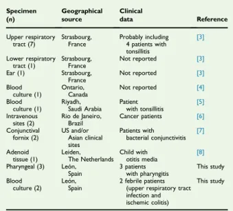
[5], from intravenous sites (catheter and blood) of cancer pa-tients [6], from the conjunctiva of patients with bacterial conjunctivitis[7]and as part of a mucosal biofilm in the adenoid tissue of a child with otitis media[8].Table 1summarizes the sources, geographical origins and clinical data of the
C. argentoratenseisolates described so far.
Data on the natural habitat and the susceptibility of
C. argentoratenseto antimicrobials are limited. Metadata of the microbiota of 18 body sites in more than 200 individuals ob-tained in the course of the Human Microbiome Project indi-cated thatC. argentoratenseis mostly present in saliva and to a lesser extent on the hard palate [9]. An additional study including 19 gastritis patients with a typical greasy white or dense yellow tongue coating revealed thatC. argentoratenseis part of this characteristic tongue-coating microbiome[10]. The
in vitro susceptibility of C. argentoratense to eight antibiotics (ampicillin, cefotaxime, ceftazidime, ciprofloxacin, erythro-mycin, fusidic acid, gentamicin and rifampin) has been deter-mined by the disk diffusion method for nine isolates[3]. Only resistance to ceftazidime was broadly present (78%) in
C. argentoratense.
Here we present data on the origin of C. argentoratense
isolates, microbiologic profiles, including identification by matrix-assisted laser desorption/ionization time-of-flight mass spectrometry (MALDI-TOF), and 16S rRNA gene sequencing, as well as data on the minimum inhibitory concentrations for 28 antibiotics as determined by Etest. Moreover, draft genome sequences of two erythromycin- and clindamycin-resistant isolates are presented, leading to the detection of theerm(X) resistance gene.
Material and Methods
Strains
Two strains were isolated from blood cultures belonging to two different patients, and three other strains were recovered from throat specimens of three different patients with phar-yngitis. All strains were obtained from patients who sought care at a hospital in the north of Spain (Complejo Asistencial Uni-versitario de León, León) from January 2003 to December 2014.
Phenotypic identification
API Coryne 2.0 (bioMérieux, Marcy l’Etoile, France), API Strep (hippurate hydrolysis), API NH (fructose fermentation) and API NE (assimilation of maltose,N-acetyl-glucosamine and phenyl-acetic acid) were used for the phenotypic characterization of the corynebacterial isolates. In addition, catalase and oxidase reactions, lipophilia, Christie Atkins Munch-Peterson (CAMP)
reaction, glucose fermentation at 42°C, growth on blood agar at 20°C and susceptibility to vibriostatic factor O/129 were tested following previously described methods[11].
MALDI-TOF was carried out with a Bruker Biotyper MALDI-TOF system (Bruker Daltonics, Leipzig, Germany). Software and library version 3.1 was used for the bacterial identification. The direct colony method, including spotting onto a MALDI-TOF target plate covered with 1μL of formic acid (100%) and 1μL of matrix, was performed as previously described[12]. Scores of1.5 and1.7 were used for genus and species identification respectively[13].
Genotypic identification
The amplification of the 16S rRNA gene and the DNA sequencing were performed according to previously described methods[14].
Antimicrobial susceptibility
Antimicrobial susceptibility testing to 28 antimicrobials was determined by Etest on Mueller-Hinton agar with 5% sheep’s blood, incubated in air at 35°C and read after 48 hours. Sus-ceptibility to antibiotics was interpreted following the 2014 recommended criteria for coryneform organisms[15].
Genome sequencing and search for antibiotic resistance genes
Technical details of genome sequencing, assembly and annota-tion of two antibiotic-resistantC. argentoratense isolates have been announced previously[16]. Both genome sequences were annotated using the RAST genome annotation server [17]. TABLE 1. Data on 21 published Corynebacterium
argentoratenseisolates from human sources
BLAST (Basic Local Alignment Search Tool) was used for the search for antibiotic resistance genes in the Antibiotic Resis-tance Genes Database[18]and in published genome informa-tion of Corynebacteriumstrains. Comparative genomic analysis was performed with the EDGAR software[19]. All-against-all comparisons were based on the BLASTP algorithm with the standard scoring matrix BLOSUM62 and an initialE-value cutoff of 1 × 10⁻⁵. Thefinal score ratio value was calculated and set to 32. Two genes were considered orthologous when revealing a bidirectional best BLAST hit with a single score ratio value exceeding the precalculated cutoff.
Nucleotide sequence accession numbers
The 16S rRNA gene sequences corresponding to thefive iso-lated strains have been deposited in the GenBank nucleotide database under accession numbers KP230551 (CNM463/05), KP230549 (CNM629/14), KP230548 (CNM630/14), KP230547 (CNM631/14) and KP230550 (CNM601/08).
Results
Isolation ofC. argentoratensefrom clinical sources
C. argentoratensewas obtained from a single blood culture in a 3-year-old boy with an upper respiratory tract infection (strain CNM463/05) and from two blood cultures in an 85-year-old woman with ischemic colitis and high fever (strain CNM601/ 08). In three other patients, aged 7 months, 4 years and 50 years and with tonsillitis,C. argentoratensewas isolated in mixed culture with normal pharyngeal flora (strains CNM629/14, CNM630/14 and CNM631/14 respectively).
Species identification by microbiologic and molecular methods
Microbiologic and genetic methods and MALDI-TOF were used to assign thefive isolates to a corynebacterial species. All iso-lates showed identical microbiologic phenotypic characteristics. After 48 hours’ incubation in air at 35°C, colonies on blood agar were nonhaemolytic, slightly rough and cream colored, and 2 mm in diameter. All isolates presented the same API profiles (2100104 at 24 hours and 2100304 at 48 hours), sug-gestingC. argentoratense(79.2%, T = 1) orC. jeikeium (93.6%, T = 1). The latter profile was doubtful because the isolates were nonlipophilic. Positive reactions were observed for cata-lase, hippurate hydrolysis, pyrazinamidase and alkaline phos-phatase. Acid was produced from glucose (24 hours), ribose (48 hours) and fructose (4 hours) but not from mannitol, su-crose, ribose, xylose, maltose, lactose and glycogen. On the other hand, the organisms were oxidase negative, urease negative and nitrate reductase negative. Negative reactions
were also observed for esculin and gelatine hydrolysis, for β-glucuronidase, β-galactosidase, α-glucosidase and N -acetyl-β-glucosaminidase activities, for the CAMP test and for the assimilation of maltose,N-acetyl-glucosamine and phenylacetic acid. The organisms grew on blood agar at 20°C and fermented glucose at 42°C within 3 days. All isolates were inhibited by the O/129 factor (150μg) showing an inhibition diameter between 18 and 27 mm.
Using MALDI-TOF, as previously described, the isolates were testedfive times recording the two values given by the system, so 50 score values were obtained ranging from 2.166 to 2.378 (mean, 2.293) and providing a reliable identification of
C. argentoratense.For comparison, twoC. jeikeiumstrains were also studiedfive times with two readings, obtaining score values ranging from 1.816 to 2.268 (mean, 1.965). The PCR fragments of the 16S rRNA gene obtained were 1200 bp (CNM463/05), 1411 bp (CNM629/14), 1399 bp (CNM630/14), 1425 bp (CNM631/14) and 1141 bp (CNM601/08) in length. The nucleotide sequence similarities with the 16S rRNA gene of the
C. argentoratensetype strain were in the range of 99.5 to 99.9%. All data clearly demonstrated that thefive isolates are members of the speciesC. argentoratense.
Antimicrobial susceptibility profiling of C. argentoratenseisolates
The antimicrobial susceptibility data for thefive isolates tested are presented inTable 2. Using the Etest method and applying the recommendations of the Clinical and Laboratory Standards Institute for antimicrobial susceptibility of coryneform organ-isms [15], the new C. argentoratense isolates were uniformly sensitive to vancomycin, linezolid, daptomycin, tetracycline, ciprofloxacin, quinupristin/dalfopristin, gentamicin and rifampin. Strain CNM631/14 showed very high minimum inhibitory concentration (MIC) values of benzylpenicillin (>256 mg/L), cefotaxime (8 mg/L) and imipenem (>32 mg/L), and MICs of cefixime were 64 mg/L for all isolates. All isolates were resistant to cotrimoxazole. Two strains (CNM463/05 and CNM601/08) were resistant to erythromycin and clindamycin.
Genome sequencing and search for antibiotic resistance genes
To identify the genes responsible for the detected erythro-mycin and clindaerythro-mycin resistance of C. argentoratense, draft genome sequences of CNM463/05 and CNM601/08 were established[16]. Prominent features of the C. argentoratense
genomes and of those from taxonomically related cor-ynebacteria are summarized inTable 3. BothC. argentoratense
from the type strain sequencing project thatC. argentoratense
has the smallest genome of a corynebacterium associated with humans [2]. The genomes of species assigned to the same taxonomic clade asC. argentoratenseare in the size range from 2.6 to 2.7 Mbp with a G+C content ranging from 58.1 to 68.6% (Table 3). These variations are indicative of the di-versity of the genusCorynebacteriumin terms of lifestyle and habitat, including human, animal, terrestrial, marine and technical environments[20]. The draft genome sequences of both isolates revealed a high grade of similarity between them (>99.9%) as well as with the genome sequence of the type strainC. argentoratenseDSM 44202 (>95.0%). A comparison of the predicted proteomes with the EDGAR software revealed that both clinical isolates and the type strain DSM 44202 share a common set of 1582 proteins. An all-against-all comparison with the proteomes of four taxonomically related species reduced this number to 1078 proteins that represent the conserved core of this diverse clade.
The antibiotic resistance geneerm(X) coding for macrolide, lincosamide and streptogramin B (MLSb) resistance was detected in the genome of both isolates, CNM 463/05 and CNM 601/08. This gene is absent in the genome of the type strain DSM 44202. It was allocated to a specific genomic re-gion with 100% similarity to the transposable element Tn5432, initially found in the R-plasmid pTP10 of Corynebac-terium striatum M82B [21,22], indicating the horizontal
transfer of an antibiotic resistance region to the clinical strains from human sources. Tn5432 is composed of two IS1249
sequences, theerm(X) gene and the transposase genetnpCX
and was previously also detected in Corynebacterium ure-alyticum [23], in Propionibacterium acnes [24] and in some
Bifidobacteriumspecies[25,26].
Discussion
The pathogenicity of C. argentoratense in humans is not well understood. Two cases of bacteraemia by this organism have been reported previously, one in a patient with tonsillitis[5] and another from a patient in whom no clinical data were provided[4]. Here we add two additional cases of bacteraemia, one of them in a patient from whom the organism was isolated from two different blood cultures.
AlthoughC. argentoratenseis not presently recognized as a cause of pharyngotonsillitis, this organism has been isolated from seven patients with tonsillitis [1] (present study); its possible role in this condition thus merits further study. Moreover, if this organism is responsible for pharyngotonsillitis, and if patients are to be empirically treated with penicillins based on a suspicion of aStreptococcus pyogenesinfection, such a treatment could be ineffective in some cases, as inferred by the antimicrobial susceptibility data obtained in this study. The TABLE 2. Antimicrobial susceptibility (MIC, mg/L) of five Corynebacterium argentoratense strains, isolated from bacteraemia (strains 1 and 2) and pharyngotonsillitis (strains 3–5), to 28 antimicrobial agents
Antimicrobial agent Strain 1 (CNM463/05) Strain 2 (CNM601/08) Strain 3 (CNM629/14) Strain 4 (CNM630/14) Strain 5 (CNM631/14)
Benzylpenicillina 1.5 4 1.5 0.75b >256
Ampicillin 1.5 1.5 2 1 >256
Cefuroxime 3 2 2 1.5 4
Cefixime > 256 >256 96 64 >256
Cefotaximea 3 4 2 1.5 8
Imipenema 0.75b 0.5b 0.75b 0.38b >32
Vancomycina 0.5b 0.75b 0.5b 0.5b 0.5b
Teicoplanin 1 1 1 0.5 1
Linezolida 0.38b 0.25b 0.5b 0.38b 0.38b
Daptomycina 0.016b 0.016b 0.016b <0.016b 0.032b
Tetracyclinea 0.5b 0.38b 0.25b 0.38b 0.5b
Tigecycline 0.64 0.023 0.094 0.094 0.094
Chloramphenicol 1.5 1 1 1 1
Ciprofloxacina 0.125b 0.25b 0.125b 0.125b 0.125b
Moxifloxacin 0.047 0.064 0.064 0.064 0.064
Levofloxacin 0.125 0.5 0.125 0.125 0.125
Erythromycina >256 12 <0.016b <0.016b <0.016b
Clarithomycin >256 16 0.016 0.016 0.016
Azithromycin >256 >256 0.125 0.094 0.125
Clindamycina >256 >256 0.094b 0.094b 0.094b
Quinupristin-dalfopristina 0.038b 0.094b 0.038b 0.038b 0.038b
Streptomycin 3 16 2 1 3
Kanamycin 12 64 8 4 12
Gentamicina 1.5b 1b 1.5b 1b 2b
Tobramycin 4 6 4 2 6
Amikacin 4 4 4 2 8
Rifampina 0.032b 0.023b 0.023b 0.023b 0.032b
Cotrimoxazolea >32 >32 >32 >32 >32
MIC, minimum inhibitory concentration.
aAntibiotics proposed for testing coryneform organisms.
isolation ofC. argentoratensefrom intravenous sites in patients with cancer[6]and from the conjunctival fornix in patients with conjunctivitis [7] is of interest and also merits further investigation.
C. argentoratense is a nonlipophilic, fermentative, nitrate reductase-, urease- and oxidase-negative corynebacterium producing acid from glucose but not from maltose and sucrose [20]. It can be identified by API-Coryne, and some additional biochemical tests may be of great help for a phenotypic iden-tification. MALDI-TOF scores of2.166 were obtained for the five isolates, which can be considered as reliable values for a significant species identification[13]. To our knowledge, there is just one published article reporting the identification of two
C. argentoratenseisolates by MALDI-TOF[27]. As occurs with many otherCorynebacteriumspecies, 16S rRNA sequencing was also a useful tool to identifyC. argentoratense[20].
The antimicrobial susceptibility of nine C. argentoratense
strains, isolated between 1995 and 1996, has been determined by a disk diffusion method [3]. Using the interpretative cate-gories recommended by the Antibiogram Committee of the French Society for Microbiology [28], the organisms were considered to be fully sensitive to ampicillin, cefotaxime, fusidic acid and rifampin. Only one of nine isolates was resistant to erythromycin and ciprofloxacin [3]. This unusual pattern of antimicrobial susceptibility, described approximately 15 years ago, agrees with the lack of typical corynebacterial antibiotics resistance genes in the genome of the type strain of
C. argentoratense[2]. In our strains, isolated from 2003 to 2014, resistance to β-lactams, erythromycin and clindamycin was found. One isolate (CNM631/14) presented high MIC values for penicillins and cephalosporins. Isolate CNM463/05 was highly resistant to erythromycin and clindamycin, and isolate CNM601/08 was moderately resistant to erythromycin and highly resistant to clindamycin. Resistance to erythromycin, clindamycin and other MLSb antibiotics is often associated with the presence of the erm(X) gene in corynebacteria [20]. Interestingly, CNM463/05 and CNM601/08 presented the MLSb resistance geneerm(X), located in the specific transposon Tn5432 previously described in C. striatum [22], as well as
C. urealyticum[23]and in the other actinobacterial genera Pro-pionibacterium[24]andBifidobacterium[25,26].
In summary, further studies on the pathogenicity, habitat and epidemiology of C. argentoratense should be carried out, including investigation of its possible role as a cause of phar-yngotonsillitis. Moreover, as observed in a growing number of members of the Corynebacterium genus [20], it seems that horizontal transfer of genes responsible for antibiotic resistance is occurring in this species, which will in future make the treatment of corynebacterial infections a real clinical challenge.
Acknowledgements
We thank A. Acedo from AC-Gen Reading Life, SL, Valladolid, Spain, for his technical support in the whole genome shotgun sequencing. Supported in part by the Gerencia Regional de Salud, Junta de Castilla y León, Spain (research project GRS 698/A/2011).
Con
fl
ict of Interest
None declared.
References
[1] Riegel P, Ruimy R, De Briel D, Prevost G, Jehl F, Bimet F, et al. Corynebacterium argentoratensesp. nov., from the human throat. Int J Syst Bacteriol 1995;45:533–7.
[2] Bomholt C, Glaub A, Graverman K, Albersmeier A, Brinkrolf K, Rückert C, et al. Whole-genome sequence of the clinical strain Cory-nebacterium argentoratenseDSM 44202, isolated from a human throat specimen. Genome Announc 2014;1:e00793–813.
[3] Riegel P, Ruimy R, Christen, Monteil H. Species identities and antimicrobial susceptibilities of corynebacteria isolated from various clinical sources. Eur J Clin Microbiol Infect Dis 1996;15: 657–62.
[4] Bernard KA, Munro C, Wiebe D, Ongsansoy E. Characteristics of rare or recently describedCorynebacteriumspecies recovered from human clinical material in Canada. J Clin Microbiol 2002;40:4375–81. TABLE 3.Features ofCorynebacterium argentoratensegenomes and taxonomically related species
Corynebacteriumspecies Strain Source
Genome
No. of genes Pseudogenes No. of proteins GenBank ID Status Size (bp) G+C%
C. argentoratense DSM 44202 Human; respiratory tract Complete 2031902 58.9 1890 55 1772 CP006365
C. argentoratense CNM 463/05 Human; respiratory tract Draft 2020912 58.9 1871 Not applicable 1643 JZEZ00000000
C. argentoratense CNM 601/08 Human; blood Draft 2014822 58.9 1878 Not applicable 1656 JZFA00000000
C. epidermidicanis DSM 45586 Dog; skin Complete 2692072 58.1 2541 Not detected 2465 CP011541
C. humireducens DSM 45392 Microbial fuel cell Complete 2681312 68.6 2595 46 2482 CP005286
C. marinum DSM 44953 Coastal sediment Complete 2607268 68.1 2457 50 2341 CP007790
[5] Babay HA, Kambal AM. Isolation of coryneform bacteria from blood cultures of patients at a university hospital in Saudi Arabia. Saudi Med J 2004;25:1073–9.
[6] Martins CAS, Faria LMD, Souza MC, Camello TCF, Velasco E, Hirata Jr R, et al. Microbiological and host features associated with corynebacteriosis in cancer patients. A five-year study. Mem Inst Oswaldo Cruz 2009;104:905–13.
[7] Hass W, Gearinger LS, Usner DW, DeCory HH, Morris TW. Integrated analysis of three bacterial conjunctivitis trials of
besi-floxacin ophthalmic suspension, 0.6%: etiology of bacterial conjunctivitis and antibacterial susceptibility profile. Clin Oph-thalmol 2011;5:1369–79.
[8] Kania RE, Lamers GEM, Vonk MJ, Dorpmans E, Struik J, Huy PTB, et al. Characterization of mucosal biofilms on human adenoid tissues. Laryngoscope 2008;118:128–34.
[9] Huse SM, Ye Y, Zhou Y, Fodor AA. A core human microbiome as viewed through 16S rRNA sequence clusters. PLoS One 2012;7: e34242.
[10] Jiang B, Liang X, Chen Y, Ma T, Liu L, Li J, et al. Integrating next-generation sequencing and traditional tongue diagnosis to determine tongue coating microbiome. Sci Rep 2012;2:936.
[11] Fernández-Natal MI, Sáez-Nieto JA, Valdezate S, Rodríguez-Pollán RH, Lapeña S, Cachón F, et al. Isolation ofCorynebacterium ureicelerivorans from normally sterile sites in humans. Eur J Clin Microbiol Infect Dis 2009;28:677–81.
[12] Alatoom AA, Cazanave CJ, Cunningham SA, Ihde SM, Patel R.
Identi-fication of non-diphtheriae Corynebacteriumby use of matrix-assisted laser desorption ionization–time offlights mass spectrometry. J Clin Microbiol 2012;50:160–3.
[13] Theel ES, Schmitt BH, Hall L, Cunningham SA, Walchak RC, Patel R, et al. Formic acid–based direct, on-plate testing of yeast and Coryne-bacteriumspecies by Bruker Biotyper matrix-assisted laser desorption ionization–time offlight mass spectrometry. J Clin Microbiol 2012;50: 3093–5.
[14] Drancourt M, Bollet C, Carlioz A, Martelin R, Gayral JP, Raoult D. 16S ribosomal DNA sequence analysis of a large collection of environ-mental and clinical unidentifiable bacteria isolates. J Clin Microbiol 2000;38:3623–30.
[15] Clinical and Laboratory Standards Institute. Methods for antimicrobial dilution and disk susceptibility testing of infrequently isolated or fastidious bacteria. approved guideline. 2nd ed. Wayne, PA: Clinical and Laboratory Standards Institute; 2014. CLSI document M45–A2. [16] Fernández-Natal MI, Soriano F, Acedo A, Hernandez M, Tauch A,
Rodríguez-Lázaro D. Draft genome sequences of the two unrelated macrolide-resistant Corynebacterium argentoratense strains CNM
463/05 and CNM 601/08, isolated from patients in the University Hospital of León, Spain. Genome Announc 2015;3(4).
[17] Aziz RK, Bartels D, Best AA, DeJongh M, Disz T, Edwards RA, et al. The RAST server: rapid annotations using subsystems technology. BMC Genomics 2008;9:75.
[18] Liu B, Pop M. ARDB—Antibiotic resistance genes database. Nucleic Acids Res 2009;37:D443–7.
[19] Blom J, Albaum SP, Doppmeier D, Pühler A, Vorhölter FJ, Zakrzewski M, et al. EDGAR: a software framework for the comparative analysis of prokaryotic genomes. BMC Bioinformatics 2009;10:154.
[20] Tauch A, Sandbote J. The family Corynebacteriaceae. In: Rosenberg E, DeLong EF, Lory S, Stackebrands E, Thompson F, editors. The pro-karyotes: Actinobacteria. 4th ed. Berlin: Springer; 2014. p. 239–77. [21] Tauch A, Kassing F, Kalinowski J, Pühler A. TheCorynebacterium xerosis
composite transposon Tn5432 consists of two identical insertion se-quences, designated IS1249,flanking the erythromycin resistance gene ermCX. Plasmid 1995;34:119–31.
[22] Tauch A, Krieft S, Kalinowski J, Pühler A. The 51,409-bp R-plasmid pTP10 from the multiresistant clinical isolateCorynebacterium striatum M82B is composed of DNA segments initially identified in soil bacteria and in plant, animal, and human pathogens. Mol Gen Genet 2000;263: 1–11.
[23] Tauch A, Trost E, Tilker A, Ludewig U, Schneiker S, Goesmann A, et al. The lifestyle ofCorynebacterium urealyticum derived from its complete genome sequence established by pyrosequencing. J Biotechnol 2008;136:11–21.
[24] Ross JI, Eady EA, Carnegie E, Cove JH. Detection of transposon Tn5432-mediated macrolide-lincosamide-streptogramin B (MLSB) resistance in cutaneous propionibacteria from six European cities. J Antimicrob Chemother 2002;49:165–8.
[25] van Hoek AH, Mayrhofer S, Domig KJ, Aarts HJ. Resistance determi-nanterm(X) is borne by transposon Tn5432inBifidobacterium ther-mophilumandBifidobacterium animalis subsp.lactis. Int J Antimicrob Agents 2008;31:544–8.
[26] Milani C, Lugli GA, Duranti S, Turroni F, Bottacini F, Mangifesta M, et al. Genomic encyclopedia of type strains of the genusBifi dobacte-rium. Appl Environ Microbiol 2014;80:6290–302.
[27] Seng P, Abat C, Rolain JM, Colson P, Lagier JC, Gouriet F, et al. Identification of rare pathogenic bacteria in a clinical microbiology laboratory: impact of matrix-assisted laser desorption ionization–time offlight mass spectrometry. J Clin Microbiol 2013;51:2182–94. [28] Société Française de Microbiologie. Communiqué 1995 du comité de