Polymorphisms of the WNT10B Gene, Bone Mineral Density,
and Fractures in Postmenopausal Women
Jose L. Perez-CastrillonÆJose M. OlmosÆDaniel N. NanÆ Jesus CastilloÆJana ArozamenaÆAntonio MonteroÆ Marı´a I. Perez-Nun˜ezÆJose A. Riancho
Received: 16 February 2009 / Accepted: 22 April 2009 / Published online: 21 May 2009
ÓSpringer Science+Business Media, LLC 2009
Abstract Wnt ligands are important regulators of skeletal homeostasis. Wnt10B tends to stimulate the differentiation of common mesenchymal precursors toward the osteo-blastic lineage, while inhibiting adipocytic differentiation. Hence, we decided to explore the association of WNT10B allelic variants with bone mineral density and osteoporotic fractures. A set of tag SNPs capturing most common variations of the WNT10B gene was genotyped in 1438 Caucasian postmenopausal women, including 146 with vertebral fractures and 432 with hip fractures. We found no association between single SNPs and spine or hip bone mineral density (BMD). In the multilocus analysis, some haplotypes showed a slight association with spine BMD (P=0.03), but it was not significant after multiple-test correction. There was no association between genotype and vertebral or hip fractures. Transcripts of WNT10B and other Wnt ligands were detected in human bone samples by real-time PCR. However, there was no relationship between genotype and RNA abundance. Thus, WNT10B is expressed in the bone microenvironment and may be an important regulator of osteoblastogenesis, but we have not
found evidence for a robust association of common WNT10B gene allelic variants with either BMD or frac-tures in postmenopausal women.
Keywords WntGene expressionAssociation study
Polymorphisms
Bone mass has a strong genetic component [1, 2]. At the tissue level, the maintenance of bone mass depends on the proper balance between bone formation and bone resorp-tion, which are modulated by a number of endocrine and paracrine factors. Following the seminal studies revealing marked skeletal abnormalities in patients with mutations of the lipoprotein receptor-related protein (LRP) 5 gene [3], the Wnt pathway has emerged as an important regulator of bone homeostasis [4, 5]. It includes different ligands, receptors, coreceptors, and inhibitors. Apart from possible variants due to alternative splicing and posttranslational modifications, at least 19 Wnt ligands have been described. They bind a membrane receptor complex formed by a frizzled protein and LRP. Ten different frizzled genes exist, whereas LRP5 and LRP6 are the most extensively studied LRP forms [6]. Binding of Wnt ligands initiates a cascade of events which mediate their cellular effects. The best-known intracellular mechanisms constitute the so-called canonical pathway, which leads to the accumulation of b-catenin [7]. Several Wnts are produced in the bone tissue and exert an influence on bone remodeling, particularly on cells of the osteoblastic lineage. However, the true physi-ological role of the various Wnt ligands and their relative importance have not been elucidated.
A number of Wnt ligands have been shown to modulate osteoblast differentiation and activity in vitro.
FABP4-J. L. Perez-Castrillon
Medicina Interna, Hospital U. Rio Hortega, Universidad de Valladolid, Valladolid, Spain
J. M. OlmosD. N. NanJ. ArozamenaJ. A. Riancho (&) Medicina Interna, Hospital U.M. Valdecilla, Universidad de Cantabria, Av Valdecilla sn, 39008 Santander, Spain e-mail: [email protected]
J. Castillo
Centro de Salud Jose Barros, Camargo, Spain
A. MonteroM. I. Perez-Nun˜ez
Traumatologı´a y Ortopedia, Hospital U.M. Valdecilla, Universidad de Cantabria, Santander, Spain
WNT10B mice, which have increased expression of WNT10B in the marrow, show an increased bone mass. On the other hand, WNT10B knockout mice have decreased trabecular bone and osteocalcin levels [8]. Those studies strongly suggest that Wnt10B is an important endogenous regulator of bone formation. The mechanism of action appears to include a shift of cell fate of the mesenchymal precursors toward the osteoblastic phenotype, while the differentiation toward the adipocytic lineage is inhibited [8, 9]. Little is known about the mechanisms regulating Wnt10B synthesis, but it has been demonstrated that loading results in increased expression of several mole-cules in the Wnt pathway, including WNT10B, thus sug-gesting its involvement in mechanotransduction [10, 11]. Those data prompted us to study the role of WNT10B as an osteoporosis candidate gene, by analyzing its expression in human bone and exploring the relationship of WNT10B polymorphisms with bone mineral density (BMD) and osteoporotic fractures.
Materials and Methods
Subjects
We studied a group of 1,006 postmenopausal women over 50 years of age (mean, 66±8 years), with an average duration of the menopause of 16 years. They were volun-teers recruited by voice and written announcements or women sent to our outpatient clinic to be studied for pos-sible osteoporosis. All participants were living in Canta-bria, a region in northern Spain with a population of about 530,000. Subjects taking bisphosphonates, corticosteroids, antiepileptics, estrogens, or other drugs known to modify bone mass, and those with non-Spanish ancestors, were excluded. The study was approved by the Institutional Committee on Ethics in Clinical Research.
We also studied 432 postmenopausal women admitted to hospital with hip fractures (mean age, 79±7 years). Women with fractures due to high-impact trauma (such as traffic accidents and falls from a height) or diseases causing secondary osteoporosis (cancer, rheumatoid arthritis, mal-absorption, severe systemic diseases, etc.) were not included.
Skeletal Studies
BMD was measured at the spine (L2–L4), femoral neck, and total hip regions by DXA (Hologic QDR4500). Lateral X-rays of the thoracic and lumbar spine were obtained in 646 women; 146 had vertebral fractures, defined as a loss of at least 25% of vertebral body height. X-rays were not obtained in some control women with a normal BMD and
absence of clinical risk factors for osteoporosis or back pain.
Genotyping
DNA was isolated from the peripheral blood or buccal swabs using standard commercial methods and quantified using the Qubit procedure (Invitrogen). We explored the Hapmap database searching SNPs in a 12-kb region including the WNT10B gene and neighboring 50 and 30regions, with a minimum allelic frequency (MAF) of 10% in the Caucasian population. Then tag SNPs capturing most gene variation were selected using Haploview soft-ware, with the ‘‘aggressive tagging’’ option and an r2
threshold of 0.8 [12]. In addition, we did an in silico search of potentially functional SNPs using the Pupa web tool [13], which explores a number of databases related to SNP function. Those SNPs selected as potentially functional were also included. The SNP set was genotyped using a Sequenom platform, at the Centro Nacional de Genotipado in Santiago de Compostela. In 699 women, randomly chosen, the rs1051886 polymorphism, which was related to bone mass in a recent study [14], was later genotyped using a Taqman assay, with the specific primers and probes designed by the manufacturer (Applied Biosystems). Ran-dom samples (about 5%) were genotyped twice to check the consistency of results, which were concordant in more than 99% of cases.
Expression of Wnt Ligands in Bone
genes (GAPDH, ACTB,b2-microglobulin, and RPL13A). The specific gene expression in bone, relative to a universal reference pool of RNA obtained from 10 human cell lines of various origins (liver, breast, testes, uterus, fat, brain, skin, plasma cells, and lymphoid; Stratagene, La Jolla, CA, USA), was calculated using the formula:
2DCt2DCt1
whereDCt1 is the difference between the gene-of-interest threshold cycle and the housekeeping threshold cycle in the bone sample, andDCt2 is the difference in the reference RNA [15].
Data Analysis
The genotyping results were first managed with the SNPator Web tool software, which allowed generating data file formats suitable for analysis by several other programs. We used HAPLOVIEW software to compute linkage dis-equilibrium (LD) measures and construct haplotypic blocks with the method of Gabriel [12]. The single-locus associ-ation between the polymorphisms and BMD was assessed assuming a linear trend between alleles and BMD. Age and weight were included as covariates in logistic regression models. The haplotypic analysis was done using sliding windows defined by three consecutive SNPs. PLINK software was used in those analysis [16]. The association with osteoporotic fractures was studied similarly with the Cochran-Armitage trend test [17]. For the hip fracture analysis, the genotype frequency distribution in women with hip fractures was compared with that of a group of women without clinical evidence of vertebral or other nontraumatic fractures after menopause (n =707), selec-ted from the control group. Gene expression was compared across genotypes using a nonparametric Kruskal–Wallis test computed with SPSS software (SPSS Inc., Chicago, IL, USA). QUANTO software was used for power analysis, assuming codominant models [18]. Nominal P-values are shown, unless otherwise stated.
Results
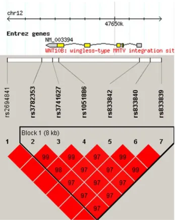
SNPs in the WNT10B gene region showed strong LD and were grouped in a single haplotypic block (Fig.1). The allelic frequencies were similar to those reported in other Caucasian populations (Table1) and there was no evidence for departure from Hardy–Weinberg equilibrium.
Association with Bone Mineral Density
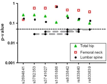
Allelic variations of the SNPs analyzed were not associated with BMD at either the lumbar spine or the hip (Fig.2). In
the multilocus analysis, several haplotypes were associated with spine BMD, with P-values between 0.03 and 0.05 (Fig.2), but they were no longer significant after multiple-test adjustment. The strongest association was found for the GTG haplotype (loci rs3782353, rs3741627, and rs833842). Spine BMD was 2.4% higher in women bearing at least one copy of the GTG haplotype as in women with no copies. The inclusion of all six SNPs in the haplotypic block established by the Gabriel method did not result in a stronger association. The polymorphisms were not associ-ated with hip BMD, hip BMC, or average femoral neck diameter.
Fig. 1 Linkage disequilibrium and resulting haplotypic block struc-ture of the WNT10B region. The numbers represent theD0 distances
Table 1 Polymorphisms included in the present study
SNP Position Location Alleles MAF rs2694841 47642801 30downstream A/G 0.03
rs3782353 47645147 30downstream A/G 0.45 rs3741627 47645854 30-UTR G/T 0.45 rs1051886 47646256 Exon 5 T/C 0.45 rs833842 47651690 50-UTR G/T 0.38 rs833840 47652417 50upstream C/G 0.39
Association with Vertebral and Hip Fractures
There was no significant association with vertebral frac-tures in the single-locus (Table2) or the haplotypic anal-ysis (not shown). Likewise, no association with hip fractures was detected in either analysis. Since women with hip fractures tended to be older than those in the control group, the analysis was repeated including only women \80 years of age (the median in the hip fracture group) and selecting age-matched controls or adjusting by age as a model covariate. No association between genotype and hip fractures was detected in those analyses.
Wnt Ligand Expression in Bone Tissue
The expression of several Wnt ligands in bone samples is shown in Fig.3. WNT3A, WNT4, WNT7A, and WNT10A were preferentially expressed in bone, in comparison with the reference RNA (nominalP-values\0.0015 and\0.05 after multiple-test correction). WNT10B expression in
bone was similar to that in reference RNA. No significant correlations existed between WNT10B expression in bone samples and the alleles present at the seven polymorphic loci studied (not shown).
Discussion
The Wnt pathway has emerged as an important regulator of skeletal development and is also involved in bone homeostasis in adult organisms. The role of WNT10B in human skeletal development has been revealed by the identification of an homozygous missense WNT10B mutation as a cause of the Split-Han/Foot malformation syndrome [19]. In general, Wnt pathway activation has an anabolic effect on adult bone, which is the consequence, at least in part, of a positive influence on the differentiation of osteoblast precursors. However, differences among the various Wnt molecules exist. Whereas some Wnt ligands stimulate differentiation toward the osteoblastic lineage, others, such as Wnt4 and Wnt5A, tend to increase the expression of adipogenesis-related genes [20]. Wnt10B has been shown to be one of the osteoblast-differentiating ligands, whereas it inhibits the differentiation of common precursors toward the adipocytic lineage, in part by sup-pressing the expression of C/EBPa and PPARc [21]. Studies with transgenic mice have shown that increased WNT10B expression is associated with a higher bone mass, an effect primarily due to increased osteoblasto-genesis [22]. Although the mechanisms controlling WNT10B expression are largely unknown, gene expression is increased by mechanical loading [10,23].
These data pointed toward WNT10B as an appealing candidate gene for osteoporosis. However, in the present study we found no consistent evidence for an association
Fig. 2 Association of WNT10B polymorphisms with BMD at the spine or the hip. NominalP-values of the single-locus and haplotype analysis (Armitage–Cochran trend test)
Table 2 Lack of association of WNT10B polymorphisms with osteoporotic fractures (nominalP-values)
SNP Vertebral fractures Hip fractures rs2694841 0.31 0.85 rs3782353 0.18 0.09 rs3741627 0.18 0.11 rs1051886 0.60 0.70 rs833842 0.27 0.28 rs833840 0.24 0.21 rs833839 0.25 0.31
between several polymorphisms of the gene and either BMD or osteoporotic fractures in Caucasian postmeno-pausal women. Thus, our results do not corroborate a recent report in men with African ancestry. In that study, Zmuda et al. sequenced the WNT10B gene and found several polymorphisms that had not been previously reported. Then they genotyped a set of eight SNPs in two groups, each one including about 1000 men with a mean age of 62 years. They found two SNPs (rs3741627 and rs1051886) to be associated with hip BMD in both groups, although the statistical significance was moderate (P\0.05). They also studied the bone structure at the distal radius by QCT and found that those SNPs were associated with BMC and bone size but not with trabecular volumetric BMD [14].
SNPs selected in the present study included tag SNPs capturing most common allelic variants of those repre-sented in the Hapmap Caucasian database, as well as some SNPs predicted to have functional consequences on gene transcription or translation. The tagging approach may result in the selection of different SNPs depending on the database used for reference. Thus, our SNP set included several SNPs different from those selected by Zmuda et al. but also the two SNPs found to be slightly associated with hip BMD in males in the study by Zmuda et al. However, in women we have not been able to demonstrate a con-sistent association between SNPs and BMD strong enough to resist multiple-test adjustment. The failure to replicate the results of Zmuda et al. suggests that the influence of WNT10B variants may depend on the gender or race of the individuals. In fact, the allelic frequencies found in this study were similar to those found in other Caucasian populations and to the frequencies reported by Zmuda et al. in a small Caucasian group but quite different from the results in individuals of African ancestry. On the other hand, there appears to be ethnicity-dependent differences in the linkage disequilibrium pattern in the Wnt10b region. Thus, in our study, as well as in the CEU (Caucasian) Hapmap population, the SNPs were grouped in a single haplotypic block. On the contrary, in populations of Afri-can origin (such as the Yoruba Hapmap population and the individuals studied by Zmuda et al.), two separate blocks can be recognized.
WNT10B expression was readily detected in human trabecular bone samples, in similar amounts to a reference mixture of RNA from 10 different cell lines. However, we did not find genotype-related differences in gene expres-sion. Similarly, we did not find an association between genotype and gross measures of skeletal size (such as height or average neck diameter of the femoral neck esti-mated from DXA output). Although BMD measurements by DXA are obviously influenced by both cortical and trabecular bone components, we do not have data on
specific measurements of the cortical bone structure by qCT. Since, according to Zmuda et al. [14], the WNT10B polymorphisms may preferentially influence the cortical bone, it could also be argued that gender and racial factors influence the power to detect the genetic affects, which could be more readily apparent in black men with thick cortices than in Caucasian postmenopausal women with thinner bones. Nevertheless, this remains speculative at the moment, and although we cannot exclude the possibility that WNT10B polymorphisms exert some influence on the cortical bone in Caucasian women, such influence is probably small, if any, as we found no association with hip fractures.
Our study has some limitations. We excluded patients with diseases that could impact bone homeostasis, but we did not exclude some subclinical disorders such as vitamin D insufficiency that might interact with the genetic factors. The age distributions of patients with hip fractures and controls were somewhat different. This might have had some influence toward the null effect, as some individuals included in the control group could fracture in the future. Nevertheless, we tried to limit this influence by performing age-adjusted analyses and subgroup age-restricted analyses (to compare age-matched patient and control groups), which failed to show an association of genotypes with fractures. With analevel of 0.05, the study had 90% power to detect an association of SNPs with bone phenotypes, if the genotypes explained at least 1% of BMD variance or were associated with odds ratios[1.3 for hip fractures or [1.5 for vertebral fractures. The sample size does not allow the exclusion of a small effect below those thresh-olds. On the other hand, we cannot exclude an influence of very rare allelic variants. But even if they have marked effects at the individual level, the results of resequencing studies [14] suggest that such variants are unlikely to play an important role at the population level. The limited number of bone samples available to study gene expression and the wide interindividual variations limited the power to analyze the genotype influence on gene expression (which could be roughly estimated to be just above 30% for 1-log differences across opposing homozygotes).
In summary, WNT10B is expressed in human bone tissue, but we have not been able to demonstrate a robust association of several common polymorphisms in the WNT10B region with BMD or fractures in postmenopausal women. These results argue against a major influence of common polymorphisms of the WNT10B gene on bone mass and the risk of osteoporosis in Caucasian postmeno-pausal women.
References
1. Ralston SH, de Crombrugghe B (2006) Genetic regulation of bone mass and susceptibility to osteoporosis. Genes Dev 20:2492–2506
2. Lei SF, Jiang H, Deng FY et al (2007) Searching for genes underlying susceptibility to osteoporotic fracture: current pro-gress and future prospect. Osteoporos Int 18:1157–1175 3. Little RD, Carulli JP, Del Mastro RG et al (2002) A mutation in
the LDL receptor-related protein 5 gene results in the autosomal dominant high-bone-mass trait. Am J Hum Genet 70:11–19 4. Glass DA, Karsenty G (2007) In vivo analysis of Wnt signaling in
bone. Endocrinology 148:2630–2634
5. Krishnan V, Bryant HU, MacDougald OA (2006) Regulation of bone mass by Wnt signaling. J Clin Invest 116:1202–1209 6. Wang HY, Liu T, Malbon CC (2006) Structure-function analysis
of Frizzleds. Cell Signal 18:934–941
7. Kikuchi A, Yamamoto H, Kishida S (2007) Multiplicity of the interactions of Wnt proteins and their receptors. Cell Signal 19:659–671
8. Bennett CN, Longo KA, Wright WS et al (2005) Regulation of osteoblastogenesis and bone mass by Wnt10b. Proc Natl Acad Sci USA 102:3324–3329
9. Zhou H, Mak W, Zheng Y et al (2008) Osteoblasts directly control lineage commitment of mesenchymal progenitor cells through Wnt signaling. J Biol Chem 283:1936–1945
10. Robinson JA, Chatterjee-Kishore M, Yaworsky PJ et al (2006) Wnt/beta-catenin signaling is a normal physiological response to mechanical loading in bone. J Biol Chem 281:31720–31728 11. Bonewald LF, Johnson ML (2008) Osteocytes, mechanosensing
and Wnt signaling. Bone 42:606–615
12. Barrett JC, Fry B, Maller J et al (2005) Haploview: analysis and visualization of LD and haplotype maps. Bioinformatics 21:263–265 13. Conde L, Vaquerizas JM, Dopazo H et al (2006) PupaSuite: finding functional single nucleotide polymorphisms for large-scale genotyping purposes. Nucleic Acids Res 34:W621–W625
14. Zmuda JM, Yerges LM, Kammerer CM et al (2009) Association analysis of WNT10B with bone mass and structure among indi-viduals of African ancestry. J Bone Miner Res 24:437–447 15. Livak KJ, Schmittgen TD (2001) Analysis of relative gene
expression data using real-time quantitative PCR and the 2(–Delta Delta C(T)) method. Methods 25:402–408
16. Purcell S, Neale B, Todd-Brown K et al (2007) PLINK: a tool set for whole-genome association and population-based linkage analyses. Am J Hum Genet 81:559–575
17. Neale BM, Ferreira MAR, Medland SE, Posthuma D (2007) Statistical genetics: gene mapping through linkage and associa-tion. Taylor and Francis, London
18. Gauderman WJ (2002) Sample size requirements for association studies of gene-gene interaction. Am J Epidemiol 155:478–484 19. Ugur SA, Tolun A (2008) Homozygous WNT10b mutation and
complex inheritance in split-hand/foot malformation. Hum Mol Genet 17:2644–2653
20. Nishizuka M, Koyanagi A, Osada S et al (2008) Wnt4 and Wnt5a promote adipocyte differentiation. FEBS Lett 582:3201–3205 21. Kang S, Bennett CN, Gerin I et al (2007) Wnt signaling
stimu-lates osteoblastogenesis of mesenchymal precursors by sup-pressing CCAAT/enhancer-binding protein alpha and peroxisome proliferator-activated receptor gamma. J Biol Chem 282:14515– 14524
22. Bennett CN, Ouyang H, Ma YL et al (2007) Wnt10b increases postnatal bone formation by enhancing osteoblast differentiation. J Bone Miner Res 22:1924–1932