Effects of constitutive deletion of opioid receptors on the basal densities of Fas and Fas associated protein with detah domain (FADD) in the mouse brain: A delta opioid tone inhibits FADD
9
0
0
Texto completo
(2) Effects of deletion of opioid receptors on the basal densities of Fas and Fas-associated protein. 367. (6.5−11.0-fold). The results suggest that μ-receptors tonically stimulate (through endogenous opioid peptides) the activation of native Fas, whereas δ-receptors tonically inhibit the expression of Fas aggregates and that of FADD and phosphorylated FADD (Ser191) in the mouse brain. These data are in line with the acute opposite modulation of Fas and FADD induced by μ- and δ-opiate agonists, and strongly support the notion of an anti-apoptotic δ-opioid tone that restrains Fas signaling. © 2006 Elsevier B.V. and ECNP. All rights reserved.. 1. Introduction The Fas receptor (APO-1/CD95) plays a major role in the physiologically regulated cell death or apoptosis (e.g., in normal brain development), and its dysregulation is a crucial component in a number of disease states, including neurodegenerative disorders (Sastry and Rao, 2000; Sharma et al., 2000). Although initially restricted to the immune system, Fas protein is widely expressed in normal tissues (WatanabeFukunaga et al., 1992), including the mammalian brain (Bechmann et al., 1999). Since Fas does not possess known enzymatic activities, proteins that interact with its cytoplasmic domain regulate the death signal. Briefly, after stimulation and trimerization (Fas/Fas-L), the receptor recruits procaspase-8 through the adaptor FADD (Fas-Associated protein with Death Domain), which transmits the death signal by activation of effector caspases leading to cell death (Algeciras-Schimnich et al., 2002; Tourneur et al., 2005). Besides the primary role of Fas/FADD in apoptosis, accumulating evidences also indicate the relevance of this system in promoting nonapoptotic signals associated with cell growth and differentiation (Budd, 2002; Tibbetts et al., 2003), including regenerative processes in neurons (Desbarats et al., 2003; Lambert et al., 2003) and the formation of new neurites (Zuliani et al., 2006). FADD carries a death domain (DD) at its C-terminal region (involved in the binding of Fas) that also displays, outside the DD, a single serine phosphorylation site (mouse/rat, Ser191; human, Ser194), which appears to be crucial for its role in the regulation of growth and proliferation (Zhang et al., 2004). In fact, phosphorylation of FADD at Ser191/194 by casein kinase Iα has been demonstrated to regulate various nonapoptotic activities of this Fas-adaptor protein (Alappat et al., 2005). Recently, the content of Fas in the rat brain (i.e., native and glycosylated receptor, and Fas aggregates) was shown to be increased after chronic treatment with opiate drugs (heroin and morphine) and during the induction of opiate withdrawal (Boronat et al., 2001; García-Fuster et al., 2003, 2004a), which demonstrated the involvement of Fas receptor in opiate addiction. Moreover, the content of native (monomeric) Fas was also upregulated by acute treatments with heroin and morphine, but not with selective δ- or κ-opioid receptor agonists, indicating that the rapid modulation of native Fas in the rat brain is related to the activation of μ-opioid receptors (García-Fuster et al., 2003, 2004a). In contrast, the content of Fas aggregates (complexes of monomers relevant in Fas signaling) was acutely modulated (reduced) only by the selective δ-agonist SNC-80 (García-Fuster et al., 2004a). On the other hand, acute treatments with μ-, δ- and κ-agonists were associated, through specific opioid receptor mechanisms, with decreases of FADD content in the rat brain, i.e., an. opposite modulation to that observed for native Fas (GarcíaFuster et al., in press). These results suggested that the three types of opioid receptors might have a role in the rapid modulation (inhibition) of this key adaptor protein in the proapoptotic or nonapoptotic function of Fas signaling. Lines of mice lacking one of the opioid receptor types (gene knock-out technology) are useful experimental tools in determining the role of these receptors in the various physiological and pharmacological effects of opioids (Kieffer and Gavériaux-Ruff, 2002), and also to unravel the possible existence of endogenous opioid tones regulating some of their functions (e.g., for the δ-opioid receptor see Nadal et al., 2006). Therefore, the aim of this study was to investigate the influence of constitutive deletion of μ-, δ- or κ-opioid receptors on the basal densities of Fas receptor and FADD coupling protein, both of which are acutely and oppositely regulated by opiate drugs in the brain. A preliminary report of this work was given at the XXVI Congress of the Spanish Society of Pharmacology (García-Fuster et al., 2004b).. 2. Experimental procedures 2.1. Opioid receptor-deficient mice Homozygous knock-out (KO) mice without μ (MOP)-, δ (DOP)- or κ (KOP)-opioid receptor and their respective wild-type (WT) littermates were used (Matthes et al., 1996; Filliol et al., 2000; Simonin et al., 1998). All mice were under a hybrid 129 SV/C57BL/6 (50%/50%) genetic background. Previous studies have shown that the genetic ablation of a specific type of opioid receptor did not result in major changes in the density of the other opioid receptor sites (Kieffer and Gavériaux-Ruff, 2002). Mice (30−35 g, 16−20 weeks old) were housed five per cage in a temperature-controlled room (21 ± 1 °C) with a 12-h light/dark cycle (light on between 8:00 and 20:00 h) and with food and water available ad libitum. Mice were acclimated to handling for 1 week, and then all animals received an acute saline injection. A total of 30 mice were used in these experiments, which were divided in three groups with the following composition: group I (five WT saline-treated and five μ-KO saline-treated), group II (five WT saline-treated, five δ-KO salinetreated) and group III (five WT saline-treated, five κ-KO salinetreated). The animals were killed by decapitation and the brains rapidly removed and frozen in liquid nitrogen. Specimens of the cerebral cortex were dissected on ice and stored at −80 °C until assays. These experiments in mice were performed according to standard ethical guidelines (European Communities Council Directive 86/609/ EEC) and approved by the local ethical committees. All efforts were made to minimize the number of animals used and their suffering.. 2.2. Immunoblot assays and quantitation of Fas and FADD proteins The mouse brain samples (crude total homogenate) were prepared (including various protease inhibitors) as described in detail.
(3) 368 previously (García-Sevilla et al., 2004). Protein concentrations were determined by the biuret reaction using bicinchoninic acid for colorimetric detection of cuprous cation (BCA, Protein Assay Reagent, Pierce Chemical Company, Rockford, IL, USA). In routine experiments, 40 μg protein of each mouse brain sample was subjected to SDS-PAGE on 10% polyacrylamide minigels (Bio-Rad Laboratories, Hercules, CA, USA). Proteins were electrophoretically transferred to nitrocellulose membranes and incubated (overnight at 4 °C) in a blocking solution containing the appropriate primary antibody: anti-Fas M-20 (affinity-purified rabbit polyclonal antibody raised against a peptide mapping the C-terminal of Fas of mouse origin; dilution 1:5000; sc-716, batches D-219 and H-301, Santa Cruz Biotechnology, CA, USA), anti-FADD H-181 (affinity−purified rabbit polyclonal antibody raised against human FADD C-terminal 28−208 residues; dilution 1:5000; sc-5559, batch C-112, Santa Cruz Biotechnology), anti-phosphorylated FADD (Ser191) (affinity-purified rabbit polyclonal antibody raised against a peptide mapping at residues surrounding Ser191 of mouse FADD; dilution 1:600; batch 12785L, Cell Signaling Technology, MA, USA) and anti-β-actin (mouse monoclonal antibody raised against a peptide mapping the Nterminal of β-actin of mouse origin; dilution 1:10,000; clone AC-15; batch no. 014K4840, Sigma Chemical Co., MO, USA). To test the selectivity of anti-Fas and anti-FADD antibodies in the mouse brain, the corresponding antigenic peptides were preincubated in excess with the antisera, which resulted in the blockade of the immunoreaction for the specific proteins (data not shown; see García-Fuster et al., 2003, 2004a, in press). In some experiments, the specificity of the anti−phosphorylated FADD antibody for the targeted epitope (Ser191) was tested on Western blots of rat brain tissue (cerebral cortex, total homogenate and cytosolic fraction) pretreated with alkaline phosphatase, as described in detail previously (GarcíaFuster et al., in press). The secondary antibody, horseradish peroxidase-linked anti-rabbit IgG or anti-mouse IgG, was incubated at 1:5000 dilution in blocking solution at room temperature for 1 h. The immunoreactivity of target proteins was detected with the Enhanced Chemiluminescence (ECL) Western Blot Detection system (Amersham International, Buckinghamshire, UK) and visualized by exposure to Hyperfilm ECL film (Amersham) for 30 s to 60 min (autoradiograms). The autoradiograms were quantitated by densitometric scanning of the immunoreactive bands (IOD units) (GS−800 Imaging Densitometer, Bio−Rad). For a direct comparison, two groups of five mice were run together in the same gel to assess for differences in opioid receptor genotypes (i.e., WT versus KO) and 40 μg of protein (within the linear range for immunolabeling of Fas and FADD) were loaded in each lane for the different blots. The experiment was quantitated and then repeated one to three times to confirm the results. This procedure allowed the evaluation of 10–20 samples for each subgroup of mice and/or protein. Finally, percent changes in immunoreactivity with respect to control (WT) samples (100%) were calculated for each experimental sample in the various gels, and the mean value was used as a final estimate. In all experiments, the content of β-actin (a cytoskeletal protein not modulated by opiate drugs; Ammon et al., 2003; Marie-Claire et al., 2004) was quantitated as a negative (loading) control.. 2.3. Data and statistical analysis All series of data were analyzed with the program GraphPad Prism™, version 3.0. Results are expressed as mean values ± standard error of the mean (S.E.M.). Two-tailed Student's t-test was used for the statistical evaluations. The level of significance was chosen as P ≤ 0.05.. 2.4. Chemicals Acrylamide (Protogel) was from Conda, S.A., Spain. Calf intestinal mucosa alkaline phosphatase (Product 79390) was from Sigma-. M.J. García-Fuster et al.. Figure 1 Effects of μ-opioid receptor genotype (WT versus μ-KO) on the basal immunodensities of Fas aggregates (∼203 kDa and 120 kDa, both quantitated as doublets), glycosylated receptor forms (∼51/48/45 kDa) and native receptor (∼35 kDa) in the mouse cerebral cortex (n = 5 for each group). The columns are mean± S.E.M. values (% immunoreactivity) of three experiments per group (i.e., 15 samples analyzed in each subgroup of mice, see Methods) and expressed as percentage of saline-treated WT mice. ⁎P= 0.025 when compared with the corresponding saline (WT) group (Student's t-test). Also note that the total content of Fas aggregates (203 plus 120 kDa forms) in μ-KO mice (IODs: 23.4 ± 2.7, n = 5) did not significantly differ (t = 1.63, P > 0.05) than that in WT mice (IODs: 29.2 ± 2.3, n = 5). Bottom: Representative immunoblot (40 μg protein) for the various forms of Fas in WT and μ-KO mice (the upper and lower parts of the same immunoblot are shown separated to illustrate the longer time exposure required to visualize native 35 kDa Fas). The apparent molecular masses of Fas were determined by calibrating the blots with prestained molecular weight markers as shown on the left-hand side..
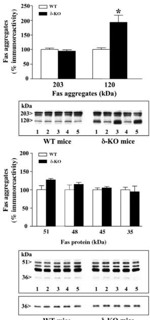
(4) Effects of deletion of opioid receptors on the basal densities of Fas and Fas-associated protein. 369. Aldrich, Germany. Other materials such as the secondary antibodies, ECL reagents and autoradiography films were purchased from Amersham International (UK) or Santa Cruz Biotechnology (USA). All other chemicals were from Sigma Chemical.. 3. Results 3.1. Basal immunodensities of Fas receptor forms in WT and opioid receptor KO mice Various specific forms of Fas were immunodetected in the mouse cerebral cortex (i.e., ∼203/120 kDa aggregates, ∼ 51/48/45 kDa glycosylated forms and ∼ 35 kDa native receptor) (Figs. 1−3), in good agreement with previous observations in rat and human brains (García-Fuster et al., 2003, 2004a). In the cerebral cortex of μ- and κ-opioid receptor KO mice, the basal immunodensities of Fas aggregates (203 kDa and 120 kDa) did not significantly differ from those in brains of their respective WT littermates (Figs. 1 and 3). In δ-opioid receptor KO mice, however, the basal immunodensity of 120 kDa Fas aggregates (putative trimeric receptors), but not that of higher-order oligomerization forms (203 kDa), was found markedly increased in the cerebral cortex (93%, t = 3.72, P = 0.009) (Fig. 2). The total content of Fas aggregates (203 plus 120 kDa forms) was also augmented in the cortex of δ-opioid receptor−deficient mice (35%, t = 2.66, P < 0.05). In μ-opioid receptor, but not in δ- or κ-, KO mice, the basal immunodensity of native Fas (35 kDa) was significantly decreased in the cerebral cortex (33%, t = 2.75, P = 0.025) (Figs. 1–3). In the corpus striatum of μ−deficient mice, native Fas was not significantly reduced (data not shown) (WT: 100 ± 6%, n = 5; μ-KO: 89 ± 12%, n = 5, P > 0.05). In μ-, δ- and κ-opioid receptor KO mice, the basal immunodensities of glycosylated Fas (51/48/45 kDa) in the cerebral cortex were similar to those in WT mice (Figs. 1−3). The constitutive deletion of opioid receptors did not alter the immunodensity of β-actin in the brain, a cytoskeletal protein that was used as a negative (loading) control (data not shown). Moreover, opioid receptor deletion did not modify the immunodensity of an unidentified peptide (∼65 kDa), not related to Fas (see García-Fuster et al., 2003), which was also quantitated in the same immunoblots (data not shown).. 3.2. Basal immunodensities of FADD protein and phosphorylated FADD (Ser191) in WT and opioid receptor KO mice In brain tissue (rat, mouse, human) FADD molecules, similarly to Fas, have the ability to aggregate being immunodetected as homodimers (about 51 kDa) in the cerebral cortex of WT and opioid receptor KO mice (Fig. 4) (see García-Fuster et al., in press). In the cerebral cortex of μ- and κ-opioid receptor KO mice, the basal immunodensities of FADD did not significantly differ from those in brains of their respective WT littermates (Fig. 4). In contrast, the basal content of FADD was found increased in the cortices of δ-opioid receptor KO mice (48%, t = 2.70, P = 0.02) (Fig. 4). As an extension of this finding, a similar increase in FADD density was also quantitated in the. Figure 2 Effects of δ-opioid receptor genotype (WT versus δ-KO) on the basal immunodensities of Fas aggregates (∼ 203 kDa and 120 kDa, both quantitated as doublets), glycosylated receptor forms (∼ 51/48/45 kDa) and native receptor (∼35 kDa) in the mouse cerebral cortex (n = 5 for each group). The columns are mean ± S.E.M. values (% immunoreactivity) of three experiments per group (i.e., 15 samples analyzed in each subgroup of mice, see Methods) and expressed as percentage of saline-treated WT mice. ⁎P = 0.009 when compared with the corresponding saline (WT) group (Student's t-test). Note that the total content of Fas aggregates (203 plus 120 kDa forms) in δ-KO mice (IODs: 30.0 ± 2.6, n = 5) was also significantly greater (35%, t = 2.66, P < 0.05) than that in WT mice (IODs: 22.3 ± 1.3, n = 5). Bottom: Representative immunoblot (40 μg protein) for the various forms of Fas in WT and δ-KO mice (the upper and lower parts of the same immunoblot are shown separated to illustrate the longer time exposure required to visualize native 35 kDa Fas). The apparent molecular masses of Fas were determined by calibrating the blots with prestained molecular weight markers as shown on the left-hand side..
(5) 370. M.J. García-Fuster et al. corpus striatum of δ-deficient mice (WT: 100 ± 6%, n = 5; δ-KO: 131 ± 6%, n = 5, t = 3.67, P = 0.006) (data not shown). In the rat cerebral cortex, the enzymatic dephosphorylation of FADD with alkaline phosphatase (total homogenate and cytosolic fraction) completely abolished the immunoreactivity of the antibody used against phosphorylated FADD at Ser191, which demonstrated the specificity of the detected phosphorylated species of FADD (about 37 kDa) (Fig. 5A). In the mouse cerebral cortex, a low level of FADD phosphorylation (Ser191) was immunodetected and the weak signals were difficult to quantitate (Fig. 5B). However, it was clearly apparent that in δ-opioid receptor KO mice the basal content of phosphorylated FADD (Ser191) in the cortex was increased compared to that in WT mice (WT, IOD units: 0.18 ± 0.03, n = 5; KO, IOD units: 1.99 ± 0.60, n = 5; 11-fold increase, P < 0.02) (Fig. 5B). In contrast, small decreases in the contents of phosphorylated FADD (Ser191) were observed in the cortex of μ- and κ-opioid receptor KO mice (Fig. 5B). Again, the constitutive deletion of opioid receptors was not associated with alterations in the immunodensities of β-actin in the brains of these animals (Fig. 5C).. Figure 3 Effects of κ-opioid receptor genotype (WT versus κ-KO) on the basal immunodensities of Fas aggregates (∼203 kDa and 120 kDa, both quantitated as doublets), glycosylated receptor forms (∼51/48/45 kDa) and native receptor (∼35 kDa) in the mouse cerebral cortex (n = 5 for each group). The columns are mean± S.E.M. values (% immunoreactivity) of two experiments per group (i.e., 10 samples analyzed in each subgroup of mice, see Methods) and expressed as percentage of salinetreated WT mice. No significant differences were quantitated between genotypes. Also note that the total content of Fas aggregates (203 plus 120 kDa forms) in κ-KO mice (IODs: 17.0 ± 0.9, n = 5) did not significantly differ (t = 0.92, P > 0.05) than that in WT mice (IODs: 19.8 ± 2.9, n = 5). Bottom: Representative immunoblot (40 μg protein) for the various forms of Fas in WT and κ-KO mice (the upper and lower parts of the same immunoblot are shown separated to illustrate the longer time exposure required to visualize native 35 kDa Fas). The apparent molecular masses of Fas were determined by calibrating the blots with prestained molecular weight markers as shown on the left-hand side.. Figure 4 Effects of μ-, δ- and κ-opioid receptor genotype (WT versus μ/δ/κ-KO) on the basal immunodensity of Fas-associated protein with death domain (FADD) in the mouse cerebral cortex (n = 5 for each group). The columns are mean± S.E.M. values (% immunoreactivity) of three to four experiments per group (i.e., 15–20 samples analyzed in each subgroup of mice, see Methods) and expressed as percentage of the corresponding saline-treated WT mice. ⁎P = 0.02 when compared with the corresponding saline (WT) group (Student's t-test). Bottom: Representative immunoblots (40 μg protein) for FADD protein in WTand μ-KO, δ-KO and κKO mice. The apparent molecular masses of Fas were determined by calibrating the blots with prestained molecular weight markers as shown on the left-hand side..
(6) Effects of deletion of opioid receptors on the basal densities of Fas and Fas-associated protein. 4. Discussion WT and opioid receptor mutant (μ-, δ- or κ-KO) mice were compared to assess (in the absence of exogenous opiates) if any endogenous opioid tone regulates the basal expression of Fas receptor and/or FADD coupling protein in the brain. The basal immunodensity of monomeric Fas (35 kDa form) was reduced in the cerebral cortex, but not in the corpus striatum, of μ-opioid receptor-deficient mice. This result may suggest that endogenous opioid peptides acting on μ−receptors tonically stimulate the activation of native Fas in the mouse cortex, which would be in agreement with the acute stimulatory effects of heroin and morphine on native Fas observed in the rat cerebral cortex (García-Fuster et al., 2003). In contrast, no opioid receptor genotype differences (WT versus μ-, δ- and κ-KO mice) were observed in the basal expression of glycosylated Fas (51/48/ 45 kDa forms). Similarly, the content of 48 kDa glycosylated Fas. 371. was shown to be unchanged in splenocytes from μ-KO mice (Wang et al., 2002). The endogenous regulation of the apoptotic Fas/FADD complex was more clearly apparent in brains of δ−opioid receptor-deficient mice. In these animals, but not in μ- and κ-receptor KO mice, the basal density of Fas aggregates (120 kDa trimeric receptors relevant in triggering receptor signaling; see Algeciras-Schimnich et al., 2002) was increased in the cerebral cortex (93%). Interestingly, the content of FADD (the adaptor protein that couples Fas to caspases and transmits the death signal; Algeciras-Schimnich et al., 2002; Tourneur et al., 2005) was also upregulated in the cortex (48%) and corpus striatum (31%) of δ-receptordeficient mice. These results suggested that δ-opioid receptors tonically inhibit the expression of trimeric Fas and FADD protein in these mouse brain structures (i.e., removal of a negative endogenous tone resulting in protein upregulation). The lack of significant changes in basal Fas/ FADD in μ- and κ-opioid receptor KO mice indicated a low tone, if any, of μ/κ-opioid system to control the expression of this apoptotic complex in the brain. In agreement with these findings, the selective δ-agonist SNC-80 decreased the content of 120 kDa trimeric Fas in the rat cerebral cortex (García-Fuster et al., 2004a) and also the density of FADD in the rat brain (García-Fuster et al., in press). The molecular mechanism by which the endogenous δ-opioid receptor tone operates modulating the basal expression of FADD in the brain is still unknown. It is known, however, that δ-opioid receptor agonists (and particularly SNC-80, a nonpeptidic and selective agonist) potently stimulate the activation of the MAPK ERK pathway through a pertussis toxin-sensitive mechanism (i.e., involving Gαi proteins) (Audet et al., 2005). Notably, the inhibitory effect of SNC-80 on FADD content in the rat brain (cerebral cortex and corpus striatum) was mediated through a mechanism fully dependent on the activation of ERK1/2 signaling (i.e., the effect was blocked by SL-327, a specific. Figure 5 (A) Representative immunoblot for the effect of dephosphorylation with alkaline phosphatase on phosphorylated FADD (Ser191) immunoreactivity in the rat cerebral cortex. Brain tissue (H: total homogenate, S2: cytosolic fraction) was incubated at 40 °C for 15 min in the absence (C, control samples) or presence of alkaline phosphatase (AP, 95 units). Samples containing the enzyme were also incubated with 100 mM sodium pirophosphate (IC, inhibited controls). The amount of protein loaded on the gel was 40 μg for all samples. Note that tissue enzymatic dephosphorylation abolished the immunoreactivity of the anti-phosphorylated FADD (Ser191) antibody, demonstrating the specificity of the detected protein band. (B) Effects of μ-, δ- and κ-opioid receptor genotype (WT versus μ/δ/κ-KO) on the basal immunodensity of phosphorylated FADD (Ser191) in the mouse cerebral cortex (n = 5 for each group, 40 μg protein) (see Results for quantitative data). These experiments were repeated with similar results (e.g., δ-opioid receptor genotype: WT, IOD units: 0.19 ± 0.05, n = 5; KO, IOD units: 1.24 ± 0.38, n = 5; 6.5-fold increase, P < 0.05). (C) Representative immunoblots (40 μg protein) for the content of β-actin in WT mice and μ-KO, δ-KO and κ-KO mice. The apparent molecular masses of target proteins were determined by calibrating the blots with prestained molecular weight markers as shown on the left-hand side..
(7) 372 and brain-penetrating MEK1/2 inhibitor) (García-Fuster et al., in press), which in turn is crucial for the induction of various anti−apoptotic effects (Wada and Penninger, 2004) including protection against Fas-mediated apoptosis (Holmström et al., 1999). In a recent in vitro study (Narita et al., 2006), SNC-80 has been shown to promote neural differentiation and neuroprotection through the release of BDNF and activation of its Trk-dependent tyrosine kinase receptor (TrkB) linked to other pathways, including MEK/ERK signaling (i.e., the effects were blocked by the MEK1/2 inhibitor PD98059). In this context, it is of interest to note that the basal content of phosphorylated FADD (Ser191) (a specific form of about 37 kDa) was also upregulated in the cerebral cortex of δopioid receptor KO mice, which is in line with the postulated role of this phosphorylated form of FADD in promoting nonapoptotic signals (Zhang et al., 2004; Alappat et al., 2005). Based on these observations and the demonstrated crosstalk between FADD and ERK (Lüschen et al., 2005), it is feasible that MEK/ERK signaling could also play a role (together with other pathways) regulating the endogenous tone of δ-opioid receptors on FADD (i.e., an inhibition mediated by enkephalins, the endogenous ligands). This possibility has been recently tested in rats with the MEK inhibitor SL-327 (10, 20 and 30 mg/kg, i.p., 90 min), but the compound by itself did not significantly alter the basal expression of FADD (expected increase) in the cerebral cortex and corpus striatum (García-Fuster et al., in press and unpublished results). SL-327 (30 mg/kg) modestly increased (15–20%) the content of monomeric FADD (about 25 kDa) phosphorylated at serine 194 (unpublished results). It should be mentioned that, whereas this range of doses for SL-327 is sufficient to counteract the inhibitory effect of SNC-80 on FADD (see above), higher doses of the MEK inhibitor (50 mg/kg) are needed to completely block ERK1/2 phosphorylation (number of activated ERK-positive neurons) in the mouse prefrontal cortex and dorsal striatum (see Valjent et al., 2006). Therefore, the possibility that MAPK ERK1/2 signaling could have a role, albeit minor, in regulating the inhibitory tone of δ-opioid receptors on FADD protein in the brain cannot be discarded. The use of KO mice has also demonstrated or confirmed the existence of endogenous opioid tones modulating some functions mediated by the various opioid receptors (e.g., behavioral alterations such as locomotion, nociception, and emotional and stress responses in KO mice; Kieffer and Gavériaux-Ruff, 2002; Martin et al., 2003; Contet et al., 2006; Nadal et al., 2006), showing in addition that the absence of a particular opioid receptor does not modify the expression of endogenous opioid peptide ligands (Kieffer and GavériauxRuff, 2002). Concerning the modulation of signaling proteins associated with opioid receptors, these genetic models have also revealed the existence of an endogenous activity of μ- and δ-opioid receptors, but not κ-receptors, regulating, in part, the phosphorylation state of the transcription factor CREB (GarcíaSevilla et al., 2004). In these genetic studies, the possibility of developmental compensations after deletion of the different opioid receptors and/or influences of the hybrid genetic background on the observed changes cannot be fully discarded. However, pharmacologic approaches in intact animals (i.e., acute in vivo effects of selective opioid receptor antagonists) have also demonstrated that endogenous opioids and/or constitutive activity of μ-opioid receptors may regulate c-Fos. M.J. García-Fuster et al. protein expression (activation of neurones) in the rat brain (Le Guen et al., 2003), and the function of excitatory synapses by modulating the density of dendritic spines in rats and mice (Liao et al., 2005). Moreover, the δ-opioid receptor system has been recently shown to regulate anxiety-like behavior in an anxiolytic (with the agonist SNC-80) and anxiogenic (with the antagonist naltrindole) manner, further suggesting the existence of an endogenous opioid tone (modulating a basal level of anxiety) mediated by δ-opioid receptors (Perrine et al., 2006; see also Filliol et al., 2000). Finally, genetic and pharmacological antagonism studies have also revealed the existence of an endogenous κ-opioid system in the brain that tonically inhibits mesoaccumbal dopaminergic neurotransmission (Chefer et al., 2005). These findings together demonstrate the inequivocal existence and relevance of endogenous opioid tones regulating many aspects of opioid functions. It is well-known that μ- and δ-opioid receptors can physically and functionally interact with each other (Matthes et al., 1998; Gomes et al., 2004), which supported the concept that activation of these receptors induces similar biological effects (Traynor and Elliot, 1993). However, the observation of some behavioral responses of δ-deficient mice opposing those of μ-deficient mice (Filliol et al., 2000; Kieffer and Gavériaux-Ruff, 2002) suggests that δ-opioid receptors may serve specific independent functions (see also Hauser et al., 2000; Chefer et al., 2004). In the current study, the comparison of mice lacking the μ- and δ-opioid-eceptor genes demonstrates that these opioid receptors can behave oppositely in regulating the apoptotic Fas/FADD complex. Thus, the μ-opioid receptor appears to be associated with a tonic stimulation of monomeric Fas receptor (immunocontent decreased in μKO mice), whereas the δ-opioid receptor would tonically inhibit the basal density of trimeric Fas as well as that of FADD and its phosphorylated form at Ser191 (immunocontents increased in δ-KO mice). This suggests possible homeostatic interactions between μ- and δ-opioid receptors in the control of Fas receptor machinery (monomeric versus trimeric Fas expression), being a major task of the δreceptor to restrain, through the input of endogenous opioid peptides, the activation of Fas signaling (trimeric Fas and FADD protein). The potential effect of μ-opioid receptor agonists in inducing apoptosis of neurons is controversial. Thus, some in vitro studies have demonstrated that morphine, through naloxone- and caspase-3-sensitive mechanisms, can induce apoptosis of enriched neuronal cell cultures (Hu et al., 2002), but other studies have documented the absence of morphine-induced apoptosis in cultured rat hippocampal neurons (Liao et al., 2005). In contrast, and in line with the current results, it has been demonstrated that activation of δ-opioid receptors can block the initiation of apoptosis (release of cytochrome c and translocation/oligomerization of Bax) induced by methamphetamine in the mouse brain in vivo (Tsao and Su, 2001) and to prevent H2O2-triggered apoptosis (activation of caspase-3) in cortical neuron/glia co-cultures of mice (Narita et al., 2006). Moreover, other in vitro studies have revealed that δ-receptor agonists can protect neocortical neurons from glutamate excitotoxicity (Zhang et al., 2000) or hypoxic stress (Zhang et al., 2002; Ma et al., 2005), and that naltrindole (a selective δ-receptor antagonist) can induce neuronal injury in normoxic.
(8) Effects of deletion of opioid receptors on the basal densities of Fas and Fas-associated protein cell cultures (Zhang et al., 2002). Furthermore, stimulation of δ-opioid receptors have been shown to promote other cytoprotective effects in various cell types, including neurons (Barry and Zuo, 2005; Narita et al., 2006). Therefore, it is tempting to conclude that possible apoptotic signals engaged by Fas activation after treatment with some opiate drugs (see Tegeder and Geisslinger, 2004; Pérez-San Emeterio et al., 2006) would be minimized by the tonic control of the δ-opioid receptor, which decreases signal transduction through the adaptor protein FADD.. Acknowledgments This work was supported by grants from Ministerio de Educación y Ciencia (SAF2004-03685, MEC/FEDER, Madrid, Spain) and Swiss National Foundation (P32-57066.99, FNSRS, Bern, Switzerland) to J.A.G.-S.; National Institutes of Health (1R01-DA016768-0111) and the European Commission (VI Programa Marco IP OJ 2004/C164, No. 005166, GENADDICT) to B.L.K. and R.M.; and MEC/FEDER (BFU200400920/BFI and GEN2003-20651-C06-04), Instituto de Salud Carlos III (04/1485, C03/06 and G03/005) and Generalitat de Catalunya (2002SGR00193) to R.M. The study was also supported by Red de Trastornos Adictivos (RETICS, Instituto de Salud Carlos III, MSC, Madrid, Spain). M.J.G.-F. was supported by a predoctoral fellowship from CSIC/MECAssociated Units (Madrid, Spain). J.A.G.-S. is a member of the Institut d'Estudis Catalans (Barcelona, Spain).. References Audet, N., Paquin-Gobeil, M., Landry-Paquet, O., Schiller, P.W., Piñeyro, G., 2005. Internalization and Src activity regulate the time course of ERK activation by delta opioid receptor ligands. J. Biol. Chem. 280, 7808–7816. Alappat, E., Feig, C., Boyerinas, B., Volkland, J., Samuels, M., Murmann, A.E., Thorburn, A., Kidd, V.J., Slaughter, C.A., Osborn, S.L., Winoto, A., Tang, W.J., Peter, M.E., 2005. Phosphorylation of FADD at serine 194 by CKIα regulates its nonapoptotic activities. Mol. Cell 19, 321–332. Algeciras-Schimnich, A., Shen, L., Barnhart, B.C., Murmann, A.E., Burkhardt, J.K., Peter, M.E., 2002. Molecular ordering of the initial signaling events of CD95. Mol. Cell. Biol. 22, 207–220. Ammon, S., Mayer, P., Riechert, U., Tischmeyer, H., Höllt, V., 2003. Microarray analysis of genes expressed in the frontal cortex of rats chronically treated with morphine and after naloxone precipitated withdrawal. Mol. Brain Res. 112, 113–125. Barry, U., Zuo, Z., 2005. Opioids: old drugs for potential new applications. Curr. Pharm. Des. 11, 1343–1350. Bechmann, I., Mor, G., Nilsen, J., Eliza, M., Nitsch, R., Naftolin, F., 1999. FasL (CD95L, Apo1L) is expressed in the normal rat and human brain. Glia 27, 62–74. Boronat, M.A., García-Fuster, M.J., García-Sevilla, J.A., 2001. Chronic morphine induces up-regulation of the pro-apoptotic Fas receptor and down-regulation of the anti-apoptotic Bcl-2 oncoprotein in rat brain. Br. J. Pharmacol. 134, 1263–1270. Budd, R.C., 2002. Death receptors couple to both cell proliferation and apoptosis. J. Clin. Invest. 109, 437–442. Chefer, V.I., Kieffer, B.L., Shippenberg, T.S., 2004. Contrasting effects of μ opioid receptor and δ opioid receptor deletion upon the behavioral and neurochemical effects of cocaine. Neuroscience 127, 497–503. Chefer, V.I., Czyzyk, T., Bolan, E.A., Moron, J., Pintar, J.E., Shippenberg, T.S., 2005. Endogenous κ-opioid receptor systems. 373. regulate mesoaccumbal dopamine dynamics and vulnerability to cocaine. J. Neurosci. 25, 5029–5037. Contet, C., Gavériaux-Ruff, C., Matifas, A., Caradec, C., Champy, M.-F., Kieffer, B.L., 2006. Dissociation of analgesic and hormonal responses to forced swim stress using opioid receptor knockout mice. Neurospsychopharmacology 31, 1733–1744. Desbarats, J., Birge, R.B., Minouni-Rongy, M., Weistein, D.E., Palerme, J.S., Newell, M.K., 2003. Fas engagement induces neurite growth through ERK activation and p35 upregulation. Nat. Cell Biol. 5, 118–125. Filliol, D., Ghozland, S., Chluba, J., Martín, M., Matthes, H.W., Simonin, F., Befort, K., Gavériaux-Ruff, C., Dierich, A., LeMeur, M., Valverde, O., Maldonado, R., Kieffer, B.L., 2000. Mice deficient for δ- and μ-opioid receptors exhibit opposing alterations of emotional responses. Nat. Genet. 25, 195–200. García-Fuster, M.J., Ferrer-Alcón, M., Miralles, A., GarcíaSevilla, J.A., 2003. Modulation of Fas receptor proteins and dynamin during opiate addiction and induction of opiate withdrawal in rat brain. Naunyn-Schmiedeberg's Arch. Pharmacol. 368, 421–431. García-Fuster, M.J., Ferrer-Alcón, M., Miralles, A., García-Sevilla, J.A., 2004a. Deglycosylation of Fas receptor and chronic morphine treatment up-regulate high molecular mass Fas aggregates in the rat brain. Eur. J. Pharmacol. 496, 63–69. García-Fuster, M.J., Miralles, A., Martín, M., Maldonado, R., GarcíaSevilla, J.A., 2004b. Fas receptor and Fas-associated death domain (FADD) in the cerebral cortex of μ-, δ-, or κ-opioid receptor knock-out mice. Methods Find. Exp. Clin. Pharmacol. 26 (Suppl. A), 78. García-Fuster, M.J., Miralles, A., García-Sevilla, J.A., in press. Effects of opiate drugs on Fas-associated protein with death domain (FADD) and effector caspases in the rat brain: regulation by the ERK1/2 MAP kinase pathway. Neuropsychopharmacology. Advance online publication: 8 February 2006, doi:10.1038/sj. npp.1301040. García-Sevilla, J.A., Ferrer-Alcón, M., Martín, M., Kieffer, B.L., Maldonado, R., 2004. Neurofilament proteins and cAMP pathway in brains of μ-, δ- or κ-opioid receptor gene knock-out mice: effects of chronic morphine administration. Neuropharmacology 46, 519–530. Gomes, I., Gupta, A., Filipovska, J., Szeto, H.H., Pintar, J.E., Devi, L.A., 2004. A role for heterodimerization of mu and delta opiate receptors in enhancing morphine analgesia. Proc. Natl. Acad. Sci. U. S. A. 101, 5135–5139. Hauser, K.F., Houdi, A.A., Turbek, C.S., Elde, R.P., Maxson III, W., 2000. Opioids intrinsically inhibit the genesis of mouse cerebellar granule neuron precursors in vitro: differential impact of μ and δ receptor activation on proliferation and neurite elongation. Eur. J. Neurosci. 12, 1281–1293. Holmström, T.H., Tran, S.E.F., Johnson, V.L., Ahn, N.G., Chow, S.C., Eriksson, J.E., 1999. Inhibition of mitogen-activated kinase signaling sensitizes HeLa cells to Fas receptor-mediated apoptosis. Mol. Cell. Biol. 19, 5991–6002. Hu, S., Sheng, W.S., Lokensgard, J.R., Peterson, P.K., 2002. Morphine induces apoptosis of human microglia and neurons. Neuropharmacology 42, 829–836. Kieffer, B.L., Gavériaux-Ruff, C., 2002. Exploring the opioid system by gene knockout. Prog. Neurobiol. 66, 285–306. Lambert, C., Landau, A.M., Desbarats, J., 2003. Fas-beyond death: a regenerative role for Fas in the nervous system. Apoptosis 8, 551–562. Le Guen, S., Gestreau, C., Besson, J.-M., 2003. Morphine withdrawal precipitated by specific mu, delta or kappa opioid receptor antagonists: a c-Fos protein study in the rat central nervous system. Eur. J. Neurosci. 17, 2425–2437. Liao, D., Lin, H., Law, P.Y., Loh, H.H., 2005. Mu-opioid receptors modulate the stability of dendritic spines. Proc. Natl. Acad. Sci. U. S. A. 102, 1725–1730..
(9) 374 Lüschen, S., Falk, M., Scherer, G., Ussat, S., Paulsen, M., AdamKlages, S., 2005. The Fas-associated death domain protein/ caspase-8/c-FLIP signaling pathway is involved in TNF-induced activation of ERK. Exp. Cell Res. 310, 33–42. Ma, M.C., Qian, H., Ghassemi, F., Zhao, P., Xia, Y., 2005. Oxygensensitive delta-opioid receptor-regulated survival and death signals: novel insights into neuronal preconditioning and protection. J. Biol. Chem. 280, 16208–16218. Marie-Claire, C., Courtin, C., Roques, B.P., Noble, F., 2004. Cytoskeletal genes regulation by chronic morphine treatment in rat striatum. Neuropsychopharmacology 29, 2208–2215. Martin, M., Matifas, A., Maldonado, R., Kieffer, B.L., 2003. Acute antinociceptive responses in single and combinatorial opioid receptor knockout mice: distinct mu, delta and kappa tones. Eur. J. Neurosci. 17, 701–708. Matthes, H.W.D., Maldonado, R., Simonin, F., Valverde, O., Slowe, S., Kitchen, I., Befort, K., Dierich, A., Le Meur, M., Dolle, P., Tzavara, E., Hanoune, J., Roques, B.P., Kieffer, B.L., 1996. Loss of morphine-induced analgesia, reward effect and withdrawal symptoms in mice lacking the μ-opioid-receptor gene. Nature 383, 819–823. Matthes, H.W.D., Smadja, C., Valverde, O., Vonesch, J.L., Foutz, A.S., Boudinot, E., Denavit-Saubié, M., Severini, C., Negri, L., Roques, B.P., Maldonado, R., Kieffer, B.L., 1998. Activity of the δ-opioid receptor is partially reduced, whereas activity of the κ-receptor is maintained in mice lacking the μ-receptor. J. Neurosci. 18, 7285–7295. Nadal, X., Baños, J.-E., Kieffer, B.L., Maldonado, R., 2006. Neuropathic pain is enhanced in δ-opioid receptor knockout mice. Eur. J. Neurosci. 23, 830–834. Narita, M., Kuzumaki, N., Miyatake, M., Sato, F., Wachi, H., Seyama, Y., Suzuki, T., 2006. Role of δ-opioid receptor function in neurogenesis and neuroprotection. J. Neurochem. 97, 1494–1505. Pérez-San Emeterio, E., Tramullas, M., Hurlé, M.A., 2006. Modulation of apoptosis in the mouse brain after morphine treatments and morphine withdrawal. J. Neurosci. Res. 83, 1352–1361. Perrine, S.A., Hoshaw, B.A., Unterwald, E.M., 2006. Delta opioid receptor ligands modulate anxiety-like behaviors in the rat. Br. J. Pharmacol. 147, 864–872. Sastry, P.S., Rao, K.S., 2000. Apoptosis and the nervous system. J. Neurochem. 74, 1–20. Sharma, K., Wang, R.X., Zhang, L.Y., Yin, D.L., Luo, X.Y., Solomon, J.C., Jiang, R.F., Markos, K., Davidson, W., Scott, D.W., Shi, Y.F., 2000. Death the Fas way: regulation and pathophysiology of CD95 and its ligand. Pharmacol. Ther. 88, 333–347. Simonin, F., Valverde, O., Smadja, C., Slowe, S., Kitchen, I., Dierich, A., Le Meur, M., Roques, B.P., Maldonado, R., Kieffer, B.L., 1998.. M.J. García-Fuster et al. Disruption of the κ-opioid receptor gene in mice enhances sensitivity to chemical visceral pain, impairs pharmacological actions of the selective κ-agonist U-50,488H and attenuates morphine withdrawal. EMBO J. 17, 886–897. Tegeder, I., Geisslinger, G., 2004. Opioids as modulators of cell death and survival—unraveling mechanisms and revealing new indications. Pharmacol. Rev. 56, 351–369. Tibbetts, M.D., Zheng, L., Lenardo, M.J., 2003. The death effector domain protein family: regulators of cellular homeostasis. Nat. Immunol. 4, 404–409. Traynor, J.R., Elliot, J., 1993. δ-Opioid receptor subtypes and cross talk with μ-receptors. Trends Pharmacol. Sci. 14, 84–85. Tourneur, L., Buzyn, A., Chiocchia, G., 2005. FADD adaptor in cancer. Med. Immunol. 4, 1–9. Tsao, L.-I., Su, T.-P., 2001. Hibernation-induced peptide and cell death: [D-Ala2,D-Leu5]enkephalin blocks Bax-related apoptotic processes. Eur. J. Pharmacol. 428, 149–151. Valjent, E., Corvol, J.-C., Trzaskos, J.M., Girault, J.-A., Hervé, D., 2006. Role of the ERK pathway in psychostimulant-induced locomotor sensitization. BMC Neurosci. 7, 20. doi:10.1186/ 1471-2202-7-20. Wada, T., Penninger, J.M., 2004. Mitogen-activated protein kinases in apoptosis regulation. Oncogene 23, 2838–2849. Wang, J., Charboneau, R., Barke, R.A., Loh, H.H., Roy, S., 2002. μ-Opioid receptor mediates chronic restraint stress-induced lymphocyte apoptosis. J. Immunol. 169, 3630–3636. Watanabe-Fukunaga, R., Brannan, C.I., Itoh, N., Yonehara, S., Copeland, N.G., Jenkins, N.A., Nagata, S., 1992. The cDNA structure, expression, and chromosomal assignment of the mouse Fas antigen. J. Immunol. 148, 1274–1279. Zhang, J., Haddad, G.G., Xia, Y., 2000. Delta-, but not mu- and kappa-, opioid receptor activation protects neocortical neurons from glutamate-induced excitotoxic injury. Brain Res. 885, 143–153. Zhang, J., Gibney, G.T., Zhao, P., Xia, Y., 2002. Neuroprotective role of delta-opioid receptors in cortical neurons. Am. J. Physiol., Cell Physiol. 282, C1225–C1234. Zhang, J., Zhang, D., Hua, Z., 2004. FADD and its phosphorylation. IUBMB Life 56, 395–401. Zuliani, C., Kleber, S., Klussmann, S., Wenger, T., Kenzelmann, M., Schreglmann, N., Martínez, A., del Río, J.A., Soriano, E., Vodrazka, P., Kuner, R., Groene, H.J., Herr, I., Krammer, P.H., Martín-Villalba, A., 2006. Control of neuronal branching by the death receptor CD95 (Fas/Apo-1). Cell Death Differ. 13, 31–40..
(10)
Figure


Documento similar
The draft amendments do not operate any more a distinction between different states of emergency; they repeal articles 120, 121and 122 and make it possible for the President to
Clinical trials on the treatment of alcoholism, with opioid receptor antagonists, have provided a new, less stringent relapse criterion, and have demonstrated their utility for
In this line, Model 10 shows that the negative relationship between debt arrears and SAH is over and beyond the effects of the current economic condition of the
On the other hand at Alalakh Level VII and at Mari, which are the sites in the Semitic world with the closest affinities to Minoan Crete, the language used was Old Babylonian,
administration of S1RA and BD-1063 also induced robust antihy- peralgesic effects to a heat stimulus in mice sensitized with PGE2 and NGF, and these were reversed by both the
Basal expression of POMC mRNA and protein, as well as proenkephalin mRNA and the derived opioid peptide Leu-enkephalin, is increased in BAMBI ⴚ/ⴚ spinal cord Given the
Otherwise, Fas supresses immune status in normal brain (Choi and Benveniste 2004). Cells of nervous system also exhibit a different susceptibility to Fas-induced cell
teriza por dos factores, que vienen a determinar la especial responsabilidad que incumbe al Tribunal de Justicia en esta materia: de un lado, la inexistencia, en el
