Accepted Manuscript
ReviewThe endocannabinoid system in mental disorders: Evidence from human brain studies
Inés Ibarra-Lecue, Fuencisla Pilar-Cuéllar, Carolina Muguruza, Eva Florensa-Zanuy, Álvaro Díaz, Leyre Urigüen, Elena Castro, Angel Pazos, Luis F. Callado
PII: S0006-2952(18)30277-6
DOI: https://doi.org/10.1016/j.bcp.2018.07.009
Reference: BCP 13192
To appear in: Biochemical Pharmacology Received Date: 29 May 2018
Accepted Date: 12 July 2018
Please cite this article as: I. Ibarra-Lecue, F. Pilar-Cuéllar, C. Muguruza, E. Florensa-Zanuy, A. Díaz, L. Urigüen, E. Castro, A. Pazos, L.F. Callado, The endocannabinoid system in mental disorders: Evidence from human brain studies, Biochemical Pharmacology (2018), doi: https://doi.org/10.1016/j.bcp.2018.07.009
This is a PDF file of an unedited manuscript that has been accepted for publication. As a service to our customers we are providing this early version of the manuscript. The manuscript will undergo copyediting, typesetting, and review of the resulting proof before it is published in its final form. Please note that during the production process errors may be discovered which could affect the content, and all legal disclaimers that apply to the journal pertain.
© 2018. This manuscript version is made available under the
CC-BY-NC-ND 4.0 license http://creativecommons.org/licenses/by-nc-nd/4.0/
1
The endocannabinoid system in mental disorders: Evidence from human brain studies
Inés Ibarra-Lecue1,2*, Fuencisla Pilar-Cuéllar2,3,4*, Carolina Muguruza1,2, Eva Florensa-Zanuy2,3,4, Álvaro Díaz2,3,4, Leyre Urigüen1,2,5 Elena Castro2,3,4, Angel Pazos2,3,4, Luis F. Callado1,2,5§
1Department of Pharmacology, University of the Basque Country UPV/EHU, Leioa,
Spain
2 Centro de Investigación Biomédica en Red de Salud Mental CIBERSAM, Spain
3 Instituto de Biomedicina y Biotecnología de Cantabria (IBBTEC); Universidad de
Cantabria-CSIC, Santander, Spain
4 Departamento de Fisiología y Farmacología, Facultad de Medicina, Universidad de
Cantabria, Santander, Spain
5Biocruces Health Research Institute, Bizkaia, Spain
§
Corresponding author: Department of Pharmacology, Medical School, University of
the Basque Country (UPV/EHU). Barrio Sarriena s/n 48940 Leioa, Bizkaia. Spain.
Email: [email protected]; telephone: (+34) 94 6015704; fax: (+34) 94 6013220
2
Abstract
Mental disorders have a high prevalence compared with many other health conditions and are the leading cause of disability worldwide. Several studies performed in the last years support the involvement of the endocannabinoid system in the etiopathogenesis of different mental disorders. The present review will summarize the latest information on
the role of the endocannabinoid system in psychiatric disorders, specifically depression, anxiety, and schizophrenia. We will focus on the findings from human brain studies regarding alterations in endocannabinoid levels, cannabinoid receptors and endocannabinoid metabolizing enzymes in patients suffering mental disorders.
Studies carried out in humans have consistently demonstrated that the endocannabinoid
system is fundamental for emotional homeostasis and cognitive function. Thus, deregulation of the different elements that are part of the endocannabinoid system may contribute to the pathophysiology of several mental disorders. However, the results reported are controversial. In this sense, different alterations in gene and/or protein expression of CB1 receptors have been shown depending on the technical approach used or the brain region studied. Despite the current discrepancies regarding cannabinoid receptors changes in depression and schizophrenia, present findings point to the endocannabinoid system as a pivotal neuromodulatory pathway relevant in the pathophysiology of mental disorders.
Keywords:
Human brain, depression, anxiety, schizophrenia, endocannabinoids, cannabinoid receptors
3
1. Introduction
Mental disorders are responsible for the largest proportion of the global burden of disease worldwide. It has been suggested that by 2030 depression will be the leading cause of disease burden globally. In this way, mood-related disorders contribute most of the non-fatal burden of mental illness followed by anxiety-related disorders, substance
abuse and schizophrenia [1]. They present a major medical, societal and economic burden that has a large impact on individuals, families and communities.
Actual knowledge about the etiology and pathophysiology of mental disorders is mainly a result of an interaction between the development of new technology and the direct study of the brain tissue of patients. Thus, the description of morphological differences,
functional deficits and molecular alterations is widely accepted today as existing in the brain of psychiatric patients due to the advance of in vivo neuroimaging techniques, genetic and genomic development, and the use of postmortem brain tissue as a key substrate of the disease [2]. Nevertheless, despite the huge economic and scientific effort developed in the last decades, the pathophysiology of mental disorders remains elusive. In this context, many studies have focused in the possible involvement of alterations of the endocannabinoid system (ECS) in the pathophysiology of mental disorders such as depression or schizophrenia. The ECS participate, in part, in the control of emotional behavior and mood through a functional coupling with monoaminergic systems in the brain [3]. These functional interactions have suggested a potential role for ECS signaling in the neurobiology of various psychiatric disorders [4-7]. The ECS is composed of two inhibitory G-protein coupled receptors (GPCRs), cannabinoid receptor 1 and 2 (CB1 and CB2, respectively), and two major endogenous ligands, N-arachidonoylethanolamine (anandamide/AEA) and 2-arachidonoylglycerol (2-AG). The ECS also includes two main metabolic enzymes, the fatty acid amide
4
hydrolase (FAAH) and the monoacylglycerol lipase (MAGL) which hydrolyze AEA and 2-AG, respectively; and two main synthetizing enzymes, N-acylphosphatidylethanolamine-phospholipase D (NAPE-PLD) and the diacylglycerol lipase (DAGL) which synthesize AEA and 2-AG, respectively. The correct interplay between all these ECS elements plays an important role in central nervous system (CNS) development, synaptic plasticity, and the homeostatic maintenance of cognitive, behavioral, emotional, developmental, and physiological processes [8, 9]. In the brain, CB1 receptors are present in GABAergic and glutamatergic neurons, exerting a presynaptic inhibitory function when they are activated by the released endocannabinoids [10, 11]. They are the most abundant G-protein coupled receptors and
are widely expressed all throughout the brain, being located in cortical, subcortical, cerebellar and brainstem structures [8]. The CB2 receptors are less numerous and were initially thought to be located mainly in the immune system; however, currently they seem to present a wider distribution in the CNS, taking part in immune-mediated responses and supporting a neuroprotective role against inflammation [12]. The two main endogenous ligands, AEA and 2-AG, are eicosanoid neuromodulatory lipids derived from membrane phospholipids, synthesized when and where they are required, and acting presynaptically on both type of cannabinoid receptors [8].
In the present review, we will summarize data obtained from human studies providing evidence about the role of the different ECS components (endocannabinoids, metabolizing/synthetizing enzymes and cannabinoid receptors) in the pathophysiology and treatment of several psychiatric disorders, with a focus on results from postmortem and living human brain studies. We will review findings from patients suffering a mood-related disorder (depression, anxiety, posttraumatic stress disorder (PTSD)) or schizophrenia compared to healthy subjects.
5
2. The endocannabinoid system and the emotional homeostasis
The ECS influences the activity of multiple brain areas involved in the regulation of the hypothalamic-pituitary-adrenal system, mood, anxiety and other related behaviors (i.e. extinction of fear learning, reward…). Indeed, the ECS enables the efficient interaction within and between brain regions that modulate cognitive and behavioral functioning.
A considerable number of studies suggest the relationship between changes in one or more components of the ECS and some of the symptoms that are present in depression and anxiety-related disorders. The ECS modulates fear and anxiety-related behaviors in both humans and rodents [13-15]. Augmented ECS signaling is usually followed by reduced conditioned fear and anxiety, whereas the opposite effect is observed when it is inhibited [16-19]. This is not surprising since the ECS is present in key structures within the brain such as prefrontal cortex (PFC), amygdala, and hippocampus [20-25].
There are also animal studies showing the correlation between CB1 receptor-deficiency and depressive/comorbid symptoms (anhedonia, anxiety, and heightened stress-response) [26-28]. In line with this relationship, chronic stress, as a pathogenic factor for depressive-behavior, has been associated with a dysfunctional endocannabinoid signaling in the brain [29]. Thus, strategies that are directed to the augmentation of the endocannabinoid signaling are reported to mitigate many of the adverse effects of chronic stress, such as anhedonia and anxiety [30-32]. The readers are directed to
comprehensive reviews on this topic that is beyond the scope of this review [33-38].
3. The endocannabinoid system and depression
Depression is one of the most prevalent major neuropsychiatric diseases, affecting 20% of the population, being almost twice as common in females as males [39]. There are
6
two main challenges to fight against depression. First, we still poorly understand its neurobiological and pathological bases. Second, there needs to be more effective antidepressant drugs overcoming the therapeutic lag between drug administration and the onset of clinical improvement, the lack of response in some patients and safety/tolerability issues [40, 41].
The implication of the ECS in depression comes from observational findings regarding the mood-related effects of cannabis in humans, though contradictory results are reported. On the one hand, the heavy use of cannabis is associated to a higher incidence of depressive disorders [42]. A recent meta-analysis showed a positive correlation between cannabis use and depression, more evident among heavy cannabis users [43]. Moreover, the abuse of cannabis has been linked to a higher risk of suicide in patients with mood disorders[44]. On contrast, other authors indicate that the use of marijuana, or its main component ∆9-tetrahidrocannabinol (THC), reduces the depressive behavior [45-48]. In addition, other authors describe that the administration of THC to patients with moderate to severe depression shows a lack of effect on mood [49], or on the
suicidal ideation [50], but an increased anxiety [49].
Curiously, the pharmacological blockade of CB1 receptors using antagonists or inverse agonists was initially suggested as a potential novel target for antidepressant treatment, according to different evidences in preclinical studies [51]. However, further studies revealed that drugs as the CB1 receptor antagonist rimonabant (SR 141716A) that was marketed to treat obesity, induced depressed mood [52, 53]. In animal studies, the pharmacological activation or blockade of CB1 receptors also give rise to contradictory results, since both approaches lead to an antidepressant-like effect [5, 51, 54-58].
Although these studies draw conflicting findings, they point to the involvement of the brain ECS in the modulation of mood and, especially, the contribution of CB1 receptors
7
to major depression have received particular attention [59]. The activation of cannabinoid receptors produces the release of stress hormones as ACTH and cortisol in the hypothalamic-pituitary-adrenal (HPA) axis [60], which has been observed following acute marijuana administration [61]. However, this presents tolerance after chronic administration [62], as the THC-induced cortisol release is blunted in frequent marijuana users [63].
Neuroimaging studies in non-cannabis users show that the administration of THC produces a reduced activation of some brain areas in response to a negative content [64, 65]. Conversely, there is an increased activation in response to a positive content, mediated by the activation of areas as prefrontal and occipital cortices, amygdala, hippocampus and orbitofrontal gyrus [65], which is associated to a reduction in the negative attentional bias, and the potentiation of positive attentional bias [65]. This type of studies have also shown that the activation of cannabinoid receptors leads to morphological changes, as the reduction in white matter (WM) observed in marijuana users that negatively correlate with the severity of the depressive disorder [66, 67]. This
decrease in WM volume has been associated with the presence of cannabinoid receptor in oligodendrocytes, the myelin-forming cells [68].
3.1 Cannabinoid receptors in depression
Human studies have corroborated the existence of an altered ECS activity associated to
major depression [34, 69]. The CB1 receptor density [70] and mRNA [71] is increased in the dorsolateral prefrontal cortex of patients with major depression, in parallel to CB1 receptor functionality (evaluated by [35S]GTPγS binding studies) [70] Mato et al., in this issue. However, no changes in CB1 immunoreactivity in the dorsolateral prefrontal cortex [72], or a reduction in CB1 immunoreactivity in glial cells in the anterior
8
cingulate cortex [73] have been reported (Table 1). Regarding the CB2 receptor, no changes have been detected in its mRNA levels in prefrontal cortex of depressed patients [71] (Table 1).
The role of the ECS in the effect of antidepressant drugs has also been evidenced in studies reporting an increased CB1 receptor expression [74], and a lack of changes in
CB1-mediated activation of Gi/o proteins in prefrontal cortex (Mato et al., in this issue) in the antidepressant-treated group. Other areas as the hippocampus only elicited increased CB1 receptor density after chronic monoamine oxidase inhibitors (MAOI) treatment [74]. On contrast, studies in human brain samples have shown a reduction in CB1 receptor immunoreactivity in the anterior cingulate cortex of patients treated with serotonin selective reuptake inhibitors (SSRIs) [73].
The presence of different single nucleotide polymorphisms in the cannabinoid receptor 1 (CNR1) gene appears to modulate either the depressive phenotype, and/or the response to antidepressant treatment. The carriers of CNR1 gene variants influences the vulnerability to suffer mental disorders, including major depression [75]. The frequency of the G allele of the CNR1 gene polymorphism rs806371 is higher in patients with major depression showing comorbid psychotic symptoms, while the haplotype C-G-T (rs806368, rs1049353, rs806371) is associated with an increased risk for melancholic and psychotic symptoms of major depression [76]. This is consistent with the melancholic depressive-like symptoms observed in animal models with pharmacological or genetic blockade of the ECS [4]. Other studies report a lower incidence of depression in Parkinsons’ disease patients carrying two long alleles of the
CNR1 gene polymorphism (AAT)n [77]. The C allele carriers of the CNR1 gene polymorphism rs2023239 present a lower incidence of major depression within a group of methadone-responder patients [78].
9
In patients with the CNR1 gene polymorphism rs1049353, carriers of one or more copies of the minor allele (AA/AG) exhibit a buffering effect to anhedonia and depression after early childhood trauma [79]. Moreover, the G allele of the rs1049353 polymorphism is associated to the resistance to the antidepressant treatment in females diagnosed with major depression that present comorbid anxiety [80]. Patients with the G allele of the CNR1 rs1049353 also present a subcortical hypo-responsiveness in the bilateral amygdala, putamen, and pallidum activity and left lateralized caudate and thalamus activity, to specific cues, which might be linked to a deficient effect on the processing of emotional and social behavior [80]. On contrast, other authors report a better response to treatment of male presenting the GG genotype [76]. In patients treated
with citalopram, TT homozygous carriers for the rs806368 and rs806371 polymorphisms, show a higher incidence of no remission, compared to the G carriers [76]. Moreover, the response in the rs806368 G carriers was different depending on the gender, presenting a better antidepressant outcome in men than in women [76].
Although the CB2 receptor subtype is less abundant than CB1 receptor subtype in brain,
some studies describe also an association with mental disorders. In this sense, the RR genotype of the Q63R polymorphism in the CNR2 gene present a higher association with depression in the Japanese population [81]. This Q63R polymorphism presents also a high incidence in patients with eating disorders (anorexia nervosa and bulimia nervosa) [82] and schizophrenia [83]. Studies in cells expressing this mutated form of the CNR2 gene showed that the functional relevance of this polymorphism is due to changes in CB2 ligand affinity, constitutive activity and a reduced 2-AG-induced adenylyl cyclase inhibition [84]. In the CNR2 gene polymorphism rs2501431, the AA carriers present a higher severity of the disease, compared to the G carriers [85].
10
The activity of cannabinoid receptors is associated to the cross-regulation that the CB1 receptors exert over the NMDA receptors mediated by their interaction via the histidine triad nucleotide binding protein 1 (HINT-1), in which a reduction in the number of CB1 receptors may be associated to the NMDA hyperfunction observed in depression [86]. In this sense, molecular studies have shown an increase in the HINT-1 protein and the NR1 subunit of the NMDA receptors in the prefrontal cortex of depressed patients, in parallel to results obtained in CB1 receptor knockout mice [86].
3.2 Endocannabinoid metabolizing enzymes in depression
One of the most frequent polymorphism of FAAH in humans is a functional non-synonymous single-nucleotide polymorphism (C385A; rs324420) associated with a reduced cellular expression of this enzyme. The C385A polymorphism of the FAAH gene, presents a greater association in A allele carriers with pathologies as depression and bipolar disorder [87]. Moreover, the presence of this polymorphism constitutes a susceptibility factor to develop depressive and anxious phenotypes in adult individuals
that have been exposed to childhood trauma [88]. The high AEA levels because the reduced FAAH activity in the A allele carriers, induce the desensitization of CB1 receptors. This reduction in CB1 receptors promotes a glutamatergic hyperactivity that, together with high cortisol levels due to childhood trauma in critic neurodevelopmental periods, results in anxious and/or depressive disorders [88]. The CC carriers of the rs324420polymorphism in the FAAH gene present a reduction in the WM integrity of fibers that connect with the anterior cingulate cortex and the orbital cortex, and greater incidence of self-reported depressive symptoms [67].
11
3.3 Endocannabinoids levels in depression
The serum content of endocannabinoids is also altered in major depression. Some authors report lower levels of the circulating endocannabinoids AEA and 2-AG in patients with depression [89, 90]. Moreover, the 2-AG levels are lower in patients with a longer duration of the depressive episode, while patients with minor depression
present higher levels of AEA [89]. On contrast, other authors report no changes in AEA and 2-AG levels in depressed women [91].
The levels of the endocannabinoids AEA and 2-AG were not modified in response to SSRIs as fluoxetine, while the chronic administration of MAOIs induced a reduction in areas as prefrontal cortex, hippocampus and hypothalamus [74]. These data are
consistent with a study that associate the beneficial effect of exercise in depression with an increase in the plasma levels of AEA, BDNF and cortisol [92].
4. The endocannabinoid system and anxiety-related disorders
Few neurochemical, molecular genetics and neuroimaging studies suggest a potential
link between dysregulation of the endocannabinoid signaling and anxiety-related behavior in both healthy and patients with mental disorders in which anxiety is a core symptom (PTSD, social phobia, agoraphobia, etc…).
4.1 Cannabinoid receptors in anxiety
Two single nucleotide polymorphism (SNP) variants (C-A and C-G) of the haplotype formed by the polymorphisms rs806368 and rs1049353 at the CNR1 gene showed a significant association with PTSD [93]. Moreover, a higher risk to suffer anxiety is observed when homozygous ‘SS’ of the polymorphism of serotonin transporter
12
HTTLPR) in the SLC6A4 promoter is combined with the homozygous ’GG’ rs2180619 of CNR1 gene [94]. This highlight the strong interaction between the serotonergic system and the ECS on anxiety disorders as extensively described in many preclinical and clinical studies, using pharmacological and genetic approaches [37, 95, 96].
Moreover, Heitland et al. [97] published the first evidence, in healthy medication-free
human subjects, of the implication of ECS in the fear extinction phenomenon, a relevant mechanism underlying the pathophysiology of human anxiety disorders. These authors describe the effect of the CNR1 gene polymorphism rs2180619 in the response to fear conditioning and extinction. They found that both homozygote (G/G) and heterozygote (A/G) G-allele carriers of this polymorphism showed a clear extinction of fear, whereas this response was absent in homozygotes (A/A).
Recent findings suggest that during childhood and adolescence, the ECS is critical to mediate a correct balance between excitatory and inhibitory neurotransmission, especially within the prefrontal cortex [98]. This makes the endocannabinoid signaling quite sensitive for developmental fluctuations due to environmental causes, which may increase the risk of anxiety and other stress-related disorders. Interestingly, a recent study show the impact of the variation in the CNR1, CNR2, and FAAH genes in a sample of children with a primary anxiety disorder diagnosis [99]. These authors nicely reported an association between two SNPs (rs12133557 and rs6454676) in the CNR1 gene and the change in symptom severity in both the entire sample and a subset of patients with fear based diagnoses [99]. Moreover, a favorable and a poorer response during the active treatment period were associated with minor allele of rs12133557 and rs6454676, respectively. Regarding the CNR2 gene, unlike to previous findings in depression [81], the rs2501431 genotype was not associated with anxiety symptoms or treatment response [99].
13
In vivo neuroimaging studies also support the existence of an abnormal CB1 receptor-mediated signaling specially in PTSD. Using positron emission tomography (PET) with [11C]OMAR, as a CB1-selective tracer, Neumeister and colleagues [100] reported a higher CB1 receptor availability in untreated individuals with PTSD, relative to control subjects (with or without lifetime histories of trauma), which was most pronounced in women. This up-regulation of CB1 receptors was present in anxiety-related brain areas, especially the amygdala-hippocampal-cortico-striatal neural circuit. In a later report, the same group [101] assessed the attentional bias to threat, which is considered one of the main endophenotypic characteristics of trauma-related mental disorders. In line with their previous findings, they reported a positive correlation between an increased CB1
receptor availability ([11C]OMAR binding) in the amygdala and increased in both attentional bias to threat and the severity of threat. Interestingly, this greater CB1 receptor availability in the amygdala was associated with lower plasma levels of AEA.
4.2 Endocannabinoid metabolizing enzymes in anxiety
Pharmacological strategies that reduce the activity of either FAAH or MAGL have been reported to reduce anxiety-like behaviors in rodents and humans [37]; however, dual FAAH/MAGL inhibitors did not reduce stress-related affective dysfunction regardless of treatment timing [102]. There are several studies linking the activity of FAAH, especially in the amygdala, with stress-reactivity and risk to suffer anxiety-disorders.
In healthy volunteers, there are some studies examining the impact of the FAAH gene polymorphism C385A (rs324420) on threat- and reward-related human brain function. Using imaging genetics, a decreased threat-related amygdala reactivity but increased reward-related ventral striatal reactivity was detected in carriers of the A allele of the
14
to threat and lower scores on the personality trait of stress-reactivity was found to be associated with carriers of a low-expressing FAAH variant (385A allele; rs324420) [104]. More recently, an enhanced fear extinction was demonstrated in both mouse and human A-allele carriers of this FAAH gene C385A polymorphism, highlighting again the association of the increased fronto-amygdala connectivity with enhanced stress-reactivity [105]. This genetic alteration appears to have functional consequences since a markedly reduced FAAH protein expression was detected in many cortico-limbic areas using the first available positron emission tomography (PET) radiotracer ([11C]CURB) in human brain [106]. Regarding the role of FAAH on PTSD, Pardini et al. [107] reported an association of the rs2295633 SNP of FAAH gene with PTSD diagnosis in
male Vietnam War veterans without lesions in the ventromedial prefrontal cortex. Even more, the C allele was present in subjects that had a more negative reported experience of trauma (Table 1).
4.3 Endocannabinoid levels in anxiety
Preclinical findings suggest that the pharmacological manipulation of endogenous either AEA or 2-AG levels under stressful conditions could represent a good strategy for treatment of anxiety-related disorders [108]. Thus, it is plausible to hypothesize the existence of altered endocannabinoid levels in the brain of patients diagnosed of psychiatric diseases in which anxiety is either core or a comorbid symptom.
Decreased plasma AEA levels are found in PTSD patients relative to healthy control subjects without trauma history [109], though elevations are also reported in chronic PTSD [110]. The AEA deficiency seems to be specific of PTSD patients since healthy subjects with lifetime histories of trauma, but without PTSD, exhibit normal AEA plasma levels [100]. As mentioned above, a positive correlation between lower plasma
15
levels of AEA and increased CB1 receptor availability in the amygdala has been reported; in addition, AEA levels were negatively associated with attentional bias to threat [101]. Intriguingly, this inverse relationship between AEA levels and anxiety appears to be disease-specific since acute stress increases the circulating levels of AEA and others endocannabinoids, in parallel with cortisol [111]. Regarding the 2-AG, reduced [112] and increased [110] levels among individuals meeting diagnostic criteria for PTSD were described. Healthy humans that were subjected to prolonged stress (520-day isolation period and simulating a flight to Mars) showed reduced blood levels of 2-AG, but not AEA [113].
All these findings suggest the interaction between the ECS and HPA axis activity in response to stress, especially at the level of the amygdala [19]. In line with this relationship, a recent imaging genetics study [114] revealed a molecular interaction between genetic polymorphisms associated with differential AEA (FAAH rs324420) and corticotrophin releasing hormone (corticotropin-releasing hormone receptor 1,
CRHR1 rs110402) signaling and amygdala function. However, in a very recent study, the levels of circulating endocannabinoids were measured in subjects with and without history of psychiatric disorder and the relationships with categorically DSM-5 defined disorders or state dimensional measures of depression or anxiety were studied. Surprisingly, neither AEA nor 2-AG levels differed as a function of any syndromal/personality disorder and neither correlated significantly with state depression or state anxiety scores [115]. Therefore, we must be cautious since peripheral endocannabinoid levels may be not well correlated with brain concentrations.
16
Schizophrenia is one of the main psychiatric syndromes together with Major Depression. It is a chronic and devastating disorder affecting 1% of the population worldwide. Individuals diagnosed with schizophrenia have impaired social and occupational functioning. Thus, the combined economic and social costs of schizophrenia place it as the world’s 15th cause of disease-related disability [116]. The clinical features of schizophrenia are clustered in positive symptoms (i.e. hallucinations and delusions); negative symptoms (i.e. social withdrawal and blunted affect) and cognitive deficits (i.e. impaired working memory and cognitive flexibility). Current antipsychotic drugs, which are the main treatment for schizophrenia, are not effective in all patients and its benefits are restricted to the amelioration of the positive symptoms,
having none or limited impact in negative symptoms and cognitive impairment.
Despite the efforts of the scientific community in the last decades to elucidate the etiological basis of schizophrenia, the etiopathogenesis of the disease remains unknown. Since many years, the predominant focus of studies concerning the biological substrates of schizophrenia has been primarily centered on unique neurotransmitters including
dopamine, serotonin, glutamate and γ-aminobutyric acid (GABA). Nonetheless, the limited efficacy of current antipsychotic drugs to treat some of the symptoms of schizophrenia has lead up researchers to investigate other potential neurotransmitter systems that may be altered in this disease.
In this sense, the ECS has become a hot-topic in schizophrenia research in the last years. Several studies starting from the 40s up to nowadays agree that the ECS represents a major neuromodulatory system participating in tones of physiological processes [117, 118]. Thereby, deregulation of the ECS has been speculated to be a proximal pathology in some forms of schizophrenia. In this sense, two ‘cannabinoid hypotheses’ of schizophrenia have been proposed [119]. The endogenous hypothesis refers to the fact
17
that deregulation of the ECS may contribute to the pathophysiology of schizophrenia, whereas the exogenous theory refers to the risk associated with cannabis abuse that could facilitate the onset of the disease in vulnerable individuals or aggravate the symptoms in schizophrenic patients. The psychotomimetic effects of cannabis plant are known since thousands of years, but the first systematic work concerning psychotic-like experiences after acute cannabis use came in the 19th century, by Jacques-Joseph Moreau [120]. In his book he described a plethora of symptoms resembling those of schizophrenia, including delusions, disorganized speech, and other psychotic symptoms. The high expression of CB1 receptors in the central nervous system, as well as the discovery that the psychoactive compound of cannabis THC actually binds to this
receptor in the brain, seem sufficient reasons to consider this system an interesting field of study in the context of psychiatric diseases, such as schizophrenia.
In this sense, different publications have reported alterations of the ECS components in the brain of patients with schizophrenia. The evidence for the implication of different elements of the central ECS in the pathophysiology and treatment of this psychiatric
disorder are presented below.
5.1 Cannabinoid receptors in schizophrenia
Several studies have investigated the status of CB1 receptors in the brain of patients with schizophrenia. Both, imaging and postmortem brain studies have reported
alterations on CB1 receptor availability, density and/or mRNA expression but with different outcomes. Thus, the integration of these results seem to be complex and certain potential confounding factors might be underlying these discrepant findings.
18
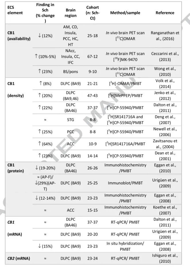
Three neuroimaging PET studies have evaluated the CB1 receptor availability in schizophrenic patients compared to controls [121-123]. The first two studies, by Wong et al. [121] and Ceccarini et al., [122] reported a generalized increase in CB1 receptor density in most brain regions of schizophrenic patients compared to controls, being statistically significant only in certain areas (Table 2). Interestingly, these studies also reported that CB1 receptor binding in certain areas correlated with the severity of positive symptoms and inversely correlated with the severity of negative symptoms [121, 122]. Opposite to these first reported imaging findings, a recent PET study by Ranganathan et al. [123], showed a significant decrease in CB1 receptor availability in patients with schizophrenia compared to healthy controls (Table 2). Moreover, this
study showed a positive global association between both positive and negative symptoms and the availability of CB1 receptors in schizophrenia [104]. Given these discordant findings related to CB1 receptor availability in schizophrenia and its association with different symptoms of the disease, the necessity of further in vivo assessments in this regard becomes clear. Explanations for the contradictory results reported in imaging studies published so far, have been discussed in detail [124]. Thus, confounding factors such as sex and age of patients and controls included in the studies, the radiotracers and the procedures used for the analysis, the influence of cannabis and/or tobacco consumption and the impact of antipsychotic medication have been proposed as variables that can account for the discrepancies of these results [124].
Regarding postmortem studies in the brain of patients with schizophrenia, also different outcomes for gene and/or protein expression of CB1 receptors have been shown depending on the technical approach used. Thus, different reports have linked schizophrenia with increased, decreased or unaltered expression/density of CB1 receptors in the postmortem human brain. Postmortem brain radioligand binding studies
19
consistently reported increased density of CB1 receptors in schizophrenia [125-131]. Six out of the eight radioligand binding studies published to date have reported an increase of CB1 receptor density in the brain of schizophrenic subjects compared to controls in areas known to be involved in schizophrenia, including the cingulate cortex and dorsolateral prefrontal cortex (Table 2). The only study that has evaluated CB1 receptor binding density in the superior temporal gyrus, a brain area particularly involved in auditory hallucinations [132], found no differences between schizophrenic subjects and controls [128]. This fact, could suggest that the alterations found in CB1 receptor density in schizophrenia might be more associated to negative and/or cognitive symptoms of the disorder, which are linked with altered cortical functions integrated by
the cingulate cortex (emotional processing and selective attention responses) and the dorsolateral prefrontal cortex (DLPC) (motivational responses and executive functions) [133]. Interestingly, the study by Dalton et al. [129] only found increased CB1 receptor binding in the DLPC of schizophrenic patients with a diagnosis of paranoid schizophrenia, characterized by the presence of prominent delusions or hallucinations, while no changes were reported in non-paranoid schizophrenic patients, indicating that also the type of diagnoses can influence the outcomes obtained. This fact highlights the relevance of the source of the diagnoses and the potential influence of changes in the clustering of schizophrenia spectrum disorders in the diagnosis manuals along time. One example of these changes, is the modification in schizophrenia definition in the Diagnostic and Statistical Manual of Mental Disorders (DSM), 5th Edition [134], respect to the previous one, that eliminates the clustering of schizophrenia classic subtypes —disorganized (hebephrenic), catatonic, paranoid, and undifferentiated— adding psychopathological dimensions instead [135]. This decision was made because the classic DSM-IV subtypes of schizophrenia provide poor description of the
20
heterogeneity of schizophrenia, low diagnostic stability, do not exhibit distinctive patterns of treatment response or longitudinal course, and are not heritable [135]. Thus, the comparisons of the outcomes between different schizophrenia subtypes should be interpreted with caution.
The potential influence of antipsychotic medication in CB1 receptor radioligand binding
studies has been also taken into account. These reports state that the increases in CB1 receptor density were not related to the antipsychotic treatment given to the patients [125-127, 129-131]. However, it must be noticed that in all the studies more than 80% of schizophrenic subjects included were under antipsychotic medication at the time of death. The evaluation of the impact of antipsychotic drugs on CB1 receptors in schizophrenia postmortem studies is difficult to overcome due to the lack of brain tissue from drug naïve patients. Nevertheless, the inclusion of a greater number of schizophrenic patients with a negative toxicology for antipsychotics at the time of death could provide new information in this regard when compared with antipsychotic-treated patients.
Opposite to radioligand binding studies, CB1 receptor immunoreactivity has been found to be decreased in the postmortem DLPC of schizophrenic subjects compared to controls, with or without changes in CB1 receptor mRNA [72, 136, 137] (Table 2). Urigüen et al. [137] reported a significant decrease in CB1 receptor immunoreactivity only in the DLPC of schizophrenic subjects that were antipsychotic-treated at the time of death without changes in those with a negative toxicology for antipsychotics. In the two reports by Eggan et al. [72, 136] decreased CB1 receptor immunoreactivity was shown in the DLPC of subjects with schizophrenia compared to controls. Authors argued that this decrease was not a consequence of the antipsychotic treatment based on the lack of statistical correlation between CB1 receptor immunoreactivity and the
21
presence of antipsychotic medication, and on the absence of alterations in CB1 expression in the brain of antipsychotic treated monkeys [72, 136]. However, more than 75% of the schizophrenic subjects included in these studies were positive for antipsychotic drugs at the time of death, making it difficult to find statistical significance when assessing the potential impact of antipsychotics. CB1 mRNA expression has also been shown decreased in the DLPC of schizophrenic subjects [136], although absence of changes have also been reported [137]. In the cingulate cortex, no alterations in CB1 receptor immunoreactivity nor CB1 receptor mRNA have been found [73] (Table 2). The reported changes in CB1 receptor inmunoreactivity in the DLPC of medicated schizophrenic subjects point to a role of the antipsychotic treatment in the
regulation of the ECS in this brain area. However, it is unknown whether this antipsychotic modulation of CB1 receptors could contribute or not to the therapeutic effects of these drugs. Taking into account all the results form postmortem studies regarding CB1 receptors in schizophrenia, showing lower or unchanged levels of mRNA, reduced immunoreactivity and higher receptor binding, two potential hypotheses have been proposed: 1) an altered trafficking of the receptor resulting in higher levels of membrane-bound CB1 receptor, and 2) a higher CB1 receptor affinity [131]. Both situations would entail a greater CB1 receptor availability, something that is not supported in all the neuroimaging studies reported to date [123].
Overall, the imaging and postmortem outcomes regarding CB1 receptor availability, density and expression in the brain of schizophrenic patients, although inconclusive, point towards a role of CB1 receptors in this pathology. Further research including functional assessment of the status of this receptor might help in understanding the potential pathophysiological consequences of the altered CB1 receptor availability, density and/or expression.
22
Little is yet known about the status of CB2 receptors in schizophrenia. To date, the only study reporting data related to CB2 receptor in the brain of schizophrenic patients found no significant correlation between the diagnosis of schizophrenia and total CB2 mRNA expression in the postmortem DLPC (BA9) [82] (Table 2). The main goal of this study was to test the association between tag SNPs in the CNR2 gene and schizophrenia. In this regard, authors showed two SNPs associated with schizophrenia that were also related to reduced function of CB2 receptors, thus concluding that people with genetically predetermined lower functioning of CB2 receptors has an increased susceptibility to suffer schizophrenia when combined with other risk factors[82]. Previously, in 2003, De Marchi and co-workers [138] reported that clinical remission in
schizophrenic patients was accompanied by a significant decrease in CB2 receptor mRNA in peripheral blood mononuclear cells. This finding does not agree with the observations of potential reduced CB2 receptor function associated with increased risk for schizophrenia [82], but authors also suggested that these peripheral changes might be related to several immunological alterations described in this pathology [138]. Nevertheless, further research is needed to support the potential role of CB2 receptors in schizophrenia.
Several genetic studies investigating different components of the ECS in patients with schizophrenia have been carried out, most of them focusing on different polymorphisms of the CNR1 gene. Nevertheless, it must be noted that all genetic studies have been carried out in peripheral blood samples, and not in the brain. As these studies goes beyond the main scope of this review, only a brief summary of the studies will be provided.
Nineteen studies have addressed different CNR1 polymorphisms in relation with schizophrenia. Five of them studied the triplet AAT in CNR1 repeat, finding no linkage
23
when comparing with the general population of schizophrenic patients from different genetic backgrounds [139-142]. Another study from Seifert et al. studied two other SNPs apart from the triplet AAT (rs6454674 and rs1049353), not finding any association with any of them [143]. Nevertheless, a nine-time repetition of the triplet AAT in CNR1 has been associated with the hebephrenic subtype of schizophrenia in two different studies, carried out in Caucasian and Japanese population [144, 145]. Ujike et al. [144] also addressed the association of rs1049353 in their cohort, but, as in the study of Seifert, they did not find any linkage. These data reflect the heterogeneity of the schizophrenia and suggest that variations in the CNR1 gene may contribute to the pathogenesis of specific subtypes of this disorder.
Some other studies have analyzed the SNP rs1049353 in schizophrenic patients [87, 146], none of them showing any significant linkage. Several other studies have failed in trying to find associations between different SNPs of CNR1 (rs806366, rs806368, rs806376, rs806379, rs806380, rs6454674, sr1535255 among other) and schizophrenia [147-150]. However, some SNPs such as rs6454674 [151], rs2023239 [152] and interactions with rs1049353, rs1535255, and rs2023239 [152] have been associated with positive and negative symptoms. At the same time, a study from Tiwari et al. have found an association between CNR1 SNP rs806378 and the weight gain as a consequence of antipsychotic treatment [153].
An interesting study evaluated interactions between CNR1 gene polymorphisms, cannabis use, cerebral volume and cognitive function [154]. They compared patients with schizophrenia or schizoaffective disorder with cannabis abuse/dependency and patients without cannabis use and observed smaller frontotemporal white matter (WM) volumes in those that smoked cannabis. They also observed associations between SNPs rs12720071, rs7766029, rs9450898 and WM volumes, as well as between SNP
24
rs12720071 and processing speed/attention and problem-solving tests. Those results suggest that the use of cannabis in association with specific CNR1 genotypes can contribute to alterations in WM and cognitive deficits in a subgroup of schizophrenic patients, which supports the hypothesis that both genetic and environmental factors could work together to determine the phenotypic expression in patients with schizophrenia.
Regarding genetic studies involving CNR2, data are critically scarce. One study has reported a close relationship between a polymorphism of the CNR2 and increased susceptibility to schizophrenia in a large Japanese population [83]. This association was also confirmed in postmortem PFC from schizophrenic and control subjects with other
ethnicities, being the risk allele also associated with low CB2 receptor mRNA levels [83]. Furthermore, culture cell experiments showed that this CNR2 gene polymorphism was linked to a lower functionality of the CB2 receptor, suggesting an increased risk of schizophrenia for people with low CB2 receptor function [83].
5.2 Endocannabinoid synthesizing and metabolizing enzymes in schizophrenia
Only a few studies have evaluated the endocannabinoid enzymes in the brain of patients with schizophrenia (Table 2). In the first study [155], quantitative polymerase chain reaction (PCR) was used to measure mRNA levels of DAGL (DAGLα and DAGLβ), MAGL, and FAAH. The mRNA level quantification of these enzymes was carried out in the prefrontal cortex Brodmann's area 9 of 42 schizophrenia subjects and matched control comparison subjects. No differences between subject groups were found in mRNA levels for endocannabinoid synthesizing and metabolizing enzymes.
In a more recent study, the same authors studied the transcript levels for the recently discovered 2-AG metabolizing enzyme, α-β-hydrolase domain 6 (ABHD6), in the
25
prefrontal cortex of schizophrenia and healthy subjects (n = 84), using quantitative PCR [156]. This study showed that ABHD6 mRNA levels were elevated in schizophrenia subjects who were younger and had a shorter illness duration relative to age-matched comparison subjects. Furthermore, age and illness duration were strongly correlated in schizophrenia subjects, which made it difficult to differentiate between their effects on ABHD6 mRNA levels.
On the other hand, Morita et al. investigated a possible relationship between the non-synonymous polymorphism in Pro129Thr (rs324420) of the FAAH gene and schizophrenia. No differences were found in a group of 260 patients with schizophrenia (127 paranoids, 127 hebephrenics and 6 not classified) as compared to 63 controls in a Japanese population, regardless of the disorder subtype [157].
5.3 Endocannabinoid levels in schizophrenia
To date, only one study has evaluated endocannabinoid levels directly in the brain of patients with schizophrenia [158]. In this postmortem study, contents of the two main endocannabinoids, 2-AG and AEA, as well as other endocannabinoid and cannabimimetic compounds were quantified in three brain regions of subjects with schizophrenia and matched controls. The study revealed an opposite pattern for the regulation of endocannabinoids in schizophrenia. Authors found increased levels of 2-AG in cerebellum, hippocampus, and DLPC, whereas decreased levels of AEA and other N-acylethanolamines —dihomo-γ-linolenoylethanolamine (LEA),
oleoylethanolamide (OEA), palmytoylethanolamide (PEA), and
docosahexaenoylethanolamine (DHEA) — were reported [158]. In this way, antipsychotic medications reduced the content of endocannabinoids in the prefrontal
26
cortex and hippocampus, but not in cerebellum, of antipsychotic-treated patients compared to antipsychotic-free subjects [158].
Before this study, several works focused their attention in the link between endocannabinoid levels, in both cerebrospinal fluid (CSF) and blood, and schizophrenia [138, 159-162]. Thus, four studies of the same research group have reported elevated
AEA levels in CSF of schizophrenic patients, with no significant differences in serum AEA levels between schizophrenic patients and controls [159-162]. Moreover, in non-medicated acute schizophrenics, a negative correlation was found between CSF AEA levels and psychotic symptoms [160]. The increases of AEA levels reported in CSF from patients with schizophrenia contrast with the reduced AEA found in postmortem human brain of schizophrenics, but the neuronal origin of CSF endocannabinoids remains conjectural and it might reflect peripheral alterations of these signaling messengers [138]. In this sense, an increase in AEA levels in the blood of patients with acute schizophrenia respect to healthy volunteers has been reported [138]. Furthermore, in schizophrenic patients, pharmacologically-induced remission of the symptoms was
accompanied by a significant decrease of blood AEA levels and of the mRNA transcripts for the degrading enzyme FAAH [138]. Thus, it has been proposed that the increased blood AEA levels observed in patients with acute schizophrenia might be due to the modified immune response observed during the course of the disease [138]. In fact, patients in initial prodromal states of psychosis with lower levels of AEA in CSF showed a higher risk for transiting to psychosis earlier [162]. Regarding the effect of antipsychotic medication on CSF endocannabinoid levels, AEA concentrations remained increased in patients that were treated with atypical antipsychotics, but not in those treated with typical ones [160].
27
In this line, a clinical trial in acute schizophrenia has evaluated the antipsychotic effects of the non-psychoactive phytocannabinoid cannabidiol (CBD) versus the atypical antipsychotic amisulpride, assessing in turn endocannabinoid serum levels along treatments [163]. Either treatment was safe and led to significant clinical improvement, but CBD displayed a markedly superior side effects profile. Results also showed that treatment with CBD, but not with amisulpride, was accompanied by a significant increase in serum AEA levels that were also associated with clinical improvement [163]. These authors suggest that inhibition of AEA deactivation may contribute to the antipsychotic effects of CBD potentially representing a completely new mechanism in the treatment of schizophrenia [163].
Besides AEA, other endocannabinoids have also been evaluated in CSF or serum of patients with schizophrenia. There is only one study which has attempted to determine 2-AG levels in CSF of schizophrenic patients [159]. However, despite being the most abundant endocannabinoid in the brain [164], significant levels of 2-AG could not be detected in any of the samples analyzed, suggesting that this endocannabinoids
concentrations are exceedingly low in CSF of both controls and schizophrenic patients. The levels of the endogenous analogues of AEA, OEA and PEA, have also been explored out of the brain in schizophrenia. In observational studies, no differences were found in CSF or serum OEA levels between controls and schizophrenic patients [159-162]. By contrast, a 2-fold increase in PEA CSF levels was found in schizophrenic patients compared to controls [159]. However, this finding was not replicated in subsequent studies [160, 161]. In previously mentioned clinical trial with CBD performed in patients with acute schizophrenia, both OEA and PEA serum levels were significantly elevated in schizophrenic patients treated with CBD, compared to those treated with amisulpride [163].
28
It is also noteworthy that CSF endocannabinoid levels have been shown to be affected depending on the history of cannabis use. Thus, markedly altered AEA concentrations (>10-fold higher) were reported in CSF of a subgroup of schizophrenic patients who had low frequency cannabis use compared to controls (with high and low frequency use), as well as compared to schizophrenic high-frequency users [161]. However, this impact of cannabis use was not observed in other studies [160, 162].
Compiling the information available from these studies is evident that the relationship between levels of endocannabinoids measured in the CSF, peripheral blood and concentrations of endocannabinoids in brain tissue is not clear yet. Thus, the understanding of functional implications of altered levels of endocannabinoids in each type of sample of schizophrenic patients remains to be elucidated.
6. Conclusions
Several evidences suggest the relationship between changes in one or more components of the ECS and some of the symptoms that are present in depression, anxiety-related disorders and schizophrenia. Indeed, recent human postmortem and in vivo neuroimaging studies are providing more knowledge about the implication of the ECS in these mental disorders. Most of the findings in depression and anxiety are related to the expression and/or functionality of CB1 receptors and FAAH in brain areas belonging to the amygdala-hippocampal-cortico-striatal neural circuit, especially the
frontal cortex in depression and the amygdala in anxiety disorders. Regarding schizophrenia, the findings in postmortem and living human brains highlight a deregulation of CB1 receptor in specific brain areas that are highly affected in this disease. The findings on peripheral endocannabinoid levels are in good consonance with
29
these adaptive changes. However, we must be cautious since peripheral endocannabinoid levels may not be well correlated with brain concentrations.
The pharmacological manipulation of the ECS is envisaged as an attractive alternative treatment for these mental disorders. For instance, drugs as the phytocannabinoid compound CBD have been reported to be effective to treat schizophrenia.
The advance in this field, together with the translational preclinical research is opening an attractive research scenario for the development of promising new pharmacological strategies based on drugs targeting the ECS to treat mental disorders.
Acknowledgements
This study was supported by the Spanish Ministry of Economy and Competitiveness (SAF2015-67457-R, MINECO/FEDER), the Plan Estatal de I+D+i 2013-2016, the Instituto de Salud Carlos III-Subdirección General de Evaluación y Fomento de la Investigación, Spanish Ministry of Economy, FEDER (PI13/01529) and the Basque Government (IT616/13). I I-L is a recipient of a Predoctoral Fellowship from the
Basque Government. E F-Z is a recipient of a Predoctoral Fellowship from the University of Cantabria. CM is a recipient of a Postdoctoral Marie Skłodowska-Curie Individual Fellowship (H2020-MSCA-IF-2016, ID 747487).
Conflict of interests
30
REFERENCES
[1] H.A. Whiteford, L. Degenhardt, J. Rehm, A.J. Baxter, A.J. Ferrari, H.E. Erskine, et al., Global burden of disease attributable to mental and substance use disorders: findings from the Global Burden of Disease Study 2010, Lancet 382(9904) (2013) 1575-86.
[2] J.J. Meana, L.F. Callado, B. Morentin, Do post-mortem brain studies provide useful information for Psychiatry?, Rev Psiquiatr Salud Ment (Barc.) 7 (2014) 3.
[3] C.H. Ashton, P.B. Moore, Endocannabinoid system dysfunction in mood and related disorders, Acta Psychiatr Scand 124(4) (2011) 250-61.
[4] M.N. Hill, B.B. Gorzalka, Is there a role for the endocannabinoid system in the etiology and treatment of melancholic depression?, Behav Pharmacol 16(5-6) (2005) 333-52.
[5] M.N. Hill, B.B. Gorzalka, Pharmacological enhancement of cannabinoid CB1 receptor activity elicits an antidepressant-like response in the rat forced swim test, Eur Neuropsychopharmacol 15(6) (2005) 593-9.
[6] D. Parolaro, N. Realini, D. Vigano, C. Guidali, T. Rubino, The endocannabinoid system and psychiatric disorders, Exp Neurol 224(1) (2010) 3-14.
[7] A.F. Carvalho, E.J. Van Bockstaele, Cannabinoid modulation of noradrenergic circuits: implications for psychiatric disorders, Prog Neuropsychopharmacol Biol Psychiatry 38(1) (2012) 59-67.
[8] R. Mechoulam, L.A. Parker, The endocannabinoid system and the brain, Annu Rev Psychol 64 (2013) 21-47.
[9] H.C. Lu, K. Mackie, An Introduction to the Endogenous Cannabinoid System, Biol Psychiatry 79(7) (2016) 516-25.
[10] S. Zou, U. Kumar, Cannabinoid Receptors and the Endocannabinoid System: Signaling and Function in the Central Nervous System, Int J Mol Sci 19(3) (2018).
[11] A.C. Howlett, F. Barth, T.I. Bonner, G. Cabral, P. Casellas, W.A. Devane, et al., International Union of Pharmacology. XXVII. Classification of cannabinoid receptors, Pharmacol Rev 54(2) (2002) 161-202.
[12] J.C. Ashton, M. Glass, The cannabinoid CB2 receptor as a target for inflammation-dependent neurodegeneration, Curr Neuropharmacol 5(2) (2007) 73-80.
31
[13] M.P. Viveros, E.M. Marco, S.E. File, Endocannabinoid system and stress and anxiety responses, Pharmacol Biochem Behav 81(2) (2005) 331-42.
[14] C.J. Riebe, F.A. Pamplona, K. Kamprath, C.T. Wotjak, Fear relief-toward a new conceptual frame work and what endocannabinoids gotta do with it, Neuroscience 204 (2012) 159-85. [15] S. Ruehle, A.A. Rey, F. Remmers, B. Lutz, The endocannabinoid system in anxiety, fear memory and habituation, J Psychopharmacol 26(1) (2012) 23-39.
[16] I. Akirav, The role of cannabinoids in modulating emotional and non-emotional memory processes in the hippocampus, Front Behav Neurosci 5 (2011) 34.
[17] O. Gunduz-Cinar, K.P. MacPherson, R. Cinar, J. Gamble-George, K. Sugden, B. Williams, et al., Convergent translational evidence of a role for anandamide in amygdala-mediated fear extinction, threat processing and stress-reactivity, Mol Psychiatry 18(7) (2013) 813-23.
[18] R.J. Bluett, J.C. Gamble-George, D.J. Hermanson, N.D. Hartley, L.J. Marnett, S. Patel, Central anandamide deficiency predicts stress-induced anxiety: behavioral reversal through endocannabinoid augmentation, Transl Psychiatry 4 (2014) e408.
[19] J.M. Gray, H.A. Vecchiarelli, M. Morena, T.T. Lee, D.J. Hermanson, A.B. Kim, et al., Corticotropin-releasing hormone drives anandamide hydrolysis in the amygdala to promote anxiety, J Neurosci 35(9) (2015) 3879-92.
[20] M. Herkenham, A.B. Lynn, M.R. Johnson, L.S. Melvin, B.R. de Costa, K.C. Rice, Characterization and localization of cannabinoid receptors in rat brain: a quantitative in vitro autoradiographic study, J Neurosci 11(2) (1991) 563-83.
[21] T. Rubino, C. Guidali, D. Vigano, N. Realini, M. Valenti, P. Massi, et al., CB1 receptor stimulation in specific brain areas differently modulate anxiety-related behaviour, Neuropharmacology 54(1) (2008) 151-60.
[22] M.N. Hill, S. Patel, P. Campolongo, J.G. Tasker, C.T. Wotjak, J.S. Bains, Functional interactions between stress and the endocannabinoid system: from synaptic signaling to behavioral output, J Neurosci 30(45) (2010) 14980-6.
[23] M.N. Hill, J.G. Tasker, Endocannabinoid signaling, glucocorticoid-mediated negative feedback, and regulation of the hypothalamic-pituitary-adrenal axis, Neuroscience 204 (2012) 5-16.
[24] T.T. Lee, B.B. Gorzalka, Timing is everything: evidence for a role of corticolimbic endocannabinoids in modulating hypothalamic-pituitary-adrenal axis activity across developmental periods, Neuroscience 204 (2012) 17-30.
[25] M. Morena, S. Patel, J.S. Bains, M.N. Hill, Neurobiological Interactions Between Stress and the Endocannabinoid System, Neuropsychopharmacology 41(1) (2016) 80-102.
[26] E. Aso, A. Ozaita, M.A. Serra, R. Maldonado, Genes differentially expressed in CB1 knockout mice: involvement in the depressive-like phenotype, Eur Neuropsychopharmacol 21(1) (2011) 11-22.
[27] L. Uriguen, S. Perez-Rial, C. Ledent, T. Palomo, J. Manzanares, Impaired action of anxiolytic drugs in mice deficient in cannabinoid CB1 receptors, Neuropharmacology 46(7) (2004) 966-73.
[28] O. Valverde, M. Torrens, CB1 receptor-deficient mice as a model for depression, Neuroscience 204 (2012) 193-206.
[29] M.N. Hill, S. Patel, E.J. Carrier, D.J. Rademacher, B.K. Ormerod, C.J. Hillard, et al., Downregulation of endocannabinoid signaling in the hippocampus following chronic unpredictable stress, Neuropsychopharmacology 30(3) (2005) 508-15.
[30] M. Bortolato, R.A. Mangieri, J. Fu, J.H. Kim, O. Arguello, A. Duranti, et al., Antidepressant-like activity of the fatty acid amide hydrolase inhibitor URB597 in a rat model of chronic mild stress, Biol Psychiatry 62(10) (2007) 1103-10.
[31] M.N. Hill, S.A. Kumar, S.B. Filipski, M. Iverson, K.L. Stuhr, J.M. Keith, et al., Disruption of fatty acid amide hydrolase activity prevents the effects of chronic stress on anxiety and amygdalar microstructure, Mol Psychiatry 18(10) (2013) 1125-35.
32
[32] E. Lomazzo, L. Bindila, F. Remmers, R. Lerner, C. Schwitter, U. Hoheisel, et al., Therapeutic potential of inhibitors of endocannabinoid degradation for the treatment of stress-related hyperalgesia in an animal model of chronic pain, Neuropsychopharmacology 40(2) (2015) 488-501.
[33] S. Gaetani, P. Dipasquale, A. Romano, L. Righetti, T. Cassano, D. Piomelli, et al., The endocannabinoid system as a target for novel anxiolytic and antidepressant drugs, Int Rev Neurobiol 85 (2009) 57-72.
[34] M.N. Hill, C.J. Hillard, F.R. Bambico, S. Patel, B.B. Gorzalka, G. Gobbi, The therapeutic potential of the endocannabinoid system for the development of a novel class of antidepressants, Trends Pharmacol Sci 30(9) (2009) 484-93.
[35] M.A. Katzman, M. Furtado, L. Anand, Targeting the Endocannabinoid System in Psychiatric Illness, J Clin Psychopharmacol 36(6) (2016) 691-703.
[36] E.M. Marco, G. Laviola, The endocannabinoid system in the regulation of emotions throughout lifespan: a discussion on therapeutic perspectives, J Psychopharmacol 26(1) (2012) 150-63.
[37] S. Patel, M.N. Hill, J.F. Cheer, C.T. Wotjak, A. Holmes, The endocannabinoid system as a target for novel anxiolytic drugs, Neurosci Biobehav Rev 76(Pt A) (2017) 56-66.
[38] T. Rubino, E. Zamberletti, D. Parolaro, Endocannabinoids and Mental Disorders, Handb Exp Pharmacol 231 (2015) 261-83.
[39] R.M. Hirschfeld, The epidemiology of depression and the evolution of treatment, J Clin Psychiatry 73 Suppl 1 (2012) 5-9.
[40] A. Adell, E. Castro, P. Celada, A. Bortolozzi, A. Pazos, F. Artigas, Strategies for producing faster acting antidepressants, Drug Discov Today 10(8) (2005) 578-85.
[41] R. Vidal, E. Castro, F. Pilar-Cuellar, J. Pascual-Brazo, A. Diaz, M.L. Rojo, et al., Serotonin 5-HT4 receptors: A new strategy for developing fast acting antidepressants?, Curr Pharm Des 20(23) (2014) 3751-62.
[42] L. Degenhardt, W. Hall, M. Lynskey, Exploring the association between cannabis use and depression, Addiction 98(11) (2003) 1493-504.
[43] S. Lev-Ran, M. Roerecke, B. Le Foll, T.P. George, K. McKenzie, J. Rehm, The association between cannabis use and depression: a systematic review and meta-analysis of longitudinal studies, Psychol Med 44(4) (2014) 797-810.
[44] A.L. Beautrais, P.R. Joyce, R.T. Mulder, Personality traits and cognitive styles as risk factors for serious suicide attempts among young people, Suicide Life Threat Behav 29(1) (1999) 37-47.
[45] H. Ashton, J. Golding, V.R. Marsh, J.E. Millman, J.W. Thompson, The seed and the soil: effect of dosage, personality and starting state on the response to delta 9 tetrahydrocannabinol in man, Br J Clin Pharmacol 12(5) (1981) 705-20.
[46] T.F. Denson, M. Earleywine, Decreased depression in marijuana users, Addict Behav 31(4) (2006) 738-42.
[47] A.J. Gruber, H.G. Pope, Jr., M.E. Brown, Do patients use marijuana as an antidepressant?, Depression 4(2) (1996) 77-80.
[48] D. Prentiss, R. Power, G. Balmas, G. Tzuang, D.M. Israelski, Patterns of marijuana use among patients with HIV/AIDS followed in a public health care setting, J Acquir Immune Defic Syndr 35(1) (2004) 38-45.
[49] J. Kotin, R.M. Post, F.K. Goodwin, 9 -Tetrahydrocannabinol in depressed patients, Arch Gen Psychiatry 28(3) (1973) 345-8.
[50] C. Price, T. Hemmingsson, G. Lewis, S. Zammit, P. Allebeck, Cannabis and suicide: longitudinal study, Br J Psychiatry 195(6) (2009) 492-7.
[51] J.M. Witkin, E.T. Tzavara, G.G. Nomikos, A role for cannabinoid CB1 receptors in mood and anxiety disorders, Behav Pharmacol 16(5-6) (2005) 315-31.