In Silico Analysis of Putative Paralytic Shellfish Poisoning Toxins Export Proteins in Cyanobacteria
10
0
0
Texto completo
(2) In Silico Analysis of PSTs Export Proteins. Figure 1. Final saxitoxin (STX) analogues synthesized by cyanobacteria (as described in [21]). Variations of the structure of saxitoxin are circled: carbamoyl group (black), sulfate group (red), sulfonate group (green) and hydroxyl group (blue). doi:10.1371/journal.pone.0055664.g001. In the past decades, the PSTs-Na+ channel binding process has been well studied and modeled [7,8], indicating that only one PST molecule is bound per Na+ channel. Sodium (Na+) is one of the most predominant soluble ions in saline soils and waters. It is also an important requirement for cyanobacterial growth [9,10] and nitrogen fixation [11]. The use of a Na+ gradient turns out to be highly efficient in cyanobacteria when taking into account that in Anabaena torulosa (brackish water strain) and Anabaena L-31 (freshwater strain), intracellular Na+ concentration remains lower than in the culture medium: 10 and 30 times lower (DyNa+ = +79.7–+90.5 mV) in Anabaena torulosa and 11 and 18 times lower (DyNa+ = +66.5–+75.7 mV) in Anabaena L-31 under extracellular sodium concentration ranges between 1 and 60 mM [11]. To date, five PSTs biosynthetic gene clusters (sxt) have been described in cyanobacteria Cylindrospermopsis raciborskii T3, Anabaena circinalis AWQC131C, Aphanizomenon sp. NH5, Lyngbya wollei and Raphidiopsis brookii D9 [12,13,14,15]. Cluster sxt size ranges between 25.7 kb (R. brookii) and 36 kb (L. wollei) (Figure 2). Among sxt genes, two have been related to toxin export: sxtF and sxtM, which encode a MATE (multidrug and toxic compound extrusion) transporter. This protein family, represented by NorM in the bacterium Vibrio parahaemolyticus, confers resistance to multiple cationic toxic agents. These proteins export norfloxacin (NOR) and other cationic toxic compounds by means of an electrochem-. PLOS ONE | www.plosone.org. ical gradient of Na+ ions, acting as a Na+/drug antiporter [16]. In clusters involved in cylindrospermopsin synthesis (cyr) cyrK is present, a gene that also encodes a MATE protein belonging to the NorM family [15]. The conserved regions G184KFGXP189 and L381RGYKD386 present in NorM of Vibrio parahaemolyticus, V. cholerae and other bacteria have been characterized as recognition motifs for Na+ and drugs respectively. The first domain was located between transmembrane (TM) domains V-VI and the second one between X-XI TM domains [16]. Both the binding of STX with the Na+ channel and the interaction of NOR with the NorM protein, have demonstrated the key role of negatively charged residues of transporter proteins in STX blocking [7] and NOR resistance [16], respectively. Although the effect of PSTs have been well documented in regards to human health, their role in phytoplankton ecology is less clear. However, these studies are subject to speculation, where PSTs could be synthesized as a chemical defense mechanism and/ or for ion transport facilitation and regulatory interactions [17]. Recently, an interaction between STX and the copper transporter Ctr1p has been proposed, suggesting that STX inhibited copper uptake [18]. In previous work, we established a relationship between the extracellular levels of Na+ and the export of PSTs in the cyanobacterium Raphidiopsis brookii D9 [19]. We proposed that PSTs in cyanobacteria could act as a protective mechanism to ensure homeostasis against extreme salt variation in the environ-. 2. February 2013 | Volume 8 | Issue 2 | e55664.
(3) In Silico Analysis of PSTs Export Proteins. Figure 2. Comparative analysis of five sxt clusters in cyanobacteria. ORFs sxtF and sxtM are shown in yellow and pink, respectively. Homologous segments are indicated in black (sxtG/H/M/I) and green (sxtSUL/DIOX/M). Putative scare sequence present in intergenic region sxtH-sxtI and also identified in the end of sxtF, sxtM sequences, is shown in red. ORFs with unknown functions are shown in black, ORFs flanking the clusters in white and the transposases element in grey. doi:10.1371/journal.pone.0055664.g002. Instituto de Investigaciones Biológicas Clemente Estable, Montevideo, Uruguay) was isolated from a shallow lake in Uruguay. C. raciborskii ITEP A3 [22] and C. raciborskii PMC0.01 [22] were obtained from the Paris Museum Collection (provided by Cécile Bernard). The isolation sites and toxin profiles are described in Table 1. Cells were grown in batch cultures in MLA medium at pH 8.4 [23] at 25uC, under continuous cool-white fluorescence lighting at a photon flux density of 75 mE s21 m22.. ment. This phenomena has been reported in a combination of evaporated and groundwater intrusions [20]. PST-producing cyanobacteria synthesized one analogue in major proportion, which was named ‘‘final analogue’’ [21]. Analogs differ in side group moieties (Figure 1), but guanidinium groups are maintained in all analogues. In this study we developed a different approach to understand why there is more than one copy of putative transporter proteins in the sequenced sxt clusters, considering previous data on PST profiles in cyanobacteria and the export of toxins in R. brookii D9. Finally, our aim was to correlate the data obtained from mutagenesis studies of the NorM protein of V. cholerae with molecular docking modeling analysis of SxtF/M proteins with specific PSTs analogues.. Genomic DNA Isolation, Amplification, Sequencing and Phylogeny DNA was extracted with the CTAB method described by Wilson [24]. For PCR amplification and the sequencing of sxtF/M from C. raciborskii ITEP A3, C. raciborskii PMC00.01, C. raciborskii MVCC14, the primers described in Table S1 were used. The PCR reaction contained 50–100 ng of genomic DNA. Reagents for each amplification were: 0.25 U Taq DNA polymerase (InvitrogenH, California, USA); 3 mL10X PCR buffer (InvitrogenH); 2.5 mM MgCl (InvitrogenH, California, USA); 0.4 mM primers; and 0.93 mM of each deoxynucleoside triphosphate (PromegaH, Madison, Wi-USA). Thermal cycling was performed in an Eppendorf Mastercycler (Westbury, NY-USA), under the follow-. Materials and Methods Isolation and Culture Conditions Raphidiopsis brookii D9 was obtained by sub-cloning from the culture SPC338 (provided by Maria Teresa de Paiva, Sao Paulo, Brazil), as originally isolated by Pedro Zagatto from a branch of the Billings water reservoir in Taquacetuba, Sao Paulo, Brazil. Cylindrospermopsis raciborskii MVCC14 (provided by S. Bonilla, PLOS ONE | www.plosone.org. 3. February 2013 | Volume 8 | Issue 2 | e55664.
(4) In Silico Analysis of PSTs Export Proteins. Table 1. PSTs profiles, presence of sxtM/F transporter genes and source of cyanobacterial strains.. PSTs profiles. Transporters. Strain. STX. sxtF. sxtM. References. CR T3(1). +. ++. +. +. [21]a; [12]b. CR PMC00.01(1). +. ++. +. +. [44]a;. +. ++. +. +. Bernard Per. Comm.a;. +. +. Fuentes-Valdés, unpub.a;. CR ITEP A3. (1). CR MVCC14 Raph D9. (2). (1). + +. GTX2/3. ++ ++. LW(3) AC AWQC131C(4) Apha NH5. (3). +. +. +. dcSTX. +. dcGTX2/3. neoSTX. C1/2. LW1–6. +. +. +. +. +. +. +. ++ ++ +. b b b. 21 a; 15 b. +. +. [ ] [ ]. ?. +. [45]a; [46]a; [14]b. +. [47]a; [13]b. +. [48]a; [13]b. a. Toxin profile. Gene sequences. Classification is not clear. + Gene or toxin is present. ++ PST analogue is synthesized in higher proportion (final analogue). This work. CR: C. raciborskii; Raph: Raphidiopsis; Apha: Aphanizomenon; AC: A. circinalis; LW: L. wollei. Sources: (1)Brazil, (2) Uruguay, (3) USA, (4) Australia. doi:10.1371/journal.pone.0055664.t001 b ?. ing conditions: initial DNA denaturation at 92uC for 2 min; 30 cycles at 94uC for 1 min, 56uC for 1 min, 72uC for 2 min and a final elongation at 72uC for 7 min. PCR primers were used for the sequencing of both DNA strands (Macrogen, Korea). All sequences were manually checked using BLASTX in addition to the National Center for Biotechnology Information (NIH, Bethesda, MD). Phylogenetic and molecular evolutionary analyses were conducted using MEGA version 5 [25]. The model was chosen based on JTT because, according to options given by MEGA5, BIC scores (Bayesian Information Criterion) were lowest in consideration to the given substitution pattern (File S2). When using different substitution models the tendency remained the same. ProTest (version 2.4) supported the substitution model in this work (data not shown) [26]. To predict protein structure, we used the programs PSIPRED (http://bioinf.cs.ucl.ac.uk/psipred/) and Predict Protein (http:// www.predictprotein.org/).. Modeling Homology modeling. The reference X-ray structure used to build the five homology models was the MATE transporter NorM from Vibrio cholerae [27] (ID code: 3MKT; Resolution: 3,65 Å; RValue: 0,312) which was obtained from the protein data bank (Research Collaboratory for Structural Bioinformatics, RCSB, http://www.rcsb.org/pdb). The homology models were built for the proteins SxtM_T3, SxtF_T3, SxtM_131C, SxtM_D9 and SxtF_D9. Sequences were aligned using Clustal W [28]. After using this alignment tool and the software MODELLER version 9v6 [29], homology models were built for all the sets of proteins. The new models were subjected to cycles of energy minimization of 500 steps, and to a further molecular dynamic of 100 ps in order to relax the conformation of the lateral chains and avoid conformation tension generated during the construction of the model. All calculations were performed using the NAMD [30] and force field charm [31]. Docking simulations. AutoDockVina [32] was used to explore the pores of all the studied proteins (SxtM_T3, SxtF_T3, SxtM_131C, SxtM_D9 and SxtF_D9). In order to study the interaction of the different toxins throughout the pore of each transporter, a technique that separates the pore into 5 separate grids maps was used, where each grid has a 10 Å superimposition with each other along the Z axis. This system was developed so that each transporter binding site could be explored thoroughly. For the construction of toxins STX, GTX2, GTX3, neoSTX, C1 and C2, density functional theory (DFT) methods [33] were used that considered B3LYP [34,35] and the 3–21 g* basis set, in order to obtain the optimized molecules. These calculations were made using gaussian03 [36]. MAESTRO graphical interface and OPLS [37] force field were used to assign partial charges for the set of proteins and toxins. Autodock Tools (ADT) was used to prepare the proteins and the ligands. All water molecules were removed and the atomic solvation parameters and fragmental volumes were assigned to the protein using the AutoDockVina program. The grid maps were calculated using Autogrid module and were centered on the Z-axis of the pore. The volume chosen. GenBank Accession Numbers The published sequences were obtained from the National Center for Biotechnology Information (NCBI) database (http:// www.ncbi.nlm.nih.gov/) under the accession numbers: ABI75096 (SxtF, C. raciborskii T3), ABI75103 (SxtM, C. raciborskii T3), ACG58379 (SxtM, A. circinalis AWQC131C), ACG63815 (SxtM, Aphanizomenon sp. NH-5), ACG63829.1 (SxtM1, Lyngbya wollei), ACG63832.1 (SxtM2, L. wollei), ACZ26231.1 (SxtM3, L. wollei), ZP_06941036 (NorM, Vibrio cholerae RC385), ZP_06305235 (SxtF, R. brookii D9), ZP_06305227 (SxtM, R. brookii D9), ADA69242.1 (MatE efflux transporter, Nostoc sp. ‘Peltigera membranacea cyanobiont’), ACZ26226.1 (SxtPER, L. wollei), ACZ26223.1 (SxtPER, Aphanizomenon sp. NH-5), ABI75130.1 (SxtPER, A. circinalis AWQC131C). The SxtF/M protein described in this study has been deposited in the GenBank database under the accession numbers JX105885 (SxtF C. raciborskii ITEP A3), JX105886 (SxtF C. raciborskii PMC00.01), JX105887 (SxtF C. raciborskii MVCC14), JX105888 (SxtM C. raciborskii ITEP A3), JX105889 (SxtM C. raciborskii PMC00.01), JX105890 (SxtM C. raciborskii MVCC14). PLOS ONE | www.plosone.org. 4. February 2013 | Volume 8 | Issue 2 | e55664.
(5) In Silico Analysis of PSTs Export Proteins. and SxtM, found by comparison against the cyanobacterial genome database, corresponds to NorM of Nostoc sp., a protein present in a different gene cluster related to secondary metabolite synthesis. By means of multiple alignments (CLUSTAL W) of SxtF and SxtM amino acid sequences from all PSTs-producing cyanobacteria and V. cholerae (VC), we identified that the motifs (G184KFGXP189) and drug characterized as Na+ 381 386 (L XGXXD ) recognition sites were conserved in SxtF and SxtM (Figure 3). The SxtM2 (L398VGLQD403) motif from Lyngbya wollei, the only cyanobacterium that does not produce STX, but the decarbamoylated form and several unique analogues (Table 1), shows a mixed sequence between the SxtM (L398IGLQD403) and SxtF (L390VGLRD395) motifs. Topology prediction of SxtF/M using the online servers PSIPRED and Predict Protein corroborated the periplasmic (TM V-TM VI) localization of the Na+ recognition and the cytoplasmic loop (TM X-TM XI) of the drug recognition domains (data not shown). The amino acid identity between NorM of VC with SxtF and SxtM of R. brookii D9 is 30% and 28%, respectively. In all PST-producing cyanobacteria, where only sxtM was identified (but not sxtF) (L. wollei, A. circinalis AWQC131C, Aphanizomenon sp. NH5) in the sxt cluster (Figure 2, Table 1), a gene encoding for a putative transporter protein, sxtPER, is present. This gene is not present in another region of the R. brookii. for the grid maps was 54654654 points with a grid-point spacing of 0.375 Å (20620620 Å). Autotors was used to define the rotatable bonds in the toxins. The Lamarckian Genetic Algorithm was used for all docking calculations. Visual inspections of the results were performed using the MGL Tools package [38].. Results Phylogenetic and Sequence Analysis We identified and sequenced the putative toxin transporter genes sxtM and sxtF, of three C. raciborskii strains (PMC00.01, ITEP A3 and MVCC14), differing in toxin profiles (Table 1, File S1). Maximum likelihood phylogenetic inference of SxtF and SxtM amino acid sequence shows a common ancestor for both transporters in all PST-producing cyanobacteria, as well as two well supported branches: one for the SxtMs and another for the SxtFs sequences (bootstrap 99%) (Figure 3). SxtF topology lacked phylogenetic resolution (identities .99), however SxtM topology grouped the strains that produce STX-neoSTX (CR ITEP A3, CR T3, CR PMC00.01) and those that produce STX-GTX2/3 (RB D9, CR MVCC14) (Table 1). It is important to note that both sxtF and sxtM were not identified in the non-PSTs producing cyanobacterial strain, Raphidiopsis sp. ITEP005 (confirmed by PCR analysis, data not shown). In addition, the sequence with highest similarity to SxtF. Figure 3. Maximum Likelihood phylogenetic tree for SxtF/SxtM and its homologues in Nostoc sp. and Vibrio cholerae. The dendrogram was inferred with a JTT amino acid substitution model, uniform rate and a bootstrap of 1000 replications (only values over 60% are shown) (MEGA version 5). The subdivisions of the tree correspond to the two different types of transporters, whose amino acid sequence in the two specific recognition motifs (Cation and Toxin), are given in the right part of the figure. The cyanobacterial branch is marked in green. SxtM recognition motifs are shown in a pink rectangle and SxtF motifs in a yellow rectangle. The toxin recognition motif of SxtM2 from L. wollei contains a non- expected amino acid sequence (orange rectangle), corresponding to an amino acid mixture: in position 399 there is a V (Valine) similar to SxtF toxin motif, and in position 402 there is a Q (Glutamine), similar to SxtM. CR: C. raciborskii; Raph: Raphidiopsis; Apha: Aphanizomenon; AC: A. circinalis; LW: L. wollei̧ PmC: Peltigera membranacea cyanobiont; VC: Vibrio cholerae. doi:10.1371/journal.pone.0055664.g003. PLOS ONE | www.plosone.org. 5. February 2013 | Volume 8 | Issue 2 | e55664.
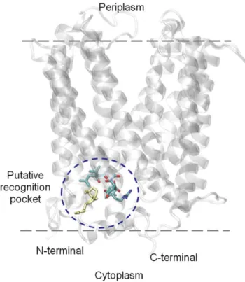
(6) In Silico Analysis of PSTs Export Proteins. D9 genome. Analysis of the SxtPER sequence of three cyanobacterial strains revealed that 10 transmembrane domains are located between amino acids 318–405, whose N- and Cterminal are located at the cytoplasm (topology according to MEMSAT3 Prediction, http://bioinf.cs.ucl.ac.uk/psipred/) (Figure S1). The identified SxtPER homologues belong to the membrane protein family Pfam00892 (DUF6). These proteins were included in the cluster of the orthologous group (COG) 0697 as permeases of the drug/metabolite transporter (DMT) superfamily [39]. According to the transporter classification database (www.tcdb.org), SxtPER is classified as part of the 2.A.7 DMT superfamily, apparently fitting within the 2.A.7.3 10 TMS Drug/ Metabolite Exporter (DME) Family. Considering the existence of experimental data in NorM proteins, but not in SxtPER homologues, only the SxtF/M proteins were considered for further analysis.. Homology Modeling Homology models were developed for the putative transporters SxtF and SxtM of C. raciborskii T3, A. circinalis AWQC131C and R. brookii D9 (SxtF_T3, SxtM_T3, SxtM_AWQC131C, SxtF_D9 and SxtM_D9). These proteins share between 27% and 30% identity with NorM of Vibrio cholerae [27], a protein with a characterized crystal structure. In order to explore the pore and binding sites of these proteins, docking simulations were performed for the systems described in Table 2. The pore of the transporter was studied by using an overlapping of 10 Å between the 5 grids of 20620620 Å along the Z-axis. The most favorable binding energy was obtained for grid 2, which contains the predicted binding domains L398XGLQD403 (SxtM) or L390VGLRD395 (SxtF) (Figure 4). The energies obtained for these pockets (binding domains) are not significantly different between the analogues synthesized for each strain (Table 2), because the values fall into the error of the method [32] (see materials and methods). When comparing amongst strains, the most favorable energy was observed in the SxtF systems of R. brookii D9 (Table 2). We analyzed the interaction of these domains with Na+ and it apparently does not affect the toxin-binding domain (Figure S2). The specific interactions in docking models are presented in Figure 5. Overall, we observed that SxtF and SxtM systems in strains D9 and T3 are similar in terms of both backbone folding and side chain geometry in the toxin-binding site.. Figure 4. Predicted protein structure of SxtF of R. brookii D9, lateral view. The model was built based on the X-ray structure of the MATE transporter NorM of Vibrio cholerae (ID code: 3MKT; Resolution: 3,65 Å; R-Value: 0,312). The location in which we obtained the most favorable binding energies (domain 390VGLRD395 in licorice representation) is shown circled in blue. STX corresponds to the yellow colored molecule. doi:10.1371/journal.pone.0055664.g004. In the SxtF_D9 system, D395 interacts directly with 1,2,3 guanidinium group of STX (Figure 5 A) and GTX3 (Figure 5 C). In the SxtM_D9 system L401 interacts with 7,8,9 guanidinium goups of STX (Figure 5 D), GTX2 (Figure 5 E) and GTX3 (Figure 5 F). In the SxtF/M_T3 systems, we observed the interaction between D395 (SxtF) with 1,2,3 guanidinium group of STX (Figure 5 G) and D403 (SxtM) with 7,8,9 guanidinium group of STX (Figure 5 I) and neoSTX (Figure 5 J). Finally, in the SxtM_131C system, direct interactions were between: Q402 with sulfocarbamoyl group of analogues C1 (Figure 5 K) and C2 (Figure 5 L); Q402 with sulfate group (C11) of GTX2; D403 with guanidinium group in C2 (Figure 5 L), GTX2 (Figure 5 M) and GTX3 (Figure 5 N).. Table 2. Binding energies of the PST analogues and putative binding sites in SxtF and SxtM.. Discussion PSTs analogues and binding energies (kcal/ mol)* Systems. GTX2. GTX3. STX. neoSTX. C1. SxtF_Raph D9. 28,3. 29,6. 28,6 NP. NP. NP. SxtM_ Raph D9. 26,9. 26,9. 28,2 NP. NP. NP. SxtF_CR T3. NP. NP. 27,5 27,1. NP. NP. SxtM_CR T3. NP. NP. 26,8 26,8. NP. NP. SxtM_AC AWQC131C. 26,9. 27,1. 2. NP. To date, transformation methods applied for genetic manipulation in PST-producing cyanobacteria have yet to be developed. The functional classification of proteins encoded by sxt genes has been explored by sequence homology [12,13,14,15] and by correlating the presence of sxt genes with toxin profiles in cyanobacteria [21]. The sxt cluster in cyanobacteria has a core of 15 genes, which has been conserved, in both sequence and synteny, among 5 clusters described to date. One of the genes present in all clusters is sxtM [40] (Fuentes-Valdés et al., unpublished data) (Table1, Figure 2). In previous work, we proposed that PSTs are exported in the cyanobacterium R. brookii D9, where SxtF/M could be involved in toxin export [19].. C2. 27,1 27,1. *Interactions obtained from docking calculations to Grid 2. Raph: Raphidiopsis brookii; CR: C. raciborskii; AC: A. circinalis. NP: Analogue is not present. (2): System was not analyzed. doi:10.1371/journal.pone.0055664.t002. PLOS ONE | www.plosone.org. 6. February 2013 | Volume 8 | Issue 2 | e55664.
(7) In Silico Analysis of PSTs Export Proteins. Figure 5. Representation view of PST-recognition sites in SxtF/M. The PST recognition region in licorice representation corresponds to: SxtF R. brookii D9 (A, B and C) for the conserved region LVGLRD; SxtM R. brookii D9 (D, E and F) for the conserved region LIGLQD; SxtF C. raciborskii T3 (G and H), for the conserved region LVGLRD; SxtM C. raciborskii T3 (I and J) for the conserved region LIGLQD; SxtM A. circinalis AWQC131C (K, L, M and N). PLOS ONE | www.plosone.org. 7. February 2013 | Volume 8 | Issue 2 | e55664.
(8) In Silico Analysis of PSTs Export Proteins. for the conserved region LIGLQD. PSTs are depicted in the surfaces representations. The name of the each PST is indicated in the bottom section of the model. The licorice representation shows: carbon atoms in cyan, oxygen in red, nitrogen in blue and sulfur in yellow. Hydrogen atoms were omitted. doi:10.1371/journal.pone.0055664.g005. they observed two branches considering that SxtM of Aphanizomenon sp. NH5 and SxtM1-SxtM2 of L. wollei were classified as a putative SxtF. Although these sequences are the most distant in the SxtM clade (Figure 3), we considered the analysis of the previously described active motif of NorM proteins in V. cholerae (VC), and assigned it to the SxtF/M classification. When we analyzed domains that are putatively involved in toxin recognition, we were able to distinguish between SxtF and SxtM domains (Figures 3; File S1), with the exception of SxtM2 of L. wollei. This may be related to the synthesis of STX derivatives named L. wollei toxins 1–6 (Lw toxin 1–6), which are only present in L. wollei. These observations suggest that these domains would maintain the drug-toxin recognition function of SxtF/M, and the differences could indicate transporter-toxin selectivity. The domain described in VC as the one responsible for Na+ recognition is conserved in all SxtF/M sequences, but is less conserved in the NorM protein of Nostoc sp. ‘Peltigera membranacea cyanobiont’, the sequence closer to the SxtF/M clade. This is evidence that positive selection has acted upon the G184KFGXP189 motif to possibly maintain the role of Na+ in transporter function. In previous studies we have analyzed the effect of Na+ on PSTs levels in R. brookii D9, concluding that Na+ plays an important role in toxin export [19], which is in concordance with the motif analysis. Comparative sequence analyses have shown that several recombination events have shaped the sxt gene clusters, especially in the sxtF/M gene family [42]. Based on comparative sequence analysis, Murray et al. [42] have suggested that a common ancestor of Nostocales possessed both sxtF and sxtM orthologs but while they were kept in R. brookii, in a common ancestor of A. circinalis and Aphanizomenon sp. NH-5, these genes were subjected to recombination events that generated a novel copy of the gene that shared the largest fraction of its sequence for either sxtM (A. circinalis) or sxtF (Aphanizomenon sp. NH-5). In both cases this sxtF/M ortholog is located between the sxtH and sxtI genes. Interestingly, our analysis shows that even though R. brookii D9 possesses both sxtF and sxtM orthologs, there is a 95 bp sequence between the sxtH and sxtI genes with high identity with the Cterminal region of the sxtF/M genes. This finding suggests that sxtF/M gene was present in this location at some point and that was subsequently lost. Whether there were up to three copies of the sxtF/M genes in a common ancestor of the Nostocales clade or that the scar between sxtH and sxtI is a remnant of an intermediate step of sxF/M recombination events, will only be clarified with the generation of novel genomic sequences from other representatives of the Nostocales clade. Despite these recombination events, the putative PST-recognition site has been conserved in SxtF and SxtM and our modeling and docking analysis results reassert the hypothesis of the involvement of L398XGLQD403 (SxtM) and L390VGLRD395 (SxtF) domains in toxin recognition. The fact that the predicted binding energies are similar for all analogues and for each analyzed cyanobacterium, could indicate that SxtF/M does not have selectivity for a final analogue. This is in agreement with our observation related to the similar toxin export behavior of STXGTX2/3 in R. brookii D9 (data not shown). Regarding the binding energies between toxins and mammalian sodium channel, values obtained by Tikhonov and Zhorov [7] are very low, which explain channel blocking due to permanent. However, unlike sxtM, the sxtF gene is only present in the Cylindrospermopsis-Raphidiopsis clade. With the aim of exploring the relationship between the presence of the SxtF and SxtM transporters and the export of PSTs analogues, we compared the presence of both predicted transporter genes and toxin profiles in eight PST-producing cyanobacteria. Our data shows no correlation between both aspects, preventing the prediction of specific analogue-transporter relationships. This might be explained by the presence of SxtF/M in all Cylindrospermopsis strains and in R. brookii D9, which differ in their toxin profiles. Furthermore, only SxtM is present in A. circinalis, Aphanizomenon sp. and L. wollei, which also posses different toxin profiles amongst each other (Table 1). In these strains we identified a second protein, SxtPER, which could act as an alternative transporter of PSTs, compensating for the lack of SxtF. All PSTs-producing cyanobacteria, except L. wollei, synthesize only one analogue in a major proportion, which we previously named the ‘‘final analogue’’ [21] (Figure 1). In the sxt cluster of R. brookii D9, which contains the minimum set of genes required to synthesize STX and GTX2/3, the last two epimers are the final analogues. Furthermore, sxtF and sxtM are transcribed [15], thereby raising the question: why does R. brookii D9 keep and express more than one transporter? On the other hand, the fact that L. wollei synthesizes the higher number of analogues in greater proportions (LW1, LW2/3 -epimers- and LW5, which constitute the 88% of the total), and contains three copies of sxtM and one copy of sxtPER [14], suggests that there is apparently a relationship between the number of final analogues and transporter proteins. The high conservation of SxtF (100% amino acid identity) may be ascribed to purifying selection and a recent horizontal gene transfer (HGT) event, as previously described for the cyr cluster [41]. Although the sxt cluster is extraordinarily conserved in cyanobacteria [42], the differences between the divergences of the SxtF and SxtM clades are notorious. Cyanobacterial phylogeny based on the 16S rRNA gene suggests that the presence of a PSTs phenotype was either gained in the toxic strains through independent HGTs or was lost from the non-toxic strains [40]; the same event has been observed in other toxic phenotypes [43]. The phylogeny of SxtMs does not completely agree with the cyanobacterial phylogenies based on the 16S rRNA gene [40] because the SxtM copies that belong to a more distant cyanobacterium, L. wollei, are distributed between the other sequences (Figure 3). Similar topologies were obtained for other sxt genes, where L. wollei sequences group together with the clade formed by Cylindrospermopsis-Raphidiopsis strains (data not shown). Evolutionary markers such as the 16S rRNA gene and the internal transcribed spacer ITS1 showed that the CylindrospermopsisRaphidiopsis clade from America are closely related, with over 99% of nucleotide identity [22]. The presence of two well-defined branches of STX-neoSTX (CR ITEP A3, CR T3, CR PMC00.01) and STX-GTX2/3 (RB D9, CR MVCC14) producers (Figure 3; Table 1) implies that the evolution of SxtM was dependent on the toxin profiles in this clade. However, posterior analyses are necessary to be able to contrast the evolution of other sequences belonging to the sxt cluster and their evolutionary markers. Murray et al., [42] showed a different topology for the SxtF/M phylogenies based on the ML method and a CpREV+ I+G+F substitution model. While analyzing amino acid residues 1–280, PLOS ONE | www.plosone.org. 8. February 2013 | Volume 8 | Issue 2 | e55664.
(9) In Silico Analysis of PSTs Export Proteins. binding between selectivity-filter region and toxin. In our study, interaction energies are higher, which allows temporal binding of the toxin with its recognition site and posterior export. In the binding of STX with the Na+ channel, glutamic and aspartic residues play a key role in channel blocking [7]. In this study we also observed interaction between aspartic residues and guanidinium groups in some PST analogues. As aforementioned, the sxt cluster has undergone intra- and interspecific recombination, and has been subject to duplication, which results in differences in PST profiles amongst cyanobacteria. This is probably the reason why transporter proteins remain without particular selectivity for any analogue. On the other hand, although the role of PSTs in the environment is unknown, the interaction between PSTs and sodium channels is highly sensitive to modifications in STX structure [8]. For example, the predicted affinity between C1 and GTX3 with SxtM of A. circinalis AWQC131C, is identical (Table 2), but in terms of toxicity in mice, GTX3 is three orders of magnitude more toxic than C1 [8]. Therefore, there would be selective pressure to maintain the conserved toxin recognition domains in the transporters in order to recognize the variety of final analogues synthesized in different cyanobacteria. Probably, differences in analogue structures are related to their target in the environment rather than the recognition by transporter proteins SxtF and SxtM.. Our results also suggest that further studies are required to evaluate toxin activity outside of the cells, where they hypothetically should be acting.. Supporting Information Predicted protein structure of SxtPER of A. circinalis AWQC131C, obtained from MEMSAT3. (TIF). Figure S1. Figure S2 Representation view of the PST-recognition site in SxtF of R. brookii D9, interacting with a sodium atom (NA). (TIF) Table S1 PCR primers used for amplification and sequencing. The primer position is based on the sxtF/M sequence of R. brookii D9. (DOC) File S1 Multiple alignment of the amino acid sequence of SxtF/ M in R. brookii D9 (Raph) and its homologs in the C. raciborskii (CR) strains ITEP A3, PMC00.01, MVCC14; L. wollei (LW); A. circinalis AWQC131C (AC), Aphanizomenon sp. NH5 (Apha); Nostoc sp. Peltigera membranacea cyanobiont (PmC) and Vibrio cholerae (VC), using CLUSTAL W. *, identical residues, :.60% homologous residues. The conserved region G184KFGXP189 is marked with gray and L381XGXXD386 in black. The SxtF sequences are in bold. (DOC). Conclusions This study suggests that the previously identified motif involved in drug recognition in the NorM protein of Vibrio cholerae, and conserved in SxtF/M (cyanobacteria), could actually participate in the recognition of PSTs. We propose that the role of Na+ in PST export described for the NorM protein of VC is maintained in the SxtF/M transporters. The binding energies obtained for all the analyzed complexes indicate that it is not possible to discriminate protein-ligand specificity through these in silico studies, and that STX and the final analogues maybe exported by both transporters (SxtF/M). Posterior studies based on mutagenic models of PSTproducing cyanobacteria or heterologous expression of SxtF/M, are necessary to confirm the conclusion shown in this study. The same in silico (modeling-docking) approximation could be employed to discover drugs capable of blocking SxtF/M, which would allow performing a broader amount of assays in cyanobacteria permitting a better understanding of toxin role under different culture conditions.. File S2 Best amino acid substitution model identified by Akaike Information Criterion (AIC). (PDF). Acknowledgments We thank Enrique Flores (Universidad de Sevilla, Sevilla, Spain) for help with in silico analyses, Daniella Spooner (P. Universidad Católica, Santiago, Chile) for language revision and Francisco Melo (P. Universidad Católica, Santiago, Chile) for their contributions to the modeling of our proteins.. Author Contributions Conceived and designed the experiments: KSL XL FGN MV. Performed the experiments: KSL XL JJF KS. Analyzed the data: KSL XL JJF KS FGN MV. Contributed reagents/materials/analysis tools: FGN MV. Wrote the paper: KSL XL JJF KS MV.. References 11. Apte SK, Thomas J (1986) Membrane electrogenesis and sodium transport in filamentous nitrogen-fixing cyanobacteria. Eur J Biochem 154: 395–401. 12. Kellmann R, Mihali TK, Jeon YJ, Pickford R, Pomati F, et al. (2008) Biosynthetic intermediate analysis and functional homology reveal a putative saxitoxin gene cluster in cyanobacteria. Appl Environ Microbiol 74: 4044–4053. 13. Mihali TK, Kellmann R, Neilan BA (2009) Characterisation of the paralytic shellfish toxin biosynthesis gene clusters in Anabaena circinalis AWQC131C and Aphanizomenonsp. NH-5. BMC Biochem 10: 8. 14. Mihali TK, Carmichael WW, Neilan BA (2011) A Putative Gene Cluster from a Lyngbya wollei Bloom that Encodes Paralytic Shellfish Toxin Biosynthesis. PLoSOne 6: e14657. 15. Stucken K, John U, Cembella A, Murillo AA, Soto-Liebe K, et al. (2010) The smallest known genomes of multicellular and toxic cyanobacteria: comparison, minimal gene sets for linked traits and the evolutionary implications. PLoS One5: e9235. 16. Singh AK, Haldar R, Mandal D, Kundu M (2006) Analysis of the topology of Vibrio cholera NorM and identification of amino acid residues involved in norfloxacin resistance. Antimicrob Agents Chemother 50: 3717–3723. 17. Cembella AD (2003) Chemical ecology of eukaryotic microalgae in marine ecosystems. Phycologia 42: 420–447. 18. Cusick KD, Minkin S, Dodani SC, Chang CJ, Wilhelm SW, et al. (2012) Inhibition of copper uptake in yeast reveals the copper transporter Ctr1p as a potential molecular target of saxitoxin. Environ Sci Technol 46: 2959–2966.. 1. Vitousek PM, Hättenschwiler S, Olander L, Allison S (2002) Nitrogen and nature. Ambio 31: 97–101. 2. Paerl HW, Paul VJ (2012) Climate change: Links to global expansion of harmful cyanobacteria. Water Res 46: 1349–1363. 3. Neilan BA, Pearson LA, Moffitt MC, Mihali KT, Kaebernick M, et al. (2008) The genetics and genomics of cyanobacterial toxicity. Adv Exp Med Biol 619: 417–452. 4. Narahashi T, Haas HG, Therrien EF (1967) Saxitoxin and tetrodotoxin: comparison of nerve blocking mechanism. Science 157 (3795): 1441–1442. 5. Wang JX, Salata JJ, Bennett PB (2003) Saxitoxin is a gating modifier of hERG K+ channels. J Gen Physiol 121: 5832598. 6. Su Z, Sheets M, Ishida H, Li FH, Barry WH (2004) Saxitoxin blocks L-type/ (Ca). J Pharmacol Exp Ther 308: 3242329. 7. Tikhonov DB, Zhorov BS (2005) Modeling P-loops domain of sodium channel: homology with potassium channels and interaction with ligands. Biophys J 88: 184–197. 8. Llewellyn LE (2006) Saxitoxin, a toxic marine natural product that targets a multitude of receptors. Nat Prod Rep 23: 200–222. 9. Allen MB, Arnon DI (1955) Studies on Nitrogen-Fixing Blue-Green Algae. I. Growth and Nitrogen Fixation by Anabaena cylindrical Lemm. Plant Physiol 30: 366–372. 10. Kratz WA, Myers J (1955) Nutrition and growth of several blue-green algae. Am J Bot 42: 282–287.. PLOS ONE | www.plosone.org. 9. February 2013 | Volume 8 | Issue 2 | e55664.
(10) In Silico Analysis of PSTs Export Proteins. 19. Soto-Liebe K, Méndez MA, Fuenzalida L, Krock B, Cembella A, et al. (2012) PSP toxin release from the cyanobacterium Raphidiopsis brookii D9 (nostocales) can be induced by sodium and potassium ions. Toxicon 60(7): 1324–1334. 20. Nielsen D L, Brock M A, Rees GN, Baldwin DS (2003) Effects of increasing salinity on freshwater ecosystems in Australia. Australian Journal of Botany 51: 655–665. 21. Soto-Liebe K, Murillo AA, Krock B, Stucken K, Fuentes-Valdés JJ, et al. (2010) Reassessment of the toxin profile of Cylindrospermopsis raciborskii T3 and function of putative sulfotransferases on synthesis of sulfated and sulfonated PSP toxins. Toxicon 56: 1350–1361. 22. Gugger M, Molica R, Le Berre B, Dufour P, Bernard C, et al. (2005) Genetic diversity of Cylindrospermopsis strains (cyanobacteria) isolated from four continents. Appl Environ Microbiol 71: 1097–1100. 23. Castro D, Vera D, Lagos N, Garcia C, Vásquez M (2004) The effect of temperature on growth and production of paralytic shellfish poisoning toxins by the cyanobacterium Cylindrospermopsis raciborskii C10. Toxicon 44: 483–489. 24. Wilson K (1990) Preparation of genomic DNA from bacteria. In: Ausubel F, Brent R, Kingston R, Seidman J, Smith J, et al. editors. Current protocols in molecular biology. Wiley Interscience, New York. pp.241–245. 25. Tamura K, Peterson D, Peterson N, Stecher G, Nei M, et al. (2011) MEGA5: Molecular Evolutionary Genetics Analysis using Maximum Likelihood, Evolutionary Distance, and Maximum Parsimony Methods. Molecular Biology and Evolution. Mol Biol Evol 28 (10): 2731–2739. 26. Abascal F, Zardoya R, Posada D (2005) ProtTest: selection of best-fit models of protein evolution. Bioinformatics 21: 2104–2105. 27. He X, Szewczyk P, Karyakin A, Evin M, Hong WX, et al. (2010) Structure of a cation-bound multidrug and toxic compound extrusion transporter. Nature 467 (7318): 991–994. 28. Thompson JD, Higgins DG, Gibson TJ (1994) ClustalW: improving the sensitivity of progressive multiple sequence alignment through sequence weighting, position-specific gap penalties and weight matrix choice. Nucleic Acids Res 22: 4673–4680. 29. Sali A, Blundell TL (1993) Comparative protein modeling by satisfaction of spatial restraints. J Mol Biol 234: 779–815. 30. James C, Phillips RB, Wang W, Gumbart J, Tajkhorshid E, et al. (2005) Scalable molecular dynamics with NAMD. J Comput Chem 26: 1781–1802. 31. Vanommeslaeghe K, Hatcher E, Acharya C, Kundu S, Zhong S, et al. (2010) CHARMM general force field: A force field for drug-like molecules compatible with the CHARMM all-atom additive biological force fields. J Comput Chem 31: 671–690. 32. Trott O, Olson AJ (2010) AutoDockVina: improving the speed and accuracy of docking with a new scoring function, efficient optimization, and multithreading. J ComputChem 31: 455–461. 33. Andzelm J, Wimmer E (1997) Density functional gaussian-type- orbital approach to molecular geometries, vibrations, and reaction energies. J Chem Phys 96: 1280–1303.. PLOS ONE | www.plosone.org. 34. Becke AD (1997) Density-functional thermo chemistry. V. Systematic optimization of exchange-correlation functional. J Chem Phys 107: 8554–8560. 35. Lee C, Yang W, Parr RG (1988) Development of the Colle- Salvetti correlationenergy formula into a functional of the electron- density. Phys Rev B Condens Matter 37: 785–789. 36. Frisch MJ, Trucks GW, Schlegel HB, Scuseria GE, Robb MA, et al. (2004) Gaussian 03, Revision C02, Gaussian, Inc, Wallingford CT. 37. Dauber-Osguthorpe P, Roberts VA, Osguthorpe DJ, Wolff J, Genest M, et al. (1998) Structure and energetics of ligand-binding to proteins: Escherichia coli dihydrofolatereductase-trimethoprim, a drug- receptor system. Proteins 4: 31– 47. 38. Sanner MF (1999) Python: A programming language for software integration and development. J Mol Graph Model 17: 57–61. 39. Livshits VA, Zakataeva NP, Aleshin VV, Vitushkina MV (2003) Identification and characterization of the new gene rhtA involved in threonine and homoserine efflux in Escherichia coli. Res Microbiol 154: 123–135. 40. Moustafa A, Loram JE, Hackett JD, Anderson DM, Plumley FG, et al. (2009) Origin of saxitoxin biosynthetic genes in cyanobacteria. PLoS One 4: e5758. 41. Jiang Y, Xiao P, Yu G, Sano T, Pan Q, et al. (2012) Molecular basis and phylogenetic implications of deoxycylindrospermopsin biosynthesis in the cyanobacterium Raphidiopsis curvata. Appl Environ Microbiol 78: 2256–2263. 42. Murray SA, Mihali TK, Neilan BA (2011) Extraordinary conservation, gene loss, and positive selection in the evolution of an ancient neurotoxin. Mol Biol Evol 28: 1173–1182. 43. Christiansen G, Molitor C, Philmus B, Kurmayer R (2008) Nontoxic strains of cyanobacteria are the result of major gene deletion events induced by a transposable element. Mol Biol Evol 25: 1695–1704. 44. Bernard C, Harvey M, Briand JF, Biré R, Krys S, et al. (2003) Toxicological comparison of diverse Cylindrospermopsis raciborskii strains: evidence of liver damage caused by a French C. raciborskii strain. Environ Toxicol 18: 176–186. 45. Onodera H, Satake M, Oshima Y, Yasumoto T, Carmichael WW (1997) New saxitoxin analogues from the freshwater filamentous cyanobacterium Lyngbya wollei. Nat Toxins 5: 146–151. 46. Carmichael WW, Evans WR, Yin QQ, Bell P, Moczydlowski E (1997) Evidence for paralytic shellfish poisons in the freshwater cyanobacterium Lyngbya wollei (Farlow ex Gomont) comb nov. Appl Environ Microbiol 63: 3104–3110. 47. Llewellyn LE, Negri AP, Doyle J, Baker PD, Beltran EC, et al. (2001) Radioreceptor assays for sensitive detection and quantitation of saxitoxin and its analogues from strains of the freshwater cyanobacterium, Anabaena circinalis. Environ Sci Technol 35: 1445–1451. 48. Mahmood NA, Carmichael WW (1986) Paralytic shellfish poisons produced by the freshwater cyanobacterium Aphanizomenon flos-aquae NH-5.Toxicon 24: 175– 186.. 10. February 2013 | Volume 8 | Issue 2 | e55664.
(11)
Figure

Documento similar
ABSTRACT Transformation of the Specialized Knowledge of Future Primary Teachers on Fraction Division
From the phenomenology associated with contexts (C.1), for the statement of task T 1.1 , the future teachers use their knowledge of situations of the personal
In the preparation of this report, the Venice Commission has relied on the comments of its rapporteurs; its recently adopted Report on Respect for Democracy, Human Rights and the Rule
H I is the incident wave height, T z is the mean wave period, Ir is the Iribarren number or surf similarity parameter, h is the water depth at the toe of the structure, Ru is the
In the previous sections we have shown how astronomical alignments and solar hierophanies – with a common interest in the solstices − were substantiated in the
Díaz Soto has raised the point about banning religious garb in the ―public space.‖ He states, ―for example, in most Spanish public Universities, there is a Catholic chapel
In the “big picture” perspective of the recent years that we have described in Brazil, Spain, Portugal and Puerto Rico there are some similarities and important differences,
For instance, the best overall accuracy of bagging in Breast with 20% noise is achieved using a 10% sampling ratio: The test error goes from 4.1% when no noise is injected to 3.5%
In particular, different versions of the training set for the base learners can be used, as in bagging (bootstrap sampling of training data), class-switching (noise injection in
