Biology and pathological implications of brown adipose tissue: promises and caveats for the control of obesity and its associated complications
20
0
0
Texto completo
(2) Pablo Tapia and others. 1146. X. XI. XII. XIII. XIV. XV.. (4) VEGF-A . . . . . . . . . . . . . . . . . . . . . . . . . . . . . . . . . . . . . . . . . . . . . . . . . . . . . . . . . . . . . . . . . . . . . . . . . . . . . . . . . . . . . . . . . . . . . . 1156 (5) IL-6 . . . . . . . . . . . . . . . . . . . . . . . . . . . . . . . . . . . . . . . . . . . . . . . . . . . . . . . . . . . . . . . . . . . . . . . . . . . . . . . . . . . . . . . . . . . . . . . . . . 1156 (6) RBP4 . . . . . . . . . . . . . . . . . . . . . . . . . . . . . . . . . . . . . . . . . . . . . . . . . . . . . . . . . . . . . . . . . . . . . . . . . . . . . . . . . . . . . . . . . . . . . . . . . 1156 (7) IGF-1 . . . . . . . . . . . . . . . . . . . . . . . . . . . . . . . . . . . . . . . . . . . . . . . . . . . . . . . . . . . . . . . . . . . . . . . . . . . . . . . . . . . . . . . . . . . . . . . . . 1156 (8) miRNAs . . . . . . . . . . . . . . . . . . . . . . . . . . . . . . . . . . . . . . . . . . . . . . . . . . . . . . . . . . . . . . . . . . . . . . . . . . . . . . . . . . . . . . . . . . . . . . 1156 The importance of BAT for metabolic regulation . . . . . . . . . . . . . . . . . . . . . . . . . . . . . . . . . . . . . . . . . . . . . . . . . . . . . . . . 1156 The impact of BAT in human health and disease . . . . . . . . . . . . . . . . . . . . . . . . . . . . . . . . . . . . . . . . . . . . . . . . . . . . . . . . 1157 Activating brown adipose tissue thermogenesis as a target to induce weight loss . . . . . . . . . . . . . . . . . . . . . . . . . 1157 Conclusions . . . . . . . . . . . . . . . . . . . . . . . . . . . . . . . . . . . . . . . . . . . . . . . . . . . . . . . . . . . . . . . . . . . . . . . . . . . . . . . . . . . . . . . . . . . . . . 1158 Acknowledgements . . . . . . . . . . . . . . . . . . . . . . . . . . . . . . . . . . . . . . . . . . . . . . . . . . . . . . . . . . . . . . . . . . . . . . . . . . . . . . . . . . . . . . . 1159 REFERENCES . . . . . . . . . . . . . . . . . . . . . . . . . . . . . . . . . . . . . . . . . . . . . . . . . . . . . . . . . . . . . . . . . . . . . . . . . . . . . . . . . . . . . . . . . . 1159. I. INTRODUCTION An estimated ∼2 billion adult people are currently overweight and more than 600 million are clinically obese worldwide (World Health Organization, 2017). Accordingly, research directed to understand the biological basis of excessive adiposity as well as to uncover novel therapeutic approaches has expanded steadily. The interplay between white and brown adipose tissue classes (WAT and BAT, respectively) has received growing attention as a relevant mechanism for the pathogenesis of obesity and its related metabolic complications (Speakman & O’Rahilly, 2012). Adipose tissue stores vast amounts of energy (∼9000 cal/kg) in highly specialized cells (Wells, 2006). While WAT mainly buffers calorie intake fluctuations, the intracellular triglycerides stored in BAT are burnt off to generate heat. BAT is a highly specialized thermogenic system that uncouples fatty acid oxidation and mitochondrial respiration from ATP synthesis to dissipate energy. This process, known as non-shivering thermogenesis, is induced in response to cold via β-adrenergic stimulation and requires uncoupling protein 1 (UCP1, thermogenin), which is present in the inner mitochondrial membrane and is recognized as a bona fide molecular marker of BAT. BAT-dependent thermogenesis has clear physiological implications for small mammals, such as rodents or newborn animals, because of their increased susceptibility to hypothermia, owing to an elevated surface-to-volume ratio (Dawkins & Hull, 1964; Cannon & Nedergaard, 2004), however, its functional relevance for adult humans remains debated. Recent correlational findings suggest that BAT activation regulates insulin sensitivity (Chondronikola et al., 2014; Lee et al., 2014b), lipid homeostasis (Chondronikola et al., 2016), and body mass/composition (Yoneshiro et al., 2013). These facts have led to proposals that pharmacological activation of BAT-dependent thermogenesis could be an effective therapy against obesity and its metabolic complications (Nedergaard & Cannon, 2010). Nevertheless, this still lacks empirical demonstration and the actual thermogenic potential of human BAT remains to be determined. Anatomically, BAT is a discrete organ located in the interscapular region of small mammals and newborn humans (Aherne & Hull, 1966; Merklin, 1974). Based on the presence of UCP1 orthologues, brown-like adipocytes have been Biological Reviews 93 (2018) 1145–1164 © 2017 Cambridge Philosophical Society. described in all newborn mammals studied to date, including ovine and bovine species (Casteilla et al., 1987; Cypess & Kahn, 2010; Nedergaard & Cannon, 2010; Birerdinc et al., 2013), and African elephant shrews (Elephantulus myurus), a species belonging to the Afrotheria, a group of animals thought to be at the base of the eutherian clade (Mzilikazi et al., 2007). Interscapular BAT (iBAT) is currently known as ‘classical BAT’ in contrast to the interspersed clusters of brown-like adipocytes found inside WAT of the supraclavicular space, mediastinum, pericardium, and perirenal/adrenal and intercostal arteries, which are denominated ‘beige’ or ‘brite (brown-in-white)’ adipose tissue (Enerback, 2010). As discussed in Section V, despite their morphological and functional similarities, classical and beige/brite adipocytes have a distinct developmental origin and regulation (Spiegelman, 2013). BAT mass diminishes with age in humans ultimately to become indistinguishable from WAT (Heaton, 1972). Seminal studies based on [18 F]-fluorodeoxyglucose-positron emission tomography-computed tomography (FDG-PET-CT) revealed that BAT is present (Cypess et al., 2009; van Marken Lichtenbelt et al., 2009; Saito et al., 2009; Virtanen et al., 2009) and is efficiently activated by cold in most adults (Cypess et al., 2012; Ouellet et al., 2012), renewing interest in understanding BAT biology. Importantly, transcriptional analysis of FDG-PETCT-detectable brown fat in adult humans revealed that these depots are composed of beige adipocytes instead of classical brown adipocytes, as they express characteristic genes of murine brite adipocytes such as CD137, TMEM26 and TBX1 but not classical brown adipocytes markers EBF3, EVA1 and FBXO31 (Spiegelman, 2013). These findings led to the conclusion that most BAT in adult humans corresponds to beige/brite fat rather than classical BAT. Herein our goal is to review key aspects of normal BAT biology as well as recent advances that link this tissue to human disease. We also discuss the potential limitations and caveats that novel obesity therapies based on BAT activation must face before moving towards clinical trials. This latter issue has been largely overlooked in the literature possibly because of the urgency of finding new pharmacological targets for obesity; however, important concerns on the potential efficacy of such therapies have been raised because.
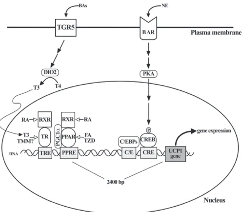
(3) Brown adipose tissue in health and disease of inconsistencies in key experimental studies (Wu et al., 2012; Spiegelman, 2013) and the absence of compelling evidence that the magnitude of BAT-dependent thermogenesis can determine sustained negative energy balance in obese humans at thermoneutral conditions (Section XII).. II. BAT MORPHOLOGY AND FUNCTION REQUIRES ADRENERGIC STIMULATION Brown adipocytes have a number of distinguishing features, including: (i) multiple and relatively small lipid droplets (LDs), (ii) large and numerous mitochondria with highly laminar cristae, and (iii) expression of UCP1 (Cannon & Nedergaard, 2004). By contrast, white adipocytes have a large unilocular LD; few, small and elongated mitochondria with randomly oriented cristae, and undetectable uncoupling activity (Frontini & Cinti, 2010). BAT thermogenesis is controlled by norepinephrine (NE), which is released from sympathetic terminals under the control of hypothalamic thermal/metabolic sensors (Lowell & Bachman, 2003). In brown adipocytes, NE activates cell-surface G-protein-coupled β-adrenoceptors (mainly β3 in mice and β1 in humans), increasing intracellular cyclic AMP (cAMP) levels (Sundin, Mills & Fain, 1984; Connolly, Nanberg & Nedergaard, 1986) and activating protein kinase A (PKA), which further amplifies the adrenergic thermogenic signal through a series of transcriptional and post-transcriptional mechanisms (Oldfield et al., 2002). PKA also promotes triglyceride lipolysis by phosphorylating LD-associated regulatory proteins and lipases, therefore coordinating fatty acid release, mitochondrial β-oxidation and thermogenesis (Fig. 1). The physiological relevance of adrenergic stimulation on BAT biology was recognized in very early studies of adipose tissue. Hausberger (1934) reported that denervation of the ‘hibernating gland’ (iBAT) transforms multilocular to unilocular adipose cells, indicating that adrenergic stimulation is required for normal BAT morphology. Correspondingly, it was noted that the adipose tissue surrounding cathecholamine-secreting pheochromocytoma tumours is composed mostly of multiloculated adipocytes (Rona, 1964), suggesting for the first time that intense paracrine adrenergic stimulation promotes the conversion of WAT to BAT in vivo. More recent experimentation has further supported the role of adrenergic stimulation in brown adipocyte differentiation. In mice, the combined deletion of all three β-adrenoceptor genes leads to thermogenically inactive brown adipocytes filled with a single LD and decreased UCP1 levels (Bachman et al., 2002). These mice fail to upregulate UCP1 expression and oxygen consumption in response to isoproterenol, a pan-β-adrenoceptor agonist, or cold exposure, and become obese under regular chow diet and standard housing conditions (Bachman et al., 2002). Moreover, mice lacking adrenaline and noradrenaline because of dopamine β-hydroxylase gene deletion, have. 1147 BAT with abnormally large LDs, are cold intolerant and are incapable of increasing UCP1 levels after cold exposure (Thomas & Palmiter, 1997). These results suggest that adrenergic stimulation is critical for BAT normal morphology and function in mice and possibly in humans, given the reported cases of pheochromocytome. Interestingly, and in contrast to the effects of cold-dependent adrenergic stimulation on BAT, pharmacological β-adrenergic activation fails to increase the uptake of FDG in BAT, while still increasing the metabolic rate in adult humans (Cypess et al., 2012; Vosselman et al., 2012). This phenomenon suggests that activation of BAT requires specific sympathetic pathways that are selectively dependent on cold stimulation and that are not activated by systemic non-selective β-adrenergic stimulation.. III. UCP1 IS REGULATED BY OVERLAPPING AND REDUNDANT PATHWAYS The understanding of BAT-dependent thermogenesis has progressed greatly in recent years. UCP1 gene expression relies on multiple and interlinked transcriptional regulators that include nuclear receptors peroxisome proliferator-activated receptor gamma (PPARγ ), retinoic acid receptors (RARs), thyroid hormone receptor (TR), and retinoid X receptor (RXR) (Fig. 2). Putative binding sites for these proteins have been identified in a 220-base pair enhancer element located 2.4 kilobases upstream of the murine UCP1 gene (Kozak, 2010). UCP1 gene expression is also dependent on the PKA target cAMP response element-binding protein (CREB), as well as members of the fibroblast growth factor (FGF), bone morphogenetic protein (BMP), and Wnt families (Lowell & Spiegelman, 2000; Cannon & Nedergaard, 2004; Fisher et al., 2012). PPARγ is essential for UCP1 enhancer activation but, interestingly, this seems to be a specific effect for brown adipocytes (Lowell & Spiegelman, 2000). In vivo studies have shown that chronic (14 days) treatment with thiazolidinedione PPARγ activators increases UCP1 at the mRNA and protein level exclusively in brown adipocytes of rats and mice (Sears et al., 1996) and whole-body transgenic overexpression of PPARγ 2 increases UCP1 levels in brown adipocytes but not in white adipocytes or skeletal muscle in mice (Kelly et al., 1998). The reason for this selective action of PPARγ on UCP1 expression in BAT is not known but likely indicates that additional transcriptional regulators are required for UCP1 gene expression specifically in brown adipocytes. An interesting new regulatory circuit for UCP1 expression links the thyroid hormone axis with circulating bile acids (BAs). BAs are cholesterol derivatives that reach the systemic circulation and activate both nuclear and cell surface receptors in cell types important for the physiological regulation of lipid, glucose and energy metabolism (Zhou et al., 2014). TGR5/Gpbar1 is a G-protein-coupled receptor Biological Reviews 93 (2018) 1145–1164 © 2017 Cambridge Philosophical Society.
(4) 1148. Pablo Tapia and others. Fig. 1. β-adrenergic receptors coordinate triglyceride lipolysis, mitochondrial fatty acid oxidation and uncoupling protein 1 (UCP1) abundance in brown adipocytes. Sympathetic stimulation of interscapular brown adipose tissue (iBAT) is the main physiological mechanism for regulating non-shivering thermogenesis in response to cold exposure. Three receptors are the most abundant adrenoceptors in murine classical brown adipocytes whereas one seems to be the main adrenoceptor in human BAT. After norepinephrine (NE) binding, β-adrenoceptor (BAR) activates adenylyl cyclase (AC) and thus increases cyclic AMP (cAMP) intracellular concentration through protein Gs-dependent mechanisms. cAMP binds to protein kinase A (PKA) and thus promotes the phosphorylation of direct PKA target proteins. These include perilipin1, a structural protein present on the lipid droplet (LD) surface and hormone-sensitive lipase (HSL), a required enzyme for LD triglyceride (TAG) lipolysis. In its non-phosphorylated state, perilipin 1 prevents the interaction between HSL as well as other lipases adipose triglyceride lipase (ATGL) and monoacylglycerol lipase (MGL) with the LD. By contrast, phosphorylated perilipin1 allows the physical interaction between these enzymes and the LD. In parallel, PKA-mediated phosphorylation of HSL triggers the translocation of this enzyme from the cytoplasm to the LD to mediate hydrolysis of sn-2,3 diacylglycerols (DAG). The resulting monoacylglycerols (MAG) are finally deacylated by MGL. The released fatty acids (FAs) are imported to the mitochondria where they are oxidized to acetyl-coenzyme A (acetyl Co-A) and ultimately fuel the oxidative phosphorylation of ADP to ATP. Adrenergic stimulation also increases the abundance of UCP1 in the inner mitochondrial membrane (IMM) by transcriptional mechanisms that also depend on cAMP levels and PKA activation. This ultimately results in dissipation of the electrochemical H+ gradient across the IMM and regulated heat generation. MGL, monoacylglycerol lipase.. present in the plasma membrane of brown adipocytes, among other cell types, and its activation by BAs increases type 2 deionidase (DIO2) levels, which converts metabolically inactive tetraiodothyronine/thyroxine (T4) into active triiodothyronine (T3) (Watanabe et al., 2006). This results in higher levels of mitochondrial UCP1 (Fig. 2), elevated uncoupled fatty acid oxidation and energy expenditure, and ultimately, protection against diet-induced obesity in mice (Watanabe et al., 2006). Consequently, TGR5 has been envisioned as a new pharmacological target for increasing thermogenesis in obese patients (Thomas, Auwerx & Schoonjans, 2008). Supporting this proposition, oral treatment with chenodeoxycholic acid, a naturally occurring BA, for 2 days increases BAT activity, as detected by FDG-PET-CT, and resting energy expenditure Biological Reviews 93 (2018) 1145–1164 © 2017 Cambridge Philosophical Society. at thermoneutral conditions in lean and healthy women (Chavez-Talavera et al., 2017). The effectiveness of this strategy to chronically elevate thermogenesis and decrease body adiposity in obese subjects remains to be determined. UCP1 gene transcription is regulated by TR isoform beta (TRβ) both in mice (Ribeiro et al., 2001) and cultured human adipocytes (Lee et al., 2012). Also, circulating levels of T4 correlate with the expression level of UCP1 and other beige/brite adipocyte gene markers in WAT depots of obese humans (Broeders et al., 2015), suggesting that thyroid hormones physiologically regulate the abundance of BAT in humans. Considering that thyroid hormone and thyromimetic compounds potently decrease body adiposity (Martagón et al., 2015) and that cardiovascular actions of thyroid hormones are dependent on TRα, the selective.
(5) Brown adipose tissue in health and disease. 1149. Fig. 2. Uncoupling protein 1 (UCP1) gene expression is regulated by multiple factors in brown adipocytes. After binding their respective ligand activators, both peroxisome proliferator-activated receptor (PPAR) and thyroid hormone receptor (TR) heterodimerize with retinod X receptor (RXR) and bind to PPAR- and TR-responsive elements (PPRE and TRE, respectively) present on an enhancer region 2400 base pairs upstream of the UCP1-encoding gene. PPARγ coactivator 1α (PGC1α) binds to PPAR and recruits transcriptional coactivators and excludes corepressors (not shown) that seem to be critical for the specific expression of UCP1 in brown adipocytes. Closer to the origin of UCP1 gene transcription, CCAAT/enhancer binding proteins (C/EBPs) and cyclic AMP response element binding protein (CREB) bind to their specific responsive sequences (C/E and CRE, respectively). In response to β-adrenergic stimulation [norepinephrine (NE) binding to β-adrenoceptors (BARs)], protein kinase A (PKA) directly phosphorylates CREB and promotes its translocation from the cytoplasm to the nucleus to trigger UCP1 transcription. Independently, G-protein coupled bile acid receptor Gpbar1 (TGR5) increases the conversion of tetraiodothyronine/thyroxine (T4) to triiodothyronine (T3) mediated by deionidase 2 (DIO2). Elevated intracellular T3 activates TR and thus increases UCP1 gene transcription.. elevation of UCP1 levels by TRβ appears to be an interesting therapeutic opportunity for increasing thermogenesis in humans. Nonetheless, formal assessments of the clinical potential of TRβ selective activation on thermogenesis, body composition, or metabolic regulation in humans are awaited. Interestingly, it was reported that pharmacological supplementation with T4 potently increased UCP1, DIO2 and other beige/brown adipocyte gene markers as well as FDG-PET-CT-detectable BAT mass, in association with improved glycaemic control, in a patient with genetic severe insulin resistance and thyroidectomy for thyroid cancer (Martínez-Sánchez et al., 2017).. IV. BROWN ADIPOCYTE DEVELOPMENT AND REGULATION Current knowledge on adipocyte differentiation (adipogenesis) largely derives from the study of established pre-adipocyte. cell lines (Rosen & MacDougald, 2006). Based on these models, adipogenesis has been conceptualized as a multi-stage process driven by the sequential activation and repression of transcription factors, co-activators, co-repressors and cell-cycle regulatory molecules (Lefterova & Lazar, 2009) (Fig. 3). PPARγ is recognized as the master regulator of white and brown adipogenesis (Tontonoz & Spiegelman, 2008) as its sole ectopic expression in non-adipogenic mouse fibroblasts triggers adipogenic differentiation and development of white adipocyte-like cells (Tontonoz, Hu & Spiegelman, 1994). In addition, multiple gene-deletion experiments in mice and the identification of mutations in the PPARγ gene in lipodystrophic patients have confirmed the key role of PPARγ in adipogenesis in vivo (Barak et al., 1999; Rosen et al., 1999; Agarwal & Garg, 2002; He et al., 2003). CCAAT/enhancer binding proteins (C/EBPs) is another group of essential adipogenic transcription factors. C/EBPβ and C/EBPδ are expressed early in adipogenesis Biological Reviews 93 (2018) 1145–1164 © 2017 Cambridge Philosophical Society.
(6) 1150. Pablo Tapia and others. Fig. 3. Transcriptional regulation of brown adipogenesis. Classical brown adipocytes reside mostly in the interscapular adipose tissue of rodents and newborn humans. They derive from mesenchymal precursors that express myogenic factor 5 (Myf5) and that also originates skeletal muscle myocytes. Adipogenic stimuli favour the conversion of these undifferentiated precursors to committed preadipocytes that progressively transform into fully differentiated brown adipocytes upon the transcriptional control of several regulatory proteins and microRNAs (miRNAs) (not shown for simplicity). Peroxisome proliferator-activated receptor γ (PPARγ ) is absolutely necessary but not sufficient for brown adipogenesis. PRD1-BF-1RIZ1 homologous-containing protein 16 (PRDM16) is a co-regulatory factor that interacts with other proteins to assemble transcriptional complexes that coordinate to suppress WAT and activate BAT adipogenesis. BMP7: bone morphogenetic protein 7; C/EBP: CCAAT/enhancer binding protein; CtBPs: C-terminal binding proteins; EHMT1: euchromatic histone lysine methyltransferase 1; EN1: Engrailed homeobox 1; EWS: Ewing sarcoma; PAX7: Paired box 7; PGC1α: PPARG coactivator 1 alpha; RXR: Retinoid X receptor; TLE3: Transducin like enhancer of split 3; YBX1: Y-Box binding protein 1.. and command the transcription of PPARγ and C/EBPα, which mediate later steps of the program and whose ectopic expression induces adipogenic differentiation in pre-adipocyte cell lines (Yeh et al., 1995). Importantly, neither PPARγ nor C/EBPs are sufficient for triggering the brown adipogenic transcriptional program. In vitro experimentation has shown that, in spite of their morphological, metabolic and developmental differences, WAT and BAT share a similar adipogenic transcriptional program (Kajimura, Seale & Spiegelman, 2010). PPARγ is necessary for both white and brown fat differentiation (Nedergaard et al., 2005), but it is clearly insufficient for a complete brown cell lineage determination. Additional factors that are selectively involved in brown adipogenesis have been recently described, including PPARγ coactivator 1α (PGC1α) (Puigserver et al., 1998), Forkhead box C2 (FOXC2) (Cederberg et al., 2001), PRD1-BF-1-RIZ1 homologous-containing protein (PRDM) 16 (Seale et al., 2007), T-box 15 (TBX15) (Gburcik et al., 2012), euchromatic histone-lysine-N-methyltransferase 1 (EHMT1) (Ohno et al., 2013) and Ewing sarcoma (EWS) (Park et al., 2013). PRDM16 is a required factor for BAT differentiation in vitro, since its gene deletion precludes brown adipogenesis in immortalized brown preadipocytes (Seale et al., 2007). Conversely, overexpression of PRDM16 in cultured preadipocytes and its transgenic expression in mouse adipose tissue induce classic BAT gene expression, including UCP1 and other mitochondrial proteins, while inhibiting typical WAT markers (Seale et al., 2007). Nonetheless, in vivo PRDM16 deletion studies suggest that this factor is not absolutely required for BAT formation since PRDM16-deficient mice, that have lethal craniofacial defects (Skarulis et al., 2010), still present substantial amounts of Biological Reviews 93 (2018) 1145–1164 © 2017 Cambridge Philosophical Society. UCP1-expressing adipocytes (Bjork et al., 2010). Similarly, PRDM16 selective suppression in mature adipocytes does not prevent the expression of classical brown adipocyte markers, including UCP1, in iBAT but largely decreases the ability of WAT to increase the expression of these markers upon cold stimulation (Seale et al., 2008), indicating that PRDM16 is a required factor for the physiological cold acclimation of the adipose tissue in mice. To promote brown adipogenesis, PRDM16, a zinc finger protein itself, does not bind to the DNA but rather interacts with other regulatory proteins, such as C-terminal-binding proteins (CtBPs) and PGC1α, to coordinately repress WAT and promote BAT gene-expression profiles (Cohen et al., 2014). On the promoter regions of WAT-selective genes PRDM16 forms complexes with corepressor CtBPs to inhibit the expression of those genes; by contrast, it forms complexes with coactivator PGC1α to activate the transcription of BAT-specific genes (Kajimura et al., 2009). Several BMP family members enhance adipogenesis in vitro; however, only BMP7 fully activates the brown adipogenic program. EWS induces Bmp7 transcription by forming a complex with Y-box binding protein 1 (YBX1) on Bmp7 promoter (Park et al., 2013). Importantly, mice with engineered deletions of either BMP7 (Tseng et al., 2008) or EWS (Park et al., 2013) display reduced iBAT mass and UCP1 expression, indicating that this regulatory circuit is physiologically relevant for these animals. The Groucho family member transducin-like enhancer of split 3 (TLE3) actively antagonizes brown adipogenesis by blocking the interaction between PRDM16 and PPARγ and reducing the transcription of BAT-specific gene determinants in vivo (Villanueva et al., 2013)..
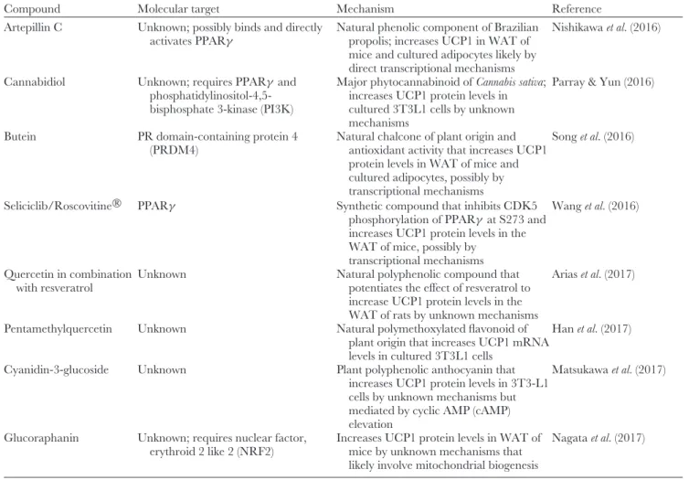
(7) Brown adipose tissue in health and disease. 1151. Micro RNAs (miRNAs) are small non-coding RNAs that control gene expression by targeting specific messenger RNAs and have been implicated in multiple biological processes and diseases, including obesity (Cruz et al., 2017). In mice, adipocyte-selective gene deletion of enzymes involved in the processing of miRNAs DICER (Mori et al., 2014) or DiGeorge syndrome critical region 8 (DGCR8) (Kim et al., 2014) results in general lipodystrophy, lower abundance of brown adipocytes in iBAT mass and impaired insulin sensitivity, indicating that normal processing of miRNAs in adipocytes is required for adipose tissue development and function. On the other hand, selective overexpression of micro RNA miR-133 in brown preadipocytes prevents their differentiation to mature brown adipocytes by targeting PRDM16 (Trajkovski et al., 2012), suggesting that specific miRNAs are physiological regulators of brown adipogenesis. Accordingly, additional miRNAs have been implicated in adipogenesis (reviewed by Cruz et al., 2017), including: miR-365, miR-346 and miR-362 (Mori et al., 2014), miR-182 and miR-203 (Kim et al., 2014), miR-193b and miR365 (Sun et al., 2011). In a survey of miRNAs that may be relevant for the development of beige/brite adipocytes in WAT depots, Giroud et al. (2016) recently found that miR-125b-5p expression is lower in BAT than in WAT and that it correlates inversely with mitochondrial biogenesis. By contrast, miR-106b and miR-93 levels were higher under diet-induced obesity and inhibited the expression of thermogenic genes in murine BAT (Wu et al., 2013), suggesting that miRNAs have a determinant role for both brown and white adipocyte abundance in vivo. Currently, the therapeutic potential of the transcriptional regulators of brown adipogenesis to increase whole body mass of brown adipocytes and hence promote non-shivering thermogenesis in humans, remain uncertain, mostly given the intrinsic difficulties of directly targeting nuclear proteins. Nonetheless, even assuming the effectiveness of such potential drugs, clinically relevant weight loss will only be obtained by the simultaneous prevention of compensatory increases of energy intake (Polidori et al., 2016). Recently, a multiplicity of natural and synthetic compounds have been claimed to promote brown adipogenesis in in vivo or in vitro systems (reviewed by Wankhade et al., 2016) (Table 1). With the exception of some thiazolidinediones (Wankhade et al., 2016) and capsinoids (Digby et al., 1998), none of these compounds have demonstrated browning effects in humans. More importantly, clinical effectiveness and security of these candidate molecules to control body mass in obese patients have yet to be demonstrated.. profiles. Interestingly, even though some early investigators argued that brown adipocytes merely corresponded to immature white adipose cells (Sheldon, 1924) other contemporary authors contended that BAT was an already mature and functional tissue (Rasmussen, 1923). Notably, compelling experimental evidence that adipose tissue has committed cellular precursors that differ from other mesodermal-derived tissues was generated early in BAT studies (Hausberger, 1955). The classical adipogenic model posits that adipose tissue has a single mesodermal origin and that a common mesenchymal stem cell (MSC) gives rise to WAT and BAT, as well as bone, muscle, and cartilage (Yoneshiro et al., 2012). Accordingly, it was thought that MSCs generate common adipose cell precursors (‘adipoblasts’) that, in turn, differentiate into committed white or brown preadipocytes under appropriate stimulatory conditions. Even though this is no longer the prevailing hypothesis (see below), the model was long supported by the simultaneous absence of both WAT and BAT depots in several lipodystrophic mouse models, including those deficient for PPARγ (Koutnikova et al., 2003), 1-acylglycerol-3-phosphate O-acyltransferase 2 (AGPAT2) (Cortés et al., 2009) and Berardinelli-Seip congenital lipodystrophy 2 (seipin) BSCL2 (Liu et al., 2014). Recent developmental and evolutionary studies show that WAT and classical BAT have, indeed, diverging origins. Temporal considerations support this idea: BAT originates early during fetal life and is morphologically and molecularly mature at birth (Cannon & Nedergaard, 1986; Cautivo et al., 2016), whereas WAT develops at mid-gestation in humans and postnatally in mice (Wang & Scherer, 2014). In addition, BAT appeared significantly later in evolution compared with WAT, along with non-shivering thermogenesis and homeothermic regulation in mammals (Gesta, Tseng & Kahn, 2007). Interestingly, WAT from different anatomical sites shows distinctive gene expression patterns (Gesta et al., 2007), and lineage-tracing studies show that, whereas WAT in the trunk derives from the mesoderm, WAT in the cephalic region derives from the neural crest and thus belongs to the ectodermal lineage (Billon et al., 2007), suggesting that white adipocytes have several distinct embryonic/developmental origins. BAT also seems to be developmentally diverse, with different cell precursors for interscapular classical iBAT and for beige/brite adipocytes (Spiegelman, 2013).. V. THE CELLULAR LINEAGE OF BAT. Using genetic fate mapping, Atit et al. (2006) first showed that in mice iBAT brown adipocytes originate from Engrailed 1 (En1)-expressing cells in the central dermomyotome, along with the dorsal dermis and epaxial skeletal muscle. Supporting these findings, Timmons et al. (2007) showed that. It was long assumed that white and brown adipocytes shared a common developmental origin owing to their similar intracellular lipid-storage capacity and gene expression. VI. INTERSCAPULAR BAT AND SKELETAL MUSCLE HAVE A CONVERGENT DEVELOPMENTAL ORIGIN. Biological Reviews 93 (2018) 1145–1164 © 2017 Cambridge Philosophical Society.
(8) Pablo Tapia and others. 1152. Table 1. Natural and synthetic compounds that increase uncoupling protein 1 (UCP1) levels in white adipose tissue (WAT) or promote brown adipose tissue (BAT) activity in vivo Compound. Molecular target. Mechanism. Thiazolidinediones Peroxisome proliferator-activated receptor Synthetic PPARγ agonist; transcriptional γ (PPARγ ) activation of brown adipogenesis Capsinoids Transient receptor potential cation channel Mechanisms for their effects on brown subfamily V member 1 (TRVP1) adipogenesis undetermined; increases UCP1 levels and BAT activity in cultured adipocytes, mice and humans, likely by increasing central adrenergic activity Irisin Unknown cell surface receptor; requires Protein product secreted by skeletal muscle Peroxisome proliferator-activated in response to exercise induces receptor α (PPARα) and p38 mitogen transcriptional activation of brown activated kinase (MAPK) adipogenesis in mice Resveratrol AMP activated protein kinase (AMPK), Natural phenol antioxidant; mechanisms sirtuin 1 (SIRT1) for its effects on brown adipogenesis unknown Melatonin Undetermined, possibly metallothionein-1 Hormone secreted by pineal gland and a natural plant product; mechanisms for (MT1/MT), RAR related orphan its effects on brown adipogenesis are receptor (ROR) unknown, possibly central activation of sympathetic system 10,12 Conjugated Unknown, possibly G-protein coupled Natural long-chain fatty acid enriched in linoleic acid receptor (GPR) 120 and GPR40 dairy products; mechanisms for its effects on brown adipogenesis unknown Glucagon-like Glucagon-like peptide 1 receptor (GLP1R) Peptide hormone secreted by peptide 1 enteroendocrine L-cells in response to feeling; hypothalamic AMPK activation that possibly increases sympathetic output β-aminoisobutyric Unknown cell surface receptor; requires Non-protein β-amino acid released by acid PPARα muscle in response to exercise; mechanisms for its effects on brown adipogenesis unknown Tamoxifen Unknown, possibly oestrogen receptor Synthetic ER antagonist; intraperitoneal (ER) in adipocytes injection increases UCP1 protein levels in subcutaneous WAT in mice after 5 days; unknown mechanism Chrysin Unknown; requires AMPK Natural flavonoid found in flowers and mushrooms that increases UCP1 protein levels in cultured 3T3L1 cells Imatinib/Gleevec® PPARγ Synthetic molecule that inhibits cyclin-dependent kinase (CDK)5-mediated PPARγ phosphorylation in S273 and increases UCP1 protein levels in subcutaneous WAT of mice β-Lapachone Micro RNA 32 (miR32) and indirectly Natural plant naphthoquinone that iodothyronine deiodinase 2 (DIO2) increases UCP1 protein levels and thermogenesis in WAT of mice Uroguanylin Guanilate cyclase 2C receptor (GUCY2C) Peptide hormone secreted by duodenal in the hypothalamus epithelial cells in response to feeding; increases UCP1 protein levels in WAT by central adrenergic activation Curcumin Unknown Natural phenolic compound of plant origin that increases UCP1 mRNA levels in cultured mouse adipocytes D-Limonene Unknown; possibly requires β3 Natural monoterpene mainly present in adrenoceptor activation citrus fruits; activates AMPK and increases UCP1 protein levels in cultured 3T3L1 cells. Biological Reviews 93 (2018) 1145–1164 © 2017 Cambridge Philosophical Society. Reference Fukui et al. (2000) Baboota et al. (2014) and Yoneshiro et al. (2012). Bostrom et al. (2012) and Zhang et al. (2014) Wang et al. (2015) and Alberdi et al. (2013) Jimenez-Aranda et al. (2013) and Tan et al. (2011) Shen et al. (2013) Lockie et al. (2012) and Beiroa et al. (2014). Roberts et al. (2014). Hesselbarth et al. (2015). Choi & Yun (2016). Choi et al. (2016a). Choi et al. (2016b) Folgueira et al. (2016). Kim et al. (2016) Lone & Yun (2016).
(9) Brown adipose tissue in health and disease. 1153. Table 1. Continued Compound. Molecular target. Mechanism. Artepillin C. Unknown; possibly binds and directly activates PPARγ. Cannabidiol. Unknown; requires PPARγ and phosphatidylinositol-4,5bisphosphate 3-kinase (PI3K). Butein. PR domain-containing protein 4 (PRDM4). Seliciclib/Roscovitine®. PPARγ. Natural phenolic component of Brazilian Nishikawa et al. (2016) propolis; increases UCP1 in WAT of mice and cultured adipocytes likely by direct transcriptional mechanisms Major phytocannabinoid of Cannabis sativa; Parray & Yun (2016) increases UCP1 protein levels in cultured 3T3L1 cells by unknown mechanisms Natural chalcone of plant origin and Song et al. (2016) antioxidant activity that increases UCP1 protein levels in WAT of mice and cultured adipocytes, possibly by transcriptional mechanisms Synthetic compound that inhibits CDK5 Wang et al. (2016) phosphorylation of PPARγ at S273 and increases UCP1 protein levels in the WAT of mice, possibly by transcriptional mechanisms Arias et al. (2017) Natural polyphenolic compound that potentiates the effect of resveratrol to increase UCP1 protein levels in the WAT of rats by unknown mechanisms Han et al. (2017) Natural polymethoxylated flavonoid of plant origin that increases UCP1 mRNA levels in cultured 3T3L1 cells Plant polyphenolic anthocyanin that Matsukawa et al. (2017) increases UCP1 protein levels in 3T3-L1 cells by unknown mechanisms but mediated by cyclic AMP (cAMP) elevation Increases UCP1 protein levels in WAT of Nagata et al. (2017) mice by unknown mechanisms that likely involve mitochondrial biogenesis. Quercetin in combination Unknown with resveratrol Pentamethylquercetin. Unknown. Cyanidin-3-glucoside. Unknown. Glucoraphanin. Unknown; requires nuclear factor, erythroid 2 like 2 (NRF2). brown preadipocytes have a gene expression profile highly similar to myogenic precursors, including elevated levels of the myogenic regulator myogenin, typically regarded as a ‘master’ transcriptional regulator of muscle development, myoblast determination protein (MyoD), and myogenic factor 5 (Myf5). Global analysis of miRNA abundance showed that myogenic miR-1, miR-133a and miR-206 were specifically expressed in brown pre- and mature adipocytes in mice but were absent in white adipocytes (Walden et al., 2009), further supporting the notion that classical brown adipocytes derive from a cell precursor shared with skeletal myocytes. Both the C2C12 myoblast cell line and primary mouse myoblasts isolated from postnatal skeletal muscle robustly differentiate into lipid-filled adipocyte-like cells when transduced with PRDM16 (Seale et al., 2008). These cells express common adipocyte markers such as PPARγ and adipocyte fatty acid binding protein 2 (aP2) as well as the BAT markers UCP1, elongation of very long chain fatty acids 3(ELOVL3) and cell death-inducing DFFA-like effector A (CIDEA), while repressing myogenic genes for MyoD and myogenin (Seale et al., 2008). Conversely, PRDM16 knockdown in primary brown fat cells potently induces. Reference. skeletal myogenesis in vitro, and its deletion in mice determines morphologically abnormal BAT, with reduced levels of brown adipocyte markers and higher expression of myogenic markers (Seale et al., 2008). Notably, myogenin-deficient mice, which are devoid of mature skeletal muscle fibres, have increased BAT mass, especially at the cervical–dorsal–interscapular region (Hasty et al., 1993), suggesting that precursor cells that are incapable of completing muscle differentiation are directed, perhaps by default, to the brown adipocyte differentiation program. By contrast, whereas iBAT brown adipocytes derive from Myf5-expressing mesenchymal precursors in mice, WAT and the pockets of brown-like adipocytes inside WAT (i.e. brite/beige fat cells) derive from precursors that do not belong to the Myf5-expressing cell lineage (Seale et al., 2008).. VII. BEIGE/BRITE AND IBAT ADIPOCYTES HAVE DIFFERENT DEVELOPMENTAL ORIGINS The actual origin of beige/brite adipocytes is still a matter of debate. Cinti (2009) and Frontini & Cinti (2010) suggested Biological Reviews 93 (2018) 1145–1164 © 2017 Cambridge Philosophical Society.
(10) 1154 that white adipocytes (or at least a specific subset of these cells) can convert reversibly into beige/brite cells. Since murine mature adipocytes can undergo reversible mammary epithelial transdifferentiation in vivo (Morroni et al., 2004; De Matteis et al., 2009), these investigators propose that analogous processes may occur within the adipose tissue. In fact, upon prolonged exposure to cold or β-adrenergic agonists, selected white adipocytes apparently do convert into brown-like fat cells (Himms-Hagen et al., 2000; Granneman et al., 2005). Similarly, thiazolidinedione PPARγ activators clearly induce beige/brite sprouts in murine WAT (Wilson-Fritch et al., 2004), possibly as a result of sirtuin 1(SIRT1)-dependent post-translational modifications of PPARγ (Qiang et al., 2012) and stabilization of the PRDM16–PPARγ interaction (Ohno et al., 2012). A second hypothesis for beige/brite adipocytes origin states that they differentiate de novo from precursor cells (preadipocytes) already present in specific areas of WAT. The invariable anatomical pattern of beige cell abundance among WAT depots (Murano et al., 2009) strongly suggests that beige preadipocytes mass is anatomically defined. Wu et al. (2012) subcloned multiple adipose cell lines from immortalized stromal cells derived from subcutaneous inguinal WAT. Comparative transcriptomic analysis showed a subgroup of adipocytes that was more similar to classical brown adipocytes (iBAT) than to white adipocytes inside WAT (Wu et al., 2012), suggesting that distinct classes of cell precursors may give rise to beige/brite cells whereas others remain committed to the white adipocyte lineage. In a clinically relevant result, the same authors found that adipocytes from biopsied BAT depots in healthy human volunteers had higher molecular similarity to murine beige/brite cells than to classic murine iBAT (high expression levels of beige markers cluster differentiation 127 (CD127), transmembrane protein 26 (TMEM26) and t-box 1 (TBX1), and lower levels of iBAT markers early B-cell factor 3 (EBF3), epithelial V-like antigen 1 (EVA1) and F-box only protein 31 (FBXO31) (Wu et al., 2012). Therefore, the most consolidated current evidence suggests that humans have at least two types of brown adipocytes: one is iBAT present in newborns and infants (Aherne & Hull, 1966; Lidell, Betz & Enerback, 2014) whereas the other corresponds to beige/brite adipose tissue, interspersed between the subcutaneous adipose tissue that surrounds the large blood vessels of the neck and supraclavicular space (Cypess et al., 2009). Nevertheless, very recent studies have found that this neat anatomical and ontogenic distinction might not be completely correct. In-depth transcriptional analysis of supraclavicular adipose tissue in humans showed simultaneous expression of markers from both iBAT [miR-206, miR-133b, lim homeobox gene 8 (LHX8), and zinc finger protein of cerebellum 1 (ZIC1)] and beige/brite adipocytes (TBX1 and TMEM26) (Jespersen et al., 2013), suggesting either coexistence of these two classes of adipose cells in that depot or, alternatively, complex gene expression and regulation patterns in human BAT. Biological Reviews 93 (2018) 1145–1164 © 2017 Cambridge Philosophical Society. Pablo Tapia and others From a functional standpoint, it remains to be determined whether classical iBAT and beige/brite adipocytes have equivalent thermogenic potentials. Beyond the physiological implications of this definition, its importance lies in the fact that beige/brite adipocytes are the likely cellular substrate of all anti-obesity therapies based on BAT activation in humans. While no direct functional comparison between iBAT and beige/brite adipocytes is available, this question can be approached by considering the relative content of UCP1 in both tissues. It has been estimated that UCP1 corresponds to 5–8% of rat iBAT mitochondrial protein (Lin & Klingenberg, 1980). Analogous quantifications are not available for human BAT or beige/brite fat cells, nevertheless, analysis of intra-abdominal BAT in three patients with pheochromocytome, in which neoplastic cells from the adrenal medulla secrete excessive amounts of catecholamines, revealed that UCP1 abundance in that adipose depot was similar to that in murine iBAT (∼30 μg/g) (Lean et al., 1986a). Importantly, the abundance of UCP1 in the BAT of normal individuals appears to be at least ten times lower that in pheochromocytome-associated BAT (Lean et al., 1986b), shedding doubts on the actual maximal thermogenic potential of BAT in adult humans as a therapeutic option for obesity. A more recent work studied UCP1 content in WAT depots of children subjected to major inflammatory and metabolic stress because of severe burning. A 37 ± 25 (mean ± SD) days after burning, UCP1 content was ∼8-fold higher in comparison with healthy control children (22.8 versus 2.7 μg/g) (Sidossis et al., 2014). Supporting the molecular evidence of acute browning of WAT in humans, total mitochondrial functional mass (estimated by citrate synthase activity) and uncoupled respiration were ∼4- and ∼3-fold increased, respectively, in the WAT of late-burn in comparison with early-burn patients (Sidossis et al., 2015). The question of how stable beige/brite adipocytes are after browning induction is equally relevant with respect to future BAT-based therapies. To investigate the kinetics of browning processes, Rosenwald et al. (2013) exposed mice to 8◦ C for 7 days and then moved them to warm (23◦ C) housing conditions. Cold stimulation increased UCP1 mRNA and protein levels as well as other brown adipocyte markers, in subcutaneous (inguinal) but not in visceral (epididymal) WAT, indicating depot-specific browning. Importantly, they found that these phenotypic changes lasted for 5 weeks after moving animals to warm conditions (Rosenwald et al., 2013). However, cell-lineage-tracing studies with a genetic labelling system that permanently tagged cells expressing UCP1 revealed that beige/brite cells persisted for at least 8 weeks after warm-housing adaptation, but this time with a white adipocyte phenotype (Rosenwald et al., 2013), suggesting long-lasting remodelling of WAT in response to cold activation. Analogous results have been generated by other investigators, which have also revealed the critical role of β-adrenergic signalling (Chabowska-Kita et al., 2015; Lee et al., 2015) and the genetic background of the mice (Guerra.
(11) Brown adipose tissue in health and disease et al., 1998; Lasar et al., 2013) for cold-induced browning activation.. VIII. WHITE AND BROWN ADIPOCYTES DERIVE FROM SPECIALIZED VASCULAR-WALL CELLS IN ADIPOSE DEPOTS The ultimate origin of white and brown preadipocytes remains to be confirmed in vivo in humans. Cell-sortingbased methods successfully identified a small subset of stromal vascular cells in the WAT of mice that exhibits elevated adipogenic and proliferative potential in vitro (Rodeheffer, Birsoy & Friedman, 2008). Moreover, these cells have the ability to differentiate spontaneously into mature WAT in lipodystrophic mice in vivo (Rodeheffer et al., 2008). Similarly, by ectopically expressing green fluorescent protein (GFP) under the transcriptional control of zinc finger protein 423 (Zfp423), a transcriptional regulator of white and brown adipogenic determination (Gupta et al., 2010), it was demonstrated that a subset of capillary pericytes and endothelial cells (ECs) in mature and developing WAT or iBAT have characteristics of preadipocytes in mice (Gupta et al., 2012). Supporting this notion, Tran et al. (2012) determined that murine ECs of both WAT and BAT have ultrastructural features of pericytes. By lineage-tracing experiments with VE-cadherin promoter-controlled reporters, these investigators found that these cells have molecular characteristics of preadipocytes, including Zfp423 expression (Tran et al., 2012). Lineage-tracing studies, using genetically labelled adipocyte progenitors with lacZ reporter gene under the transcriptional control of PPARγ promoter in mice, confirmed that the vascular wall of adipose tissue capillaries is a physiological niche of adipocyte precursors (Tang et al., 2008). These findings agree with the hypothesis that adipose progenitors reside in or near capillary vessels in adipose tissue, however, they appear to contradict the idea of a common cellular progenitor for myocytes and brown adipocytes. Interestingly, a subgroup of cells in the murine dorsal aorta expresses myogenic and endothelial markers, including Myf5 and VE-cadherin (De Angelis et al., 1999), suggesting that Myf5+ progenitor cells also reside in the capillary wall of both adipogenic and myogenic niches.. IX. BAT IS A SECRETORY ORGAN BAT depletion by specific inactivation of the insulin receptor gene in UCP1-expressing adipocytes impairs insulin secretion and triggers diabetes with no associated changes in insulin resistance, WAT mass, or levels of circulating free fatty acids and triglycerides (Guerra et al., 2001), suggesting that BAT can regulate β-cell function and mass, perhaps through still-unidentified secreted factors. Although these seminal findings have not yet been independently confirmed, they launched a search for new endocrine roles of BAT. Indeed,. 1155 brown adipocytes secrete several factors that influence critical tissues for metabolic regulation and that might help to explain the metabolic actions claimed for BAT activation (reviewed in Villarroya et al., 2017). (1) FGF21 Circulating fibroblast growth factor 21 (FGF21) is secreted by the liver (Markan et al., 2014) and its physiological effects are mediated by fibroblast growth factor receptor 1 (FGFR1) and β-Klotho co-receptor (Ogawa et al., 2007). Pharmacological administration of FGF21 decreases body mass, increases energy expenditure, and lowers blood glucose and insulin levels in mice and non-human primates (Gimeno & Moller, 2014). Active BAT and brite/beige adipocytes are able to release FGF21 (Chartoumpekis et al., 2011; Lee et al., 2014c). In humans, levels of circulating FGF21 correlate with BAT activity after acute exposure to cold (Hanssen et al., 2015), suggesting that BAT-derived FGF21 could also reach the circulation. Nonetheless, it seems more plausible that adipose-derived FGF21 works as an autocrine/paracrine factor, increasing the expression of UCP1 and other thermogenic genes, and thus promoting browning (Fisher et al., 2012; Lee et al., 2014a). (2) BMP8B BMPs belong to the transforming growth factor β (TGF-β) superfamily and some members are relevant regulators of adipogenesis (Tseng et al., 2008). BMP8B-deficient mice have a reduced metabolic rate and increased susceptibility to diet-induced obesity (Whittle et al., 2012), suggesting a role for this protein in the regulation of BAT mass/activity. BMP8B is expressed in BAT mature adipocytes along with thermogenic markers and it enhances lipolysis in brown adipocytes via a P38 mitogen-activated protein kinase (MAPK)/CREB-dependent pathway (Whittle et al., 2012). BMP8B also mediates WAT browning through mechanisms that rely on AMP-activated protein kinase (AMPK) inhibition and increased signalling through orexin receptor 1 in specific hypothalamic nuclei (Martins et al., 2016). (3) NRG4 Neuregulin 4 (NRG4) is a member of the epidermal growth factor (EGF) family and is expressed in the murine lung, heart, and adipose tissues, with the highest expression level in BAT (Pfeifer, 2015). NRG4 expression in BAT is increased by cold exposure (Rosell et al., 2014) and its gene deletion determines increased body mass, insulin resistance and fatty liver production in mice (Wang et al., 2014). Conversely, by hydrodynamic in vivo transfection, hepatic NRG4 overexpression prevents high-fat-diet-induced mass gain and decreases obesity-induced chronic inflammation and insulin resistance in mice (Ma, Gao & Liu, 2016). These and other results led to suggestions that NRG4 is a potential therapeutic tool to increase BAT in obesity, Biological Reviews 93 (2018) 1145–1164 © 2017 Cambridge Philosophical Society.
(12) Pablo Tapia and others. 1156 although the relevance for this molecule in human BAT physiology remains unknown (Christian, 2015). (4) VEGF-A Vascular endothelial growth factor A (VEGF-A) is a key regulator of angiogenesis and tissue remodelling (Neufeld et al., 1999). Its selective transgenic overexpression in white adipocytes increases vascularization, mitochondrial mass, UCP1 levels and thermogenic capacity in mice (Sun et al., 2012). Transgenic overexpression of VEGF-A in classical brown adipocytes further increased UCP1 levels in iBAT and both basal and cold-stimulated non-shivering thermogenesis in mice (Sun et al., 2014). Although the physiological translation of these results remains unknown, it is interesting to note that cold exposure increases iBAT angiogenesis in association with increased expression levels of VEGF-A, suggesting a role for VEGF-A in normal iBAT thermogenic capacity (Xue et al., 2009) and WAT browning (Sun et al., 2012). (5) IL-6 Interleukin-6 (IL-6) is a proinflammatory cytokine produced by immune and non-immune cells, mainly in the skeletal muscle and adipose tissue; however, the role of this cytokine in obesity remains controversial (Ma et al., 2015; Braune et al., 2017). Interestingly, transplantation of BAT that is deficient in IL-6 prevents the metabolic benefits of increasing BAT mass in mice (Stanford et al., 2013), suggesting that IL-6 is an essential mediator of BAT actions on insulin sensitivity and glucose regulation. (6) RBP4 Retinol binding protein 4 (RBP4) is a serum protein involved in the transport of vitamin A and has been associated with insulin resistance in humans (Graham et al., 2006). It is mainly produced by the liver and WAT (Asha et al., 2016), and although it remains unknown whether it plays a pathogenic role in human insulin resistance, its transgenic overexpression or direct injection causes insulin resistance in mice (Yang et al., 2005). To address the role of RBP4 in insulin resistance, Zemany et al. (2014) specifically deleted Stra6, the gene encoding the high-affinity receptor for RBP4, in white adipocytes. They found that Stra6-deficient mice were leaner and had increased energy expenditure relative to wild-type controls, in association with elevated UCP1 mRNA levels in WAT, suggesting enhanced browning (Zemany et al., 2014). Intriguingly, it was described that BAT may contribute to RBP4 circulating levels via cAMP-, PPARα- and PPARγ -mediated pathways (Rosell et al., 2012), however, the physiological significance of these findings remains unknown. (7) IGF-1 Insulin like growth factor 1 (IGF-1) is produced by the liver in response to growth hormone and insulin stimulation. IGF-1 Biological Reviews 93 (2018) 1145–1164 © 2017 Cambridge Philosophical Society. deficiency consistently associates with insulin resistance and therefore it is a prospective pharmacological target for this condition (reviewed in Aguirre et al., 2016). In streptozotocin-induced diabetic mice, BAT transplantation results in higher IGF-1 levels and improved glucose regulation (Gunawardana & Piston, 2012), suggesting that BAT can be a physiological source of circulating IGF-1. (8) miRNAs Recent studies suggest that, in addition to their direct roles in adipogenesis, miRNAs can be secreted in exosomes by brown adipocytes and thus possibly exert regulatory roles in distant tissues (Zemany et al., 2014). In vitro, adrenergic stimulation increases the release of miRNA-loaded exosomes by cultured murine brown adipocytes and iBAT explants by five- and ninefold, respectively (Chen et al., 2016), indicating that miRNA secretion could be physiologically regulated. Adipocyte-specific gene deletion of DICER, an enzyme necessary for the processing of miRNAs, results in lower levels of circulating exosomal miRNAs, indicating that BAT and WAT are physiological sources of miRNAs in the blood (Thomou et al., 2017).. X. THE IMPORTANCE OF BAT FOR METABOLIC REGULATION Mouse models of BAT (Lowell et al., 1993) and UCP1 (Enerback et al., 1997) ablation provided evidence that stimulated studies on the physiological role of BAT in energy homeostasis and body composition, particularly its roles in thermogenic adaptation to cold exposure. Genotoxic deletion of brown adipocytes by transgenic expression of diphtheria toxin-A under the transcriptional control of UCP1 promoter resulted in overt obesity and insulin resistance in mice, importantly without changes in food intake (Lowell et al., 1993). Interestingly, studies of UCP1-deficient mice did not replicate these results, shedding doubt on the functional role of UCP1-driven thermogenesis on body composition, since resting energy expenditure and body mass remained unaffected in mice lacking UCP1 (Enerback et al., 1997). Subsequent reports claimed that UCP1-deficient animals spontaneously developed obesity when housed under thermoneutral conditions (∼30◦ C) (Feldmann et al., 2009), possibly indicating that differences in metabolic efficiency, masked by ‘normal’ housing conditions (18–22◦ C), may underlie the absence of obesity found by Enerback et al. (1997). However, other investigators could not replicate these findings. Liu et al. (2003) showed that in congenic C57BL/6J mice, a lack of UCP1 paradoxically resulted in protection from diet-induced obesity when the animals were housed at 20◦ C. This response is possibly due to the use of less metabolically efficient alternative mechanisms for non-shivering thermogenesis, as suggested by the fact that body mass differences were not observed when animals.
(13) Brown adipose tissue in health and disease were housed at thermoneutral conditions (Liu et al., 2003; Anunciado-Koza et al., 2008). Therefore, it is currently thought that BAT-dependent thermogenesis can have at least two distinct functional roles: adaptive thermoregulation and metaboloregulation. Whereas the former has reached scientific consensus (Enerback et al., 1997; Hofmann et al., 2001; Meyer et al., 2010), the latter, i.e. that BAT thermogenesis burns off excess calories in states of positive energy balance to maintain energy homeostasis and protect against obesity and insulin resistance, remains controversial (Kozak, 2010). The known inverse correlation between BAT mass and total adiposity in humans (Cypess et al., 2009; van Marken Lichtenbelt et al., 2009; Saito et al., 2009) favours the metaboloregulation hypothesis as does the observation that total mass of beige/brite adipocytes, which is strongly dependent on genetic factors (Almind et al., 2007; Xue et al., 2007), also inversely correlates with the susceptibility to develop obesity in mice (Almind et al., 2007). In contradiction is the likelihood that obesity was a very rare condition in premodern humans, owing to food scarcity and elevated energy expenditure (Hayes et al., 2005), therefore burning off excess calories by means of BAT activation is in clear conflict with evolutionary arguments. Nevertheless, although the physiological actions of BAT on basal metabolic regulation are controversial, the capacity of activated BAT to generate heat by dissipating metabolic energy, either by cold exposure or adrenergic stimulation, and thus increase whole-body energy expenditure, is beyond doubt (Dawkins & Hull, 1964). In high-fat-fed mice, intermittent exposure to severe cold (4◦ C for 1–8 h, 3× a week for 10 weeks) consistently increases whole-body and iBAT heat generation but, since there are compensatory increases in food intake, no changes in body mass are detectable (Ravussin et al., 2014). Notably, in spite of its lack of effects on body adiposity, cold exposure improves glucose tolerance and insulin sensitivity in obese mice (Ravussin et al., 2014). In agreement with the above observations, pharmacological β-adrenergic activation results in a substantial reduction of adiposity in rats, dogs and mice, likely by stimulating BAT-mediated energy dissipation (Robidoux, Martin & Collins, 2004) and BAT transplantation to genetically obese mice decreases body mass and hepatic steatosis, improves adipose tissue hypertrophy and inflammation, and increases whole-body insulin sensitivity (Liu et al., 2015). However, it is equally important to recognize that metaboloregulatory roles for BAT in humans remain hypothetical. Human populations have a broad distribution of BAT mass (as determined by FDG-PET-CT scan), which consistently correlates negatively with body adiposity. This association, combined with larger BAT mass in cold than in warm seasons (Saito et al., 2009) and its acute activity regulation by cold exposure (van Marken Lichtenbelt et al., 2009; Saito et al., 2009), do suggest that BAT may be physiologically implicated in whole-body energy balance in humans. This has inspired the suggestion that the excessive. 1157 periods spent in thermoneutral environments in modern human populations is a determinant of obesity (Johnson et al., 2011). Blondin et al. (2015) showed that cold exposure can increase energy expenditure by 82% and raise plasma free fatty acid concentration by 1.4-fold in association with a 2.3-fold elevation of BAT oxidative activity in lean healthy men, suggesting that WAT lipolysis is physiologically linked to BAT metabolic activation and that BAT-dependent thermogenesis could be substantial in humans. Reinforcing the notion that BAT activity increases fat mobilization and oxidation in humans, Hibi et al. (2016) showed that patients with FDG-PET-CT-detectable BAT after acute exposure to cold have higher diet-induced thermogenesis and a lower respiratory quotient than subjects without detectable BAT. It is noteworthy that in spite of these and analogous correlation-based studies, actual demonstrations of the physiological significance of BAT as a determinant of energy expenditure in adult humans are lacking.. XI. THE IMPACT OF BAT IN HUMAN HEALTH AND DISEASE Besides its effects on body composition/mass regulation, BAT may have a direct impact on other determinants of disease in humans. This idea was inspired by the observation that pharmacological or genetic activation of BAT, or its surgical implantation, improve circulating levels of glucose (Stanford et al., 2013), triglycerides (Bartelt et al., 2011) and cholesterol (Berbée et al., 2015) as well as whole-body insulin sensitivity (Cederberg et al., 2001; Liu et al., 2013; Stanford et al., 2013) in mice, independently of significant changes in body mass. Whether these findings extrapolate to humans and can be used in clinical applications to treat diseases connected with obesity and insulin resistance remains to be determined. Important questions to be answered are: (i) what is the relative power of BAT activation to reduce both body adiposity and insulin resistance in comparison with currently available therapies, and (ii) what are the risks associated with chronic BAT activation?. XII. ACTIVATING BROWN ADIPOSE TISSUE THERMOGENESIS AS A TARGET TO INDUCE WEIGHT LOSS The observed inverse correlation between body mass index and BAT activity (see Sections I and X) lead us to investigate the extent to which maximally activated BAT can influence whole-body energy expenditure. Eventually, enhanced thermogenesis may prevent weight gain (or even induce weight loss) if a compensatory increase in energy intake does not take place. However, the precise mass and metabolic activity of human BAT remains undetermined. This is partly because Biological Reviews 93 (2018) 1145–1164 © 2017 Cambridge Philosophical Society.
(14) Pablo Tapia and others. 1158 Table 2. Resting metabolic activity of principal organs in humans Organ Heart Kidneys Brain Liver Skeletal muscle Adipose tissue Residual mass. Resting metabolic activity (kcal/kg/day) 440 440 240 200 13 5 7. of the diffuse anatomical distribution of this tissue but also, because of great heterogeneity in published PET-CT-based detection protocols and intrinsic variability in BAT mass, even at the individual level (Pardo et al., 2017). For example, in one study only considering individuals with FDG-PET-CT-detectable BAT (81 out of 1644 patients), the volume of BAT ranged between ∼12 and ∼300 ml; i.e. 11–285 g, assuming a tissue density of 0.95 g/ml in normal volunteers (Gerngross et al., 2017). These authors suggested that the total mass of BAT could have been greatly underestimated, by a factor of ∼4, in adult humans and thus its total thermogenic capacity could also be underappreciated. Classical studies have estimated that maximally activated BAT can produce up to 300 W/kg tissue of heat in comparison with 1–2 W/kg tissue by other organs (Power, 1989). If we speculate that whole-body metabolically active BAT is 2–3 times the mass reported in the supraclavicular and neck by FDG-PET of healthy adults (60 g) (Virtanen et al., 2009) and assume that BAT may have a resting metabolic activity similar to the heart or the kidneys, the two organs with the highest reported resting metabolic activity in humans (Muller et al., 2011) (Table 2), then total BAT mass (∼180 g) should account for only ∼80 kcal/day. Another calculation, taking a total BAT mass of ∼200 g and a resting metabolic activity equivalent to the liver (Table 2) (Wang et al., 2012), provides a thermogenic yield of only 40 kcal/day. Our estimations of the contribution of BAT to whole-body thermogenesis is in sharp contrast with previous analyses based on animal findings (Rothwell & Stock, 1983; Rosenbaum & Leibel, 2010) that speculated that 50 g of maximally stimulated BAT could account for up to 20% of total energy expenditure in humans. For an adult human with a resting metabolic rate of 1500 kcal/day, 50 g of BAT therefore would account for an energy expenditure of 300 kcal/day, giving a specific metabolic activity of 6000 kcal/kg of BAT per day, or 13-fold higher than the resting metabolic activity of heart or kidneys which seems highly unlikely. An absolute magnitude of dissipated energy of 40–80 kcal/day seems of negligible importance for long-term energy balance and body-mass determination (Weinsier, Bracco & Schutz, 1993; Butte & Ellis, 2003; Biological Reviews 93 (2018) 1145–1164 © 2017 Cambridge Philosophical Society. Swinburn et al., 2006), although it may be that such small but persistent energy imbalances may have triggered the observed increased body mass of westernized populations over the last three decades (Hill, 2006). A direct role of BAT on thermogenesis and body mass appears to be strongly limited by the small mass of this tissue and the seemingly low plausibility of maintaining BAT permanently activated for prolonged periods of time. Nonetheless, therapeutic activation of BAT could still be beneficial to metabolic health by increasing the clearance rate of circulating lipids and glucose and thus preventing ectopic toxic lipid buildup in tissues, which is a leading cause of insulin resistance (ter Horst et al., 2017). It is also possible that BAT activation could increase whole-body energy expenditure beyond the limits imposed by its own mass by means of secreted factors that regulate thermogenesis in distant target tissues.. XIII. CONCLUSIONS (1) The existence of BAT in infants has been known for a long time, but the demonstration of metabolically active BAT in adults was only documented in 2009. This finding has reinvigorated the investigation of different aspects of BAT, mainly driven by the expectation of developing new therapeutic tools against obesity. (2) In small mammals, BAT-dependent thermogenesis is pivotal to prevent hypothermia; however, its physiological relevance in larger organisms, such as adult humans, remains uncertain. It has been proposed, mostly based on animal studies, that BAT may help to prevent or reverse excessive adiposity and improve glucose and triglyceride clearance from blood; however, direct and convincing evidence from human studies is lacking. (3) The cellular lineage and the developmental pathways of BAT are diverse in humans. Interscapular BAT, which is abundant in newborns and infants, is similar to classical BAT of small rodents. By contrast, beige/brite adipose tissue, the most abundant form of BAT in adult humans, is developmentally closer to white adipose tissue. Recent findings suggest that both types of brown adipocytes can coexist in some BAT depots of adults. (4) BAT can also secrete several protein and RNA factors that are likely to influence thermogenic activity by autocrine mechanisms or regulate insulin sensitivity in distant tissues by endocrine actions. (5) Accurate quantification of the mass and specific thermogenic activity of BAT in living humans remains a technical challenge. Furthermore, metabolically active BAT detected by FDG-PET-CT methods seems to be highly variable, at both the inter-individual and individual level, possibly owing to intrinsic properties of this tissue but also to the high heterogeneity of the detection protocols used. (6) Our estimation of the contribution of BAT to whole-body resting thermogenesis indicates that it is unlikely that BAT has a significant role in the determination of.
Figure


+3

Documento similar
BRS-3 seems to play a role in glucose metabolism, not only in normal subjects, but also in obese patients and patients with T2D. Furthermore, its ligand is capable of improving
The draft amendments do not operate any more a distinction between different states of emergency; they repeal articles 120, 121and 122 and make it possible for the President to
H I is the incident wave height, T z is the mean wave period, Ir is the Iribarren number or surf similarity parameter, h is the water depth at the toe of the structure, Ru is the
Evolution of the NVDI and GNDVI spectral indices of a point associated with vegetation (green striped lines: ndvi-veg, gndvi-veg), of a point associated with soil (brown dotted
Since such powers frequently exist outside the institutional framework, and/or exercise their influence through channels exempt (or simply out of reach) from any political
Data are shown as fold-induction relative to control (untreated cells) values for each panel. Significant differences in comparison to controls are shown by * p<0.05. Regulation
Ribeiro, M.O., et al., Expression of uncoupling protein 1 in mouse brown adipose tissue is thyroid hormone receptor-beta isoform specific and required for adaptive
Body weight, daily food intake and weights of retroperitoneal visceral adipose tissue, lumbar subcutaneous adipose tissue, interscapular brown adipose tissue, heart, kidneys,
