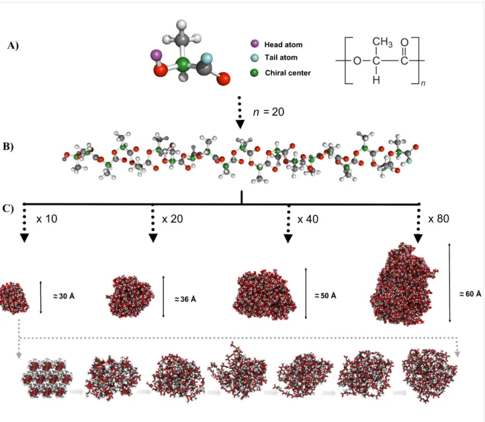
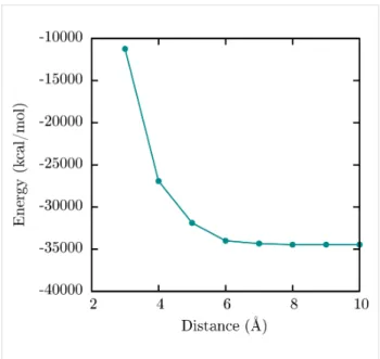
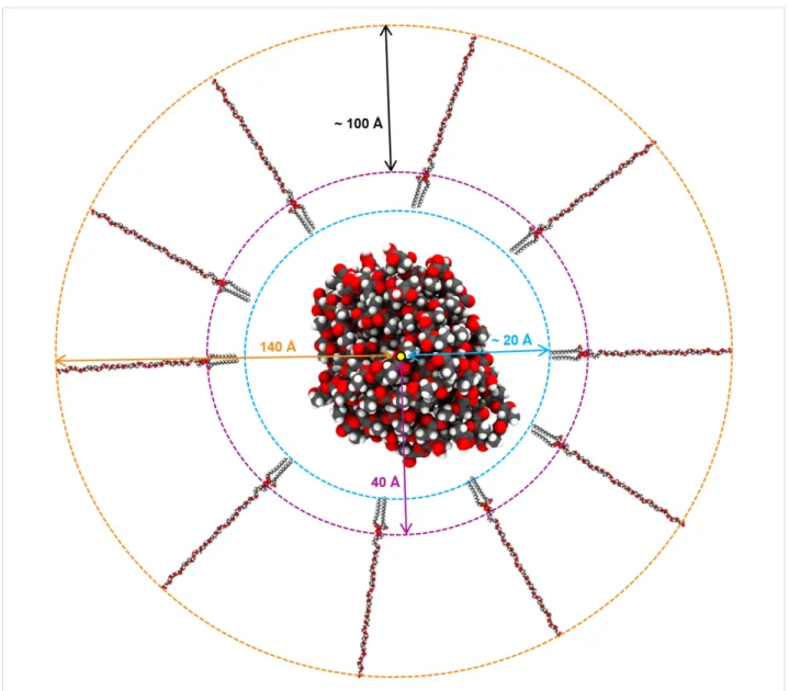
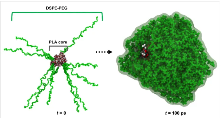
Atomic level characterization and cilostazol affinity of poly(lactic acid) nanoparticles conjugated with differentially charged hydrophilic molecules
Texto completo
Figure


Documento similar
(a) Immunoblottings showing phosphorylation of the indicated molecules in lysates of J77 Jurkat T cells pretreated with vehicle (DMSO) or Aurora A inhibitor (MLN8237) and conjugated
Characterization of optical frequency comb based measurements and spectral purity transfer for optical atomic clocks.. soutenue le 15
Incorporation of DMSA-SPION into MCF-7 cells can be followed by bright field microscopy after 24 h incuba- tion (Figure 1A), where SPION are observed inside living cells, distributed
Of special concern for this work are outbreaks formed by the benthic dinoflagellate Ostreopsis (Schmidt), including several species producers of palytoxin (PLTX)-like compounds,
Figure 4.23: Use of the tunable transparency to follow the interaction between graphene C vacancies and the Cu(111) substrate underneath. a) STM image showing a G/Cu(111) sample
In this thesis, TERRA molecules have been investigated at the single-molecule level by atomic force microscopy (AFM), in the imaging mode, and optical tweezers (OT), as
Atomic force microscopy (AFM) fulfills these requirements providing images of individual virus particles under physiological conditions, along with the characterization of a variety
Polymer (Guildf).. 1: SEM images of the fracture surfaces of PHBV/PLA blends with diisocyanates.. 2: DSC curves of neat PHBV, PHBV/PLA and PHBV/PLA blends with a 1:20 molar ratio of
