An understanding of the prostate cancer pathophysiology for identification of biomarkers that support an early diagnosis
144
0
0
Texto completo
(2) Introduction Prostate Cancer (CaP) is a condition whose etiology is multifactorial, it goes through genetic, infectious, as well as environmental, among others. Currently, PCa represents the most frequent cancer among men and the second in mortality, however, there are still multiple gaps in the knowledge of this condition. One of them is related to a molecular biomarker that allows early and accurate diagnosis of PCa. We have the Prostate Specific Antigen (PSA), however, its diagnostic performance alone is very low, and it must always be associated with digital rectal examination to obtain the best of both diagnostic methods. Problem statement Over the years, the focus of prostate cancer has been on the treatment. A patient with the condition is identified and they are offered invasive and non-invasive procedures that can generate adverse deleterious effects for the quality of life. Some efforts have been made for prevention without obtaining clear results according to the available evidence, and related to food and medication. Other efforts have focused on early diagnosis, with emphasis on the measurement of prostatespecific antigen (PSA) and digital rectal examination, however, there are already systematic reviews and meta-analyzes that show how population screening is not suggested of opportunity, given the large population that would have to sift to prevent a 10-year death (1). On the other hand but complementary, PSA and digital rectal examination, although they are fundamental elements, have an operative performance (Sensitivity, specificity, predictive values and probability reasons) that is not high and therefore, has room for false positives and negatives that would limit the diagnostic suspicion of prostate cancer. There are some biomolecules that have been studied both in Prostatic Hyperplasia and in Prostate Cancer, however its validity in the general population is still limited. There is only one identified genomic variation (AR-V7) that shows the probability of resistance to hormonal management with enzalutamide and abiraterone in patients with castration-resistant prostate cancer and therefore would require the use of chemotherapy(2–4). However, there is no biomolecule or genomic variant that can be distributed at the time as an early detection test or risk for the development of prostate cancer, which supports the realization of this work. Justification Prostate cancer (PC) is the second most common type of cancer in the world's male population (5). It is estimated that one in seven men will be diagnosed at the end of their life with CP, and one in 38 men will die as a result of this in the long term. Prostate Cancer is diagnosed more frequently as a consequence of the introduction in 1980 of the Prostate Specific Antigen (PSA) test as a diagnostic tool (3). During the last decade, there has been a notable decrease in mortality from prostate cancer in developed countries, but it has not been evidenced in developing countries. GLOBCAN in 2008 reported that in northern European countries (Denmark, Norway and Sweden) the diagnosis has. 2.
(3) increased by 8.2% per year, with a mortality rate falling since 2000 of 3.1% per year (6). Similar data were evidenced in the United States and Canada, in which the incidence in the diagnosis of PC remains stable, but with a decrease in mortality of 4.3% and 3.1% respectively. On the other hand, in developing countries, mortality has increased (although there are trends towards an increase in diagnosis, mortality has increased in countries such as Colombia, 3.4% per year, Costa Rica, 3.4% year, Chile 2.8% per year and Cuba 5.5% per year) (6,7). With respect to the global context, Colombia has one of the lowest incidences in Latin America (6). Additionally, the mortality rate from prostate cancer has decreased in the last four years (7) and the largest number of cases reported originate in the cities of Bogotá, Valle and Antioquia (The most populated regions with the largest number of Urologists) (7). On the other hand, when considering an appetite for the treatment of localized prostate cancer, which is the focus of the present work, there are currently studies that show that there are no differences in 10-year mortality when comparing monitoring, radical surgery of prostate and radiotherapy (8). Even when classifying the patient according to the risk of disease progression (D'amico) (9), it is evidenced that when comparing radical prostatectomy with clinical observation, there are no differences at 10 years of follow-up for the specific cancer mortality outcome (10) in localized low risk PC. This is important to note because we still have gaps in knowledge. For several decades, urologists have performed radical surgeries that generate adverse events such as erectile dysfunction, urinary incontinence and urethral stricture. Given that we have elements of uncertainty such as those already demonstrated, we could consider the search for markers that identify which patients do not require a surgical procedure or those that have the risk, could be evaluated the possibility of genetic manipulation for prevention of prostate cancer. Recent and interesting investigations have been carried out that, although they are not yet conclusive, do encourage us to continue searching for risk markers and genomic alterations that allow us to classify and treat our patients even before developing the disease. Inhibitory markers such as mitochondrial autophagy have been identified, related to the poor prognosis in prostate cancer. Epigenetics have identified alterations in DNA methylation and microRNA expression that occur in the transition from the normal cell, precancerous, primary and metastatic tumors. The circulating DNA fragments are another important strategy to generate the genetic profile and sequencing of the genes of patients with prostate cancer(11). In such a way that we still have gaps in the knowledge of localized prostate cancer that deserve to be evaluated in works of this type.. 3.
(4) Objectives: •. To describe the frequency of Inherited DNA-repair genes and their variants associated to Prostate Cancer in a Cancer-Free Southwest Colombian population. •. To identify the association between the TMPRSS2:ERG fusion gene, their variants and the onset of localized prostate cancer.. •. To describe the frequency of allelic variants of TMPRSS2 gene in this population. •. To analyze the metabolomic profile in patients with malignant disturbances of the prostate compared with non-cancer patients.. 4.
(5) Chapter 1: An updated and global review on prostate cancer Type of article: Narrative Review Authors: Herney Andrés García-Perdomo MD, MSc, EdD, PhD, FACS (1,2); James Alejandro Zapata-Copete MD (2,3); Adalberto Sanchez BSc PhD (1,4) Affiliations 1. Associate professor, Universidad del Valle- Cali, Colombia 2. UROGIV research group. Universidad del Valle, Cali, Colombia 3. Epidemiology Department, Universidad Libre- Cali, Colombia 4. School of Basic Sciences- Universidad del Valle- Cali, Colombia Introduction Prostate cancer given its frequency in the populations is a pathology of importance in public health not only national, but of high global impact (12). Given the difficulties of the Health System, the limited availability of specialists and the high prevalence of this condition, the knowledge of this condition should be the domain of any general practitioner, and should not remain in specialized medicine areas such as urology and oncology. However, the treatment in an integral way, must be given by centers of excellence in cancer (13). Objective To obtain an updated view through a detailed and up-to-date review of the epidemiology, risk factors, classification, diagnosis and treatment of prostate cancer. Methods A search was performed in Embase and Medline (through OVID) from January 2000 to March 2017. The keywords used were: "prostate" OR "prostate, neoplasm" AND "diagnosis" OR "treatment”. Additionally, an exhaustive manual evaluation of the bibliography provided in the articles found was also made. Results Epidemiology Prostate cancer (PCA) is the most frequent neoplasm in men in the world, and represents the second cause of cancer death in this population in the United States (14). It has an incidence of 131.5 / 100,000 inhabitants (14) with a race distribution of 123 / 100,000 inhabitants in the white and 208 / 100,000 inhabitants in the African American population (15). It is estimated that 1 in 7 men will be diagnosed throughout their life with PCA, and 1 in 38 men will die as a consequence of it (16). The GLOBCAN study reported that in the Northern European countries (Denmark, Norway and Sweden) the diagnosis of PCA has increased 8.2% per year, accompanied by a declining mortality of 3,1% per year since 2000 (6). In the United States and Canada, similar data have been found, with a stable incidence but with a decrease in mortality of 4.3% and 3.1% respectively; however, 5.
(6) in developing countries, mortality has increased (6,7). Regarding the national epidemiology, Colombia has one of the lowest incidences of PCA in Latin America and a proportion of 28% between incidence and mortality for it, very close to the world average of 28.6% and lower than countries like Ecuador (40.41%), Cuba (46.65%) and Peru (37.74%) (6). Mortality has decreased in the last four years (7) and the regions with the highest number of CAP patients reported are Bogotá, Valle and Antioquia (The most populated regions and with the largest number of Urologists) (7,16). Risk factors Race Black patients have a higher prevalence of PCA (17), furthermore, they present at younger ages with greater tumor volume, a greater prostatic antigen level and worse prognosis (18,19). Some authors associate these results with social inequities and difficulties in accessing health services, to which this population is exposed (20), however there is much more evidence that supports this factor as a risk factor for PCA. On the other hand, lower rates have been found in Asians, which has been additionally related to diet, lifestyles and environmental factors (21). Family History Approximately 10-15% of men with PCA have at least one relative with history of this condition (17,22). It has been estimated that having a relative of first degree of consanguinity with PCA increases the RR 2 to 4 times and is 5 times higher if there are two relatives with that diagnosis. Inflammation Chronic inflammation is considered a risk factor because it leads to cellular hyperproliferation, which generates an alteration in antioxidant levels, in DNA repair and apoptosis. It has also been found that the history of a sexually transmitted infection has an OR 1.5 (23) and history or current prostatitis an OR of 1.57 (24). Despite being one of the strongest hypotheses, it is still unclear the mechanism that would lead to producing the PCA or if it is a sufficient cause for its development. Oxidative Stress Some studies have suggested that reactive oxygen species (such as superoxide or peroxide) create an environment of mutagenesis conducive to the initiation of PCA (25,26). This element could be related to the hypothesis of chronic inflammation. Androgens There is evidence that an increase in testosterone levels increases the incidence of PCA, although a dose-response relationship or a concentration of which the risk is increased has not been found; additionally, a higher risk of PCA has not been found in patients with hypogonadism treated with testosterone replacement therapy (27). Estrogens It has been shown that estrogens that can predispose and even cause PCA. It is necessary to emphasize that 17β-estradiol has already been classified as a carcinogen, especially in breast and endometrial cancer. It is believed that the effect of estrogen in PCA is caused by direct mutations, through regulation by epigenetic effects or by endocrine disruption (28). Diet Several studies have suggested that a diet low in fat, calcium, with an increase in the consumption of vitamin E and lycopene, as well as regular exercise, could behave as protective factors for the development of PCA, on the other hand, the high intake of saturated fats of animal origin and red meat have been described as risk factors; however, the findings are not consistent between the different studies. For example, in the SELECT study, the protective factor for the use of vitamin E and selenium was not demonstrated (29). 6.
(7) Increase in levels of insulin-like growth factor (IGF-1) Insulin-like growth factor is a mitogenic and antiapoptotic factor. High levels imply more PCA risk (30). However, other studies do not find it as a risk factor. Genetic Alterations have been found in suppressor genes such as p53, PTEN, these are related to increased incidence, progression and aggressiveness of the PCA. Among other altered genes have been found: the RAS oncogene, EIF3S3, BCL2 (anti-apoptosis), EGFR, FGFR2c, ERBB2, BRCA 2, MET among many others under study. Some mutations in Chromosome 1 (family PCA risk) and 8 (sporadic cancer), among others. Genetic polymorphisms have been demonstrated in some enzymes such as: • 5 alpha reductase (higher in black race) • Vitamin D receptor (VDR) which has been recognized as a protective factor, however it is decreased in black patients, thus increasing the risk of PCA. • Androgen receptor (AR): Increases the risk of family PCA. • Telomerase: It is a risk factor for sporadic cancer. Much attention has been focused on the BRCA2 gene (Breast Cancer susceptibility protein type 2), which has an autosomal dominant inheritance pattern with incomplete dominance. Codifies for a protein of the same name, whose function is to act as a center by recruiting regulatory proteins, in order to repair double-strand breaks by homologous recombination, in addition it facilitates the repair of simple chains promoting the formation of the RAD51-ssDNA complex (Simple chain of DNA) (31). Historically, the BRCA2 gene has been linked to breast cancer, however recent findings indicate that it may play an important role in PCA. It has not been possible to identify with certainty the mechanism by which the mutations in this gene predispose to the development of the PCA, nevertheless by its function it is deduced that alterations of this predispose to a minor repair of the damages of the genome, which could result in alterations of the cell cycle and consequently a greater cellular proliferation. It has been found that patients with mutations of the BRCA2 gene generally present a higher incidence of PCA (32), more advanced stages (T3-T4), more aggressive phenotypes and lower survival despite receiving a local treatment with similar curative intent (33). Obesity Some authors suggest that obesity plays a role in the development of PCA. It is believed that the resistance to insulin produced by obesity leads to an elevation of this hormone, which due to its anabolic capacity could generate cancer development or its progression (34). It is believed that the obese are less likely to have high Prostate-Specific Antigen (PSA) and therefore less likely to perform biopsy and thus, less likely to diagnose PCA; This, together with the associations with the circulating levels of metabolic and sexual hormones, leads to the suggestion of obesity as a risk factor for aggressive PCA (35). Alcohol The relationship of alcohol intake with PCA is controversial. Rota et al in a meta-analysis (50 casecontrol studies and 22 cohorts) with a total of 52,899 cancer cases, found no evidence between alcohol intake and PCA, there were even no statistical differences in the high-risk group intake (> = 4 alcoholic drinks per day) (36). Smoke The carcinogenic capacity of tobacco is known as well as the mechanism by which genetic damage is generated. In PCA, an increase in incidence has not been described, however it has been found. 7.
(8) that it can generate higher rates of death due to PCA, although it is modest, it could have an impact at public health level because it is a modifiable risk factor (37). Natural History In autopsies, a 30-40% prevalence of PCA has been found in men older than 50 years, on the other hand, <5% in those under 30 years and approximately 60-70% in older people 79 years (38). It has been calculated that 1.5% of these are detectable by clinic every year. The PCA is progressive and its biological aggressiveness is directly related to the degree of cellular differentiation (Gleason Scale), the TNM, the PSA value, among other factors. Histopathologic Classification For classification of histopathology, the Gleason classification system is used, which exposes the degree of cell differentiation found in the prostatic stroma. It consists of two values, the first is the degree found more frequently and the second the next, thus a final value is obtained (For example: 4 + 5 = 9), the score goes from 2 to 10. In the case where the two values are in the same proportions, the most undifferentiated is placed first. The new classification of Gleason performed by the American College of Pathology relates the score with the prognosis of each group (see Table 1) (39). Table 1. Gleason classification of the American College of Pathology. Adapted from Epstein et al.(39). Classification 1. Score <= 6. 2. 3+4=7. 3. 4+3=7. 4. 8 (4 + 4 ; 3 + 5; 5 + 3). 5. 9 o 10. Characteristics Only well differentiated glands. Predominantly well differentiated glands with less component of poorly differentiated, fused or cribiform glands. Predominantly poorly differentiated, fused or cribriform glands with less component of differentiated glands The different ways are: Only poorly differentiated, fused or cribiform glands; predominantly well differentiated glands and minor component that lacks glands; Predominant lack of glands and less component of well-differentiated glands. It lacks gland formation (or with necrosis) with or without glands, poorly differentiated, fused or cribriform glands.. Pathologic and clinical classification It is based in TNM classification 2016 (Table 2) (40). Table 2. TNM classification for Prostate Cancer. Adapted from American Joint Committee on Cancer (40). 8.
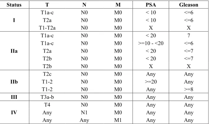
(9) TX T0 T1 T1a T1b T1c T2 T2a T2b T2c T3 T3a T3b T4. TNM Classification for Prostate Cancer Primary tumor, (T) clinical The primary tumor cannot be evaluated There is no evidence of primary tumor Primary tumor is not clinically apparent (not visible, not palpable) Incidental tumor in 5% or less of resected prostate tissue Incidental tumor in more than 5% of resected prostate tissue Tumor identified by needle biopsy (due to elevation of the PSA). Primary tumor confined to the prostate Tumor compromises <50% of a lobe Tumor that compromises> 50% of a lobe Tumor compromises both lobes The tumor extends beyond the prostatic capsule (Invasion to the prostatic apex or to the prostatic capsule is classified as T2). Unilateral or bilateral extracapsular extension. The tumor involves seminal vesicles Fixed tumor or tumor that invades adjacent structures different from the seminal vesicles: bladder neck, external sphincter, rectum, levator ani and / or pelvic wall.. * The tumor detected by biopsy in one or both prostatic lobes, which is not palpable or visible by imaging, is classified as T1c. * Positive margins should be indicated as R1 (residual microscopic disease). Regional lymph nodes (N) Nx Regional metastasis not evaluable. N0 There are no regional metastases. N1 Metastasis in one or several regional nodes. Distant metastasis (M) Mx Distant metastasis not evaluable. M0 There is no distant metastasis. M1 Remote metastasis M1a To non-regional lymph nodes M1b To bone M1c To another site When there is more than one site of metastasis it is classified as M1c.. Staging The staging of the PCA is shown in table 3. For this, it must be taken into account that if the PSA or the Gleason value are not available, the classification must be determined by the T and/or the PSA value or the Gleason score available.. 9.
(10) Table 3. Prostate cancer staging. Status I. IIa. IIb III IV. T T1a-c T2a T1-T2a T1a-c T1a-c T2a T2b T2b T2c T1-2 T1-2 T3a-b T4 Any Any. N N0 N0 N0 N0 N0 N0 N0 N0 N0 N0 N0 N0 N0 N1 Any. M M0 M0 M0 M0 M0 M0 M0 M0 M0 M0 M0 M0 M0 M0 M1. PSA < 10 < 10 X < 20 >=10 - <20 < 20 < 20 X Any >=20 Any Any Any Any Any. Gleason <=6 <=6 X 7 <=6 <=7 <=7 X Any Any >=8 Any Any Any Any. Risk classification for localized carcinoma: The risk classification for localized carcinoma used historically for PCA is the D'amico Classification (see Table 4) (41). However, during the last years different changes have been generated according to the heterogeneous prognosis that can be presented with different factors, for which modifications and a new classification suggested by the National Comprehensive Cancer Network (NCCN) have been made (see Table 5) (42). Table 4. D’Amico Classification for localized carcinoma. Adapted from D’Amico et al (41). Risk Low Intermediate High. PSA <= 10 10 a 20 >20. Gleason <= 6 7 >= 8. TNM <= T2a T2b >= T2c. Table 5. New classification for localized carcinoma suggested for National Comprehensive Cancer Network (42). Risk. PSA. Gleason. TNM. Very low. <10. <= 6. T1c. Low. <10. <= 6. T1-T2a. Others Less than 3 cores of the positive biopsy, all with <50% of the core compromised; PSA density <15 ng / ml / gr. 10.
(11) Intermediate High. 10 a 20 >20. 7 8 to 10. T2b-T2c T3a. Diagnosis Currently, prostate-specific antigen (PSA) in conjunction with digital rectal examination are mainly diagnostic methods used in the clinic to detect prostate cancer, however, these have low diagnostic performance, both individually and together (16). Prostate-Specific Antigen (PSA) PSA, also called kallikrein III is a 34kDA glycoprotein which is almost exclusive of prostatic epithelial cells. It circulates bound to alpha-1-antichymotrypsin and alpha-2-macroglobulin and its duty is to divide semenogelin I and II into smaller polypeptides, thus preventing the formation of the seminal clot (43–47). It is found in prostatic fluid at concentrations of 1,000,000 ng / mL, under normal conditions a small amount, less than 4ng / mL is released into the bloodstream, but in a neoplastic process these levels rise (47). For this reason, it is considered to perform a prostate biopsy on men with a serum PSA level greater than 4ng / mL (47). However, it has also been found elevated in other pathologies such as breast cancer, renal cell carcinoma, ovarian cancer and adrenal neoplasia (48). Similarly, other urological diseases such as benign prostatic hyperplasia (BPH), prostatitis, cystitis, instrumentation and surgery of the recent urinary tract may be elevated (1,47). It should be noted that digital rectal examination does not increase PSA values. According to the American Cancer Society (ACS), the PSA sensitivity for reference values of 4ng/ml and 3ng/ml for cancer diagnosis is 21 and 32% respectively. A specificity of 91% for cutoff values of 4ng / ml and 85% for PSA values of 3ng/ml (49). In the PLCO study, regarding PCA, men between 55-74 years were evaluated, who underwent annual screening with PSA for 13 years, as a result it was obtained that screening with PSA does not lead to a decrease in the incidence of PCA (RR 1.09, 95% CI 0.87-1.36) (50). Another large study was the ERSPC where PSA screening was carried out for 11 years to men from certain European countries, assessing mortality by PCA, the results indicated a relative reduction in mortality rates of 21% (RR 0.79, 95% CI 0.68 to 0.91) (51). A Cochrane meta-analysis conducted in 2011 summarized the results of 5 population experiments with a total of 341,351 participants and showed that screening with PSA is effective for the detection of PCA (RR 1.35, 95% CI 1.06-1.72) However, this test did not decrease mortality (RR 0.95, 95% CI 0.85-1.07) (52), so that population screening for PCA is currently not recommended. However, there is not enough evidence to determine the best measure for screening in public health, for now it is suggested opportunity screening, men between 50 and 70 years (according to the life expectancy of the population) who enter the urologist's office and patients with risk factors (black race and family members with prostate cancer). It is of vital importance to clarify that in deciding the start of the search for the diagnosis of PCA, rectal examination should be performed in conjunction with PSA. Other biomarkers in the PCA diagnosis The failure of the PSA has led to the need to identify new biomarkers, with greater sensitivity and specificity, which allow an early diagnosis of PCA to be achieved (16). PSA, due to being elevated 11.
(12) in both benign conditions (benign prostatic hyperplasia) and in conditions of malignancy (53), has led to expensive and unnecessary biopsies being requested from patients who did not require it from the beginning (53). As a consequence, other techniques and molecules have been explored to make a more specific diagnosis, such as PCA3, microglobulin, mucins, among others (16). Some of these were included in a detailed review previously published by Esquivel Parra et al (16) and it is suggested to review to deepen the topic. Other tools that try to decrease the number of unnecessary biopsies that have not yet been successful and are used for patients with PSA values between 4 and 10 ng/ml are: The Free PSA/Total PSA Ratio: Raises the specificity for the diagnosis of PCA, in those cases with the mentioned levels in the presence of doubt as to the indication for biopsy. PSA can transit in serum freely (fPSA) or accompanied by protease inhibitors (cPSA) in order to avoid proteolysis. When adding the fPSA and cPSA results in the total serum PSA (tPSA), a large part of this, around 70-90% can be linked to alpha-1-antichymotrypsin, in smaller proportion to alpha-2-macroglobulin, alpha-1 antitrypsin or a protein C inhibitor (46). Consequently, about 1030% of the total PSA (tPSA) circulates freely (fPSA) (16). With indexes <0.07, the probability of PCA is almost 90%. A limit value is not yet defined, however the use of 0.20 is recommended to decide between biopsy or follow-up, the values above this suggest a diagnosis and adequate treatment for benign prostatic hyperplasia, on the other hand, the realization is suggested of biopsy in values lower than this due to the high probability of PCA. The PSA density (total PSA/volume of the prostate in cubic centimeters (cc) determined by ultrasound) before values > 0.15 ng /ml/cc should be biopsied since they suggest adenocarcinoma. The PSA velocity > 0.75 ng/ml/year (used in follow-up after prostatectomy or initial PSA > 4ng/ml) suggests the presence of cancer; at present it has been lowered even to values like 0.35 ng/ml/year (for PSA <4 ng / ml). Although all the tools are available to raise the specificity of PSA, prostate biopsy is currently considered in all patients with PSA> 4 ng / ml, however some authors suggest that taking a single PSA value of <2.5 ng/ml (It can be applied for patients under 50 years of age, since studies are needed to confirm its value). Transrectal prostate ultrasound guided biopsy: It is the gold standard for the diagnosis of PCA. The samples are taken in the prostatic periphery, which is the site with the highest frequency of carcinoma; usually for the first biopsy, at least 6 cylinders should be taken for each lobe, in case of having a negative biopsy with persistence of elevated PSA, a saturation biopsy will be performed (> 10 - 12 samples/lobe). There are studies that show that prostate biopsy requires adequate analgesia/anesthesia and the use of antibiotic prophylaxis. Transrectal ultrasound of the prostate is only indicated, if it is accompanied by a biopsy, it should not be done in another condition. The indications for prostate biopsy are PSA >4ng/ml and the presence of alterations in the prostatic surface (nodule or stone prostate) predominantly, although there are variants that are not the subject of this review. Nuclear medicine Bone scintigraphy should be performed initially, in patients with PSA> 10 ng/ml or in those classified as intermediate and high risk patients. The probability that patients with PSA levels <10 have a positive scintigraphy is below 1%. On the other hand, a PSA> 49 has a LR + (positive likelihood ratio) >6, which means that there is a 6 times higher probability of finding a positive scintigraphy in patients with PCA than in patients without PCA (54). 12.
(13) Computed tomography (CT) Abdominal CT (like magnetic resonance imaging) indirectly evaluates nodal invasion by measuring the diameter of the lymph nodes. However, its sensitivity is low and microscopic invasion can not be detected. Using a threshold of 10 mm, the sensitivity is <40%. The median sensitivity, specificity, negative predictive value and estimated positive predictive value are 7%, 100%, 85% and 100%, respectively. Although fine needle aspiration biopsy (BACAF in Spanish) can be a good complement in cases with positive images, the difficulty of reaching the ganglia due to its position, makes it not very sensitive for staging and has a false negative rate of 40%. %. For CT (like MRI), the detection of microscopic lymph node invasion is <1% in patients with a Gleason score <8, PSA <20ng / ml, or localized disease. Its use should be reserved for high risk patients. Although bone CT has low specificity, its use over other techniques to evaluate bone metastases is preferred. It is recommended to be performed in symptomatic patients regardless of PSA levels, Gleason score or clinical stage. (55). Magnetic resonance (MRI) of prostate/pelvic The T2-weighted image is the most useful for local staging in MRI. At 1.5 T (Tesla), MRI has low sensitivity to detect extraprostatic extension of carcinoma (22-82%) or invasion of seminal vesicles (0-71%), but greater specificity (61-100% and 62-100). %, respectively). The overall accuracy of MRI to distinguish T1 / T2 stages from T3 is 50-85%. These results are due to the fact that MRI cannot detect the microscopic extra-prostatic extension, in such a way that its sensitivity increases with the radius of extension within the periprostatic fat (55). The use of the endorectal probe improves the accuracy of the stage at 1.5T, and a better precision has been demonstrated in the combined use of endorectal and external probes compared to the use only of external one. The high field strength allows a high resolution T2-WI and the results at 3T seem better than at 1.5T, although the experience of the reader is still of utmost importance, the precision of the RM to 3T varies between 67% and 93% depending on of the reader's experience. Even the prediction of the pathological stage by MRI can be improved when combined with clinical data. Given its low sensitivity for focal extra-prostatic (microscopic) extension, multiparametric prostate MRI is not recommended for local staging in low-risk patients. However, it may be useful in planning treatment in selected low-risk patients (56). PET/CT Positron emission tomography of 11C- or 18F-choline (PET) / CT has a good specificity for lymph node metastases, but the sensitivity is 10-73%. In a meta-analysis of 609 patients, the sensitivity and specificity of PET/CT for pelvic node metastases were 62% (95% CI, 51-66%) and 92% (95% CI: 89 -94%) (57). Because of its low sensitivity, PET/CT is not recommended for initial staging in lymph node metastases. Currently, studies are underway with psmaPET-CT (prostate-specific membrane antigen-PET CT). The PET / CT of 18F-choline shows a superior sensitivity to conventional bone CT when evaluating bone metastasis. It is not clear whether 11C-choline PET / CT is more sensitive than conventional bone scan, but has greater specificity, with fewer indeterminate lesions. However, the cost-effectiveness of these interventions has not yet been evaluated. Therefore, bone CT is preferred based on availability and cost.. 13.
(14) Treatment The treatment of choice depends on the stage of the tumor at the time of diagnosis. Six treatment modalities could be used: • Surgical • External conformational radiation therapy • Brachytherapy • Hormonotherapy • Active surveillance. • Observation Localized prostate cancer < cT2c, Nx, M0 The approach to localized prostate carcinoma depends on the level of risk previously called by D'Amico and the NCCN guidelines, as follows: • Very low risk: Any treatment modality can be performed. Good results have been observed with observation and active surveillance to avoid overtreatment. However, in patients with life expectancy greater than 10 years, surgical treatment could be considered. • Low risk: As in the previous group, any treatment modality can be performed. Life expectancy also plays an important role, since surgical treatment is probably not justified in patients with <10 years of age. In those who have a life expectancy> 10 years, who are considered candidates for surgical treatment, radical prostatectomy is the treatment of choice, without the need to perform lymphadenectomy that has been shown to be more effective when compared to radiotherapy (58). • Intermediate risk: Retropubic radical prostatectomy with bilateral pelvic lymphadenectomy is the management of choice in these patients. Usually in patients with life expectancy greater than 10 years. As any surgical procedure can have complications such as bleeding, infection, however, the most important for the patient are urinary incontinence and erectile dysfunction, clarifying that the frequency of these has decreased along with the improvement of the surgical technique and the preservation of the neurovascular bundles together with the external sphincter. Another modality of treatment is external radiotherapy which can be performed in these patients, it can be 3D conformational, with modulated intensity (74-80 Gy), or low rate brachytherapy (preferable for low risk patients). All have related complications, similar to surgery such as incontinence, erectile dysfunction, proctitis and/or cystitis. • High Risk: In this group of patients the treatment is performed in the same way as in the intermediate risk patient Locally advanced prostate cancer cT3-4, Nx, M0 One of the therapeutic options is radical retropubic prostatectomy with bilateral pelvic lymphadenectomy in selected young patients, with clinical stage T3, gleason < 8 and PSA less than 20 ng/ml, this given that up to 25% may be overstaged. Another option that could be performed is external radiotherapy combined with hormonal therapy (neoadjuvant, concurrent and adjuvant LHRH analogues (for 1-3 years)). Advanced prostate cancer; Any T, N1, M1 Hormone therapy (surgical or medical) is the treatment of choice in patients with advanced prostate cancer. The drugs currently used for medical orchiectomy are the LHRH analogues (leuprolide. 14.
(15) acetate, goserelin acetate, and triptorelin acetate) and the gonadotropin-releasing hormone (GnRH) receptor antagonists (Degarelix), whose use it must be by specialized personnel. These can be added to emerging medications such as Abiraterone (inhibitor of testosterone synthesis), Enzalutamide (Androgen receptor inhibitor) and Radio-223, when the patient is called castrationresistant (CRPC) (9). The criteria to be defined as resistant to castration are (9): Testosterone level below 50ng/dl or 1.7nmol/l plus • Biochemical progression: Three consecutive elevations of the PSA, at least one week apart and resulting in two increments of 50% above the nadir and a PSA> 2, or; • Radiological progression: Appearance of new lesions: two or more lesions in a bone scan or a soft tissue lesion using the RECIST criteria (Response Evaluation Criteria in Solid Tumors). Antiandrogens are used as adjuvants in the treatment, there are two types: steroids and nonsteroids. Among the non-steroids are flutamide, bicalutamide, among others, however its indications, dosage and monitoring should be performed by the specialist (9). In those patients with metastatic and castration-resistant PCA, different classifications can be made: 1. According to the functional status and 2. According to the presence of symptoms or visceral metastasis. For those with impaired functional status, even the treatment is under investigation given the poor prognosis of the patients. For those with good functional status, we have different treatments among which are the new generation antiandrogens and chemotherapy. Asymptomatic or mildly symptomatic patients may receive: Abiraterone, Enzalutamide and Radio 223, on the other hand, those who are symptomatic and/or have visceral metastases, are candidates for chemotherapy (Docetaxel or Cabazitaxel (As a second line)), whose management is given by the specialist in oncology and/or in interdisciplinary meetings (9). Discussion The present review on PCA presents an updated view on different aspects of prostate cancer. It is a very frequent pathology, with an incidence that varies between being stable or increasing and a prevalence that is clearly increasing, probably due to the improvement for diagnosis and treatment. With regard to risk factors, we have classically recognized as race, age and genetic factors clearly recognized as such, those that have not yet been able to find the role that plays as food and some that were formerly believed were factors protective and now aim to play a carcinogenic role as estrogens. It is important to perform an adequate classification and clinical, anatomical and pathological staging, since the process depends on the approximation and use of imaging and radiology techniques, as well as timely, individualized and indicated treatment based on evidence-based medicine. The genetic characterization and the microbiology of the PCA is what most efforts and researches aim for and point to, since tests with greater sensitivity and specificity are required that lead to an earlier, more accurate diagnosis and, if possible, to reach therapies more specific and less morbid Conclusion We have made a journey through the conditions of risk, screening, diagnosis, new biomarkers and treatment of prostate cancer. Several elements have changed in recent years, mainly about the. 15.
(16) understanding of the physiology of cancer, the associated factors, the search for new biomarkers in each of the stages of cancer and various elements related to the treatment, however, much research there is ongoing on the prevention, diagnosis and treatment of this condition so important, relevant and relevant to men around the world.. 16.
(17) Chapter 2: Molecular alterations associated with prostate cancer Type of article: Narrative Review Authors: Herney Andrés García-Perdomo MD, MSc, EdD, PhD, FACS (1,2); James Alejandro Zapata-Copete MD (2,3); Adalberto Sanchez BSc PhD (1,4) Affiliations 1. Associate professor, Universidad del Valle- Cali, Colombia 2. UROGIV research group. Universidad del Valle, Cali, Colombia 3. Epidemiology Department, Universidad Libre- Cali, Colombia 4. School of Basic Sciences- Universidad del Valle- Cali, Colombia Introduction Recently, the quantity and quality of tools available for the genetic study of cancer and the whole genome have increased, with even greater detail available for the exome alone. Many molecular signaling pathways provide negative or positive regulatory signals that control cell proliferation in a way that attempts to preserve cell number and homeostasis, but this process is completely altered in cancer cells (59,60). Normal cells must acquire at least eight attributes to transition from a normal cell to a cancer cell. These attributes include the following: 1. Genetic instability and mutation, 2. Autonomous growth, 3. Insensitivity to internal and external anti-proliferative signals, 4. Resistance to apoptosis and other forms of induced cell death, 5. Unlimited cell division potential, 6. Ability to form new blood vessels (Angiogenesis), 7. Local invasive behavior that enables the distinction of benign and malignant neoplasms, and 8. Evasion of the immune system. Additionally, cancer cells require energy for autonomous growth and unlimited replication. Tumor-associated inflammatory mediators also cause preneoplastic cells to progress to invasive cancer cells; finally, cancer cells gain the ability to metastasize, that is, to migrate and colonize organs or tissues (59,61,62). The purpose of this article was to review the main biomolecular mechanisms associated with prostate cancer. Therefore, the somatic genetic alterations that are involved in the pathogenesis of prostate carcinoma progression are shown in Figure 1 (63,64). The objective of this review was to describe some of the different biological mechanisms associated with prostate cancer Methods We performed a systematic literature search in Medline via Ovid, Scopus (Includes Embase) and Lilacs from the inception to nowadays with the following keywords: Prostate neoplasms; Prostate cancer; Molecular Medicine; Genomics; pathways; cell cycle. We included reviews, systematic reviews, basic science studies and analytical studies, which tried to explain the molecular disturbances associated with prostate cancer. According to the heterogeneity expected, we synthesized information based on the molecular mechanism. Information about most promising biomarkers associated with prostate cancer can be found elsewhere (16).. 17.
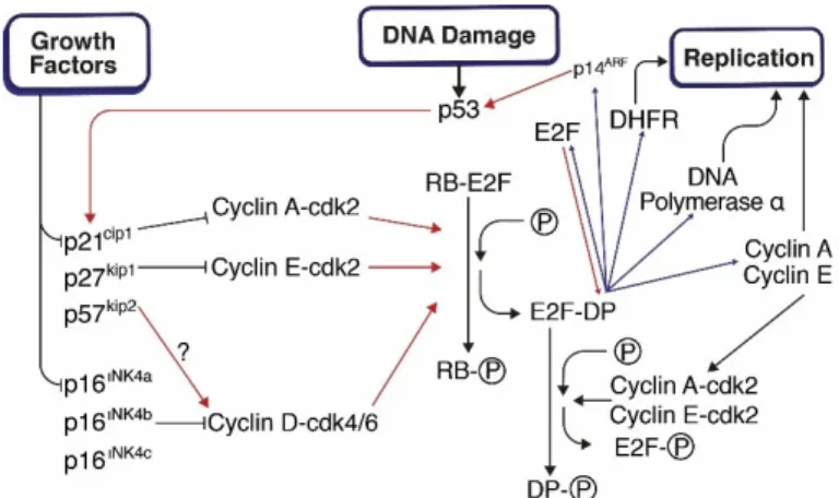
(18) Tumor suppressor genes and oncogenes Suppressor genes negatively regulate cell growth, and therefore, they play an important role in the normal cell cycle, DNA repair and cell signaling. The loss of the function of both alleles of a suppressor gene leads to carcinogenesis; thus, different pathways can result in cancer, such as 1. Homozygous gene deletion, 2. Loss of one allele and mutational inactivation of the second, 3. Mutations in both alleles, or 4. Loss of one allele and epigenetic inactivation of the second allele (e.g., DNA Methylation)(59). The two best characterized suppressor genes thus far are the retinoblastoma gene (RB1) and the TP53 gene, which are described later. Oncogenes are positively associated with cell proliferation and are the mutated form of normal genes (proto-oncogenes). Two such oncogenes are MYC and MET. MYC is responsible for the regulation of cell proliferation. This amplified gene is frequently present in prostate cancer (PCa), and its expression in prostatic cells has been associated with immortalization(65). In contrast, MET has been reported in renal cell carcinoma (RCC), primarily in the hereditary type(66). The mechanisms by which a proto-oncogene can become an activated oncogene are as follows: 1. Proto-oncogene mutation, 2. Gene amplification and 3. Chromosomal rearrangement. An example of the latter mechanism is the translocation that leads to the fusion of the TMPRSS2 gene with the ERG oncogene in a large proportion of PCa cases(67). Figure 2 schematically represents the cell cycle and describes how a cell in G0 is allowed to proliferate based on a signal, is duplicated in S phase, the phase in which DNA is synthesized, and subsequently segregates its genomic complement, which results in two daughter cells in a process called M phase (Mitosis). These two processes are separated by two critical gaps termed Gap 1 and Gap 2. The entire cycle lasts approximately 24 hours, and each phase depends on the previous one. In addition, some mechanisms function to verify the integrity of the DNA. If any alterations are found, the cell attempts to repair the damage, but if repair is not possible, the cell enters an active process termed apoptosis, which will be described later. The loss of the ability to respond to DNA damage leads to genetic instability, increases the mutation rate and mutates genes associated with cancer, thereby contributing to carcinogenesis and progression of the disease(68,69). Retinoblastoma protein (RB1) RB1 is important for controlling the R-point, which is a decisive point in late G1 phase during which the cell is committed to undergo division. Thus, if this control is lost, the cell continues to proliferate. All of the above events are due to inactivation of the RB1 pathway, which is mutated in at least 30% of bladder and prostate tumors, although RB1 mutations have not been strongly associated with these cancer types. It has also rarely been associated with renal carcinoma(70,71). Cyclin-dependent kinase inhibitors The temporal sequence of events during the cell cycle is dependent on cyclins and cyclindependent kinases (CDKs). CDKs phosphorylate protein substrates that are involved in the execution of specific activities in each phase. In contrast, cyclin-dependent kinase inhibitors (CDKIs) bind directly to CDKs and suspend their activity and their ability to phosphorylate other proteins(72). CDKIs belong to one of two classes: the Cip / Kip Family, which includes the CDKN1A (p21), CDKN1B (p27) and CDKN1C (p57) proteins, and the INK4 (inhibits CDK4) family, which includes the INK4B (p15), INK4A (p16), INK4C (p18) and INK4D (p19) proteins. The p16 protein binds to CDKs 4 and 6 and inhibits their interaction with cyclin D1; normally,. 18.
(19) active CDK4 and 6 mediate the passage of the cell through G1 phase via the phosphorylation of RB1(59,73). The latter has also been associated with bladder cancer (by deletion of INK4A) and with renal cancer, and p16 inactivation has been shown to occur by hypermethylation of the DNA (epigenetic mechanism)(74). In prostate cancer, hypermethylation of INK4A is typically seen in 60% of cases, although INK4B is rarely inactivated (75). Decreased CDKN1B has been correlated with decreased overall survival and disease-free survival after radical prostatectomy. In addition to positive CDKN1B in prostate biopsies, it has been associated with increased biochemical recurrence, and in mice, absence is associated with prostatic hyperplasia (76,77). Tumor suppressor TP53 TP53 is a suppressor protein that plays an important role in response to cellular damage. It signals a halt to the cycle or leads to damage repair pathways (Figure 3), but if repair is not possible, the cell will undergo apoptosis. This suppressor is often mutated in genitourinary cancers. Additionally, Figure 4 shows the possible causes of alterations in TP53 and its responses in the cell cycle. TP53-induced apoptosis is mediated by Bcl-2 through an intrinsic pathway, and alterations in the regulation of this pathway have direct relevance in the etiology of cancer. This pathway is associated with the activation of transcriptional genes and the inhibition of other genes that block the cascade. On the one hand, TP-53 is dependent on the activation of the Apaf-1/caspase-9 pathway, but on the other hand, Bax (Bcl-2 family) is not essential for TP-53-dependent apoptosis. In addition, different tumor suppressive pathways are associated with TP-53, and some examples are the response to DNA damage, cell senescence and apoptosis, and thus, it is logical to consider that TP-53 is frequently mutated in cancer (59,78). Methylation of DNA The covalent modification of the C5 position of the cytosine by a methyl group is mediated by DNA methyltransferase and results in the formation of 5-methylcytosine, which is an epigenetic modification that occurs in vertebrates and is part of normal and essential embryological development. As vertebrates have evolved, CpG dinucleotides have become depleted within the genome, but in some areas, this depletion is not seen; these areas are termed CpG islands and represent approximately 1% of the genome. The epigenetic properties of methylation can affect the genetic activity without alterations in the DNA sequence and represent an alternative means to inactivate the gene apart from a mutation or deletion. The three major pathways that DNA methylation uses to result in genetic alterations include the following: 1. Inherent mutational effects of 5-methylcytosine, 2. Epigenetic effects of the promoter on transcription and 3. Activation and potential induction of a gene due to instability of the chromosome by DNA hypomethylation(59). DNA methylation and prostate cancer Glutathione S transferases are a family of enzymes that are responsible for the detoxification of a large group of xenobiotics that catalyze the nucleophilic attack of reduced glutathione in potentially harmful electrophilic compounds. The aberrant methylation of the CpG island at the glutathione S transferase pi (GSTP1) locus is the most frequent somatic alteration reported in PCa (79). This methylation has been detected in up to 90% of PCa and in 70% of Prostatic. 19.
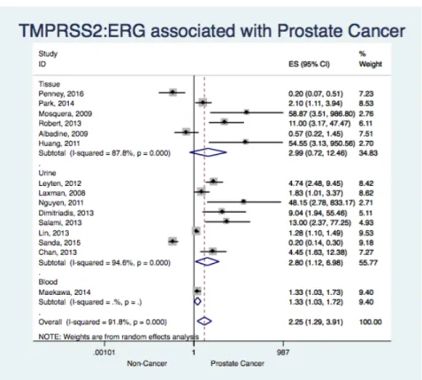
(20) Intraepithelial Neoplasia (PIN), however it might be present in normal or hyperplastic tissue (79). Due to oxidative stress, this aberrant methylation leads to the overexpression of GSTP1 in prostatic epithelial columnar cells. These findings are also associated with worse clinical outcomes(80). The gene of the ras association domain of the familial protein 1 isoform A (RASSF1A) is located on chromosome 3p21. This is a suppressor gene that is methylated in 60-70% of prostate carcinomas, and it has been observed that this alteration is more frequent in high-grade tumors than in less aggressive tumors(81,82). The methylation patterns are not consistent since they can consist of either hypo or hypermethylation, and in addition, the methylation is conserved in all metastases, suggesting an alteration that follows clonal selection(83,84). DNA damage and repair Cancer is fundamentally a genetic disease. Alterations in different genes will lead to the activation of different pathways associated with cancer, leading to changes in tumor suppressor genes and oncogenes through epigenetic, mutational and copy number distortions. To counteract these elements, the cell employs different defense mechanisms including the use of free radicals such as alpha tocopherol, vitamin C, carotenoids, bilirubin and urate and protective enzymes such as superoxide dismutase, glutathione peroxidase and glutathione transferase. In addition to the previously described methylation of GSTP1, associations have been found between the polymorphisms of this gene and the risk of biochemical recurrence in patients with PCa (85). The cell additionally employs a series of mechanisms termed the DNA damage response (DDR), which involves a number of genes. DDR relies on the replication machinery, as well as on specific mechanisms such as repairs of base cleavage, nucleotide cleavage, double helix rupture and imbalance (59). Chromosomal abnormalities Deletions of chromosomal segments are frequently found, although gains and amplifications are seen more frequently in cases of advanced disease(69). These changes have been demonstrated through cytogenetic techniques such as genomic hybridization, fluorescence in situ hybridization or detection of microsatellites. Cytogenetic methods detect numerical changes, while molecular analytic methods identify recombinations that do not lead to changes in copy number(69). The most frequently altered autosomes are 8, 13, 7, 10, 16, 6 and 17, likely in this order. In addition, gains or amplification of parts of the X chromosome and losses of the Y are also observed. A decreased copy number and loss of heterozygosity of chromosome 8p are also consistent in previous studies (observed in approximately 50% of cases). Specific alterations are observed in each of the chromosomes, but special attention must be paid to the functional impact each alteration may have on the tumor phenotype and the indication or expression of the tumor suppressor genes or oncogenes in the affected regions (69). Recurrent genetic rearrangements in PCa Recurrent gene fusions have been identified, primarily between the androgen-regulated gene TMPRSS2 and ERG, which is a member of the ETS (E26 Transforming sequence) family. This fusion occurs in 90% of all fusions that involve ETS genes in prostate cancer(86). The other fusions occur as a result of more complex types of translocations(67,87). In 60% of cases, the fusion occurs due to a deletion of the sequence that separates the two genes (3 Mb).. 20.
(21) These rearrangements can be readily identified through reverse transcription polymerase chain reaction (RT-PCR) or by multicolor fluorescence in situ hybridization (FISH). At present, several clinical studies have evaluated these markers in urine and blood, while other studies have evaluated the expression of the ERG protein using a simpler method (immunostaining)(88–90). The TMPRSS2-ERG fusion status is considered a possible diagnostic marker, although its prognostic significance is still unclear, which is a fundamental part of patient follow-up(91,92). The TMPRSS2 gene can be merged with other members of the ETS family including ETV1, ETV4 and ETV5(93,94). Studies have been performed with different technologies including next generation RNA sequencing; in these studies, it was found that some fusions are single events or events that occur in only one patient, which might imply that we actually know very little about PCa (95). PTEN and PI3K/mTOR The PI3K/mTOR (mammalian target of rapamycin) pathway plays an important role in cell growth, proliferation and oncogenesis in PCa (96–98). PTEN is a negative regulator of this pathway. In retrospective studies, it has been shown how the loss of PTEN and consequently, the activation of the mTOR pathway lead to a poor prognosis in PCa (63). PTEN deletions have been found in up to 20% of patients with PCa and have been associated with earlier biochemical relapse, metastasis, resistance to castration, presence of ERG gene fusions and the accumulation of nuclear TP53(63). Association with telomeres A potential association between telomere length and prostate cancer has been found. Initially, this association was found only in studies with small sample sizes, but subsequently, some studies with larger sample sizes were performed in which associations were observed between a short telomere length and decreased overall survival and increased biochemical recurrence. These findings have been consistent even when adjusted for age, Gleason score and lymph node involvement. It has been proposed that cancer that develops from these areas can lead to greater genotype and phenotype heterogeneity, as well as to an increase in aggressiveness(99). Some studies have even suggested that the risk of death is increased up to 14 times in patients with short telomeres compared with patients with long telomeres(100). Apoptosis Apoptosis is an orderly, energy-requiring process in which the cellular content is degraded and condensed into an apoptotic body that is finally digested by neighboring cells or macrophages(101). The positive or negative signals of the apoptotic process finally converge in a family of proteases termed cysteine proteases with aspartic acid specificity. Caspases total at least 13, and some of them are initiators (caspase-8, caspase-9, caspase-10), whereas others are executioners (caspase-3, caspase-6 and caspase-7). Caspases are derived from procaspases, which are larger inactive forms that require proteolytic cleavage in order to become active. They are frequently activated by other caspases (initiator) to generate an activating cascade of executioner caspases. The latter type attacks different anti-apoptotic intracellular proteins such as Bcl-2 and Bcl-XL. They not only destroy their anti-apoptotic functions but also release carboxyl-terminal fragments to remove the cell(102). They also degrade DNA repair and replication proteins such as DNA-PKcs and replication factor C, leading to nuclear dysregulation.. 21.
(22) Nuclear proteins such as laminin, NuMa and SAF-A are fragmented and undergo nuclear dissolution and nuclear condensation (milestones in the cell that lead to apoptosis). Proteolysis of cytoskeletal proteins such as keratin and actin also occurs, which leads to the destruction of the integrity of the internal structure. A final step is the breakdown of cell-to-cell interaction proteins such as beta-catenin and focal adhesion kinase, which precipitates the phenotypic and irreversible changes associated with apoptosis(102). Global defects in apoptosis In both PCa and PIN, a high level of apoptosis is seen, although compared with other malignancies, PCa has low apoptotic activity along with increased replication. As PCa progresses, it is unclear whether androgen-resistant cells have an increased or decreased apoptosis rate because both have been found in patients with castration-resistant prostate cancer(103). In contrast, an advanced infiltrative tumor whose DNA is mutated and that is fast-growing may have a high rate of apoptosis despite the protective mechanisms that the cell has acquired. Apoptosis can be initiated by two pathways: the intrinsic and the extrinsic pathways (Figure 5). The intrinsic pathway monitors conditions within the cell and responds to various forms of stress. Pro-apoptotic signals can originate from damaged and unrepaired DNA or from the lack of signals from the cell surface (cell-cell or cell-matrix contacts, including hormones or diminished growth factors). Mitochondria and the Bcl-2 family are major components of the intrinsic pathway. The Bcl-2 family contains 12 pro-apoptotic proteins including Bax, Bak, Bok, Bik, Bas, Bid and Bim. It also contains six pro-survival proteins including Bcl-2, Bcl-XL, Bcl-W and Mcl1(104). Each protein in the family responds to different stimuli; however, their primary function is to increase the permeability of the mitochondrial membrane(105). Subsequently, cytochrome c is released from the intermembrane space into the cytoplasm, where it binds to Apaf-1 proteins and forms the apoptosome complex. Caspase-9 is activated, which then activates the entire cascade described above. In addition, other activations occur such as ones that involve Bid, which is regulated by initiating caspases in the cytosol. Caspase-8 allows the dimerization of Bid with Bax or Bcl-2. This active form of Bax inhibits Bcl-2, which leads to apoptosis. This process can be blocked by anti-apoptotic proteins and by the IAP proteins that inhibit specific caspases(59). The extrinsic pathway mediates apoptosis after receiving external signals from surface receptors called "death receptors", such as TNFR1 (tumor necrosis factor receptor 1) and Fas receptor. The death receptor domain is located in its intracellular region and allows binding to adapter proteins that also contain a death domain (RIP, TRADD, FADD). Additionally, these proteins have an effector domain that binds to the caspase recruitment domain (CARD) of the initiating caspase(106). Subsequently, the initiator caspase is cleaved and is able to activate the cascade. The most well-known death receptor is CD95 or Fas, but this receptor does not appear to have a direct effect on the etiology of cancer. In contrast, IGF-1 can activate the PI3K / AKT antiapoptotic pathway and stimulate the expression of Bcl-like proteins along with Bax suppression. In addition, the expression of IGF binding proteins may also be altered in PCa (69,107,108).. 22.
(23) Androgens and prostate cancer Most treatments for PCa are based on androgenic suppression, but they are rarely curative. To cure the disease, different mechanisms should be considered and are described as follows (Figure 6): 1. Some carcinomas do not express androgen receptor (AR) in some cases because the gene is silenced by a hypermethylated promoter; 2. Several peptide growth factors and cytokines such as fibroblast growth factor 7 (FGF7), epidermal growth factor (EGF) and interleukin-6 (IL-6) can activate the AR synergistically with or independently of a steroid ligand; 3. In some carcinomas, somatic mutations in the AR alter its receptor specificity and cause it to respond to estrogen, progesterone, dehydroepiandrosterone or synthetic anti-androgens; 4. Amplification of the AR gene can occur in up to 30% of cases, even in the presence of depletion, which leads to increased sensitivity to a minimal androgen level; 5. Different coactivating proteins have been identified as mediators of the effects of AR on chromatin structure, as well as transcriptional initiators and their interactions with other signaling pathways. All of the circumstances described above could lead to androgen-independent tumor growth(69). Expression profiles With the advent of the analysis of gene expression patterns by cDNA or oligonucleotide microarrays, research related to the diagnosis, prognosis and new therapeutic markers has become increasingly important. For example, it has been found that hepsin is not a good marker since its down-regulation increases tumor heterogeneity in prostate carcinomas(109). Another marker is the P504S protein, which is identical to Alpha-Methyl-Acyl-CoA Racemase (AMACR); the latter is a peroxisomal enzyme that is involved in the metabolism of branched-chain amino acids, which might be useful for the differentiation of hyperplasia and atrophy from cancer(110,111). Epigenetics / Environmental factors Lifestyle and dietary habits have been found to be triggers of the oncogenic cascade in PCa. For example, dietary carcinogens, estrogens and oxidants act as a trigger for chronic inflammatory changes within the prostatic tissue and thus act as a promoter of PCa (63,112,113). It has been suggested that the intake of red meat (formation of heterocyclic aromatic amines and polycyclic aromatic hydrocarbon, which have carcinogenic properties) or animal fat is a risk factor for PCa. However, when prevention studies on both of these micronutrients and other elements of the diet were performed, the suppression of those elements was not found to prevent prostate cancer(29). Additionally, sexually transmitted diseases as part of a system that triggers chronic inflammation in epithelial cells have been associated with the development of PCa (Figure 7)(114–116). Given the change from persistent oxidative stress, a survival response is generated by glutathione S transferase, cyclooxygenase-2 and other mediators. In general, the epigenetic silencing of multiple genes occurs, including the silencing of a fundamental gene, GSTP1, which is found throughout all stages of prostate cancer progression(63). Conclusions The natural history of prostate cancer involves numerous genetic and molecular alterations that cause the normal prostatic epithelial cell to become cancerous and resistant to castration. Different biological mechanisms have been associated with the development of prostate cancer, such as alterations in tumor suppressor genes, oncogenes (TP53, RB1, among others) and CDKIs; DNA methylation; chromosomal alterations and rearrangements; changes in PTEN and PI3K / mTOR; global defects in apoptosis; alterations in the AR; and epigenetic mechanisms. These are not the. 23.
(24) only mechanisms, but they have been found to be associated with prostate cancer at a higher frequency than others. Similarly, the development of prostate cancer does not have a unique etiology, but rather, it is predominantly multifactorial and can be explained by the different mechanisms described here. Legends Figure 1. Natural history of prostate cancer and the molecular alterations involved. Figure 2. Cell cycle scheme. Figure 3. TP53-dependent repair mechanisms 24.
(25) Figure 4. Causes of alterations in TP53 and its response in the cell cycle. Figure 5. Intrinsic and extrinsic pathways of apoptosis. 25.
(26) Figure 6. Mechanisms of altered androgen signaling in prostate carcinoma* *1: The androgen receptor (AR) is absent in some carcinomas. 2: Growth factors activate the AR or a coactivator in a ligand-independent fashion. 3: AR mutations alter ligand specificity and affinity. 4: AR expression is increased by gene amplification. 5: Coactivators are differentially expressed or mutated.. 26.
(27) Figure 7. Epigenetic factors associated with prostate cancer. 27.
(28) Chapter 3. A general overview of biomarkers for the screening and early diagnosis of prostate cancer Type of article: Narrative Review Authors: Luisa Maria Esquivel Parra; Ana María Caicedo Bolaños; Juan Manuel Guaitarilla Soto; Herney Andrés García-Perdomo Introduction Currently, prostate-specific antigen combined with digital rectal examination are the screening methods mainly used in the clinic to detect prostate cancer, however, these have low diagnostic performance, both individually and as a whole. The importance of finding new biomarkers, which are more sensitive and specific for screening and early diagnosis of prostate cancer, has been especially reinforced by the PSA failure. The fact that the latter is elevated in a benign condition such as prostatic hyperplasia and also in conditions of malignancy (53), has led to the requesting costly and unnecessary biopsies to patients who did not require it from the beginning (53). As a consequence of this, other techniques and molecules have been explored to make a more specific diagnosis, such as PCA3, microglobulin and mucins, among others. The aim of the present work was to describe some of the new biomarkers involved in the screening and early diagnosis of prostate cancer. Epidemiology Prostate cancer (PCA) is the second most common type of cancer in the world's male population (5). It is estimated that 1 in 7 men will be diagnosed throughout their life with PCA, and 1 in 38 men will die as a result of it. Similarly, in the United States 6 out of 10 men diagnosed with prostate cancer are older than 65 (117). It is the type of cancer that is most often diagnosed as a result of the introduction in 1980 of the Prostate-Specific Antigen (PSA) test as a diagnostic tool (3). It is estimated that by the year 2030 there will be 1.7 million new cases of PC in the world, with an expected mortality of 499,000 cases (29.3%) (7). During the last decade, there has been a notable decrease in mortality from prostate cancer in developed countries. The GLOBOCAN study reports that the incidence of PC is variable in the global context: Rates are higher in countries such as Australia/New Zealand, North America (ASR) 111.6 and 97.2 per 100,000 respectively) and in Western and Northern Europe, because screening with PSA and subsequent biopsy are performed routinely (118). In countries of North America (United States and Canada) the mortality by PCA has decreased to 4.3% and 3.1% respectively, and in countries such as Denmark, Norway and Sweden (Northern Europe) the mortality rates have declined from the year 2000 to 3.1% per year (6); however, in developing countries, mortality has increased (although there are trends towards increased diagnosis, mortality has increased in countries such as Colombia, 3.4% per year, Costa Rica, 3.4% per year, Chile 2.8% per year and Cuba 5.5% per year) (6,7). With regard to the global context, Colombia has one of the lowest PCA incidences in Latin America and a 28% share of incidence and mortality for it, very close to the. 28.
(29) world average of 28.6% and lower than countries like Ecuador ( 40.41%), Peru (37.74%) and Cuba (46.65%) (6). Finally, in Colombia the mortality rate from prostate cancer has decreased in the last four years (7) and the highest number of reported cases originate in cities such as Bogotá, Valle and Antioquia (The most populated regions with the largest number of Urologists ) (7). Biomarkers in PCA diagnosis What we had? Prostatic-Specific Antigen The history of use of the Prostate-Specific Antigen (PSA) dates from the 1980s, when it was used in the follow-up of patients diagnosed with prostate cancer PCA (119), subsequently, for the year 1994 FDA (US - Food and Drug Administration) approved the use of PSA along with digital rectal examination as screening methods for PCA (47). The PSA, also called kallikrein III is a glycoprotein of 34kDa produced almost exclusively by the epithelial cells of the prostate gland, which circulates bound to alpha 1-antichymotrypsin and alpha 2-macroglobulin and its duty is to split semenogelin I and II in smaller polypeptides, thus avoiding formation of the seminal clot (43–47). Under normal conditions a small amount, less than 4ng/mL, is released into the bloodstream, but in a neoplastic process these levels rise (47). For this reason, it is considered to perform a prostate biopsy on men with a serum PSA level greater than 4ng / mL (47). However, PSA has also been found elevated in other pathologies such as breast cancer, renal cell carcinoma, ovarian cancer and adrenal neoplasia (48). Similarly, benign prostatic hyperplasia (BPH), prostatitis, cystitis, perineal trauma and recent urinary tract surgery may elevate it (1,47). PSA can be in serum freely (fPSA) or accompanied by protease inhibitors (cPSA) in order to avoid proteolysis. When adding the fPSA and cPSA results in the total serum PSA (tPSA), a large part of this, around 70-90% can be linked to alpha-1-antichymotrypsin, in smaller proportion to alpha2-macroglobulin, alpha-1 antitrypsin or a protein C inhibitor (46). Consequently, about 10-30% of total PSA (tPSA) circulates freely (fPSA), this free form of PSA is characterized by assuming three specific molecular forms (120). One of them is predominantly in the transition zone of the prostate in patients with BPH and is called BPSA (121), the second representation is called intact PSA (iPSA) and finally there is the proPSA (pPSA), found in its most in the peripheral zone of the prostate gland, which is associated with prostate cancer (122). According to the American Cancer Society (ACS), the PSA sensitivity for reference values of 4ng / ml and 3ng / ml for cancer diagnosis is 21 and 32% respectively. A specificity of 91% for cut-off values of 4ng / ml and 85% for PSA values of 3ng / ml (49). In the USA, the study PLCO was performed to evaluate the incidence of ovarian, colorectal, pulmonary and prostate cancer. In the case of PCA, men between 55-74 years were evaluated, who underwent annual screening with PSA for 13 years, as a result it was obtained that screening with PSA does not lead to a decrease in the incidence of PC (RR 1.09 , 95% CI 0.87-1.36) (50). Another large study was the ERSPC (The European Randomized Study of Screening for Prostate Cancer), where PSA was screened for 11 years to men from certain European countries, evaluating the mortality by PC, the results indicated a relative reduction in the rates of Mortality of 21% (RR 0.79, 95% CI 0.68 to 0.91) (51).. 29.
(30) A Cochrane meta-analysis conducted in 2011 summarized the results of 5 population experiments with a total of 341,351 participants and showed that screening with PSA is effective for the detection of prostate cancer (RR 1.35, 95% CI 1.06-1.72) , however, this test did not decrease mortality (RR 0.95, 95% CI 0.85-1.07) (52). Another meta-analysis of the year 2010 obtained similar results, aimed to show evidence of the benefits of screening with the prostate-specific antigen, for which results were taken from 6 experiments with a total of 387,286 participants. The results showed that screening with PSA is related to an increased probability of diagnosing prostate cancer (RR 1.46, 95%, CI 1.21-1.77), but as in the previous study, no decrease in PCA mortality was observed. (RR 0.88, 95% CI 0.71- 1.09) (123), so that population screening for prostate cancer is not recommended at present. This suggests the importance of determining other more specific biomarkers, which lead to a decrease in unnecessary biopsies, as well as an early detection of prostate cancer and thus improve patient survival. What is more recent? Prostate Cancer Gen 3 (PCA3) The PCA3 (Long non-coding RNA prostate cancer associated 3 gene test) has been implicated in a significant number of investigations that reflect the rationale for the study of prostate cancer (124). This is involved in the survival of the cancer cell, this effect is achieved in part by modulating the signaling to the androgen receptor (AR) and exerts its main function in the nucleus and in the microsomal fraction of the cell (125). Taking this test requires that the process be divided into two large parts; First a prostate massage must be performed and then the urine sample is obtained, this will facilitate the appearance of the biomarker in the urine (125). The studies have shown promising results, but controversial at the same time for different reasons. They are promising because it has been found that the measurement of this biomarker can differentiate between cancer and chronic prostatitis/benign prostatic hyperplasia (53). In part, the reason why this is possible is related to the high expression of the molecule in cancer tissue 60 to 100 times more than in inflamed tissue but without neoplasia and it should also be noted that this was observed in 95% of patients (124). Christos K et al., (53) mention that this biomarker is useful to differentiate between benign prostatic hyperplasia (BPH) from localized cancer, however, they suggest that the use of the molecule for screening should not be unique, but rather, should be a panel of new biomarkers that provide greater diagnostic precision (124). The use of Beta 2 microglobulin (B2M) and intestinal mucin (MUC3) associated with PCA3 (53) is also mentioned. It was known in this study that the values taken into account to say if the increase of PCA3 is relevant are: 195 DU (with this reference value the diagnostic accuracy is improved up to 77%) (53). Although the advantages of the use of PCA3 for the diagnosis of prostate cancer have been elucidated and the idea of using it in therapeutic programs as a support has been supported (124); there is a counterpart that discusses the clinical validity of the findings found in the different studies. This controversy is based on the fact that there are few studies that provide a greater knowledge of the biomarker and those that currently exist are criticized that have used small population samples that make the final result less reliable. More studies are needed that include a larger proportion of patients and are followed up over a longer period of time (124). As a result of the above, it has been found that there is a failure to demonstrate that the associations between PCA3 and prostate cancer have any relevance in the patient's prognosis (125). According to this. 30.
Figure


+7


Documento similar