Oxford phase 3 unicondylar knee arthroplasty through a minimally invasive approach: long term results
6
0
0
Texto completo
(2) 834. International Orthopaedics (SICOT) (2013) 37:833–838. There are only a few studies reporting the clinical results and survival rates of the Oxford phase 3 UKA through a minimally invasive exposure; most of the articles report on short- and mid-term results [5, 6, 17, 18]. In this study, we evaluated the long-term results of patients who underwent UKA with the Oxford cemented meniscal-bearing unicondylar knee prosthesis through a minimally invasive approach including a clinical, functional and radiographic assessment.. Patients and methods During the period 1999–2005 a total of 416 patients underwent the Oxford phase 3 unicompartmental knee replacement procedure (Biomet, Warsaw, IN, USA) in our institution. The inclusion criteria for this revision study were: – – – –. Surgery performed by the same surgeon Complete preoperative clinical history, including data corresponding with American Knee Society (AKS) score items Patients alive in 2012 Absence of previous surgery for osteoarthritis in the affected knee. The number of patients who fulfilled the above criteria was 402 (511 unicompartmental arthroplasties). The indication for surgery followed the recommended standards established in the literature [19, 20]: medial compartment osteoarthritis (489 cases) or avascular necrosis (22 cases); presence of full thickness cartilage loss with bone eburnation in the medial compartment in the case of arthritic patients; intact anterior cruciate ligament; if varus deformity, correctable at 20° flexion indicating that the medial collateral ligament is functionally normal; presence of full thickness cartilage in the lateral compartment, but a chondral ulcer on the medial side of the lateral femoral condyle can be ignored. On the other hand, the patient’s age, activity, weight and the presence of chondrocalcinosis and patellofemoral arthritis were not considered contraindications to the operation. The cemented Oxford phase 3 UKA consists of cobaltchromium-molybdenum spherical femoral and flat tibial components and a fully congruent polyethylene mobile bearing device [21]. All operations were performed through a minimally invasive approach [22]. The technique was carried out under tourniquet in the affected limb and via a short paramedian incision running from the medial pole of the patella to the medial border of the anterior tibial tuberosity. This exposure did not compromise the extensor mechanism or the suprapatellar pouch. Before cementing, pulsed lavage was used to rinse the subchondral bone. Antibiotic. prophylaxis with a second-generation cephalosporin was used preoperatively in all cases and full weight-bearing was allowed after removal of drainage after 24 hours. In the year 2012, as in the preoperative stage, patients were evaluated as follows: –. – – –. Clinical and functional assessment using the AKS score [23]. Overall results of UKA according to this scale were assessed using Insall’s criteria [23]. A knee score of 100–85 was excellent, 84–70 good, 69–60 fair and below 60 poor. Range of movement of the operated knee. Subjective patient evaluation was categorised as very satisfied, satisfied, uncertain and dissatisfied. Radiographic assessment included measuring the tibiofemoral angle and examining anteroposterior and lateral radiographs of both knees in a standing view for any bony change, loosening, wear or dislocated components.. We were granted permission from the Hospital Ethics Commission and received informed consent from all patients. The need for a sample size of 242 patients to detect differences of 20 % in the clinical and radiological outcomes of each evaluation was estimated. For statistical analysis the t test of comparison of means for paired data was used. Prior to the study of data obtained, the normal distribution of the different variables considered was verified and clinical characteristics of the patients included in the evaluation were analysed. The mean age in the preoperative period was 59.02 years (standard deviation 8.63), the female to male ratio was 2.5:1 and the mean body mass index was 27.08 kg/m2 (standard deviation 4.80). The mean duration of symptoms until surgery was 45.08 months (standard deviation 8.83). The presence of other pathological conditions was: diabetes in 52 patients (13.04 %), rheumatoid arthritis in eight patients (2.17 %) and peripheral vascular disease in 72 patients (17.91 %).. Results The following post-operative outcomes refer to a follow-up period averaging 10.38 years (standard deviation 0.97) and are in relation to the subgroup of 125 knees (95.45 %) that did not undergo re-intervention to total knee prosthesis (Table 1). In this manner the mean AKS knee score before surgery was 51.5 points (range 26–68). This value improved to 90.2 (range 72–100) points at follow-up. The average preoperative AKS knee function score was 50.7 points (range 30–70). It improved to 88.6 points (range 65–100) at ten years after surgery. Overall results of UKA according to the AKS using.
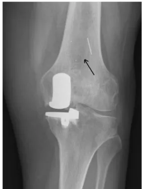
(3) International Orthopaedics (SICOT) (2013) 37:833–838. 835. Table 1 Outcome of UKA Value. AKS knee score (100)a Pain (50) Mobility (25) Stability (25) AKS knee function (100) a Walking (50) Stairs (50) Deductions (−20) X-ray tibiofemoral angle b. Preoperative period. Follow-up, 10 years after surgery. p. 51.5 (12.4). 90.2 (7.82). 0.006*. 25.70 (4.51) 13.87 (1.23) 11.93 (1.13) 50.7(18.5). 45.10 (7.85) 20.62 (3.24) 24.58 (4.28) 88.6 (17.8). 0.000* 0.000* 0.000* 0.007*. 28.35 (5.14) 30.85 (5.33) −8.50 (3.16) −6.8° (4.2). 47.30 (8.19) 44.75 (7.17) −3.45 (2.44) 3.2° (3.1). 0.000* 0.000* 0.997 0.000*. Mean value and SD; n=126 knees *Statistically significant results (p value<0.05) a. Points. b. Varus: minus value; valgus: positive value. Insall’s criteria showed among those patients an excellent or good outcome for 492 knees (96.28 %), fair for 11 (2.15 %) and poor for eight (1.57 %) in the post-operative long term. The mean preoperative active flexion of the evaluated knees was 105.5° (range 85–135°), with 461 cases in the range of 85–110° and 49 in the range of 110–135°. In the long term, the mean active flexion significantly increased to 130.9° (range 110–140°) (p<0.0001). There was no limitation in knee extension post-operatively or before the intervention. Regarding the radiological assessment no femoral or tibial component showed radiological loosening. There were radiolucent lines less than two millimetres thick around 29 tibial components (5.68 %) without symptomatology. Seventy-six patients (14.87 %) had minor osteoarthritic changes affecting the lateral compartment, graded as Ahlbäck 1, without symptoms. Preoperative varus alignment was on average, 6.8° (range 14° varus to 2° varus). Post-operative long-term radiographic measurements showed that the position of the femoral components was within acceptable ranges with a mean of 3° valgus (range 6° valgus to 7° varus). There was no posterior protrusion of the femoral component. The position of the tibial components was also within acceptable ranges with a mean of 1.5° varus (range 3° varus to 3° valgus) and a mean posterior inclination of 4° (range 3–6°). All of the tibial components showed full congruency with the medial, lateral, anterior and posterior planes. Post-operative tibiofemoral alignment was on average of 3.2 grades of valgus (range 2° varus to 8° valgus) Complications registered were as follows: pulmonary embolism (one case, 0.2 %), deep venous thrombosis (six cases,. 1.2 %), post-operative stiffness that required mobilisation under anaesthesia (two cases, 0.4 %) and bearing dislocations (two cases, 0.4 %) (Fig. 1). In relation to the knees that underwent revision to TKA (27 cases, 5.3 %), the mean time of re-intervention was 3.6 years after the primary surgery (range 1.5–7.4 years). The reasons for the revision surgery and outcomes are presented in Table 2. Figure 2 shows the survival curve of the unicompartmental knee arthroplasties, with the point of failure defined as revision of the prosthesis or any component part. The overall level of satisfaction in the long term of all patients included in our study (132) was very satisfied in 154 cases (38.31 %), satisfied in 203 (50.49 %), uncertain in 55 (4.23 %) and dissatisfied in 28 cases (6.97 %).. Discussion This study reports the outcome of patients who satisfied the recommended indications for the Oxford phase 3 unicompartmental knee replacement [24]: anteromedial osteoarthritis, spontaneous necrosis, the medial and anterior cruciate ligament should be functionally normal and the lateral tibiofemoral compartment should not be significantly affected. In anteromedial osteoarthritis there should be bone on bone contact. As the outcome shown in our evaluation is favourable, these indications seem to be appropriate. However,. Fig. 1 Bearing dislocation in a UKA 10 months after surgery because of a mismatch between the tibial and femoral implants. Black arrow shows the polyethylene component in an anterior and superior position.
(4) 836. International Orthopaedics (SICOT) (2013) 37:833–838. Table 2 Summary of revision surgery for UKA No. of cases. Mean time from UKA replacement (range). Complication. Treatment. Outcome at follow-upa. 15. 30.8 months (9–68). Infection. Total knee replacement: two-stage revision with antibiotics. 2 8. 12.5 months (8–17) 31.8 months (13–53). Bearing dislocation Persistent pain. Bearing exchange Total knee replacement. 4. 43.2 months (31–75). Aseptic loosening of tibial component. Total knee replacement. Good result: 12 cases Fair result: 3 cases Good result: 2 cases Fair result: 1 case Poor result: 7 cases Good result: 3 cases Fair result: 1 case. a. AKS outcome using Insall’s criteria. despite the fact that clinical outcomes are encouraging, it has been stated in the literature [6] that more than one third of the patients that have undergone a total knee replacement could have benefited from a unicompartmental replacement. In accordance with the previous studies [25, 26], our longterm assessment confirms that overall good results may be achieved using a minimally invasive approach for the implantation of a mobile bearing unicompartmental prosthesis. Pandit and co-authors [6] report a 97.3 % survival rate of this implant at seven years and 96 % of their patients have a good or excellent AKS score [23] at five years with a mean flexion of 133°. In our series, we have found similar clinical outcomes, with an excellent or good outcome in 96.3 % of the revised patients with a mean active flexion of 131° ten years after surgery. Complications of unicompartmental knee replacement are relatively few. However, in most of the cases there is controversy about the causes of complications and appropriate treatment [27, 28]. Known complications of UKA are polyethylene wear and breakage, dislocation of the polyethylene spacer, aseptic loosening, infection, lateral compartment osteoarthritis, proximal tibial fracture, limited motion and unexplained severe pain [28]. In our series local complications described made Fig. 2 Survival rate of UKA. reference to post-operative stiffness that required mobilisation under anaesthesia (two cases, 0.4 %), bearing dislocations (two cases, 0.4 %), aseptic loosening of the tibial component (four cases, 0.8 %), infection (15 cases, 2.9 %) and persistent unexplained pain (eight cases, 1.6 %); these rates are not very dissimilar from previous studies [21, 25, 27–29]. Bearing dislocation is a peculiar complication to mobile bearings, as in the case of Oxford phase 3 prostheses and primarily occurs shortly after surgery [29]. This fact is probably be attributed to a technical error during surgery. A mismatch between the tibial and femoral component, and/or size of the bearing itself, might lead to failure [21]. Saragaglia et al. [30] consider that the navigation of only the tibial bone cut is a reasonable option and invaluable in the positioning of mobile bearing UKA, where the risk of overcorrection should not be underestimated. It has been reported that UKA conversion to TKA is associated with poorer clinical outcome as compared to primary TKA [31]. The most common cause of revision surgery in our series was infection. These data might have been influenced by the high rate of diabetes registered in the individuals studied. In fact, 11 of the 15 cases revised because of infection were in patients affected by diabetic disease. The complication with the worst results was unexplained pain..
(5) International Orthopaedics (SICOT) (2013) 37:833–838. This suggests that in those cases patients should be thoroughly investigated and carefully followed up, and revision for unexplained pain must be avoided as it might not be warranted. Compared to previous authors, a low incidence of radiolucency was found at the ten-year follow-up [6]. Like others, we conclude that these radiolucent lines have no clinical relevance [6, 21]. To reduce the incidence of radiolucency and to improve long-term fixation Clarius et al. [32] recommend the routine use of pulsed lavage. To sum up, long-term follow-up results of UKA through a minimally invasive exposure demonstrate predictably good outcomes comparable with those of total knee replacement [6, 21, 24]. We believe that with appropriate surgical technique, adequate patient selection and prosthetic design a trained surgeon should achieve good outcomes with the added advantages of a minimally invasive approach. High volume for this demanding technique [33, 34] is important in our opinion.. Conflict of interest No benefits in any form have been received or will be received from a commercial party related directly or indirectly to the subject of this article. The authors declare that they have no conflict of interest.. References 1. Çullu E, Aydoğdu S, Alparslan B, Sur H (2005) Tibial slope changes following dome-type high tibial osteotomy. Knee Surg Sports Traumatol Arthrosc 13:38–43 2. Kesmezacar H, Erginer R, Ögüt T, Seyahi A, Babacan M, Tenekecioğlu Y (2005) Evaluation of patellar height and measurement methods after valgus high tibial osteotomy. Knee Surg Sports Traumatol Arthrosc 13:539–544 3. Aslan H, Ersan O, Baz AB, Duman E, Aydın E, Ateş Y (2007) Midterm results of Oxford phase 3 unicondylar knee arthroplasty for medial osteoarthritis. Acta Orthop Traumatol Turc 41:367–372 4. Laurencin CT, Zelicof SB, Scott RD, Ewald FC (1991) Unicompartmental versus total knee arthroplasty in the same patient. A comparative study. Clin Orthop Relat Res 273:151–156 5. Emerson RH Jr, Higgins LL (2008) Unicompartmental knee arthroplasty with the Oxford prosthesis in patients with medial compartment arthritis. J Bone Joint Surg Am 90:118–122 6. Pandit H, Jenkins C, Barker K, Dodd CA, Murray DW (2006) The Oxford medial unicompartmental knee replacement using a minimally-invasive approach. J Bone Joint Surg Br 88:54–60 7. Murray DW, Goodfellow JW, O’Connor JJ (1998) The Oxford medial unicompartmental arthroplasty: a ten-year survival study. J Bone Joint Surg Br 80:983–989 8. Repicci JA, Eberle RW (1999) Minimally invasive surgical technique for unicondylar knee arthroplasty. J South Orthop Assoc 8:20–27 9. Goodfellow JW, O’Connor J, Murray DW (2002) The Oxford meniscal unicompartmental knee. J Knee Surg 15:240–246 10. Rajasekhar C, Das S, Smith A (2004) Unicompartmental knee arthroplasty. 2- to 12-year results in a community hospital. J Bone Joint Surg Br 86:983–985 11. Svärd U (2009) Long-term results after partial knee arthroplasty with the Oxford Knee. Dissertation, University of Gothenburg. 837 12. Svärd UC, Price AJ (2001) Oxford medial unicompartmental knee arthroplasty. A survival analysis of an independent series. J Bone Joint Surg Br 83:191–194 13. Skowroński J, Jatskewych J, Długosz J, Skowroński R, Bielecki M (2005) The Oxford II medial unicompartmental knee replacement. A minimum 10-year follow-up study. Ortop Traumatol Rehabil 7:620–625 14. Argenson JN, Parratte S, Bertani A, Flecher X, Aubaniac JM (2008) Long-term results with a lateral unicondylar replacement. Clin Orthop Relat Res 466:2686–2693 15. Kendrick BJ, Longino D, Pandit H, Svard U, Gill HS, Dodd CA, Murray DW, Price AJ (2010) Polyethylene wear in Oxford unicompartmental knee replacement: a retrieval study of 47 bearings. J Bone Joint Surg Br 92:367–373 16. Psychoyios V, Crawford RW, O’Connor JJ, Murray DW (1998) Wear of congruent meniscal bearings in unicompartmental knee arthroplasty: a retrieval study of 16 specimens. J Bone Joint Surg Br 80:976–982 17. Kim KT, Lee S, Park HS, Cho KH, Kim KS (2007) A prospective analysis of Oxford phase 3 unicompartmental knee arthroplasty. Orthopedics 30(Suppl):15–18 18. Carr A, Keyes G, Miller R, O’Connor J, Goodfellow J (1993) Medial unicompartmental arthroplasty. A survival study of the Oxford meniscal knee. Clin Orthop Relat Res 295:205–213 19. Kozinn SC, Scott R (1989) Unicondylar knee arthroplasty. J Bone Joint Surg Am 71:145–150 20. Langdown AJ, Pandit H, Price AJ et al (2005) Oxford medial unicompartmental arthroplasty for focal spontaneous osteonecrosis of the knee. Acta Orthop 76:688–692 21. Lisowski LA, van den Bekerom MP, Pilot P, van Dijk CN, Lisowski AE (2011) Oxford Phase 3 unicompartmental knee arthroplasty: medium-term results of a minimally invasive surgical procedure. Knee Surg Sports Traumatol Arthrosc 19:277–284 22. Murray DW, Goodfellow JW, O’Connor JJ, Dodd CA (1999) Oxford unicompartmental knee: manual of the surgical technique. Biomet, Bridgend, pp 1–40 23. Insall JN, Dorr LD, Scott RD, Scott WN (1989) Rationale of the Knee Society clinical rating system. Clin Orthop Relat Res 248:13–14 24. Goodfellow JW, Kershaw CJ, Benson MK, O’Connor JJ (1988) The Oxford Knee for unicompartmental osteoarthritis. The first 103 cases. J Bone Joint Surg Br 70:692–701 25. Pandit H, Jenkins C, Gill HS, Barker K, Dodd CA, Murray DW (2011) Minimally invasive Oxford phase 3 unicompartmental knee replacement: results of 1000 cases. J Bone Joint Surg Br 93:198– 204 26. Pietschmann MF, Wohlleb L, Weber P et al (2013) Sports activities after medial unicompartmental knee arthroplasty Oxford III-what can we expect? Int Orthop 37:31–37 27. Vardi G, Strover AE (2004) Early complications of unicompartmental knee replacement; the Droitwich experience. Knee 11:389– 394 28. Kim KT, Lee S, Bae EH et al (2005) Short-term results and early complications of minimally invasive unicompartmental knee arthroplasty. J Korean Knee Soc 17:119–126 29. Lewold S, Goodman S, Knutson K, Robertsson O, Lidgren L (1995) Oxford meniscal bearing knee versus the Marmor knee in unicompartmental arthroplasty for arthrosis. A Swedish multicenter survival study. J Arthroplasty 10:722–731 30. Saragaglia D, Picard F, Refaie R (2012) Navigation of the tibial plateau alone appears to be sufficient in computerassisted unicompartmental knee arthroplasty. Int Orthop 36:2479–2483 31. Järvenpää J, Kettunen J, Miettinen H, Kröger H (2010) The clinical outcome of revision knee replacement after unicompartmental knee arthroplasty versus primary total knee.
(6) 838 arthroplasty: 8–17 years follow-up study of 49 patients. Int Orthop 34:649–653 32. Clarius M, Hauck C, Seeger JB, James A, Murray DW, Aldinger PR (2009) Pulsed lavage reduces the incidence of radiolucent lines under the tibial tray of Oxford unicompartmental knee arthroplasty: pulsed lavage versus syringe lavage. Int Orthop 33:1585–1590. International Orthopaedics (SICOT) (2013) 37:833–838 33. Mercier N, Wimsey S, Saragaglia D (2010) Long-term clinical results of the Oxford medial unicompartmental knee arthroplasty. Int Orthop 34:1137–1143 34. Luscombe KL, Lim J, Jones PW, White SH (2007) Minimally invasive Oxford medial unicompartmental knee arthroplasty. A note of caution! Int Orthop 31:321–324.
(7)
Figure

Documento similar
Objective: To study the clinical and radiological outcomes of patients with extra-articular phalangeal and metacarpal fractures who were treated with minimally invasive
The purpose of this study was to calculate prevalence of coronal malalignment in cementless, fully coated with hydroxialapatyte (HA) femoral stems focusing in their long- term
The results of hallux valgus X-ray correction by Chev- ron osteotomy using minimally invasive techniques con- firm that this is a safe and effective procedure for the
Conclusions: We recommend using posterior-stabilized plus implants in deformities <20°, with sufficient collateral li- gaments and no bone defects; constrained prosthesis in
The scarf-like procedure has been effective with good to excellent results in the AOFAS (American Orthopaedic Foot and Ankle Society) scale; however, there are reports on
Key words: Knee hinged prosthesis; bilateral knee deformity; posterior prosthetic dislocation; implant rupture; hinged prosthesis failure; uncoupling; revision in knee
Minimally invasive percutaneous plate osteosynthesis (MIPPO) technique applied in the treatment of humeral shaft distal fractures through a lateral approach.. Minimal invasive
Study inclusion criteria were: patients with advanced knee Osteoarthritis, associated or not with nonunion, who had undergone TKA, with a history of knee fracture (distal femoral
