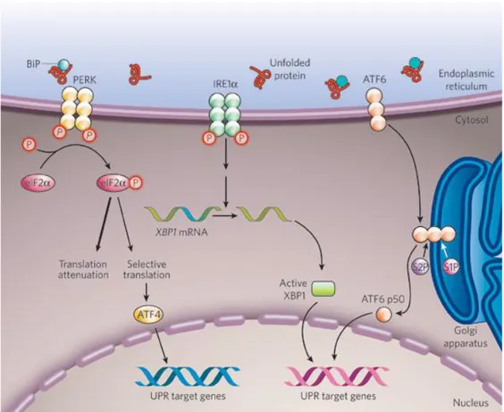
The Two Faces of Janus: Unfolded Protein Response Autophagy in Cell Death and Survival
Texto completo
Figure


Documento similar
The expansionary monetary policy measures have had a negative impact on net interest margins both via the reduction in interest rates and –less powerfully- the flattening of the
A short CDS length, together with a slightly better AUG context and a shorter 59 UTR and 39 UTR could explain the high translation rates for a subset of mRNA in NIH3T3 cells (Table
Most of them aim to increase the functionality of the defective enzyme or protein by gene therapy, enzyme replacement, pharmacological chaperones, cell based therapy and
“The Fission Yeast Pmk1+ Gene Encodes a Novel Mitogen-Activated Protein Kinase Homolog Which Regulates Cell Integrity and Functions Coordinately with the Protein Kinase C
To reinforce the role of SKF in nuclear accumulation of Nrf2 in response to APAP, we treated wild-type hepatocytes with the Figure 3 Effect of PTP1B deficiency in stress and
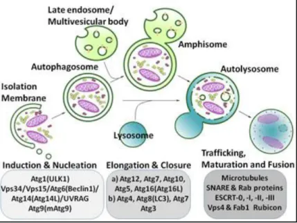
TipC and the chorea-acanthocytosis protein VPS13A regulate autophagy in Dictyostelium and human HeLa cells.. Sandra Muñoz-Braceras a , Rosa Calvo a & Ricardo
In HeLa cells stably expressing GFP-LC3, a high proportion of VPS13A- depleted cells showed an accumulation of puncta at the perinuclear region regardless of the
In this review, we have highlighted the ER stressors that activate the UPR and subsequently EMT, suggesting that the UPR may be an additional upstream signal for the induction of
