ELECTRONIC STRUCTURE OF CAROTENOIDS IN NATURAL AND ARTIFICIAL PHOTOSYNTHESIS
18
0
0
Texto completo
(2) 18. Carotenoids. nature and it is fundamental for life existence. Solar energy conversion to chemical fuels using green methodologies may be approached with photosynthesis [1] since this natural process is the main user of solar energy in our planet. This natural process uses effectively the largest exploitable renewable energy resource. Solar energy provides our planet with more energy per hour than the total energy consumed by human activities in 1 year. In other words, direct conversion of solar energy into chemical fuels represents an optimal approach to address the globally growing energy demand in a sustainable way [1–2]. Photosynthesis if reproduced may address a lot of our environmental problems derived from energy conversion. In this way, mimicking photosynthesis has become a subject of great interest in the scientific world, and this global research trend has given origin to a recently created term, artificial photosynthesis [1–3]. This concept refers to a chemical process that replicates the natural pro‐ cess of photosynthesis; it mainly studies the process to convert sunlight, water, and carbon dioxide into carbohydrates and oxygen. This process aims to emulate natural ways by using man‐made devices to convert and store solar energy using chemical fuels as feedstock [3]. To absorb the visible light part of the solar radiation (350–700 nm), green plants use chlorophyll a as the main light absorber along with a number of accessory pigments such as xanthophylls, carotenoids, and a modified form of chlorophyll, called chlorophyll b. Chlorophyll a absorbs in the blue‐violet, orange‐red spectral regions while the accessory pigments cover the inter‐ mediate yellow‐green‐orange part [3–4]. Carotenoids are important in photosynthesis, and with the mimicking of this natural pro‐ cess, they have raised their importance due to the fundamental need for renewable energy sources such as artificial photosynthesis [5]. There are other fields in which carotenoids are important as well, such as food or health. Fruits and vegetables are the principal sources of carotenoids and play an important role in diet due to vitamin A activity [5–6]. In addition to this, carotenoids are also important for antioxidant activity, intercellular communication, and immune system activity [6–8]. Epidemiological studies reported that the consumption of diets rich in carotenoids is associated with a lower incidence of cancer, cardiovascular diseases, age‐related macular degeneration, and cataract formation [9–10]. Deficiency of carotenoids results in clinical signs of conjunctiva and corneal aberrations, including xerophthalmia, night blindness, corneal ulceration, scarring, and resultant irreversible blindness [11]. Carotenoids are classified in carotenes and xanthophylls. Carotenes contain only a parent hydrocarbon chain without any functional group, such as α‐carotene, β‐carotene, and lyco‐ pene. Xanthophylls contain oxygen as the functional group, including lutein and zeaxanthin [12]. In plant tissues, carotenoids are typically located in chromoplasts (specific organelles) inside cells. Substructures composed of lipids, proteins, and carotenoids are being synthesized during chromoplast development, and depending on their morphology, they can be classi‐ fied as crystalline, globular, fibrillar, membranous, or tubular‐type chromoplasts [13–15]. Carotenoids are embedded in a complex structural organization. Carotenes and xanthophylls contain more than seven conjugate bonds that enable visible light absorption and from here, they have the capability to participate in the photosynthesis [6]. For light energy to be trans‐ formed into chemical energy, the electronic structure is fundamental in understanding how natural photosynthesis occurs and how this process can be associated with clean energy.
(3) Electronic Structure of Carotenoids in Natural and Artificial Photosynthesis http://dx.doi.org/10.5772/67636. eneration through artificial photosynthesis. Due to their chemical structure, carotenoids are g tetraterpenoids so they have a long chain of conjugated double bonds; for this reason, these micronutrients are highly lipophilic [13, 16–18]. There are different determination methods to find out the basic chemical structure of carot‐ enoids. Their structure is based on eight isoprenoid units with a conjugated double‐bond system, which makes isomeric forms very common [19]. In addition, double bonds in the carbon chain make carotenoids susceptible to reactions, such as oxidation and isomerization (cis‐trans), especially due to light, heat, acids, and oxygen [20]. Cyclization, hydrogenation, dehydrogenation, or additions of lateral groups, among others, are some modifications that lead to an extremely complex variety of compounds with common structures [19]. Moreover, carotenoids can be found in nature both in their free form and also in a more stable, esterified form with fatty acids [21]. The high variability in their chemical structure and their poor stability greatly contribute to the difficulty of carotenoid analysis. Also, there is a lack of commercially available standards and other important reasons [22] that make it difficult to have more analytical methods to identify and to measure carotenoids in real samples [21–23]. Another analytic alternative is related to theoretical methods. There is a wide chart of choices to model and simulate these compounds that range from macro‐, micro‐, and atomic to sub‐ atomic methodologies. In this work, we use density functional theory (DFT) to learn more about carotenoids. We use DFT to model and simulate carotenoids’ ground states as well as excited states and analyze their electronic structure. Fundamentally, carotenoids have a strong absorption of visible light in the blue and green region of the solar spectrum. Most carotenoids found in photosynthetic organisms have the characteristic colors yellow, orange, and red. The lowest excited single state in most pigment molecules represents the lowest energy, which optically allows a one‐photon transition from the ground state [13]. This chap‐ ter provides numerical data to parameterize some of the more important properties of carot‐ enoids. It provides a good insight about their important role in both natural and artificial photosynthesis, and since these results relate to its more basic features, it can be useful for other applications as well, such as in the food and health industries.. 2. Computational methods and details All calculations were carried out employing Gaussian 09 program suite [24]. This chapter was developed with computational calculations employing electronic structure methods using density functional theory (DFT). Then, a vibrational frequencies’ calculation was car‐ ried out to corroborate a global minimum. These calculations, geometry optimization, and vibrational frequencies were performed in the gaseous phase, using a methanol‐like solvent. Molecular orbitals for the different carotenoids were obtained with energy calculations using the B3LYP/6‐31G(d)//B3LYP/6‐31+G(d,p) theoretical method [25]. Excited states in the gaseous phase were carried out for all six carotenoid variants within this work and in the solvent phase for xanthophylls molecules. These later calculations allowed us to obtain molecular orbitals and absorption states. The same set of calculations used in carotenoids was applied. 19.
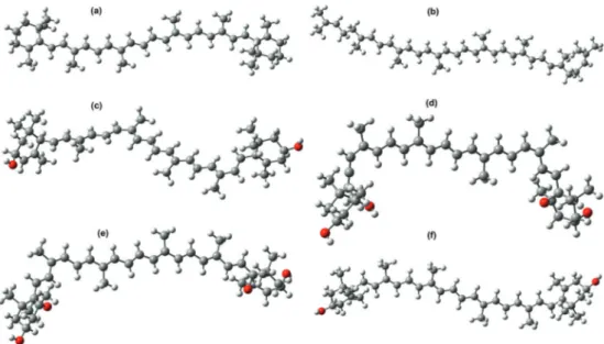
(4) 20. Carotenoids. to chlorophyll a in the gaseous phase, with the objective of forming one of the main diades that has been found as a participant in natural photosynthesis. CAM‐B3LYP [26] functional was used in all excited states’ calculations using the time‐dependent density functional theory (TDDFT). Molecular orbitals data was processed to obtain orbitals’ diagrams and the absorp‐ tion spectra with Chemissan code [27].. 3. Results and discussions In this section, calculations results are displayed and analyzed. The first set of results contains ground states data; first, the geometric structures are displayed and next the energy results are displayed, including molecular orbitals, energy gap, and relevant chemical properties. In the second set of results are included excited states data with their corresponding molecular orbital diagrams and absorption spectra based on TDDFT calculations. 3.1. Carotenoid structures Carotenoids included in this work are displayed in Figure 1. Beta‐carotene and lycopene are carotenes with the characteristic of belonging to the hydrocarbons group, which means that their structure includes only carbon and hydrogen atoms. The rest are four carotenoids that belong to the xanthophylls which characterize themselves by containing within their struc‐ ture carbon and hydrogen with oxygen atoms bonded to the six‐carbon ring.. Figure 1. Geometry optimization of carotenoid structures: (a) Beta‐carotene, (b) lycopene, (c) lutein, (d) neoxanthin, (e) violaxanthin, (f) zeaxanthin..
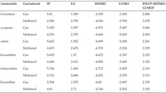
(5) Electronic Structure of Carotenoids in Natural and Artificial Photosynthesis http://dx.doi.org/10.5772/67636. 3.1.1. Carotenoids ground states The ground states energy results allow us to obtain data related to electronic conduction capa‐ bilities of the selected molecules in their ground states. For these calculations, the interpreta‐ tion scheme of the difference between the Highest Occupied Molecular Orbital (HOMO) and the Lowest Unoccupied Molecular Orbital (LUMO) was applied, where HOMO and LUMO are frontier orbitals located in the valence band and in the conduction band, respectively. Resulting values for these orbitals relate to ionization energy in the case of HOMO, which means that a lower ionization energy corresponds to a higher HOMO energy. In fact, a lower energy for LUMO is associated with a higher electronic affinity. These effects were explained in more detail in our published work [28] and can be studied by further reading Table 1 as shown ahead in this section. Frontier molecular orbitals’ values are shown in Table 1, and Figure 2 displays molecular orbitals obtained for the six selected carotenoid structures in their gaseous phase that will be discussed next. The diagram is divided into two sections: the first belongs to carotenes and the second to xanthophylls. The HOMO‐LUMO energy gap is obtained by calculating the difference between frontier orbitals’ energy values. Beta‐carotene and lycopene energy gap results have a small difference. Then, it is observed that these compounds present a similar trend in their HOMO orbitals. Based on these results, xanthophylls have a higher ionization energy which derives in the capability present in one of these variants to form a diade inte‐ grated system configured by chlorophyll a‐xanthophyll. Carotenoids. Gas/solvent. IP. EA. HOMO. LUMO. B3LYP (HOMO‐ LUMO)*. β‐Carotene. Gas. 5.41. 1.549. -4.529. -2.443. 2.086. Methanol. 4.504. 2.709. -4.636. -2.558. 2.078. Gas. 5.429. 1.597. -4.553. -2.487. 2.066. Methanol. 4.533. 2.755. -4.668. -2.605. 2.063. Gas. 5.665. 1.502. -4.699. -2.458. 2.241. Methanol. 4.615. 2.678. -4.753. -2.524. 2.229. Gas. 5.652. 1.37. -4.672. -2.347. 2.325. Methanol. 4.668. 2.633. -4.805. -2.483. 2.322. Gas. 5.744. 1.484. -4.772. -2.459. 2.313. Methanol. 4.716. 2.684. -4.852. -2.539. 2.313. Gas. 5.594. 1.555. -4.68. -2.487. 2.193. Methanol. 4.61. 2.71. -4.744. -2.562. 2.182. Lycopene. Lutein. Neoxanthin. Violaxanthin. Zeaxanthin. Results are in electron Volts (eV). *Absolute values. Table 1. Global chemical reactivity parameters (IP and EA), energy levels (HOMO/LUMO), and energy gap (HOMO‐ LUMO) in the gaseous phase, and methanol as a solvent for all B3LYP/6‐31+G(d,p)‐analyzed carotenoids.. 21.
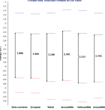
(6) 22. Carotenoids. Figure 2. Ground states molecular orbitals for selected carotenoid structures. The calculation was carried out in the gaseous phase using DFT with the B3LYP/6‐31+G(d,p) theoretical method.. From xanthophylls displayed in the molecular orbitals diagram, one can see that zeaxanthin has the narrower HOMO‐LUMO energy gap. A narrow HOMO-LUMO energy difference benefits energy transfer process. Photosynthesis requires that plants containing chlorophyll a capture light to transform it into chemical energy. The role that light plays consists of producing a luminous excitation that impacts an electron, allowing this charged particle to jump from a ground state of inferior energy to an excited state with higher energy and later return to the lower energy state. This process is known as excited states in the electronic structure. In photosynthesis, carotenoids play the role of the aforementioned accessory pigments. Excited states analysis explains xanthophylls’ performance as pigments that are part of the photosynthetic process. Figure 3 displays a molecular orbital diagram for excited states corresponding to all mol‐ ecules within our chapter. This diagram, as occurred for ground states, provides information discussed previously in this section..
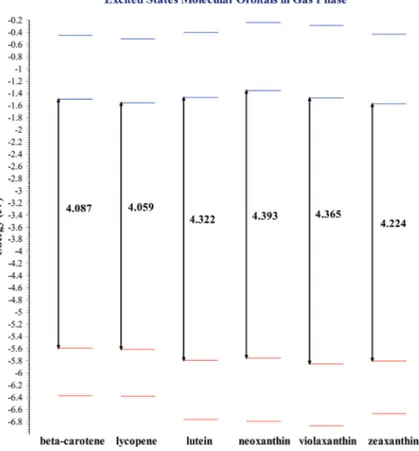
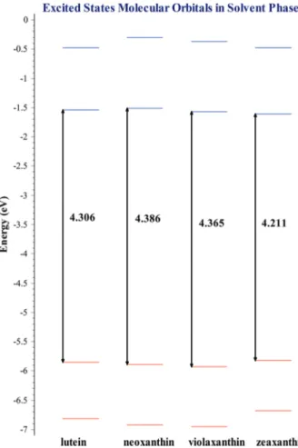
(7) Electronic Structure of Carotenoids in Natural and Artificial Photosynthesis http://dx.doi.org/10.5772/67636. Figure 3. Excited states molecular orbitals for selected carotenoid structures. The calculation was carried out in the gaseous phase using TDDFT with the CAM‐B3LYP/6‐31+G(d,p) theoretical method.. 3.1.2. Carotenoids excited states molecular orbitals As occurs in ground states, in excited states, zeaxanthin is the molecule with the narrower energy gap from the xanthophylls‐type carotenoids which means this is the molecule that can be excited more easily. Other data important to consider in excited states analysis is the UV‐Vis absorption spectra. Absorption spectra enable us to identify the wavelength in which a pigment absorbs sunlight and thus locate the reference electromagnetic radiation working range and the visible light required to favor photosynthesis. Now, depending on the solvent used, sunlight absorption may have a benefit. In general, the absorption extends in larger wavelength when the solvent is employed if compared to the absorption results in the gaseous phase. Figure 4 displays the diagram of excited states molecular orbitals with the use of solvents, which in this case is methanol. This excited states calculation was carried out only for xanthophylls because zeaxanthin is the accessory pigment used to form the diade with chlorophyll a, considering that the latter is the main photosyn‐ thetic pigment. Molecular orbitals shown so far, both in the gaseous phase and in the solvent phase correspond to those involved in the main absorption peak found.. 23.
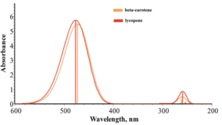
(8) 24. Carotenoids. Figure 4. Excited states molecular orbitals for selected carotenoid structures. Calculations were carried out in methanol as a solvent with TDDFT using the CAM‐B3LYP/6‐31+G(d,p) theoretical method.. If one observes the molecular orbitals diagram corresponding to the molecules in methanol as a solvent, one can see that zeaxanthin is the molecule with the narrower energy gap as occurred in the gaseous phase calculations. Another important observation is how violaxan‐ thin keeps the same values in both calculations, in the gaseous phase and in the solvent phase. 3.1.3. Carotenoids excited states UV‐Vis spectra Absorption spectra for carotenoids‐type beta‐carotene and lycopene for the gaseous phase are shown in Figure 5. In this figure, a high coincidence in the absorption maximum peak between beta‐carotene with 472 nm and lycopene with 476 nm is observed. According to these calculations, the maximum absorption peak for beta‐carotene is surpassed by 20 nm. Meanwhile for lycopene, a better approximation with the experimental value of 470 nm is obtained..
(9) Electronic Structure of Carotenoids in Natural and Artificial Photosynthesis http://dx.doi.org/10.5772/67636. Figure 5. UV‐Vis absorption spectra for carotenoids‐type beta‐carotene and lycopene. Calculations were carried out with TDDFT using the CAM‐B3LYP/6‐31+G(d,p) theoretical method.. 3.1.4. Xanthophylls excited states UV‐Vis spectra Absorption spectra for the gaseous phase and with the solvent for xanthophylls‐type carot‐ enoids are displayed in Figure 6. According to the figures, one can observe that the varia‐ tion in absorption results in the gaseous phase with respect to the solvent phase is different only by a wavelength displacement. Zeaxanthin is the carotenoid from xanthophylls with an absorption in a longer wavelength, similar to lutein. The difference between them is the absorbance value where zeaxanthin has the bigger absorbance. 3.2. Diades Diades structures are formed with two molecules, one from the group of the main photo‐ synthetic pigments and the other from the accessory pigments group. For this work, we used chlorophyll a‐carotenoid. In the formation of the system, chlorophyll a‐zeaxanthin was employed, and the chlorophyllide a, due to the phytol, lacks contribution in the photochemi‐ cal activity related to light absorbance in chlorophyll a but brings about some benefit with some computational cost reduction. In the next paragraphs, we discuss our results for the selected diade. Our discussion for these systems is a relative view between ground states and excited states that will enable us to understand their electronic structure properties. Our discussion for this system is a relative view between ground states and excited states that will enable us to understand their electronic structure properties. For the intermolecular energy transfer study, photosynthetic pigments’ diade formed by chlo‐ rophyll a‐zeaxanthin was modeled and analyzed. For construction of the diade system, we used chlorophyllide a, and zeaxanthin was centered, and to build this diade, the individual optimized structures were used. Once the diade was modeled, the system was subject to a geometric optimization using the B3LYP/6‐31G (d) theoretical method in the gaseous phase.. 25.
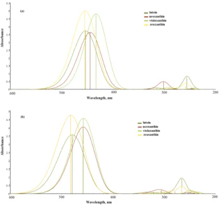
(10) 26. Carotenoids. Figure 6. UV‐Vis absorption spectra for xanthophylls‐type carotenoid structures. Calculations are in (a) the gaseous phase and (b) by using methanol as a solvent with TDDFT using the CAM‐B3LYP/6‐31+G(d,p) theoretical method.. For the determination of the intermolecular distances between chlorophyllide a and zeaxan‐ thin, we performed a series of prior work to determine the best choice with distances between 5.0 and 9 Å (all calculations included vibrations frequencies analysis to make sure that the global minima was reached). Since electronic properties of selected photosynthetic pigments depend of nature in both cases, the ground and the excited states, we moved forward with molecular orbitals analysis for iso‐ lated molecules and diade systems (in both ground and excited states). TDDFT was used, with Tamm‐Dancoff approximation (TDA) and the CAMB3LYP/6‐31+G(d,p) theoretical method. After HOMO/LUMO energy analysis, UV‐Vis absorption spectra were obtained for isolated molecules and their corresponding diade systems. The same theoretical level was used with same functional set. 3.2.1. Diade molecular orbitals (HOMO/LUMO energies) Results for molecular orbitals and HOMO‐LUMO energies corresponding to chlorophyll a, zeaxanthin, and the respective diade formed by them, in their ground and excited states, are shown in Figure 7(a) and (b), respectively. This way of organizing the diagrams enables one.
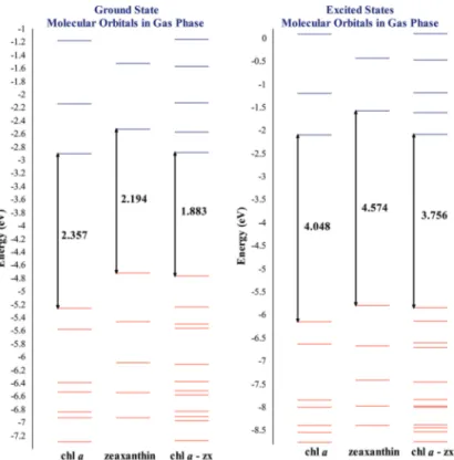
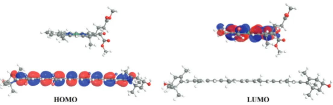
(11) Electronic Structure of Carotenoids in Natural and Artificial Photosynthesis http://dx.doi.org/10.5772/67636. Figure 7. Molecular orbitals for chlorophyllide a, zeaxanthin, and their corresponding diade in both ground and excited states. Calculations for ground states were obtained using DFT with B3LYP/6‐31+G(d,p) and excited states were obtained using the TDDFT scheme with TDA approximation employing the CAMB3LYP/6‐31+G(d,p) theoretical method. Energies in eV.. to compare both, ground and excited states, and obtain an easy way to compare the results. For the specific case of diade formed by chlorophyllide a and zeaxanthin in its ground state, when analyzed separately, chlorophyllide a presented an energy gap of 2.357 eV. When the diade was analyzed, the energy gap decreased to 1.883 eV. Meanwhile, at the excited state, chlorophyllide a's result had 4.048 eV (isolated), and for the diade, it was obtained as 3.756 eV. Figure 7 shows an energy gap for chlorophyllide a which decreased in both energetic states (ground and excited), presenting a difference between the isolated chlorophyllide a and those integrated into a diade system of 0.474 and 0.292 eV, respectively. This indicates in a general way that chlorophyllide a has a higher reactivity when it is found in the diade system, and this is independent of the energetic state. For the diade system, it was found that the HOMO molecular orbital is provided by the cor‐ responding carotenoid zeaxanthin, while the LUMO orbital corresponds to chlorophyllide a. Figure 8 displays both HOMO and LUMO orbitals for diade chlorophyllide a/zeaxantina, indi‐ cating in general that carotenoids are responsible for luminous energy absorption, by means of HOMO molecular orbitals, and interact with LUMO from chlorophyllide a.. 27.
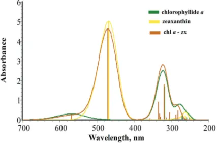
(12) 28. Carotenoids. Figure 8. Molecular orbitals HOMO and LUMO's graphical representations for the diade systemchlorophyllide a/zeaxanthin.. 3.2.2. Absorption UV‐vis spectra for chlorophyllide a and chlorophyll a From excited states energy calculations, UV‐vis absorption spectra were determined for each of the two selected molecules including integrated systems in the diade. In Figure 9 are displayed UV‐vis absorption spectra for experimental and theoretical (corresponding to experimentation similar to calculations within this work) frameworks for chlorophyll a. Chlorophyllide a presents a similar structure than chlorophyll a, except that in chlorophyl‐ lide a, the phytol lateral chain is removed which does not present any π bonding (conjugate double bonds). Therefore, it does not present photochemical reactivity [29]. In this way, the. Figure 9. UV‐Vis absorption spectra for both chlorophyll a (experimental data) and for chlorophyllide a. Chlorophyll a is analyzed in diethylic ether and chlorophyllide a is analyzed in the gaseous phase using TDDFT with CAM‐B3LYP/6‐31+G(d,p)..
(13) Electronic Structure of Carotenoids in Natural and Artificial Photosynthesis http://dx.doi.org/10.5772/67636. comparison between experimental data for chlorophyll a (with the phytol lateral chain) and theoretical data for chlorophyllide a (without phytol) is in agreement. In Figure 9 are shown experimental values for the more characteristic chlorophyll a absorp‐ tion bands [30], which are 670 y 420 nm for Qy and Soret bands, respectively. For chlorophyl‐ lide a, the theoretical values found were 568 y 320 nm, respectively. As can be seen in Figure 9, chlorophyllide a presents characteristic absorption maximum peaks. These peaks are displaced of approximately 100 nm in chlorophyllide a with respect to chlorophyll a values. This difference between experimental and theoretical values may be attributed mainly to the solvent effect. As mentioned in this section, to obtain experimental values, the samples used diethylic ether as a solvent while for theoretical values, and calcu‐ lations were made in the gaseous phase. Another contributor is the methodological differ‐ ence, in concrete, in the computational method employed (which relies on approximations). However, characteristic absorption peaks for these molecules were consistent in all cases. Regarding theoretical UV‐Vis absorption spectra (theoretical), calculations were developed for the two molecules integrated in the diades, and their diagram was based on the excited states’ results. In Figure 10, chlorophyllide a absorption spectra, zeaxanthin, and their correspond‐ ing diades are displayed. Here, one can observe that Qy bands for isolated chlorophyllide a and the corresponding band for the diade are practically identical (in both axes). Meanwhile, the Soret band corresponding to the diade presents absorption values slightly over the cor‐ responding values for chlorophyllide a alone (the values being the same for the wavelength in both cases). Concretely for zeaxanthin in the diade, this pigment contributes to a slight dis‐ placement in the x‐axis of 468.6 a 470.5 nm with respect to isolated zeaxanthin. Furthermore,. Figure 10. UV‐Vis absorption spectra for chlorophyllide a, zeaxanthin, and their corresponding diade (chlorophyllide a/zeaxanthin). Calculations were carried out under the TDDFT scheme using the CAMB3LYP/6‐31+G(d,p) theoretical method.. 29.
(14) 30. Carotenoids. isolated zeaxanthin presents in this particular peak an absorbance slightly above with respect to the one measured in its corresponding diade. Then, it may be observed how integrated diade systems can cover a wider absorption spectra than isolated chlorophyll a.. 4. Conclusions Theoretical calculations allowed us to predict UV‐Vis absorption spectra for carotenoid struc‐ tures selected for this work, achieving a good accuracy with experimental results. Within the selected carotenoids that were analyzed, zeaxanthin is the pigment with better electronic properties to form an integrated system, chlorophyll a‐carotenoid. Excited states molecular orbitals analysis for the diade system formed by chlorophyllide a‐zea‐ xanthin allowed one to observe the specific role played by chlorophyll a as a photosynthetic pigment by its LUMO contribution to the integrated system on the one hand and on the other hand, the role of zeaxanthin as an accessory pigment with the contribution made to HOMO. Analysis of pigments related to the natural photosynthetic process allowed us to set a basis on how these pigments can be used in potential artificial photosynthetic applications and to develop new materials for alternative energy applications. Electronic structure data are fundamental information for any material and may help to develop systems with carotenoids and accessory pigments in other fields such as the health or food industries.. Acknowledgements The authors thank the National Council of Science and Technology (CONACYT, for its acro‐ nym in Spanish) for the financial support in the development of this scientific research to the basic science project, No. 158307. FTR and JMB thank CONACYT for a scholarship to support their doctoral studies.. Author details Manuel Flores-Hidalgo1, Francisco Torres-Rivas2, Jesus Monzon-Bensojo2, Miguel EscobedoBretado1, Daniel Glossman‐Mitnik3 and Diana Barraza‐Jimenez1,2* *Address all correspondence to: dianabarraza@ujed.mx 1 Department of Chemical Sciences, Juarez University of Durango State, Durango, Mexico 2 Food and Development Research Center, A. C. Delicias Unit, Chih, Mexico 3 Advanced Materials Research Center, Chihuahua, Chih, Mexico.
(15) Electronic Structure of Carotenoids in Natural and Artificial Photosynthesis http://dx.doi.org/10.5772/67636. References [1] Su J, Vayssieres L. A place in the sun for artificial photosynthesis? ACS Energy Lett 2016; 1: 121-135. doi:10.1021/acsenergylett.6b00059 [2] Demmig‐Adams B, Stewart JJ, Burch TA, Adams WW, III. Insights from placing photo‐ synthetic light harvesting into context. J Phys Chem Lett 2014; 5: 2880-2889. doi:10.1021/ jz5010768 [3] Tachibana Y, Vayssieres L, Durrant JR. Artificial photosynthesis for solar water‐splitting. Nat Photonics 2012; 6: 511-518. doi:10.1038/nphoton.2012.175 [4] Kalyanasundaram K, Graetzel M. Artificial photosynthesis: biomimetic approaches to solar energy conversion and storage. Curr Opin Biotechnol 2010; 21: 298–310. doi:10.1016/j. copbio.2010.03.021 [5] Saini RK, Nile SH, Park SW. Carotenoids from fruits and vegetables: chemistry, analy‐ sis, occurrence, bioavailability and biological activities. Food Res Int 2015; 76: 735–750. doi:10.1016/j.foodres.2015.07.047 [6] Haskell MJ. Provitamin A carotenoids as a dietary source of vitamin A. In: Tanumihardjo S.A, editors. Carotenoids and Human Health. 1st ed. New York: Springer; 2013. p. 249–260. doi: 10.1007/978‐1‐62703‐203‐2_15 [7] Skibsted LH. Carotenoids in antioxidant networks colorants or radical scavengers. J Agric Food Chem 2012; 60(10): 2409–2417. doi:10.1021/jf2051416 [8] Stephensen CB. Provitamin A carotenoids and immune function. In: Tanumihardjo S.A, editors. Carotenoids and Human Health. 1st ed. New York: Springer; 2013. p. 261-270. doi: 10.1007/978‐1‐62703‐203‐2_16 [9] Meyers KJ, Mares JA, Igo RP, Truitt B, Liu Z, Millen AE, et al. Genetic evidence for role of carotenoids in age‐related macular degeneration in the carotenoids in age‐related eye disease study (CAREDS). Invest Ophthalmol Vis Sci 2014; 55(1): 587–599. doi:10.1167/ iovs.13‐13216 [10] Sharoni Y, Linnewiel‐Hermoni K, Khanin M, Salman H, Veprik A, Danilenko M, et al. Carotenoids and apocarotenoids in cellular signaling related to cancer: a review. Mol Nutr Food Res 2012; 56(2): 259–269. doi:10.1002/mnfr.201100311. [11] Sommer A. Vitamin A deficiency and clinical disease: a historical overview. J Nutr 2008; 138(10): 1835–1839. [12] Namitha KK, Negi PS. Chemistry and biotechnology of carotenoids. Crit Rev Food Sci Nutr 2010; 50(8): 728–760. doi:10.1080/10408398.2010.499811 [13] Lemmens L, Colle I, Van Buggenhout S, Palmero P, Van Loey A, Hendrickx M. Carotenoid bioaccessibility in fruit‐ and vegetable‐based food products as affected by product (micro) structural characteristics and the presence of lipids: a review. Trends Food Sci Technol 2014; 38: 125–135. doi:10.1016/j.tifs.2014.05.005. 31.
(16) 32. Carotenoids. [14] Bartley GE, Scolnik PA. Plant carotenoids: pigments for photoprotection, visual attrac‐ tion, and human health. Plant Cell 1995; 7: 1027–1038. doi:10.1105/tpc.7.7.1027 [15] Vishnevetsky M, Ovadis M, Vainstein A. Carotenoid sequestration in plants: the role of carotenoid‐associated proteins. Trends Plant Sci 1999; 4: 232–235. doi:10.1016/ S1360‐1385(99)01414‐4 [16] Britton G. Structure and properties of carotenoids in relation to function. FASEB J 1995; 9: 1551–1558. [17] Rodriguez‐Amaya DB, Kimura M. Harvest plus handbook for carotenoid analysis. Washington, DC: International Food Policy Research Institute and International Center for Tropical Agriculture; 2004. [18] Hashimotoa H, Sugaia Y, Uragamia C, Gardiner AT, Cogdell RJ. Natural and artificial light‐harvesting systems utilizing the functions of carotenoids. J Photochem Photobiol C Photochem Rev 2015; 25: 46–70. doi:10.1016/j.jphotochemrev.2015.07.004 [19] Amorim‐Carrilho KT, Cepeda A, Fente C, Regal P. Review of methods for analysis of carotenoids. Trends Anal Chem 2014; 56: 49–73. doi:10.1016/j.trac.2013.12.011 [20] Provesi JG, Dias CO, Amante ER. Changes in carotenoids during processing and storage of pumpkin puree. Food Chem 2011; 128: 195–202. doi:10.1016/j.foodchem. 2011.03.027 [21] Herrero M, Cacciola F, Donato P, Giuffrida D, Dugo G, Dugo P, et al. Serial coupled columns reversed‐phase separations in high‐performance liquid chromatography: tool for analysis of complex real samples. J Chromatogr A 2008; 1188: 208–215. doi:10.1016/j. chroma.2008.02.039 [22] Van Breemen RB, Dong L, Pajkovic ND. Atmospheric pressure chemical ionization tandem mass spectrometry of carotenoids. Int J Mass Spectrom 2012; 312: 163–172. doi:10.1016/j.ijms.2011.07.030. [23] Cacciola F, Donato P, Beccaria M, Mondello L. Advances in LC-MS for food analysis. LC GC Europe 2012; 25(5):15–24 [24] Frisch MJ, et al. Gaussian 09, Revision A.1. Wallingford CT: Gaussian Inc.; 2009. [25] Becke AD. Density functional thermochemistry‐III‐the role of exact exchange. J Phys Chem A 1993; 98(7): 5648–5652. doi:10.1063/1.464913 [26] Zhao Y, Truhlar DG. Density functionals with broad applicability in chemistry. Acc Chem Rev 2008; 41: 157–167. doi:10.1021/ar700111a [27] Chemissian, A Computer Program to Analyse and Visualise Quantum-Chemical Cal culations (L. Skripnikov, 2012). [28] Torres‐Rivas F, Flores‐Hidalgo MA, Glossman‐Mitnik D, Barraza‐Jimenez D. Geometric description and electronic properties of the principal photosynthetic pigments of higher plants: a DFT study. J Mol Model 2015; 21: 256. doi:10.1007/s00894‐015‐2796‐9.
(17) Electronic Structure of Carotenoids in Natural and Artificial Photosynthesis http://dx.doi.org/10.5772/67636. [29] Sundholm D. Comparison of the electronic excitation spectra of chlorophyll a and pheo‐ phytin a calculated at density functional theory level. Chem Phys Lett 2000; 317: 545– 552. doi:10.1016/S0009‐2614(99)01428‐1 [30] Strain HH, Thomas MR, Katz JJ. Spectral absorption properties of ordinary and fully deuteriated chlorophylls a and b. Biochim Biophys Acta 1963; 75: 306–311. doi:10.1016/0 006‐3002(63)90617‐6. 33.
(18)
(19)
Figure
+7
Documento similar
Finally, the representation of the amplitudes in impact parameter space is introduced highlighting the role played by the DL corrections in the eikonal phase, having as a consequence
The purpose of our molecular dynamics is to depict the evolution the population of di�erent electronic excited states following the electronic excitation of the system as a function
When the coupling is negligible (Fig. 1a), two clearly distinguishable excited states exist: the molecular exciton, characterized by the excitonic PES (orange line), and the
Micromagnetic simulations identify the modes excited in the B state as spin waves emitted by the vertex magnetic charges and propagating and interfering along the edge
The synthesis and analysis of these properties were then featured by (i) the composition, geometry, and electronic and mag- netic structure of the exposed surfaces at the
20 geometry and electronic properties of Ca 10 V 6 O 25 crystal in the fundamental and excited electronic.. 21 states (singlet
By means of electronic structure calculations, we show that self- induced catalysis emerges without any external stimuli through the interaction of the molecular permanent
Here, we demonstrate attosecond transient absorption spectroscopy (ATAS) as a viable probe of the electronic and nuclear dynamics initiated in excited states of a neutral molecule by
