A study on the lateralization of the effect of
musical imagery on spontaneous
otoacoustic emissions
A. Ramos-Amézquita
1G. Pérez-Acosta
2E. Castro-Sierra
3Abstract: It has been suggested that different pathways through the brain are
followed depending on the type of information that is being processed. Although it is now known that there is a continuous exchange of information through both hemispheres, language is considered to be processed by the left hemisphere, where Broca’s and Wernicke’s areas are located. On the other hand, music is thought to be processed mainly by the right hemisphere. According to Sininger Y.S. & Cone-Wesson, B. (2004), there is a similar but contralateral specialization of the human ears; due to the fact that auditory pathways cross-over at the brainstem. A previous study showed an effect of musical imagery on spontaneous otoacoustic emissions (SOAEs) (Perez-Acosta and Ramos-Amezquita, 2006), providing evidence of an efferent influence from the auditory cortex on the basilar membrane. Based on these results, the present work is a comparative study between left and right ears of a population of eight musicians that presented SOAEs. A familiar musical tune was chosen, and the subjects were trained in the task of evoking it after having heard it. Samples of ear-canal signals were obtained and processed in order to extract frequency and amplitude data on the SOAEs. This procedure was carried out before, during and after the musical image creation task. Results were then analyzed to compare the difference between SOAE responses of left and right ears. A clear asymmetrical SOAEs response to musical imagery tasks between left and right ears was obtained. Significant changes of SOAE amplitude related to musical imagery tasks were only observed on the right ear of the subjects. These results may suggest a predominant left hemisphere activity related to a melodic image creation task.
1Laboratorio de Cibernética, Facultad de Ciencias, UNAM.
2Programa de Maestría y Doctorado en Música, Escuela Nacional de Música,
UNAM,.
1. Background
Hemispheric specialization
An opinion commonly shared is that the analytic sequential functions of language became, through the development of verbal communication, the target of the left hemisphere. On the other hand, the right hemisphere has emerged as being more suited for the analysis of synthetic, holistic relations. This phenomenon could be explained by taking under consideration two different operational modes coexisting in the brain. The fist involves a sequential analysis of subparts while the other involves the spatial integration of momentaneous patterns of neural activity (Roederer, J., 1975). In accordance to this, it has been suggested that left and right auditory regions may be individually specialized for processing sounds based on acoustic properties. The left hemisphere process preferentially rapidly changing signals (temporal resolution), and tonal stimuli are best processed by the right hemisphere auditory areas (spectral resolution) (Sininger, Y. S. & Cone-Wesson, B., 2004). For all that stated above, it has been commonly suggested that it is the right hemisphere the one responsible for the processing of musical information, while the left hemisphere takes care of the processing of language.
However, recent observations of the hemispheric responses of people specialized in some facet of musical art, have shown left hemisphere activity providing evidence of the existence of some analytical process happening.
Otoacoustic emissions
Otoacoustic emissions are acoustic signals that can be registered in the external ear canal.
In 1978, Kemp submitted an extraordinary report in which he presented his findings on acoustic signals originated in the cochlea; otoacoustic emissions (OAEs) (Kemp, 1978, 1979). He later found that OAEs emerged either as a result of external stimulation or in a spontaneous manner (spontaneous otoacoustic emissions, SOAEs).
(SOAEs) (Perez-Acosta and Ramos-Amezquita, 2006), providing evidence of an efferent influence from the auditory cortex on the basilar membrane. It is also widely presumed that there might be a close relation between hearing sensitivity and SOAEs (Norton et al., 1988; McFadden and Mishra, 1993). In the music domain, Braun has reported interesting observations regarding the relation of SOAE frequencies to critical bands (1997) and their possible relation to pitch extraction (2000).
Mental Imagery
Thanks to the use of different imaging techniques [positron emission tomography (PET), functional magnetic resonance imaging (fMRI), magnetoencephalography (MEG) and transcranial magnetic stimulation (TMS)], it is now widely known that brain activity triggered by the creation of a mental image appears to be almost the same as that triggered by perception of the actual stimulus. That is, when you imagine a face or a place, the visual cortex is activated almost in the same way as if you were actually seeing that face or place (O’Craver et al., 2000). The same occurs with images of movement. These findings have been of particular interest in the music domain aiming to enhance the importance of mental practice (Meister et al., 2004; Pascual-Leone et al., 1995 and 1999), as it has been found that to imagine oneself playing an instrument activates the motor cortex in a very similar way than during practice itself.
Several studies have shown that auditory imagery activates most of the cerebral substrates activated when listening to sound (Halpern, 2001; Yoo et al., 2001; Schürmann et al., 2002; Zatorre et al., 2004; Kraemer et al., 2005). These substrates include bilateral primary and secondary auditory cortices, supplementary motor area (SMA) and right dorsolateral frontal cortex. There are some differences between some activated substrates depending mainly on the experimental design; nonetheless, they all coincide on the primary and secondary auditory cortex in the temporal lobes being activated.
2. Aims
The aim of this study was to elucidate whether the efferent auditory cortex effect on the cochlea, due to a musical imagery task, showed any lateralization preference.
3. Materials and Method
Subjects
The study involved 8 subjects (aged 20–36 years; mean age 27.4), of which, 3 were male and 5 female, with no history of auditory pathology and with normal audiometric measurements (only one subject manifested acoustic trauma, level I, apparently due to constant loud sound exposure, around the 4000-Hz band. However, since no SOAEs were present near this band, he was considered eligible for the study). The subjects were either professional musicians or music students with over 10 years of training in music listening (mean 12.3) (3 singers, 2 pianists, 1 guitarist, 1 accordionist, 1 electrical bassist). Audiometric analyses were conducted using a portable Beltone clinical audiometer. Hearing thresholds were measured at 125, 250, 500, 1000, 2000, 4000 and 8000 Hz.
Subjects were volunteers recruited from Escuela Nacional de Música of the National Autonomous University of Mexico (UNAM).
In each recording session, subjects were seated in a double-walled, soundproof chamber (Cabina Sonoamortiguada, Electromédica y Acústica, S. A.), located at Clínica Lomas Altas in Mexico City. Each subject was instructed to sit quietly throughout the test period.
All participants gave written informed consent.
Apparatus
For SOAE recordings, the ear canal sound pressure was measured with an ILO-292 Otodynamics microphone system, developed by Kemp. The probe was fitted into the external ear canal via an adaptable plastic ear plug. A Fast Fourier Transform (FFT) of the signal was performed, and a frequency and amplitude analysis was carried out using the ILO system. The FFT of the acoustic signal was calculated over a frequency span of 0-6 kHz. The SOAEs were recorded using the synchronized technique, in which a click is presented to the ear and SOAEs time-locked to the click are averaged (Hall, 1999). This procedure was carried out after a training session.
Training
b) Musical image creation task: a familiar musical tune was chosen. The selected melody was a traditional Mexican tune, “Marcha de Zacatecas”. This tune (a simple melodic line) was recorded with a flute on a single track. Training consisted of listening to the tune until all subjects reported that they were able to evoke it. They were asked to indicate the beginning and the end of the task of evoking in order to measure how long it lasted (which should be very similar to the actual duration of the melody). The tune lasted 1min 03sec. The entire average duration measured was 1min 03sec ± 03sec.
Recording Sessions
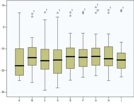
All the subjects were asked not to take any caffeine or alcohol for at least 12 hour previous to testing. Subjects were trained for an hour, ca. 2 hours before the recording session. The recording session consisted of 9 different conditions, and SOAEs were measured during each one of them. The first condition (condition A) consisted of a baseline recording with no task associated to it; subjects were asked only to sit quietly during this recording with their eyes open. For condition B, subjects were asked to concentrate on their breathing with their eyes closed. For conditions C and D, subjects were asked to perform the imagery task with their eyes closed. Conditions E and F consisted of the same imagery task with eyes open. For condition G, subjects were asked to concentrate again on their breathing with their eyes closed. Condition H consisted of a second baseline recording with no task (eyes open). The last recording (Final condition) was made 2 hours after the second baseline recording.
4. Results
The vast majority of SOAE recordings obtained lay in the frequency range of 1 to 2 kHz. This is the frequency range reported in the present work. Each SOAE of each subject was considered as independent. Since this study is observational in nature, a 10% significance value was considered for the tests (p < 0.1).
difference was found between any other (Table 2). Independent sample t -tests were performed to compare the change in SOAE amplitude between right and left ear for each possible pair of conditions; results are shown in Table 3. The pairs of conditions for which a significant difference was found are: A-B, A-C, A-D, A-E, D-E, D-F, D-G, D-H.
5. Conclusions
A clear asymmetrical SOAEs response to auditory imagery tasks between left and right ears was found. Although significant SOAE amplitu-de changes were found for both ears, significant differences related to the musical imagery tasks perfomed were found only for right ears.
It should be noted that in numerous studies regarding SOAEs, a noticable right ear and female prevalence has been found (Hall, 2000). These reports are consistent with the data obtained in the present work (4 left ear SOAEs vs. 20 right ear SOAEs). The variances of both groups proved to be equal, however, the scarcity of left ear SOAEs might have an effect on the results presented; this could explain in particular the unexpected mean amplitude leap form the baseline measurement to the subsecuent conditions for the left ear data. On the other hand, no other significant difference was found between any other task. In particular, no significant difference was found between any of the musical imagery conditions against any condition other than the baseline measurement. This gives rise to the possibility that no real effect was produced on the left ear SOAEs.
It is a well known fact that ascending and descending central auditory pathways cross-over at the superior olivary nucleus in the brainstem, sending most of the information to the contralateral side. Considering the above stated, it might be suggested that the efferent effect from the auditory cortex on the peripheral auditory system, triggered by a melodic image creation task, shows a right ear lateralization that could be due to a predominant left hemisphere activity. As stated in section 1, it seems possible that the musical analysis performed by musically trained subjects is lateralized to their “dominant” (left) hemisphere (Lafarga, 2000). Nevertheless, the validity of this statement must still to be further substantiated.
Results reported here should be considered as applicable only to the subjects of the present study, representing a particular population.
Fig. 1. Right ears. Estimated Marginal Means (dB) vs. Recording conditions.
longer periods and to analyze the changes in SOAE behavior related to a possible learning process. Another important suggestion is to change the order of the tasks to see if the changes are in fact related to the tasks and do not depend on the time elapsed.
6. Acknowledgements
Thanks are due to Valeria Lapilover, M.S., and Gonzalo Corvera, M.D., from Clínica Lomas Altas, for their providing the means and assistance to carry out measurements of SOAEs, to Adrián Poblano, M.D., Ph.D., and Carmina Arteaga, M.D., from Instituto Nacional de Rehabilitación de Mexico, for providing means and assistance to undertake audiometric analyses, to Alfonso Reyes, M.S., from Hospital Infantil de México Federico Gómez, for aid in carrying out statistical analyses and to Pablo Padilla from Facultad de Ciencias UNAM for all his comments and guidance on different matters.
Table 3. Significant differences between left and right ears (p < 0.1)
*Note: t -tests were perfomed considering the mean difference of SOAE amplitudes between the
REFERENCES
Besson, M. & Schön, D. (2004). Comparison between language and music. In I. Peretz & R. Zatorre (Org.). The cognitive neuroscience of music (pgs. 269-293). Oxford: Oxford University Press.
Castro-Sierra, E. (1996). Actividad auditiva cortical cerebral medida por técni-cas de magnetoencefalografía. Ciencia y Desarrollo, 21(127), 42-51. Chen, W., Kato, T., Zhu, X-H., Adriany, G. & Uðurbil, K. (1996). Functional
mapping of human brain during music imagery processing.
NeuroImage, 3, S205.
Hall, J.W. (2000). Spontaneous otoacoustic emissions (SOAEs). In
Handbook of Otoacoustic Emissions (pgs. 67-92). San Diego: Sin-gular Publishing Group.
Halpern, A.R. (2001). Cerebral substrates of music imagery. Ann NY Acad Sci, 930, 179-92.
Kemp, D.T. (1978) Stimulated acoustic emissions from within the human auditory system. JASA, 64, 1386-1391.
Kraemer, D.J.M., Macrae, C.N., Green, A.E. & Kelley, W.M. (2005). Sound of silence activates auditory cortex. Nature, 434, 158.
Lafarga, M.M. (2000, December). Desarrollo musical y desarrollo neuroló-gico. Paper presented in Congreso Mundial de Lecto-escritura. Va-lencia, España.
Maison, S., Micheyl, C. & Collet, L. (2001). Influence of focused attention on cochlear activity in humans. Psychophys, 38, 35-40.
McFadden, D. & Mishra, R. (1993). On the relation between hearing sensitivity and otoacoustic emissions. Hear Res, 97, 208-13.
O’Craven, K.M. & Kanwisher, N. (2000). Mental imagery of faces and places activates corresponding stimulus-specific brain regions. J Cogn Neurosci, 12(6), 1013-23.
Pascual-Leone, A., Nguyet, A.D. & Cohen, L., G. et al. (1995). Modulation of muscle responses evoked by transcranial magnetic stimulation during the acquisition of new fine motor skills. J Neurophysiol, 74, 1037-45. Pascual-Leone, A., Tarazona, A.F. & Catala, M.D. (1999). Applications of transcranial magnetic stimulation in studies on motor learning.
Electroencephalogr Clin Neurophysiol Suppl, 51, 157-161. Penner, M.J., Glotzbach, L. & Huang, T. (1993). Spontaneous otoacoustic
emissions: Measurement and data. Hear Res, 68, 229-37.
Perrot, X., Micheyl, C., Khalfa, S. & Collet, L. (1999). Stronger bilateral efferent influences on cochlear biomechanical activity in musicians than in non-musicians. Neurosci Lett, 262, 167-70.
Roederer, J. (1975). Superposition and successions of complex tones and the perception of music. In Introduction to the physics and psychophysics of music (pgs. 165-170). Denver: Springer-Verlag. Schürmann, M., Raij, T., Fujiki, N. & Hari, R. (2002). Mind’s ear in a musician:
where and when in the brain. NeuroImage, 16(2), 434-40.
Sininger, Y.S. & Cone-Wesson, B.(2004). Asymmetric cochlear processing mimics hemispheric specialization. Science, 305, 1581.
Whitehead, M.L. (1991). Slow variations of the amplitude and frequency of spontaneous otoacoustic emissions. Hear Res, 53, 269-280.
Yoo, S., Lee, Ch.U. & Choi, B.G. (2001). Human brain mapping of auditory imagery: Event-related functional MRI study. NeuroReport, 12, 3045-3049.