Open Access
Research
Replication by the Epistasis Project of the interaction
between the genes for IL-6 and IL-10 in the risk of Alzheimer's
disease
Onofre Combarros*
1, Cornelia M van Duijn
2, Naomi Hammond
3,
Olivia Belbin
4, Alejandro Arias-Vásquez
2, Mario Cortina-Borja
5,
Michael G Lehmann
6, Yurii S Aulchenko
2, Maaike Schuur
2,7, Heike Kölsch
8,
Reinhard Heun
8,9, Gordon K Wilcock
10,11, Kristelle Brown
4,
Patrick G Kehoe
11, Rachel Harrison
11, Eliecer Coto
12, Victoria Alvarez
12,
Panos Deloukas
3, Ignacio Mateo
1, Rhian Gwilliam
3, Kevin Morgan
4,
Donald R Warden
6, A David Smith
6and Donald J Lehmann
6Address: 1Neurology Service and Centro de Investigación Biomédica en Red sobre Enfermedades Neurodegenerativas (CIBERNED), Marqués de
Valdecilla University Hospital (University of Cantabria), 39008 Santander, Spain, 2Department of Epidemiology, Erasmus MC University Medical
Center, Rotterdam, the Netherlands, 3The Wellcome Trust Sanger Institute, Hinxton, Cambridge CB10 1SA, UK, 4School of Molecular Medical
Sciences, Institute of Genetics, University of Nottingham, Queens Medical Centre, Nottingham NG7 2UH, UK, 5Centre for Paediatric
Epidemiology and Biostatistics, Institute of Child Health, University College London, 30 Guilford Street, London WC1N 1EH, UK, 6Oxford Project
to Investigate Memory and Ageing (OPTIMA), University Department of Physiology, Anatomy and Genetics, South Parks Road, Oxford OX1 3QX, UK, 7Department of Neurology, Erasmus MC University Medical Center, Rotterdam, the Netherlands, 8Department of Psychiatry, University of
Bonn, Bonn, Germany, 9Department of Psychiatry, Derby City General Hospital, Uttoxeter Road, Derby DE22 3NE, UK, 10Nuffield Department
of Medicine, University of Oxford, Level 4, John Radcliffe Hospital, Oxford OX3 9DU, UK, 11Dementia Research Group, Institute of Clinical
Neurosciences, University of Bristol, Frenchay Hospital, Frenchay, Bristol BS32 8BE, UK and 12Genética Molecular, Hospital Central de Asturias,
Oviedo, Spain
Email: Onofre Combarros* - combarro@unican.es; Cornelia M van Duijn - c.vanduijn@erasmusmc.nl; Naomi Hammond - nh4@sanger.ac.uk; Olivia Belbin - Belbin.Olivia@mayo.edu; Alejandro Arias-Vásquez - a.ariasvasquez@umcn.nl; Mario Cortina-Borja - m.cortina@ich.ucl.ac.uk; Michael G Lehmann - mike@worldinneed.co.uk; Yurii S Aulchenko - i.aoultchenko@erasmusmc.nl; Maaike Schuur - m.schuur@erasmusmc.nl; Heike Kölsch - heike.koelsch@iqwig.de; Reinhard Heun - heun@gmx.com; Gordon K Wilcock - Gordon.Wilcock@ndm.ox.ac.uk;
Kristelle Brown - Kristelle.Brown@nottingham.ac.uk; Patrick G Kehoe - Patrick.Kehoe@bristol.ac.uk; Rachel Harrison - Rachel.Harrison@bristol.ac.uk; Eliecer Coto - eliecer.coto@sespa.princast.es;
Victoria Alvarez - victoria.alvarez@sespa.princast.es; Panos Deloukas - panos@sanger.ac.uk; Ignacio Mateo - mateonacho@hotmail.com; Rhian Gwilliam - rgl@sanger.ac.uk; Kevin Morgan - Kevin.Morgan@nottingham.ac.uk; Donald R Warden - donald.warden@dpag.ox.ac.uk; A David Smith - david.smith@pharm.ox.ac.uk; Donald J Lehmann - donald.lehmann@pharm.ox.ac.uk
* Corresponding author
Abstract
Background: Chronic inflammation is a characteristic of Alzheimer's disease (AD). An interaction
associated with the risk of AD has been reported between polymorphisms in the regulatory regions of the genes for the pro-inflammatory cytokine, interleukin-6 (IL-6, gene: IL6), and the anti-inflammatory cytokine, interleukin-10 (IL-10, gene: IL10).
Methods: We examined this interaction in the Epistasis Project, a collaboration of 7 AD research
groups, contributing DNA samples from 1,757 cases of AD and 6,295 controls.
Published: 23 August 2009
Journal of Neuroinflammation 2009, 6:22 doi:10.1186/1742-2094-6-22
Received: 9 July 2009 Accepted: 23 August 2009
This article is available from: http://www.jneuroinflammation.com/content/6/1/22 © 2009 Combarros et al; licensee BioMed Central Ltd.
This is an Open Access article distributed under the terms of the Creative Commons Attribution License (http://creativecommons.org/licenses/by/2.0), which permits unrestricted use, distribution, and reproduction in any medium, provided the original work is properly cited.
Results: We replicated the interaction. For IL6 rs2069837 AA × IL10 rs1800871 CC, the synergy
factor (SF) was 1.63 (95% confidence interval: 1.10–2.41, p = 0.01), controlling for centre, age, gender and apolipoprotein E ε4 (APOEε4) genotype. Our results are consistent between North Europe (SF = 1.7, p = 0.03) and North Spain (SF = 2.0, p = 0.09). Further replication may require a meta-analysis. However, association due to linkage disequilibrium with other polymorphisms in the regulatory regions of these genes cannot be excluded.
Conclusion: We suggest that dysregulation of both IL-6 and IL-10 in some elderly people, due in
part to genetic variations in the two genes, contributes to the development of AD. Thus, inflammation facilitates the onset of sporadic AD.
Background
Alzheimer's disease (AD) is accompanied by a chronic inflammatory process, including activation of microglia and astrocytes that express pro-inflammatory cytokines [1,2]. It is unclear to what extent this inflammation is a reaction to the pathology of AD, and to what extent it con-tributes to the onset or progression of the disease.
Two multi-functional cytokines, interleukin-6 (IL-6) and interleukin-10 (IL-10), may be relevant to this question. 6 is a potent pro-inflammatory cytokine [3], while IL-10 acts to limit inflammation in the brain [4]. Both are produced by activated microglia and astrocytes [3,4]. Two single nucleotide polymorphisms (SNPs), rs1800795 (-174G/C) and rs1800896 (-1082G/A), in the regulatory regions of the genes, IL6 and IL10, respectively, have been widely studied. However, the ongoing AlzGene meta-analyses of the two SNPs [5]http://www.alzforum.org/ res/com/gen/alzgene/ are both currently negative (4 July 2009): pooled odds ratios for Caucasians in single-locus analyses of IL6-174C versus G alleles = 0.93 (95% confi-dence interval: 0.79–1.08, 14 studies) and of IL10-1082G versus A alleles = 0.91 (0.74–1.11, eight studies). Our own meta-analyses, both by allele and by genotype, confirmed these results and also indicated a high degree of heteroge-neity among the studies (p < 0.05 in seven out of eight analyses, data not shown). Such inconsistencies may be due to study differences, e.g. in design or technical or ana-lytical approach. However, the heterogeneity remained in six out of eight analyses after altogether four studies with controls in Hardy-Weinberg disequilibrium were removed. Alternatively, the heterogeneity may reflect true population differences, such as in interactions with other factors, including other genes. Such diverse results have been described as a marker of epistasis [6,7], i.e. where the effect of one polymorphism depends on the genotype at another locus.
Infante et al (2004) [8] reported an interaction between
IL6-174G/C and IL10-1082G/A associated with the risk of
AD. We therefore set out to replicate this result in the Epistasis Project.
The Epistasis Project
Sporadic Alzheimer's disease (AD) is a complex disease, with over 50% heritability [9]. This suggests that its study requires the investigation of interactions between risk fac-tors, particularly genetic factors. The Epistasis Project stud-ies such interactions, mainly those between distinct genetic loci, i.e. epistasis. The project is a collaboration of seven AD research groups: Bonn, Bristol, Nottingham, OPTIMA, Oviedo, Rotterdam and Santander (Table 1). The project aims: first, to replicate genetic interactions that have been reported to affect the risk of sporadic AD; second, to explore other polymorphisms in the relevant genetic regions, ultimately to reveal the true risk loci. The overriding object is to gain insights into AD causality. However, interactions can only be reliably studied with sufficient statistical power and careful study design. The project therefore has these
characteristics:-1. Power
The project has 1,757 cases of AD and 6,295 elderly con-trols. These numbers give 99.9% power to detect an SF [10] of 2 between two polymorphisms, each with a minor allele frequency of 20%, when controlling for individual centres (2 below). They give 89.8% power or 48.6% power to detect SFs of 1.5 or 1.25, respectively. Quality control of genotyping reduces the numbers somewhat, depending on the polymorphism.
2. Selection of sample-sets
Only sample-sets drawn from narrow geographical regions with relatively homogeneous, Caucasian popula-tions have been chosen, from seven AD research centres (Table 1 and Additional file 1).
3. Sample characterisation
Cases of AD are either confirmed at autopsy as "definite" or "probable" by CERAD criteria [11], or clinically diag-nosed as "probable AD" by NINCDS-ADRDA criteria [12]. Controls are either screened as free of cognitive impair-ment, or confirmed at autopsy as free of pathology con-sistent with AD or other dementias.
4. Matching of cases and controls
Only cases and controls drawn from the same region are compared, thus controlling for geographical differences, e.g. between North and South Europe.
5. Candidate interactions
Interactions are studied that have prior evidence of an association with AD and a plausible biological hypothe-sis. Interactions with age (± 75 years), gender and apoli-poprotein E ε4 (APOEε4) genotype are also examined.
6. Analytical methods
Logistic regression and SF [10] analyses are used. All anal-yses are controlled for age, gender and APOEε4 genotype. All pooled analyses are also controlled for centre. There is thus no question of controls from one centre (e.g., Rotter-dam) being compared with AD cases from another region. Controlling for centre also reduces the relative influence of large subgroups, such as Rotterdam controls, in the pooled result. Where pooled analyses yield significant results, the seven individual centres are also examined for associations with AD, for heterogeneity, and for power to detect the interaction.
Table 1 gives the basic characteristics of the seven sample-sets. See Additional file 1 for further information.
To select the interactions for study, a survey of over 100 published claims and suggestions of epistasis in sporadic AD was undertaken [13]. The interactions finally chosen are involved in various networks that are widely consid-ered to contribute to the development of AD: lipid metab-olism [14], β-amyloid metabmetab-olism [15], oxidative stress [16], inflammation [1], insulin metabolism [17] and homocysteine metabolism [18]. Positive results should therefore deliver insights into the causes of AD.
Methods
Basic information on the 1,757 cases of AD and the 6,295 controls from the seven centres is given above and in Table 1, and fuller details are provided in Additional file 1.
Genotyping for the six centres other than Rotterdam was performed at the Wellcome Trust Sanger Institute, using the iPLEX Gold assay (Sequenom Inc.). Whole genome amplified DNA was used for 82% of samples; genomic DNA was used for the 18% of samples that were not suit-able for whole genome amplification. A Sequenom iPLEX, designed for quality control purposes, was used to assess genotype concordance between genomic and whole genome amplified DNA for 168 individuals. Assays for all SNPs were designed using the eXTEND suite and
MassAR-Table 1: Sample-sets used in the Epistasis Project
Group Geographical Region* Subjects† Numbers % women p (AD vs controls) Median age‡
(interquartile range)
p (AD vs controls)
Bonn Bonn and Mainz, Germany
Clinical AD 259 61.4 71.0 (65.1–77.5)
Screened controls 232 56.9 0.31 67.8 (64.4–76.1) 0.11
Bristol South-West England Autopsy AD 200 45.0 80.4 (75.6–86.0)
Autopsy controls 57 40.4 0.55 77.9 (72.8–82.9) 0.12
Nottingham Cambridge, England Autopsy AD 104 48.1 78.7 (70.8–84.6)
Autopsy controls 107 34.6 0.051 72.5 (65.6–80.0) < 0.001
OPTIMA Oxford, England Autopsy AD 163 54.6 79.5 (72.4–85.2)
Clinical AD 87 54.0 79.1 (73.4–84.8)
Screened controls 261 53.6 0.93 78.9 (72.9–83.8) 0.54
Oviedo Asturias, Spain Clinical AD 202 69.8 78.4 (74.8–82.8)
Screened controls 131 61.8 0.15 69.7 (63.5–74.5) < 0.001 Rotterdam Rotterdam (Ommord), the Netherlands Clinical AD 391 74.9 86.6 (82.2–91.6) Screened controls 5,111 57.7 < 0.001 76.9 (71.7–83.0) < 0.001
Santander Cantabria, Spain Clinical AD 351 65.0 75.5 (70.8–78.9)
Screened controls 396 68.9 0.27 80.9 (74.7–85.6) < 0.001
Totals Total AD 1,757 62.4 79.0 (73.0–85.2)
Total controls 6,295 57.7 76.9 (71.3–83.0)
AD = Alzheimer's disease
*from which the samples were drawn.
†Clinical AD cases were diagnosed as "probable AD" by NINCDS-ADRDA criteria [12]; autopsy AD cases were diagnosed as "definite" or
"probable" by CERAD criteria [11]; screened controls were free of cognitive impairment; autopsy controls were free of pathology consistent with AD or other dementias; all controls were ≥ 60 years of age.
RAY Assay Design software version 3.1 (Sequenom Inc.). Samples were amplified in multiplexed PCR reactions before allele specific extension. Allelic discrimination was obtained by analysis with a MassARRAY Analyzer Com-pact mass spectrometer. Genotypes were automatically assigned and manually confirmed using MassArray Typer-Analyzer software version 4.0 (Sequenom Inc.). Gender markers were included in all iPLEX assays as a quality con-trol metric for confirmation of plate/sample identity. Genotyping of SNPs rs1800871, rs2069837 and rs3024505 was carried out using the KASPar technology by KBioscience http://www.kbioscience.co.uk.
Genotyping in the Rotterdam cohort was done on Version 3 Illumina-Infinium-II HumanHap550 SNP array (Illu-mina, San Diego, USA) and additionally, SNPs were imputed using MACH software http:// www.sph.umich.edu/csg/abecasis/MACH/ with HapMap CEU Release 22 as a reference [19]. The reliability of imputation was estimated for each imputed SNP with the ratio of expected and observed dosage variance (O/E ratio). Only samples with high-quality extracted DNA were genotyped; 5974 were available with good quality genotyping data; 5502 of these had reliable phenotypes. For the Epistasis Project, 52 genotyped SNPs and 116 imputed SNPs were selected.
We assessed associations with logistic regression models and SF analysis [10], controlling for age, gender, APOEε4 and study centre, using R Version 2.6.1 (R Foundation for Statistical Computing, Vienna, Austria). Heterogeneity between centres was controlled by fitting a fixed effect cor-responding to contrasts between the baseline centre and the six other centres (having compared models with fixed-and rfixed-andom-effect terms in centre, goodness of fit was measured using Akaike's Information Criterion, which favoured using fixed effects only). Where the overall SF was significant at p < 0.05, the seven individual centres and the two geographical regions, North Europe and
North Spain, were also examined. Power calculations were by SF analysis. Meta-analyses were performed using the random-effect method of DerSimonian and Laird [20] and the heterogeneity test of Armitage [21]. Comparisons of allelic frequencies between North Spain and North Europe were by Fisher's exact test. We compared the medi-ans of the age distributions of AD and control groups using the Wilcoxon-Mann-Whitney test. Linkage disequi-librium data were estimated using the R genetics library http://cran.r-project.org/web/packages/genetics/
index.html. All tests of significance were two-sided.
Results
We replicated the interaction reported by Infante et al (2004) [8] between IL6 rs1800795 (-174G/C) and IL10 rs1800896 (-1082G/A) (below). In the Rotterdam cohort, we examined altogether five IL6-related SNPs, rs1800797, rs1800795, rs2069837, rs2069840 and rs2069845, and four IL10-related SNPs, rs1800896, rs1800871,
rs3024498 and rs3024505. In that preliminary study, we found that the strongest interactions were seen between
IL6 rs2069837 (intron 2 A/G) and IL10 rs1800871
(-819C/T) and between IL6 rs2069837 and IL10 rs3024505 (3' C/T) (data not shown). We therefore studied the five SNPs shown in Table 2 in all seven sample-sets.
Hardy-Weinberg analysis was performed for the five SNPs of Table 2 in both cases and controls of the Rotterdam samples, genotyped by Rotterdam, and of the samples from the other six centres, genotyped by the Sanger Insti-tute. As expected by chance, one of these 20 analyses resulted in disequilibrium: IL6-174G/C in AD cases of the six centres (p = 0.02). The two IL6 SNPs were in linkage disequilibrium (LD), as were the three IL10 SNPs (Table 2). The allelic frequencies differed significantly between North Europe (Bonn, Bristol, Nottingham, OPTIMA and Rotterdam) and North Spain (Oviedo and Santander) in four out of the five SNPs (Table 2).
Table 2: Studied SNPs
Gene SNP Minor allele frequency in controls LD in controls
North Europe North Spain Difference (p) With D' r2 IL6 rs1800795 -174G/C 41.1% (C) 32.8% (C) < 0.0001 rs2069837 0.998 0.055 rs2069837 Intron 2 A/G 7.3% (G) 9.3% (G) 0.03 IL10 rs1800896 -1082G/A 50.7% (G) 43.1% (G) < 0.0001 rs1800871 0.999 0.296 rs3024505 0.988 0.191 rs1800871 -819C/T 22.5% (T) 26.7% (T) 0.002 rs3024505 0.999 0.058 rs3024505 3' C/T 16.6% (T) 14.2% (T) 0.06
SNP = single nucleotide polymorphism; LD = linkage disequilibrium; D' = ratio of observed LD to maximum possible LD; r = correlation coefficient;
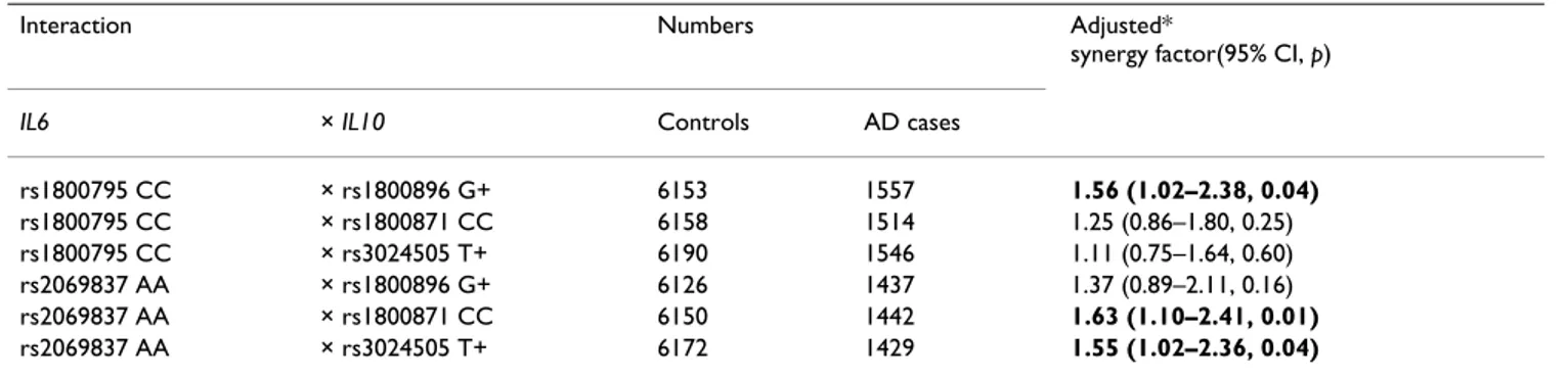
We examined the potential associations with AD of the six interactions generated between the two IL6 SNPs and the three IL10 SNPs in our overall dataset (Table 3), control-ling for centre, age, gender and APOEε4 genotype (as in all association analyses). Three of the six interactions were associated with AD: SF p < 0.05. The first interaction shown in Table 3 is effectively identical to that reported by Infante et al 2004 [8]; we have merely reversed the first genotype, i.e. CC rather than GC + GG, to give an SF of 1.56, rather than its inverse, 0.64, for easier comparison with the other interactions. The two IL6 genotypes shown in Table 3 were in LD (p < 0.0001), as were the three IL10 genotypes (p < 0.0001). The interaction between IL6 intron 2 AA and IL10-819CC was slightly the strongest: SF = 1.63 (95% confidence interval = 1.10–2.41, p = 0.01). All further analysis was therefore restricted to that interac-tion.
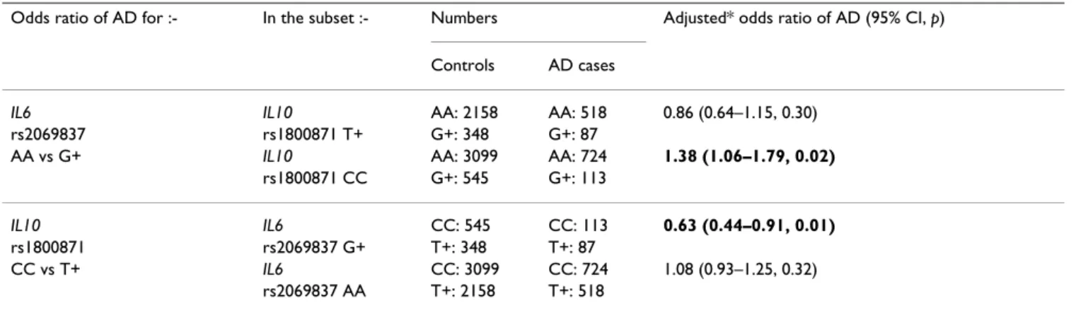
Table 4 shows the effect of each of the two factors in that interaction on the association with AD of the other factor. The presence of IL10-819CC changed the association of
IL6 intron 2 AA with AD from negative (odds ratio = 0.86, p = 0.30) to risk (odds ratio = 1.38, p = 0.02), while the
presence of the latter removed the protective association of the former (odds ratio = 0.63, p = 0.01, changed to 1.08,
p = 0.32).
There was significant heterogeneity between the centres, and the interaction was only significant in one of the seven centres, Rotterdam, with an SF of 3.0 (95% confi-dence interval = 1.6–5.8, p = 0.0007). But the power to detect an SF of 1.6 was low in each centre, ranging from below 10% to 40%. The results for North Europe (SF = 1.7, p = 0.03) and for North Spain (SF = 2.0, p = 0.09) were consistent (Table 5).
Results were similar for men as for women (data not shown). The interaction was slightly stronger in older sub-jects: SF for > 75 years = 1.71 (1.05–2.78, 0.03); SF for <
75 years = 1.27 (0.56–2.87, 0.57). However, there was no 3-way interaction with age. Nor did we find a significant interaction with APOEε4.
Discussion
In our whole dataset of over 7,500 samples, we have rep-licated both the interaction reported by Infante et al 2004 [8] and also that found in our preliminary study in the Rotterdam dataset, between SNPs of IL6 and IL10 (Table 3). Both interactions gave significant SFs of approximately 1.6. There was significant heterogeneity between centres, which was unsurprising since only two, Rotterdam and Santander, had > 20% power to detect these interactions. But the results for the two main regions, North Europe and North Spain, were consistent (Table 5). We conclude that this is likely to be a true effect, but not a very strong one. As in any example of true epistasis, the presence or absence of one factor critically influenced the effect of the other (Table 4).
Further replication will require a dataset at least as large, with appropriate statistical control for differences between individual sample-sets. Underpowered studies are unhelpful, since chance can produce misleading results in such cases. This criticism may apply to a previ-ous study of this interaction that had power of < 2% to detect it. Even the large Rotterdam Study, with 395 cases of AD and 5,111 controls, only had 40% power to detect this interaction. Thus, further replication may require a meta-analysis.
IL-6 in age-related decline and in AD
There is much evidence that chronic, low-grade overex-pression of IL-6 contributes to age-related decline. Blood levels of IL-6 can rise with age in humans [22-24]. Raised levels are associated with various age-related conditions, including risk of cognitive decline in some studies [25,26], but not in all [27]. Raised levels have been con-sistently associated with increased mortality in several
Table 3: Potential interactions between IL6 SNPs and IL10 SNPs in the risk of AD
Interaction Numbers Adjusted*
synergy factor(95% CI, p)
IL6 × IL10 Controls AD cases
rs1800795 CC × rs1800896 G+ 6153 1557 1.56 (1.02–2.38, 0.04) rs1800795 CC × rs1800871 CC 6158 1514 1.25 (0.86–1.80, 0.25) rs1800795 CC × rs3024505 T+ 6190 1546 1.11 (0.75–1.64, 0.60) rs2069837 AA × rs1800896 G+ 6126 1437 1.37 (0.89–2.11, 0.16) rs2069837 AA × rs1800871 CC 6150 1442 1.63 (1.10–2.41, 0.01) rs2069837 AA × rs3024505 T+ 6172 1429 1.55 (1.02–2.36, 0.04)
IL6 and IL10 = the genes for interleukin-6 and interleukin-10, respectively; SNPs = single nucleotide polymorphisms; AD = Alzheimer's disease; CI =
confidence interval; G+ and T+ group the genotypes, GA+GG and CT+TT, respectively; APOEε4 = apolipoprotein E ε4. *All analyses controlled for centre, age, gender and APOEε4 genotype.
large prospective studies [28-31]. Raised levels have also been associated with various conditions considered to be risk factors for dementia and/or AD: subclinical and clin-ical cardiovascular disease and the risk thereof [32-34], type 2 diabetes and its risk [35,36], psychological stress [37] and the damage following stroke (IL-6 also increased in CSF) [38,39].
Further, raised blood levels of IL-6 have often been asso-ciated with AD [40-42] and also with the risk of AD [43], although not in all studies [44,45]. Post-mortem studies, although small, have generally reported increased IL-6 levels or changed IL-6 distribution in AD brain [46-49], with one exception [50]. IL-6 was found not only in plaques, but also around the bodies of isocortical neu-rones, only in AD [46]. This evidence and that above sug-gest that dysregulation of IL-6 contributes to the development of AD.
A potential interaction between IL6 and IL10
Both IL-6 and IL-10 are produced by activated microglia and astrocytes (reviewed in [3,4]). However, in contrast to IL-6, IL-10 acts to limit inflammation in the brain. IL-10 inhibits the production of IL-6 [51,52] and its receptor [53]. Thus, certain combinations of genetic variants of IL6
and IL10, i.e. those associated with high production of IL-6 combined with those associated with low production of IL-10, may contribute to the dysregulation of inflamma-tion. High heritability (> 50%) has been reported for in
vitro stimulated production of both IL-6 and IL-10 [54].
The functions of two of our studied SNPs, IL6 intron 2 A/ G and IL10 3' C/T have not yet been investigated. Thus we cannot rule out that they might have effects on transcrip-tion or post-transcriptranscrip-tional processing. Alternatively, they may be in linkage disequilibrium with other, functional variants. On the other hand, studies of IL6-174G/C [55,56], of IL10-819C/T [57] and of IL10-1082G/A [58-60] have reported that these SNPs affect transcription or are in linkage disequilibrium with those that do. How-ever, it may be premature to designate any particular alle-les as high or low producers, since there have been contrasting results for the effects of IL6-174G/C and IL10-1082G/A on transcription, on in vitro stimulated produc-tion and on blood levels of the respective proteins. But most studies have associated the IL10-1082GG genotype at least with higher in vitro stimulated production of IL-10 [61-64], although not all, [65,66] and results have varied with experimental conditions [64,66]. It appears that sev-eral SNPs in the regulatory regions of each gene affect transcription in an interactive and tissue-specific manner
Table 4: Odds ratios of Alzheimer's disease for two IL6 and IL10 SNPs, stratified by each other
Odds ratio of AD for :- In the subset :- Numbers Adjusted* odds ratio of AD (95% CI, p) Controls AD cases
IL6 IL10 AA: 2158 AA: 518 0.86 (0.64–1.15, 0.30)
rs2069837 rs1800871 T+ G+: 348 G+: 87
AA vs G+ IL10 AA: 3099 AA: 724 1.38 (1.06–1.79, 0.02)
rs1800871 CC G+: 545 G+: 113
IL10 IL6 CC: 545 CC: 113 0.63 (0.44–0.91, 0.01)
rs1800871 rs2069837 G+ T+: 348 T+: 87
CC vs T+ IL6 CC: 3099 CC: 724 1.08 (0.93–1.25, 0.32)
rs2069837 AA T+: 2158 T+: 518
IL6 and IL10 = the genes for interleukin-6 and interleukin-10, respectively; AD = Alzheimer's disease; CI = confidence interval; G+ and T+ group the
genotypes, GA+GG and CT+TT, respectively; APOEε4 = apolipoprotein E ε4. *All analyses controlled for centre, age, gender and APOEε4 genotype.
Table 5: Interaction between IL6 rs2069837 AA vs G+ and IL10 rs1800871 CC vs T+, by region
Region Power* Adjusted† synergy factor (95% CI, p) in the risk of AD
North Europe‡ 64% 1.7 (1.05–2.6, 0.03)
North Spain‡ 37% 2.0 (0.9–4.4, 0.09)
All 76% 1.6 (1.1–2.4, 0.01)
IL6 and IL10 = the genes for interleukin-6 and interleukin-10, respectively; G+ and T+ group the genotypes, GA+GG and CT+TT, respectively; CI =
confidence interval; AD = Alzheimer's disease; APOEε4 = apolipoprotein E ε4. *To detect a synergy factor of 1.6 (as in the overall dataset) at 0.05.
†All analyses controlled for centre, age, gender and APOEε4 genotype.
[56]. Indeed, we note that in our dataset the 2-SNP com-bination of IL10 -1082G+/-819CC has a slightly stronger interaction with IL6 than that of either SNP alone (data not shown).
Conclusion
We conclude that an interaction between IL-6 and IL-10 is plausible; that dysregulation of the two genes contributes to chronic low-grade inflammation in some elderly peo-ple and thus to the risk of AD; and that certain combina-tions of genetic variants in the regulatory regions of the two genes are conducive to this dysregulation. But in view of the linkage disequilibrium in the region of each gene (Table 2), we cannot conclude that we have yet found the true risk polymorphisms. However, we suggest that our results are consistent with the contribution of inflamma-tion to the onset of AD.
Abbreviations
AD: Alzheimer's disease; APOEε4: apolipoprotein E ε4; CERAD: Consortium to Establish a Registry for Alzhe-imer's Disease; CSF: cerebrospinal fluid; IL-6: interleukin-6; IL6: the gene for interleukin-interleukin-6; IL-10: interleukin-10;
IL10: the gene for interleukin-10; NINCDS-ADRDA:
National Institute of Neurological, Communicative eases and Stroke-Alzheimer's Disease and Related Dis-eases Association; OPTIMA: the Oxford Project to Investigate Memory and Ageing; SF: synergy factor; SNP: single nucleotide polymorphism.
Competing interests
The authors declare that they have no competing interests.
Authors' contributions
All authors contributed to the design of the study. In addi-tion, ADS and DJL set up the Epistasis Project, with the help of the other authors. ADS and DJL decided on the strategy of the Epistasis Project, with the help of CMvD, OC, KM, PK, R Heun, MC-B, DRW and EC. ADS, DJL, CMvD, OC, KM, PK, R Heun, MC-B, DRW and EC chose the genetic interactions to study. OC produced the hypothesis for this study. KM and OB gave extensive advice on the choice of SNPs to study. DJL made the final selection of polymorphisms. HK, R Harrison, KM, DRW, EC and IM provided DNA for genotyping. CMvD, YSA, AA-V and MS provided the data for the preliminary study. DRW gave technical advice throughout. RG and NH were responsible for the genotyping of 6 sample-sets. AA-V was responsible for the Rotterdam genotyping. MC-B and DJL decided on the analytical approach. MC-B and YSA advised on statistics throughout. DJL and MGL performed the analysis. DJL drafted the manuscript. OC submitted the manuscript and is responsible for correspondence. All authors read the manuscript, studied it critically for its intellectual content and approved the final draft.
Additional material
Acknowledgements
We are most grateful to the Moulton Charitable Foundation for a grant to fund the Epistasis Project and to all those who have provided support for the individual clinical studies. GW was partly supported by the NIHR Bio-medical Research Centre, Oxford. UCL Institute of Child Health receives funding from the Department of Health's NIHR Biomedical Research Cen-tres funding scheme. The Centre for Paediatric Epidemiology and Biostatis-tics also benefits from funding support from the Medical Research Council in its capacity as the MRC Centre of Epidemiology for Child Health (G0400546).
References
1. Akiyama H, Barger S, Barnum S, Bradt B, Bauer J, Cole GM, Cooper NR, Eikelenboom P, Emmerling M, Fiebich BL, et al.: Inflammation and Alzheimer's disease. Neurobiol Aging 2000, 21:383-421. 2. Rogers J, Webster S, Lue L-F, Brachova L, Civin WH, Emmerling M,
Shivers B, Walker D, McGeer P: Inflammation and Alzheimer's disease pathogenesis. Neurobiol Aging 1996, 17:681-686. 3. Gruol DL, Nelson TE: Physiological and pathological roles of
interleukin-6 in the central nervous system. Mol Neurobiol 1997, 15:307-339.
4. Strle K, Zhou JH, Shen W-H, Broussard SR, Johnson RW, Freund GG, Dantzer R, Kelley KW: Interleukin-10 in the brain. Crit Rev Immu-nol 2001, 21:427-449.
5. Bertram L, McQueen MB, Mullin K, Blacker D, Tanzi RE: Systematic meta-analyses of Alzheimer disease genetic association studies: the Alzgene database. Nature Genetics 2007, 39:17-23. Accessed on 14 July, 2009.
6. Moore JH, Williams SM: Traversing the conceptual divide between biological and statistical epistasis: systems biology and a more modern synthesis. BioEssays 2005, 27:637-646. 7. Wade MJ: Epistasis, complex traits, and mapping genes.
Genet-ica 2001, 112–113:59-69.
8. Infante J, Sanz C, Fernández-Luna JL, Llorca J, Berciano J, Combarros O: Gene-gene interaction between interleukin-6 and inter-leukin-10 reduces AD risk. Neurology 2004, 63:1135-1136. 9. Bergem ALM, Engedal K, Kringlen E: The role of heredity in
late-onset Alzheimer disease and vascular dementia – a twin study. Arch Gen Psychiatry 1997, 54(3):264-270.
10. Cortina-Borja M, Smith AD, Combarros O, Lehmann DJ: The syn-ergy factor: a statistic to measure interactions in complex diseases. BMC Res Notes 2009, 2(1):105.
11. Mirra SS, Heyman A, McKeel D, Sumi SM, Crain BJ, Brownlee LM, Vogel FS, Hughes JP, van Belle G, Berg L: The Consortium to Establish a Registry for Alzheimer's Disease (CERAD). Part II. Standardization of the neuropathologic assessment of Alzheimer's disease. Neurology 1991, 41:479-486.
12. McKhann G, Drachman D, Folstein M, Katzman R, Price D, Stadlan EM: Clinical diagnosis of Alzheimer's disease: report of the NINCDS-ADRDA work group under the auspices of Depart-ment of Health and Human Services task force on Alzhe-imer's disease. Neurology 1984, 34:939-944.
13. Combarros O, Cortina-Borja M, Smith AD, Lehmann DJ: Epistasis in sporadic Alzheimer's disease. Neurobiol Aging 2009, 30:1333-1349.
14. Carter CJ: Convergence of genes implicated in Alzheimer's disease on the cerebral cholesterol shuttle: APP, choles-terol, lipoproteins, and atherosclerosis. Neurochem Int 2007, 50:12-38.
Additional file 1
The seven centres of the Epistasis Project. Click here for file
[http://www.biomedcentral.com/content/supplementary/1742-2094-6-22-S1.doc]
15. Hardy J, Selkoe DJ: The amyloid hypothesis of Alzheimer's dis-ease: progress and problems on the road to therapeutics.
Sci-ence 2002, 297:353-356.
16. Perry G, Nunomura A, Hirai K, Zhu X, Pérez M, Avila J, Castellani RJ, Atwood CS, Aliev G, Sayre LM, et al.: Is oxidative damage the fun-damental pathogenic mechanism of Alzheimer's and other neurodegenerative diseases? Free Rad Biol Med 2002, 33:1475-1479.
17. Craft S: The role of metabolic disorders in Alzheimer disease and vascular dementia. Arch Neurol 2009, 66:300-305.
18. Smith AD: The worldwide challenge of the dementias: a role for B vitamins and homocysteine? Food Nutr Bull 2008, 29:S143-S172.
19. Ikram MA, Seshadri S, Bis JC, Fornage M, DeStefano AL, Aulchenko YS, Debette S, Lumley T, Folsom AR, van den Herik EG, et al.: Genomewide association studies of stroke. N Engl J Med 2009, 360:1718-1728.
20. DerSimonian R, Laird N: Meta-analysis in clinical trials. Control Clin Trials 1986, 7:177-188.
21. Armitage P: Statistical Methods in Medical Research. Oxford: Blackwell Scientific Publications; 1983.
22. Ershler WB: Interleukin-6: a cytokine for gerontologists. J Am Geriatr Soc 1993, 41:176-181.
23. Hager K, Machein U, Krieger S, Platt D, Seefried G, Bauer J: Inter-leukin-6 and selected plasma proteins in healthy persons of different ages. Neurobiol Aging 1994, 15:771-772.
24. Young DG, Skibinski G, Mason JI, James K: The influence of age and gender on serum dehydroepiandrosterone sulphate (DHEA-S), IL-6, IL-6 soluble receptor (IL-6 sR) and trans-forming growth factor β1 (TGF-β1) levels in normal healthy blood donors. Clin Exp Immunol 1999, 117:476-481.
25. Weaver JD, Huang M-H, Albert M, Harris T, Rowe JW, Seeman TE: Interleukin-6 and risk of cognitive decline. Neurology 2002, 59:371-378.
26. Yaffe K, Lindquist K, Penninx BW, Simonsick EM, Pahor M, Kritch-evsky S, Launer L, Kuller L, Rubin S, Harris T: Inflammatory mark-ers and cognition in well-functioning African-American and white elders. Neurology 2003, 61:76-80.
27. Dik MG, Jonker C, Hack CE, Smit JH, Comijs HC, Eikelenboom P: Serum inflammatory proteins and cognitive decline in older persons. Neurology 2005, 64:1371-1377.
28. Cohen HJ, Harris T, Peiper CF: Coagulation and activation of inflammatory pathways in the development of functional decline and mortality in the elderly. Am J Med 2003, 114:180-187.
29. Harris TB, Ferrucci L, Tracy RP, Corti M-C, Wacholder S, Ettinger WHJ, Heimovitz H, Cohen HJ, Wallace R: Associations of ele-vated interleukin-6 and C-reactive protein levels with mor-tality in the elderly. Am J Med 1999, 106:506-512.
30. Reuben DB, Cheh AI, Harris TB, Ferrucci L, Rowe JW, Tracy RP, See-man TE: Peripheral blood markers of inflammation predict mortality and functional decline in high-functioning commu-nity-dwelling older persons. J Am Geriatr Soc 2002, 50:638-644. 31. Volpato S, Guralnik JM, Ferrucci L, Balfour J, Chaves P, Fried LP,
Har-ris TB: Cardiovascular disease, interleukin-6, and Har-risk of mor-tality in older women: the Women's Health and Aging Study. Circulation 2001, 103:947-953.
32. Cesari M, Penninx BW, Newman AB, Kritchevsky SB, Nicklas BJ, Sut-ton-Tyrrell K, Rubin SM, Ding J, Simonsick EM, Harris TB, et al.: Inflammatory markers and onset of cardiovascular events: results from the Health ABC study. Circulation 2003, 108:2317-2322.
33. Cesari M, Penninx BW, Newman AB, Kritchevsky SB, Nicklas BJ, Sut-ton-Tyrrell K, Tracy RP, Rubin SM, Harris TB, Pahor M: Inflamma-tory markers and cardiovascular disease (The Health, Aging and Body Composition [Health ABC] study). Am J Cardiol 2003, 92:522-528.
34. Ridker PM, Rifai N, Stampfer MJ, Hennekens CH: Plasma concen-tration of interleukin-6 and the risk of future myocardial inf-arction among apparently healthy men. Circulation 2000, 101:1767-1772.
35. Pradhan AD, Manson JE, Rifai N, Buring JE, Ridker PM: C-reactive protein, interleukin 6, and risk of developing type 2 diabetes mellitus. JAMA 2001, 286:327-334.
36. Testa R, Olivieri F, Bonfigli AR, Sirolla C, Boemi M, Marchegiani F, Marra M, Cenerelli S, Antonicelli R, Dolci A, et al.:
Interleukin-6-174G>C polymorphism affects the association between IL-6 plasma levels and insulin resistance in type 2 diabetic patients. Diabetes Res Clin Pract 2006, 71:299-305.
37. Kiecolt-Glaser JK, Preacher KJ, MacCullum RC, Atkinson C, Malarkey WB, Glaser R: Chronic stress and age-related increases in the proinflammatory cytokine IL-6. Proc Natl Acad Sci USA 2003, 100:9090-9095.
38. Castellanos M, Castillo J, García MM, Leira R, Serena J, Chamorro A, Dávalos A: Inflammation-mediated damage in progressing lacunar infarctions: a potential therapeutic target. Stroke 2002, 33:982-987.
39. Tarkowski E, Rosengren L, Blomstrand C, Wikkelsö C, Jensen C, Ekholm S, Tarkowski A: Early intrathecal production of inter-leukin-6 predicts the size of brain lesion in stroke. Stroke 1995, 26:1393-1398.
40. Baranowska-Bik A, Bik W, Wolinska-Witort E, Martynska L, Chmielowska M, Barcikowska M, Baranowska B: Plasma β amyloid and cytokine profile in women with Alzheimer's disease.
Neuro Endocrinol Lett 2008, 29:75-79.
41. Bermejo P, Martín-Aragón S, Benedí J, Susín C, Felici E, Gil P, Ribera JM, Villar ÁM: Differences of peripheral inflammatory markers between mild cognitive impairment and Alzheimer's dis-ease. Immunol Lett 2008, 117:198-202.
42. Licastro F, Pedrini S, Caputo L, Annoni G, Davis LJ, Ferri C, Casadei V, Grimaldi LME: Increased plasma levels of interleukin-1, interleukin-6 and α-1-antichymotrypsin in patients with Alzheimer's disease: peripheral inflammation or signals from the brain? J Neuroimmunol 2000, 103:97-102.
43. Engelhart MJ, Geerlings MI, Meijer J, Kiliaan A, Ruitenberg A, van Swi-eten JC, Stijnen T, Hofman A, Witteman JC, Breteler MM: Inflam-matory proteins in plasma and the risk of dementia: the Rotterdam Study. Arch Neurol 2004, 61:668-672.
44. Ravaglia G, Forti P, Maioli F, Chiappelli M, Montesi F, Tumini E, Mariani E, Licastro F, Patterson C: Blood inflammatory markers and risk of dementia: the Conselice Study of Brain Aging. Neurobiol
Aging 2007, 28:1810-1820.
45. van Duijn CM, Hofman A, Nagelkerken L: Serum levels of inter-leukin-6 are not elevated in patients with Alzheimer's dis-ease. Neurosci Lett 1990, 108:350-354.
46. Bauer J, Strauss S, Schreiter-Gasser U, Ganter U, Schlegel P, Witt I, Yolk B, Berger M: Interleukin-6 and α-2-macroglobulin indicate an acute-phase state in Alzheimer's disease cortices. FEBS
Lett 1991, 285:111-114.
47. Hampel H, Haslinger A, Scheloske M, Padberg F, Fischer P, Unger J, Teipel SJ, Neumann M, Rosenberg C, Oshida R, et al.: Pattern of interleukin-6 receptor complex immunoreactivity between cortical regions of rapid autopsy normal and Alzheimer's dis-ease brain. Eur Arch Psychiatry Clin Neurosci 2005, 255:269-278. 48. Luterman JD, Haroutunian V, Yemul S, Ho L, Purohit D, Aisen PS,
Mohs R, Pasinetti GM: Cytokine gene expression as a function of the clinical progression of Alzheimer disease dementia.
Arch Neurol 2000, 57:1153-1160.
49. Wood JA, Wood PL, Ryan R, Graff-Radford NR, Pilapil C, Robitaille Y, Quirion R: Cytokine indices in Alzheimer's temporal cor-tex: no changes in mature IL-1β or IL-1RA but increases in the associated acute phase proteins IL-6, alpha 2-macroglob-ulin and C-reactive protein. Brain Res 1993, 629:245-252. 50. Lanzrein AS, Johnston CM, Perry VH, Jobst KA, King EM, Smith AD:
Longitudinal study of inflammatory factors in serum, cere-brospinal fluid, and brain tissue in Alzheimer disease: inter-leukin-1beta, interleukin-6, interleukin-1 receptor antagonist, tumor necrosis factor-alpha, the soluble tumor necrosis factor receptors I and II, and alpha1-antichymot-rypsin. Alzheimer Dis Assoc Disord 1998, 12:215-227.
51. Heyen JR, Ye S, Finck BN, Johnson RW: Interleukin (IL)-10 inhib-its IL-6 production in microglia by preventing activation of NFκ B. Brain Res Mol Brain Res 2000, 77:138-147.
52. Szczepanik AM, Funes S, Petko W, Ringheim GE: 4, 10 and IL-13 modulate Aβ (1–42)-induced cytokine and chemokine production in primary murine microglia and a human mono-cyte cell line. J Neuroimmunol 2001, 113:49-62.
53. Sawada M, Suzumura A, Hosoya H, Marunouchi T, Nagatsu T: Inter-leukin-10 inhibits both production of cytokines and expres-sion of cytokine receptors in microglia. J Neurochem 1999, 72:1466-1471.
Publish with BioMed Central and every scientist can read your work free of charge "BioMed Central will be the most significant development for disseminating the results of biomedical researc h in our lifetime."
Sir Paul Nurse, Cancer Research UK Your research papers will be:
available free of charge to the entire biomedical community peer reviewed and published immediately upon acceptance cited in PubMed and archived on PubMed Central yours — you keep the copyright
Submit your manuscript here:
http://www.biomedcentral.com/info/publishing_adv.asp
BioMedcentral
54. Posthuma D, Meulenbelt I, de Craen AJM, de Geus EJC, Slagboom PE, Boomsma DI, Westendorp RGJ: Human cytokine response to ex vivo amyloid-β stimulation is mediated by genetic factors. Twin Res Hum Genet 2005, 8:132-137.
55. Fishman D, Faulds G, Jeffery R, Mohamed-Ali V, Yudkin JS, Humphries S, Woo P: The effect of novel polymorphisms in the inter-leukin-6 (IL-6) gene on IL-6 transcription and plasma IL-6 levels, and an association with systemic-onset juvenile chronic arthritis. J Clin Invest 1998, 102:1369-1376.
56. Terry CF, Loukaci V, Green FR: Cooperative influence of genetic polymorphisms on interleukin 6 transcriptional regulation. J
Biol Chem 2000, 275:18138-18144.
57. Rad R, Dossumbekova A, Neu B, Lang R, Bauer S, Saur D, Gerhard M, Prinz C: Cytokine gene polymorphisms influence mucosal cytokine expression, gastric inflammation, and host specific colonisation during Helicobacter pylori infection. Gut 2004, 53(8):1082-1089.
58. Crawley E, Kay R, Sillibourne J, Patel P, Hutchinson I, Woo P: Poly-morphic haplotypes of the interleukin-10 5' flanking region determine variable interleukin-10 transcription and are associated with particular phenotypes of juvenile rheuma-toid arthritis. Arthritis Rheum 1999, 42:1101-1108.
59. Rees LE, Wood NA, Gillespie KM, Lai KN, Gaston K, Mathieson PW: The interleukin-10 -1082 G/A polymorphism: allele fre-quency in different populations and functional significance.
Cell Mol Life Sci 2002, 59:560-569.
60. Reuss E, Fimmers R, Kruger A, Becker C, Rittner C, Hohler T: Dif-ferential regulation of interleukin-10 production by genetic and environmental factors – a twin study. Genes Immun 2002, 3:407-413.
61. Koss K, Satsangi J, Fanning GC, Welsh KI, Jewell DP: Cytokine (TNFα, LT α l and IL-10) polymorphisms in inflammatory bowel diseases and normal controls: differential effects on production and allele frequencies. Genes Immun 2000, 1:185-190.
62. Suárez A, Castro P, Alonso R, Mozo L, Gutiérrez C: Interindividual variations in constitutive interleukin-10 messenger RNA and protein levels and their association with genetic polymor-phisms. Transplantation 2003, 75:711-717.
63. Turner DM, Williams DM, Sankaran D, Lazarus M, Sinnott PJ, Hutch-inson IV: An investigation of polymorphism in the interleukin-10 gene promoter. Eur J Immunogenet 1997, 24:1-8.
64. Yilmaz V, Yentür SP, Saruhan-Direskeneli G: IL-12 and IL-10 poly-morphisms and their effects on cytokine production. Cytokine 2005, 30:188-194.
65. Huizinga TWJ, Keijsers V, Yanni G, Hall M, Ramage W, Lanchbury J, Pitzalis C, Drossaers-Bakker WK, Westendorp RGJ, Breedveld FC, et al.: Are differences in interleukin 10 production associated
with joint damage? Rheumatology 2000, 39:1180-1188.
66. Warlé MC, Farhan A, Metselaar HJ, Hop WCJ, Perrey C, Zondervan PE, Kap M, Kwekkeboom J, Ijzermans JNM, Tilanus HW, et al.: Are cytokine gene polymorphisms related to in vitro cytokine production profiles? Liver Transpl 2003, 9:170-181.