Midkine regulates amphetamine induced astrocytosis in striatum but has no effects on amphetamine induced striatal dopaminergic denervation and addictive effects : functional differences between pleiotrophin and midkine / Esther Gramage [et al ]
30
0
0
Texto completo
(2) ABSTRACT Midkine (MK), a neurotrophic factor with important roles in survival and differentiation of dopaminergic neurons, is upregulated in different brain areas after administration of different drugs of abuse suggesting MK could modulate drugs of abuse-induced pharmacological or neuroadaptative effects. To test this hypothesis, we have studied the effects of amphetamine administration in MK genetically deficient (MK-/-) and wild type (MK+/+) mice. In conditioning studies, we found that amphetamine induces conditioned place preference (CPP) similarly in both MK-/- and MK+/+ mice. In immunohystochemistry studies, we found that amphetamine (10 mg/Kg, 4 times, every 2 h) causes a similar striatal dopaminergic denervation in both MK-/- and MK+/+ mice. However, we detected a significant increase of Glial Fibrillary Acidic Protein (GFAP) positive cells in the striatum of amphetamine-treated MK-/mice compared to MK+/+ mice, suggesting an enhanced amphetamine-induced astrocytosis in absence of endogenous MK. Interestingly, the levels of expression of the MK receptor, Receptor Protein Tyrosine Phosphatase (RPTP) β/ζ, in the striatum were not found to be changed by the drug administration or the mouse genotype. In a similar manner the phosphorylation levels of RPTP β/ζ substrates with important roles in survival of dopaminergic neurons, Fyn kinase and TrkA, and of the MAP kinases ERK1/2, were unaffected by the drug or the genotype. The data clearly suggest that endogenous MK limits amphetamine-induced astrocytosis through Fyn-, TrkA- and ERK1/2-independent mechanisms and identify previously unexpected functional differences between MK and pleiotrophin, the only other member of the MK family of growth factors, in the modulation of effects of drugs of abuse.. Keywords: RPTP β/ζ, TrkA, Fyn, pleiotrophin, neurotoxicity, drug abuse.. 2.
(3) The neurotoxic effects of amphetamine and its derivatives have been extensively characterized and constitute a global concern since the numbers of abusers of these drugs are yearly increasing. Amphetamine and its derivatives are drugs of abuse with high addictive potential which induce severe neurotoxic effects within the central nervous system (Yamamoto and Bankson 2005), such as proliferation of astrocytes in striatum (Pu and Vorhees 1995; Krasnova et al., 2005), apoptosis of striatal neurons and destruction of striatal dopaminergic terminals (Krasnova et al., 2005; Granado et al., 2008a, b; Granado et al., 2010). In contrast to its derivatives, methamphetamine and 3,4-methylenedioxymethamphetamine (MDMA), amphetamine has been shown to induce astrocytosis in striatum and a significant decrease of striatal tyrosine hydroxylase (TH) contents without exerting any effect on the dopaminergic neurons of the substantia nigra (Pu and Vorhees 1995; Krasnova et al., 2005; Yamamoto and Bankson 2005). However, we recently observed a significantly enhanced loss of striatal TH in mice genetically deficient in pleiotrophin (PTN) (Gramage et al., 2010a), a cytokine known to be highly upregulated in the nucleus accumbens of amphetamine-treated rats (Le Greves, 2005), which led us to study the numbers of TH+ neurons in the substantia nigra of amphetamine-treated PTN-/- and PTN+/+ mice uncovering a significant loss of dopaminergic neurons only in PTN-/- mice (Gramage et al., 2010a). Thus, PTN seems to be a single genetic factor whose endogenous levels may underlie differences in individuals vulnerability to suffer amphetamine-induced neurotoxic effects. This hypothesis is supported by the significantly enhanced amphetamine-induced astrocytosis in striatum of PTN-/- mice compared to PTN+/+ mice (Gramage et al., 2010b). Interestingly, it was also found that PTN-/- mice maintain amphetamineseeking behaviours for a longer period of time than PTN+/+ mice (Gramage et al.,. 3.
(4) 2010b) suggesting that PTN endogenous levels regulate amphetamine addictive effects in addition to the drug neurotoxic effects. Midkine (MK) is the only other member of the PTN developmentally regulated gene family (Kadomatsu et al., 1988; Milner et al., 1989) and is highly redundant in structure and function with PTN (Herradon et al., 2005; Herradon and Ezquerra, 2009; Muramatsu, 2011). Midkine and PTN have been shown to exert similar repair functions within the central nervous system in a wide range of different diseases such as brain infarction, nerve injury, Parkinson’s disease or Alzheimer’s disease (Martin et al., 2011; Ooboshi, 2011; Muramatsu, 2011). Interestingly, both PTN and MK have been shown to be survival factors for dopaminergic neurons (Kikuchi et al., 1993; Hida et al., 2003; Jung et al., 2004; Hida et al., 2007; Gramage et al., 2008; Ohgake et al., 2009), the main neuronal type affected in addictive disorders (Nestler, 2004). These PTN and MK survival effects on dopaminergic neurons have been related to the common mechanism of action of both cytokines (Herradon et al., 2009). One receptor that initiates PTN/MK signaling is the trans-membrane receptor protein tyrosine phosphatase (RPTP) β/ζ (Meng et al., 2000). The interaction of PTN/MK with RPTPβ/ζ inactivates the endogenous tyrosine phosphatase activity of RPTPβ/ζ, thereby initiating a sharp and rapid increase in the steady state levels of tyrosine phosphorylation of substrates of RPTPβ/ζ (Meng et al., 2000; Pariser et al., 2005a,b,c; Fukada et al., 2006). Two of the substrates of RPTPβ/ζ whose levels of tyrosine phosphorylation are increased by MK/PTN and have been related to dopaminergic survival are Fyn kinase (Pariser et al., 2005a) and TrkA (Shintani and Noda, 2008). Fyn has been shown to activate ERK1/2 signaling pathway by increasing the phosphorylation levels of ERK1/2 (Lovatt et al., 2006), suggesting Fyn could serve the PTN/RPTPβ/ζ signaling pathway to. 4.
(5) phosphorylate ERK1/2 and, through this mechanism, exert its protective effects on dopaminergic neurons (Gramage et al., 2010a). Very interestingly, PTN and MK have been shown to be highly upregulated in different brain areas of rodents and humans treated with different drugs of abuse such as nicotine, alcohol, amphetamine, cannabis and morphine (see review by Herradon et al., 2009), suggesting the possibility that these cytokines may limit the neurotoxic effects of these drugs of abuse and/or the neuroadaptations underlying their addictive effects. As mentioned before, this hypothesis has been recently confirmed when amphetaminetreated PTN-/- mice showed exacerbated amphetamine-induced neurotoxic and addictive effects (Gramage et al., 2010a; Gramage et al., 2010b). However, despite the evidences pointing to a similar role of MK in drug addiction, studies directly addressing the possible roles of MK as a mediator of the addictive and neurotoxic effects of drugs of abuse were lacking. To fill this gap in knowledge, we have now comparatively studied the addictive and neurotoxic effects of amphetamine in MK genetically deficient (MK-/-) mice and MK+/+ mice.. 5.
(6) 2. EXPERIEMNTAL PROCEDURES 2.1. Midkine genetically deficient (MK-/-) mice MK-/- mice, generated as previously described by using a basic vector to target a part of exon 1, intron 1 and a part of exon 2 of MK (Nakamura et al., 1998; Ezquerra et al., 2005; Ezquerra et al., 2006), were kindly donated by Dr. Thomas F. Deuel (The Scripps Research Institute, La Jolla, CA). Male MK-/- and MK+/+ mice on a 129/Ola x C57BL/6J background were used at 8-10 weeks of age. The genotypes of the MK-/mice were confirmed with the polymerase chain reaction using as primers 5’-ATC GGT TCC AAG TCC TCC CTC CGT C-3’ forward and 5’-CAC CTT CCT CAG TTG ACA AAG ACA AGC-3’ reverse to generate from genomic DNA extracted from tails of MK-/- and MK+/+ mice a cDNA of ~0.7 kb. All the animals used in this study were maintained in accordance with European Union Laboratory Animal Care Rules (86/609/ECC directive) and the protocols were approved by the Animal Research Committee of USP-CEU.. 2.2. Conditioned Place Preference (CPP) 2.2.1. Apparatus The apparatus used consisted of two Plexiglas square compartments of the same size (20 cm long x 14 cm high x 27 cm wide). One compartment had black plexiglas floor and walls and the other had black plexiglas floor and white walls. During the amphetamine and saline-paired sessions, the compartments were closed by a removable black guillotine door.. 2.2.2. CPP procedure. 6.
(7) The procedure selected for this study was based on a modification of the method previously used in our laboratory (Morales et al. 2007). The procedure to evaluate amphetamine-induced conditioning consisted of a 5-day schedule with three phases: preconditioning (day 1), conditioning (days 2-4) and testing (day 5). During preconditioning, MK+/+ mice and MK-/- mice were free to explore the two compartments for a 30-min period; their behaviour was monitored to calculate the time spent in each compartment. Placement was counterbalanced within each treatment group such that half the animals started in one chamber and half started in the other. As expected and previously shown (Gramage et al., 2010b), the compartment with white walls was the less-preferred compartment by both mouse genotypes (15 - 30 % of total time). In experiments to evaluate amphetamine-induced CPP, the conditioning phase consisted of a 3-day schedule of double conditioning sessions. The first one involved a morning session starting at 8 am, in which animals received a single injection of saline i.p. (10 ml/kg) and were immediately confined to the initially preferred compartment for 30 min. In the evening session starting at 3 pm, the animals were injected (i.p.) with amphetamine 1 mg/kg (MK+/+, n = 16; MK-/-, n = 8) and 3 mg/kg (MK+/+, n = 13; MK-/-, n = 16), doses within the range known to induce CPP effects (0.25 – 5 mg/Kg) (see review by Tzschencke 2007), and confined to the initially less-preferred chamber for 30 min. On the following two days, the procedure used was the same but the order of the treatments (morning/evening) was changed to avoid the influence of circadian variability. The testing phase was carried out on the 5th day of the schedule. In this phase the animals freely moved throughout the apparatus, exactly as in the preconditioning phase. The time spent in each compartment was also registered. The percentage of time-. 7.
(8) spent (stay) in the less-preferred compartment was then calculated in all cases and the difference between the time spent in the drug-paired compartment in this phase and the time spent in the same compartment in the preconditioning was considered as indicative of the degree of conditioning induced by amphetamine. In order to evaluate the capability of each mouse genotype to maintain amphetamine-seeking behaviour, once amphetamine preference was established in the testing phase, the animals were returned to their cages and were neither injected nor reexposed to the CPP apparatus for a 4-day period (5 days after last amphetamine administration). After that period of time (day 9 of the procedure), place preference was re-examined by conducting a new 30-min free choice test.. 2. 3. Immunohistochemistry studies MK-/- and MK+/+ mice (n = 5-6/group) received 4 injections (i.p.) of amphetamine (10 mg/Kg) or saline (control, 10 ml/Kg), allowing between injections a 2 hour interval. This regimen of administrations of amphetamine is known to cause significant damage to striatal dopaminergic terminals (Krasnova et al., 2001). Four days after the animals received the first administration of amphetamine or saline (control), they were euthanized and brains rapidly removed and conserved in p-formaldehyde for 7 days and transferred to a solution of 0.1 M phosphate buffer containing 0.02% sodium azide for storage at 4 ºC. The brains were cut at a thickness of 30 µm using a vibratome (Leica, Wetzlar, Germany) and striatal freefloating sections were processed as follows: After endogenous peroxidase blocking by 0.3% H2O2, the sections were washed with PBS and placed in a blocking solution containing 5% normal horse serum and 0.3% Triton X-100 in PBS (STPBS) for 60 min. Sections were then incubated overnight at 4 ºC with anti-GFAP (1:1000 in STPBS) or anti-TH antibodies (1:1000 in STPBS), both. 8.
(9) of them purchased from Millipore (Madrid, Spain). The sections were then rinsed in PBS three times for 10 min and incubated for 30 minutes with the biotinylated secondary antibodies in PBS at room temperature. The sections were rinsed in PBS three times for 5 minutes and then the avidin–biotin reaction was performed using Vectastain Elite ABC peroxidase kit following the protocol suggested by the manufacturer. The immunoreactivity was visualized using 0.06% diaminobenzidine and 0.03% H2O2 diluted in PBS. The sections were rinsed 5 minutes with phosphate buffer and mounted on gelatin/chrome alume-coated slides, air-dried overnight, dehydrated through graded ethanols, cleared in xylene, and coverslipped with DPX medium. Photomicrographs were captured with a digital camera coupled to an optical microscope (DMLS, Leica, Solms, Germany). The counting of GFAP-positive cells was accomplished with the help of the software Scion Image 4.02 (Scion Corporation, Frederick, MD, USA). As previously performed (Gramage et al., 2010b), cell counts were made in standardized areas (325 x 435 µm) obtaining the mean from three sections per animal. Striatal TH-positive fiber staining was assessed by optical density (OD) measurements after digitalized images of TH immunostained striatal sections were collected. ODs were measured using Image-Pro Plus software (Version 3.0.1; Media Cybernetics, Silver Spring, MD). For each animal, the nonspecific background correction in each section was done by subtracting the OD value of the corpus callosum from the striatal OD value obtained from the same section.. 2. 4. Western blots RPTP β/ζ and DAT After sacrifice, the striatum from saline- and amphetamine-treated MK-/- and MK+/+ mice (n = 3/group) different from those used in immunohistochemistry studies. 9.
(10) were rapidly dissected, frozen in dry ice and stored to -80ºC until the protein extraction procedure. Tissue samples were homogenized in RIPA buffer and protein extracted in presence of protease inhibitors. Total protein was quantified by the Bradford protein assay (Pierce, Rockford, IL). Equilibrated protein samples were mixed with loading buffer (60mM Tris pH 6.8, 10% glycerol, 5% SDS, 0.65% β-mercaptoethanol, and 0.01% bromophenol blue), boiled for 5 minutes, and loaded onto 10% polyacrylamide gels. The gels were transferred to nitrocellulose membranes that were blocked with 50mM Tris, 150mM NaCl, 0.1% Tween-20 (TBS-T) and 5% non-fat milk for 1 hour. In some experiments, membranes were probed overnight at 4ºC with anti-RPTPβ/ζ antibodies (Santa Cruz, Santa Cruz, CA) at a 1:1000 dilution, and re-probed for one hour at room temperature with anti-actin antibodies (Chemicon, Temecula, CA) at a 1:2000 dilution. In other experiments, membranes were probed overnight at 4ºC with anti-dopamine transporter (DAT) antibodies (Chemicon, Temecula, CA) at a 1:1000 dilution, and re-probed for one hour at room temperature with anti-actin antibodies as above. Signal was detected with horseradish peroxidase (HRP) conjugated secondary antibodies diluted 1:5000 (Santa Cruz, Santa Cruz, CA), and the immunoreactive proteins visualized using the ECL enhanced method (Amersham, San Francisco, CA).. ERK1/2, Fyn, TrkA Protein from striatal samples from saline- and amphetamine-treated MK-/- and MK+/+ mice (n = 4-6/experimental group) was extracted as above but, in this case, in presence of a phosphatase inhibitor cocktail (Sigma, Madrid, Spain). Total protein was quantified as above and equilibrated protein samples mixed with loading buffer were loaded onto 10% polyacrylamide gels. The gels were transferred to nitrocellulose membranes that were blocked in 5% phosphoblocker reagent (Cell biolabs, San Diego,. 10.
(11) CA), and then probed with anti-phospho-ERK1/2 (Cell Signaling, Danvers, MA) (1:1000), anti-phospho (Y530)-Fin (Acris, Herford, Germany) (1:500) and anti-phospho (Y490)-TrkA (1:500) antibodies (Cell signaling, Danvers, MA) and reprobed with antiERK1/2 (Cell Signaling, Danvers, MA) (1:2000), anti-Fin Acris, Herford, Germany) (1:1000) and anti-TrkA (1:1000) antibodies (Cell signaling, Danvers, MA) to confirm the identity of the proteins. After 3 washes in TBS-T, the membranes were incubated for one hour with appropriate secondary antibodies conjugated with horseradish peroxidase (Santa Cruz, Santa Cruz, CA) diluted 1:5000 in TBS-T with 5% non-fat milk for 30 min. The membranes were washed 3 times in TBS-T and the immunoreactive proteins were visualized using the ECL Enhanced method according to the manufacturer’s instructions. We quantified phospho-ERK1, phospho-ERK2, phosphoFyn and phospho-TrkA protein levels by densitometry in each animal sample as above. Total ERK1, ERK2, Fyn and TrkA protein levels were measured by densitometry to normalize phospho-ERK1, phospho-ERK2, phospho-Fyn and phospho-TrkA levels.. 2. 5. Statistics In the CPP studies, the percentage of time spent in the drug-paired compartment (less-preferred compartment) by animals from experimental groups was compared by two-way analysis of varianze (ANOVA) with repeated measures followed by Bonferroni’s post-hoc tests, considering experimental phase (days 1, 5 and 9) and genotype (MK-/-, MK+/+) as factors. Significance was considered at the 0.05 level. Data obtained from image analysis of striatal immunostaining and Western Blots were analysed using two-way ANOVA. Relevant differences were analyzed pair-wise by post-hoc comparisons with Bonferroni’s post-hoc tests, considering genotype (MK-/, MK+/+) and treatment (saline, amphetamine) as variables. P < 0.05 was considered as. 11.
(12) statistically significant. All statistical analyses were performed using graphpad prism 4 program.. 12.
(13) 3. RESULTS 3.1. Place Conditioning ANOVA revealed a significant effect of experimental phase in comparative place conditioning studies in MK-/- and MK+/+ mice conditioned with 1 mg/Kg amphetamine (F (2, 69) = 6.595; P = 0.0024) and 3 mg/Kg amphetamine (F (2, 80) = 7.362; P = 0.0012). Post-hoc comparisons uncovered that amphetamine (1 and 3 mg/Kg) induced a significant conditioned place preference in both MK+/+ and MK-/mice (Figs. 1A, 1B). Both genotypes showed similar increases in the time spent in the amphetamine-paired compartment on the testing phase (day 5) compared to preconditioning (basal) values (day 1). Five days after the last injection of amphetamine (3 mg/Kg), both MK+/+ and MK-/- mice did not continue exhibiting drug-seeking behaviour because the time spent in the amphetamine-paired compartment (day 9) was not found to be statistically different from preconditioning values (day 1) (Fig. 1B). Similar results were observed with a lower dose of amphetamine (1 mg/Kg). However, in this case mice from both genotypes showed a clear trend to continue the search for the compartment in which they were injected with the drug during the conditioning phase (Fig. 1A).. 3.2. Amphetamine-induced loss of tyrosine hydroxylase in the striatum of Midkine knockout mice and wild type mice As it was previously confirmed in comparisons between wild type mice and PTN-/- mice (Gramage et al., 2010a), amphetamine induced similar and moderate hyperthermic effects in MK-/- and wild type mice (data not shown). Since one of the consequences of amphetamine administration is the loss of dopaminergic terminals in the striatum (Bowyer et al., 1998), we analyzed in immunohistochemistry studies the. 13.
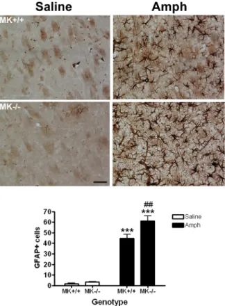
(14) TH expression in the striatum of MK-/- and MK+/+ mice treated with either amphetamine (10 mg/Kg, 4 times, every 2h) or saline (control). ANOVA revealed a striking effect of the drug (F(1,18) = 25.92; P < 0.001) (Fig. 2). However, differences between genotypes were lacking (F(1,18) = 0.6184). Thus, amphetamine caused a significant and similar depletion of TH contents in the striatum of both MK+/+ and MK-/- mice compared with saline-treated mice (Fig. 2). These data were confirmed by similar depletions of DAT levels in amphetamine-treated MK+/+ and MK-/- mice as assessed in Western blots (Fig. 3). The data confirm that amphetamine causes degeneration of dopaminergic terminals in the striatum. Furthermore, these data suggest that endogenous MK is not a key factor for TH expression in the mouse striatum in normal condition and does not affect the severity of amphetamine-induced neurotoxicity in the dopaminergic terminals of the mouse striatum.. 3.3. Enhanced amphetamine-induced astrocytosis in the striatum of Midkine knockout mice Given that the development of reactive astrocytosis in response to treatment with different drugs of abuse is considered an indirect marker of neuronal damage (Ridet et al. 1997; Alonso et al. 2007), we performed immunohistochemical analysis of GFAPpositive cells in the striata of MK-/- and MK+/+ mice. Very few GFAP-positive astrocytes were observed in the striata of saline-treated mice (Fig. 4), being these cells characterized by small cell bodies as well as very fine and short processes. In contrast, after amphetamine administrations, the astrocytes developed large densely stained cell bodies as well as long and extensive processes (Fig. 4). In addition, the numbers of GFAP+ cells in amphetamine-treated mice increased many fold in both genotypes when compared to the saline-treated groups (Fig. 4). ANOVA revealed a significant effect of. 14.
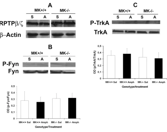
(15) the drug on the number of GFAP+ cells (F(1,20) = 244.6; P < 0.001), a significant effect of the genotype (F(1,20) = 7.708; P = 0.0117) and a significant interaction between factors (F(1,20) = 5.067; P = 0.0358). Very interestingly, post-hoc comparisons of the number of amphetamine-induced GFAP+ cells in both genotypes uncovered a significant increase in the number of astrocytes in the striata of MK-/- mice compared to the MK+/+ mice (Fig. 4), suggesting that MK is a novel endogenous modulator of amphetamine-induced astrocytosis in this brain area.. 3.4. Phosphorylation levels of Fyn and TrkA, two substrates of the MK receptor RPTPβ/ζ, in the striatum of Midkine knockout mice Since Fyn and TrkA are two identified substrates of RPTPβ/ζ with known roles in survival of dopaminergic neurons (Gramage and Herradon, 2011), we aimed to determine the phosphorylation levels and total protein levels of these substrates in the striata of saline- and amphetamine-treated MK+/+ and MK-/- mice. First, we found that RPTPβ/ζ total levels of expression were unchanged in saline- and amphetamine-treated mice and, furthermore, were not affected by the genotype (Fig. 5A). Striatal levels of Fyn phosphorylated in Y530, residue that has been related to Fyn activation (Zhu et al., 2007), and total Fyn protein levels were found unaffected by treatment or genotype (Fig. 5B). In a similar manner, striatal levels of TrkA phosphorylated in Y490, a relevant residue in TrkA-mediated survival of dopaminergic cells (Ashcroft et al., 1999), and total TrkA protein levels were found to be unaffected by treatment or genotype (Fig. 5C). The data suggest that MK modulation of amphetamine-induced astrocytosis in striatum is independent of Fyn- and TrkA-mediated mechanisms.. 3.5. Phosphorylation levels of ERK1/2 in the striatum of Midkine knockout mice. 15.
(16) Recently, we found that the significantly enhanced amphetamine-induced astrocytosis and loss of striatal TH contents in amphetamine-treated PTN-/- mice correlated with striking decreases in the phosphorylation levels of ERK1/2 (Gramage et al., 2010a). Since MK is highly redundant in function with PTN and the data presented here demonstrate a significantly enhanced amphetamine-induced astrocytosis in striatum of MK-/- mice, we aimed to determine the phosphorylation levels of ERK1/2 in this brain area of MK-/- mice. Interestingly, we found that the phosphorylation levels of ERK1/2 and total ERK1/2 protein levels were not affected by treatment or genotype (Fig. 6), suggesting that the significantly enhanced amphetamine-induced astrocytosis in striatum of MK-/- mice does not depend on the phosphorylation status of ERK1/2. Importantly, these data suggest unexpected differences in the mechanisms triggered by PTN and MK to modulate amphetamine-induced neurotoxic effects in the striatum.. 16.
(17) 4. DISCUSSION Recently, we have shown that wild type mice extinguish amphetamine-seeking behaviour before than PTN-/- mice (Gramage et al., 2010b) and show diminished amphetamine-induced neurotoxic effects in the nigrostriatal pathway compared to PTN/- mice (Gramage et al., 2010a), uncovering for the first time a higher vulnerability of mice lacking endogenous PTN to the pharmacological and neurotoxic effects of amphetamine. Pleiotrophin is highly redundant in structure and function with MK, the only other member of the PTN developmentally regulated gene family (Deuel et al., 2002; Kadomatsu and Muramatsu, 2004). Interestingly, both PTN and MK are highly upregulated after administration of different drugs of abuse including psychostimulants such as nicotine and amphetamine (see reviews by Herradon et al., 2009; Gramage and Herradon, 2011). The previous reports mentioned above (Gramage et al., 2010a, b) clearly suggest that upregulation of PTN in the nucleus accumbens after amphetamine administration (Le Greves, 2005) serves to modulate amphetamine rewarding effects and to prevent amphetamine neurotoxic effects. In contrast, studies performed to assess the potential roles of MK on amphetamine-induced rewarding and neurotoxic effects that are presented here demonstrate unexpected differences in the roles of PTN and MK on drug-induced addictive and neurotoxic effects. First, we have found that amphetamine-induced conditioned place preference was effectively similar in both genotypes at the two doses tested (1 and 3 mg/Kg) as assessed on the testing phase of the CPP paradigm (day 5). More interestingly, both genotypes showed a similar extinction rate of amphetamine-seeking behaviour on day 9 of the CPP experimental paradigm identifying significant behavioural differences between MK-/- and PTN-/mice since the latter genotype was previously shown to be unable to extinguish amphetamine (3 mg/Kg)-seeking behaviour in identical experimental conditions. 17.
(18) (Gramage et al., 2010b). It has to be noted that amphetamine administration induces a significant upregulation of PTN in the nucleus accumbens (Le Greves, 2005). However, administration of different drugs of abuse including psychostimulants induce MK expression in other brain areas such as prefrontal cortex and hippocampus (FlatscherBader and Wilce, 2006; Ezquerra et al., 2007; Flatscher-Bader and Wilce, 2008; Herradon et al., 2009). Since drug-induced neuroadaptations in the nucleus accumbens are key to develop addictive behaviours (Russo et al., 2010) and, more importantly, for relapse to drug-seeking (Self and Nestler, 1998), it seems reasonable to hypothesize that the impact of amphetamine-induced upregulation of PTN levels in nucleus accumbens in the maintenance of amphetamine-seeking behaviour will be more significant than drug-induced modulation of MK levels in other brain areas. In a similar manner, amphetamine-induced striatal TH loss was significantly enhanced in PTN-/- mice compared to wild type mice (Gramage et al., 2010a) whereas, in the present report, we show that this amphetamine neurotoxic effect was found to be similar in MK-/- and wild type mice. In contrast, amphetamine-induced astrocytosis was found to be significantly enhanced in the striatum of PTN-/- mice (Gramage et al., 2010b) and, in the present report, in the MK-/- mice striatum. Collectively, these data identify for the first time critical differences in the modulatory roles of the otherwise functionally redundant cytokines PTN and MK on drugs of abuse neurotoxic effects. Whereas PTN seems to be an overall regulator of a wide variety of amphetamine effects (Gramage et al., 2010a; Gramage et al., 2010b), which led us to suggest PTN as a novel therapeutic target to treat amphetamine addictive and neurotoxic effects (Gramage and Herradon, 2011), MK seems to specifically regulate amphetamine-induced striatal astrocytosis. Differences in the temporal regulation of PTN and MK in situations of brain damage could underlie the differential modulation of amphetamine-induced. 18.
(19) neurotoxicity by endogenous PTN and MK. Both PTN and MK are usually upregulated in astrocytes after nerve injury (Takeda et al., 1995; Wang et al., 1998; Mochizuki et al., 1998; Yeh et al., 1998; Kim et al., 2010). However, the temporal regulation of expression of both cytokines differs. Midkine is upregulated in earlier stages than PTN after nerve damage. For instance, MK expression is induced in the early stages after ischemic insult in the adult brain (Wada et al., 2002), and is upregulated in astrocytes one day after brain infarction, peaking 4 days after injury and returning to normal levels 7 days after infarction (Wang et al., 1998), pattern that is observed as well after transient forebrain ischemia (Mochizuki et al., 1998). In contrast, in similar animal models, PTN is upregulated in later stages after injury and in different types of cells further than astrocytes, such as microvasculature and macrophages (Yeh et al., 1998). In addition, its elevated levels of expression remain at least for 14 days (Yeh et al., 1998). Whether or not, these differences in the temporal induction of expression of PTN and MK after different insults may underlie the significant differences in amphetamine-induced neurotoxic effects in PTN-/- and MK-/- mice will require further tests at different time points. All in all, it is important to note that astrocytosis has been considered as a parameter reflecting amphetamine-induced neurotoxicity in neural tissues (Thomas et al., 2004; Guillot et al., 2008; Thomas et al., 2008) suggesting that endogenous MK counteracts this very specific amphetamine neurotoxic effect. In support of a relevant role of MK on amphetamine-induced neurotoxic effects, this cytokine has been shown to block kainic acid-induced neuronal death in hippocampus (Kim et al., 2010) and to exert important effects on survival and differentiation of dopaminergic neurons in different contexts (Kikuchi et al., 1993; Sotogaku et al., 2007; Ohgake et al., 2009), results that became promising when the MK knockout mouse was suggested to be an. 19.
(20) animal model of early features of Parkinson’s disease (Prediger et al., 2011). It is very important to note that defects in dopaminergic functions were previously described in MK-/- mice (Ohgake et al., 2009). In that report, lower levels of dopamine and its receptors were found in striatum of MK-/- mice compared to wild type mice. However, in our present immunohistochemistry studies, it is shown that striatal TH levels are similar in saline-treated MK-/- and MK+/+ mice, suggesting deficiencies in the enzymatic activity of TH in MK-/- mice could underlie the lower levels of dopamine found in MK-/- mice (Ohgake et al., 2009). As mentioned in the introduction section of this manuscript, MK binds to RPTP β/ζ and through its interaction with this receptor abrogates its phosphatase activity and, as a result, increases the levels of phosphorylation of substrates of RPTP β/ζ such as Fyn kinase (Pariser et al., 2005a) and TrkA (Shintani and Noda, 2008). Interestingly, these substrates are known to play survival roles on different types of neurons and other neural cells (Herradon and Ezquerra, 2009). However, in our present studies in striata of MK-/- and MK+/+ mice treated with amphetamine, we did not observe changes in the phosphorylation of relevant residues for cell survival actions of Fyn and TrkA (Ashcroft et al., 1999; Zhu et al., 2007), suggesting the phosphorylation status of these substrates is not critical in the modulation of amphetamine-induced astrocytosis by MK. In addition, we did not observe significant changes in the levels of expression of RPTP β/ζ, suggesting regulation of the levels of this MK receptor is not involved in MK effects on amphetamine-induced neurotoxicity. Since previous data from our lab suggested that deficiencies in the control of phosphorylation of ERK1/2 in PTN-/- mice could underlie the enhanced amphetamineinduced loss of dopaminergic terminals and astrocytosis in the striatum compared to wild type mice, we also tested this possibility in the striatum of amphetamine-treated. 20.
(21) MK-/- and MK+/+ mice. In contrast to what it was observed in PTN-/- mice in identical experimental conditions (Gramage et al., 2010a), we did not find genotype differences in the phosphorylation levels of ERK1/2 in the striatum, suggesting enhanced amphetamine-induced astrocytosis in the striatum of MK-/- mice is independent of the levels of phosphorylation of ERK1/2. In summary, in this work we demonstrate for the first time a specific regulation of amphetamine-induced striatal astrocytosis by MK which seems to be independent of Fyn-, TrkA- and ERK1/2-related mechanisms. In addition, we have identified previously unexpected phenotypical differences between MK-/- and PTN-/- mice challenged with different regimens of amphetamine administrations. Further studies are needed to better understand the potential relevance of the modulation of amphetamineinduced astrocytosis by MK on the overall amphetamine effects.. Acknowledgements: This work has been supported by grants SAF2007-61528 and SAF2009-08136 from Ministerio de Ciencia e Innovación of Spain to GH. Esther Gramage is supported by fellowship AP2008-00726 from the Spanish Ministerio de Ciencia e Innovación. Priya Ramanah is a student from the University of Surrey (UK) supported by an Erasmus fellowship.. 21.
(22) REFERENCES Alonso E, Garrido E, Diez-Fernandez C, Perez-Garcia C, Herradon G, Ezquerra L, Deuel TF, Alguacil LF (2007), Yohimbine prevents morphine-induced changes of glial fibrillary acidic protein in brainstem and alpha2-adrenoceptor gene expression in hippocampus. Neurosci Lett 412:163-167. Ashcroft M, Stephens RM, Hallberg B, Downward J, Kaplan DR (1999), The selective and inducible activation of endogenous PI 3-kinase in PC12 cells results in efficient NGF-mediated survival but defective neurite outgrowth. Oncogene 18:4586-4597. Bowyer JF, Frame LT, Clausing P, Nagamoto-Combs K, Osterhout CA, Sterling CR, Tank AW (1998), Long-term effects of amphetamine neurotoxicity on tyrosine hydroxylase mRNA and protein in aged rats. J Pharmacol Exp Ther 286:10741085. Deuel TF, Zhang N, Yeh HJ, Silos-Santiago I, Wang ZY (2002), Pleiotrophin: a cytokine with diverse functions and a novel signaling pathway. Arch Biochem Biophys 397:162-171. Ezquerra L, Herradon G, Nguyen T, Silos-Santiago I, Deuel TF (2005), Midkine, a newly discovered regulator of the renin-angiotensin pathway in mouse aorta: significance of the pleiotrophin/midkine developmental gene family in angiotensin II signaling. Biochem Biophys Res Commun 333:636-643. Ezquerra L, Herradon G, Nguyen T, Silos-Santiago I, Deuel TF (2006), Midkine is a potent regulator of the catecholamine biosynthesis pathway in mouse aorta. Life Sci 79:1049-1055. Ezquerra L, Perez-Garcia C, Garrido E, Diez-Fernandez C, Deuel TF, Alguacil LF, Herradon G (2007), Morphine and yohimbine regulate midkine gene expression in the rat hippocampus. Eur J Pharmacol 557:147-50 Flatscher-Bader T, Wilce PA (2006), Chronic smoking and alcoholism change expression of selective genes in the human prefrontal cortex. Alcohol Clin Exp Res 30:908-15. Flatscher-Bader T, Wilce PA (2008), Impact of Alcohol Abuse on Protein Expression of Midkine and Excitatory Amino Acid Transporter 1 in the Human Prefrontal Cortex. Alcohol Clin Exp Res 32:1849-1858. Fukada M, Fujikawa A, Chow JP, Ikematsu S, Sakuma S, Noda M (2006), Protein tyrosine phosphatase receptor type Z is inactivated by ligand-induced oligomerization. FEBS Lett 580:4051-4056. Gramage E, Alguacil LF, Herradon G (2008), Pleiotrophin prevents cocaine-induced toxicity in vitro. Eur J Pharmacol 595:35-38. Gramage E, Herradon G (2011), Connecting Parkinson's disease and drug addiction: common players reveal unexpected disease connections and novel therapeutic approaches. Curr Pharm Des 17:449-461. Gramage E, Putelli A, Polanco MJ, Gonzalez-Martin C, Ezquerra L, Alguacil LF, Perez-Pinera P, Deuel TF, Herradon G (2010b), The neurotrophic factor pleiotrophin modulates amphetamine-seeking behaviour and amphetamineinduced neurotoxic effects: evidence from pleiotrophin knockout mice. Addict Biol 15:403-412. Gramage E, Rossi L, Granado N, Moratalla R, Herradon G (2010a), Genetic inactivation of Pleiotrophin triggers amphetamine-induced cell loss in the substantia nigra and enhances amphetamine neurotoxicity in the striatum. Neuroscience 170:308-316.. 22.
(23) Granado N, Ares-Santos S, O'Shea E, Vicario-Abejon C, Colado MI, Moratalla R (2010), Selective vulnerability in striosomes and in the nigrostriatal dopaminergic pathway after methamphetamine administration : early loss of TH in striosomes after methamphetamine. Neurotox Res 18:48-58. Granado N, Escobedo I, O'Shea E, Colado I, Moratalla R (2008a), Early loss of dopaminergic terminals in striosomes after MDMA administration to mice. Synapse 62:80-84. Granado N, O'Shea E, Bove J, Vila M, Colado MI, Moratalla R (2008b), Persistent MDMA-induced dopaminergic neurotoxicity in the striatum and substantia nigra of mice. J Neurochem 107:1102-1112. Guillot TS, Shepherd KR, Richardson JR, Wang MZ, Li Y, Emson PC, Miller GW (2008), Reduced vesicular storage of dopamine exacerbates methamphetamineinduced neurodegeneration and astrogliosis. J Neurochem 106:2205-2217. Herradon G, Ezquerra L (2009), Blocking receptor protein tyrosine phosphatase beta/zeta: a potential therapeutic strategy for Parkinson's disease. Curr Med Chem 16:3322-3329. Herradon G, Ezquerra L, Gramage E, Alguacil LF (2009), Targeting the pleiotrophin/receptor protein tyrosine phosphatase beta/zeta signaling pathway to limit neurotoxicity induced by drug abuse. Mini Rev Med Chem 9:440-447. Herradon G, Ezquerra L, Nguyen T, Silos-Santiago I, Deuel TF (2005), Midkine regulates pleiotrophin organ-specific gene expression: evidence for transcriptional regulation and functional redundancy within the pleiotrophin/midkine developmental gene family. Biochem Biophys Res Commun 333:714-721. Hida H, Jung CG, Wu CZ, Kim HJ, Kodama Y, Masuda T, Nishino H (2003), Pleiotrophin exhibits a trophic effect on survival of dopaminergic neurons in vitro. Eur J Neurosci 17:2127-2134. Hida H, Masuda T, Sato T, Kim TS, Misumi S, Nishino H (2007), Pleiotrophin promotes functional recovery after neural transplantation in rats. Neuroreport 18:179-183. Jung CG, Hida H, Nakahira K, Ikenaka K, Kim HJ, Nishino H (2004), Pleiotrophin mRNA is highly expressed in neural stem (progenitor) cells of mouse ventral mesencephalon and the product promotes production of dopaminergic neurons from embryonic stem cell-derived nestin-positive cells. Faseb J 18:1237-1239. Kadomatsu K, Muramatsu T (2004), Midkine and pleiotrophin in neural development and cancer. Cancer Lett 204:127-143. Kadomatsu K, Tomomura M, Muramatsu T (1988), cDNA cloning and sequencing of a new gene intensely expressed in early differentiation stages of embryonal carcinoma cells and in mid-gestation period of mouse embryogenesis. Biochem Biophys Res Commun 151:1312-1318. Kikuchi S, Muramatsu H, Muramatsu T, Kim SU (1993), Midkine, a novel neurotrophic factor, promotes survival of mesencephalic neurons in culture. Neurosci Lett 160:9-12. Kim YB, Ryu JK, Lee HJ, Lim IJ, Park D, Lee MC, Kim SU (2010), Midkine, heparinbinding growth factor, blocks kainic acid-induced seizure and neuronal cell death in mouse hippocampus. BMC Neurosci 11:42. Krasnova IN, Ladenheim B, Cadet JL (2005), Amphetamine induces apoptosis of medium spiny striatal projection neurons via the mitochondria-dependent pathway. Faseb J 19:851-853.. 23.
(24) Krasnova IN, Ladenheim B, Jayanthi S, Oyler J, Moran TH, Huestis MA, Cadet JL (2001), Amphetamine-induced toxicity in dopamine terminals in CD-1 and C57BL/6J mice: complex roles for oxygen-based species and temperature regulation. Neuroscience 107:265-274. Le Greves P (2005), Pleiotrophin gene transcription in the rat nucleus accumbens is stimulated by an acute dose of amphetamine. Brain Res Bull 65:529-532. Lovatt M, Filby A, Parravicini V, Werlen G, Palmer E, Zamoyska R (2006), Lck regulates the threshold of activation in primary T cells, while both Lck and Fyn contribute to the magnitude of the extracellular signal-related kinase response. Mol Cell Biol 26:8655-8665. Martin YB, Herradon G, Ezquerra L (2011), Uncovering new pharmacological targets to treat neuropathic pain by understanding how the organism reacts to nerve injury. Curr Pharm Des 17:434-448. Meng K, Rodriguez-Pena A, Dimitrov T, Chen W, Yamin M, Noda M, Deuel TF (2000), Pleiotrophin signals increased tyrosine phosphorylation of beta betacatenin through inactivation of the intrinsic catalytic activity of the receptor-type protein tyrosine phosphatase beta/zeta. Proc Natl Acad Sci U S A 97:2603-2608. Milner PG, Li YS, Hoffman RM, Kodner CM, Siegel NR, Deuel TF (1989), A novel 17 kD heparin-binding growth factor (HBGF-8) in bovine uterus: purification and N-terminal amino acid sequence. Biochem Biophys Res Commun 165:10961103. Morales L, Perez-Garcia C, Herradon G, Alguacil LF (2007), Place conditioning in a two- or three-conditioning compartment apparatus: a comparative study with morphine and U-50,488. Addict Biol 12:482-484. Mochizuki R, Takeda A, Sato N, Kimpara T, Onodera H, Itoyama Y, Muramatsu T (1998), Induction of midkine expression in reactive astrocytes following rat transient forebrain ischemia. Exp Neurol 149:73-78. Muramatsu T (2011), Midkine: a promising molecule for drug development to treat diseases of the central nervous system. Curr Pharm Des 17:410-423. Nakamura E, Kadomatsu K, Yuasa S, Muramatsu H, Mamiya T, Nabeshima T, Fan QW, Ishiguro K, Igakura T, Matsubara S, Kaname T, Horiba M, Saito H, Muramatsu T (1998), Disruption of the midkine gene (Mdk) resulted in altered expression of a calcium binding protein in the hippocampus of infant mice and their abnormal behaviour. Genes Cells 3:811-822. Nestler EJ (2004), Molecular mechanisms of drug addiction. Neuropharmacology 47 Suppl 1:24-32. Ohgake S, Shimizu E, Hashimoto K, Okamura N, Koike K, Koizumi H, Fujisaki M, Kanahara N, Matsuda S, Sutoh C, Matsuzawa D, Muramatsu H, Muramatsu T, Iyo M (2009), Dopaminergic hypofunctions and prepulse inhibition deficits in mice lacking midkine. Prog Neuropsychopharmacol Biol Psychiatry 33:541-546. Ooboshi H (2011), Gene therapy as a novel pharmaceutical intervention for stroke. Curr Pharm Des 17:424-433. Pariser H, Ezquerra L, Herradon G, Perez-Pinera P, Deuel TF (2005a), Fyn is a downstream target of the pleiotrophin/receptor protein tyrosine phosphatase beta/zeta-signaling pathway: regulation of tyrosine phosphorylation of Fyn by pleiotrophin. Biochem Biophys Res Commun 332:664-669. Pariser H, Herradon G, Ezquerra L, Perez-Pinera P, Deuel TF (2005b), Pleiotrophin regulates serine phosphorylation and the cellular distribution of beta-adducin through activation of protein kinase C. Proc Natl Acad Sci U S A 102:1240712412.. 24.
(25) Pariser H, Perez-Pinera P, Ezquerra L, Herradon G, Deuel TF (2005c), Pleiotrophin stimulates tyrosine phosphorylation of beta-adducin through inactivation of the transmembrane receptor protein tyrosine phosphatase beta/zeta. Biochem Biophys Res Commun 335:232-239. Prediger RD, Rojas-Mayorquin AE, Aguiar AS, Jr., Chevarin C, Mongeau R, Hamon M, Lanfumey L, Del Bel E, Muramatsu H, Courty J, Raisman-Vozari R (2011), Mice with genetic deletion of the heparin-binding growth factor midkine exhibit early preclinical features of Parkinson's disease. J Neural Transm, In Press. Pu C, Vorhees CV (1995), Protective effects of MK-801 on methamphetamine-induced depletion of dopaminergic and serotonergic terminals and striatal astrocytic response: an immunohistochemical study. Synapse 19:97-104. Ridet JL, Malhotra SK, Privat A, Gage FH (1997), Reactive astrocytes: cellular and molecular cues to biological function. Trends Neurosci 20:570-577. Russo SJ, Dietz DM, Dumitriu D, Morrison JH, Malenka RC, Nestler EJ (2010), The addicted synapse: mechanisms of synaptic and structural plasticity in nucleus accumbens. Trends Neurosci. 33:267-76. Self DW, Nestler EJ (1998), Relapse to drug-seeking: neural and molecular mechanisms. Drug Alcohol Depend. 51:49-60. Shintani T, Noda M (2008), Protein tyrosine phosphatase receptor type Z dephosphorylates TrkA receptors and attenuates NGF-dependent neurite outgrowth of PC12 cells. J Biochem 144:259-266. Sotogaku N, Tully SE, Gama CI, Higashi H, Tanaka M, Hsieh-Wilson LC, Nishi A (2007), Activation of phospholipase C pathways by a synthetic chondroitin sulfate-E tetrasaccharide promotes neurite outgrowth of dopaminergic neurons. J Neurochem 103:749-760. Takeda A, Onodera H, Sugimoto A, Itoyama Y, Kogure K, Rauvala H, Shibahara S (1995), Induction of heparin-binding growth-associated molecule expression in reactive astrocytes following hippocampal neuronal injury. Neurosci 68:57-64. Thomas DM, Francescutti-Verbeem DM, Kuhn DM (2008), The newly synthesized pool of dopamine determines the severity of methamphetamine-induced neurotoxicity. J Neurochem 105:605-616. Thomas DM, Walker PD, Benjamins JA, Geddes TJ, Kuhn DM (2004), Methamphetamine neurotoxicity in dopamine nerve endings of the striatum is associated with microglial activation. J Pharmacol Exp Ther 311:1-7. Tzschentke TM (2007), Measuring reward with the conditioned place preference (CPP) paradigm: update of the last decade. Addict Biol 12:227-462. Wada M, Kamata M, Aizu Y, Morita T, Hu J, Oyanagi K (2002), Alteration of midkine expression in the ischemic brain of humans. J Neurol Sci 200:67-73. Wang S, Yoshida Y, Goto M, Moritoyo T, Tsutsui J, Izumo S, Sato E, Muramatsu T, Osame M (1998), Midkine exists in astrocytes in the early stage of cerebral infarction. Brain Res Dev Brain Res. 106:205-9 Yamamoto BK, Bankson MG (2005), Amphetamine neurotoxicity: cause and consequence of oxidative stress. Crit Rev Neurobiol 17:87-117. Yeh HJ, He YY, Xu J, Hsu CY, Deuel TF (1998), Upregulation of pleiotrophin gene expression in developing microvasculature, macrophages, and astrocytes after acute ischemic brain injury. J Neurosci 18:3699-707. Zhu S, Bjorge JD, Fujita DJ (2007), PTP1B contributes to the oncogenic properties of colon cancer cells through Src activation. Cancer Res 67:10129-10137.. 25.
(26) FIGURES. Fig. 1. Amphetamine-induced place preference. (A) Results are presented as the mean ± SEM of the percentage of the time spent by MK+/+ and MK-/- mice in the drug-paired (less-preferred) compartment during preconditioning (day 1) and testing phase (day 5), and 5 days after the last amphetamine (1 mg/Kg) injection (day 9). (B) Results are presented as the mean ± SEM of the percentage of the time spent by MK+/+ and MK-/- mice in the drug-paired (less-preferred) compartment during preconditioning (day 1) and testing phase (day 5), and 5 days after the last amphetamine (3 mg/Kg) injection (day 9). * P < 0.05; ** P < 0.01 vs. preconditioning (day 1).. 26.
(27) Fig. 2. TH expression in striatum of MK-/- and MK+/+ mice after amphetamine administration. Photomicrographs illustrate amphetamine-induced dopaminergic damage. TH-immunostained striatal sections of mice, 4 days after saline or amphetamine (Amph) treatment. Graph represents the optical densitometry (OD) measures of TH-ir in the striatum, data show mean ± SEM (n = 5 animals per group). ** P < 0.01 vs. Sal. * P < 0.05 vs. Sal. Scale bar = 200 µm.. 27.
(28) Fig. 3. DAT expression after amphetamine administration in striatum of MK-/and MK+/+ mice. Western blot assays using a DAT antiserum of striatal proteins extracts prepared 4 days after amphetamine (A) or saline (S) treatment. Anti-DAT antiserum revealed a specific band at ~ 70-75 kDa. Loading control shown in the bottom panel was reacted with β-actin antiserum. 3-4 different samples of every experimental group were tested and densitometry measures did not show statistical differences between groups.. Fig. 4. Amphetamine induces astrocytosis in the striatum. Photomicrographs are from GFAP-immunostained striatal sections of saline- or amphetamine (Amph)-treated MK-/- and MK+/+ mice. The graph represent quantification of data (mean ± S.E.M) obtained from the counts of GFAP-positive cells in the striatum (n = 5 animals per group). Scale bar = 50 µm. *** P < 0.001 vs. Sal. ## P < 0.01 vs. MK+/+.. 28.
(29) Fig. 5. Levels of phosphorylation of Fyn and TrkA in the striatum of MK-/- mice after amphetamine administration. Total RPTPβ/ζ protein levels (A), the levels of phosphorylation of Fyn (B) and the levels of phosphorylation of TrkA (C) in saline (S)and amphetamine (A)-treated MK-/- and MK+/+ mice (n = 4-6/group) were determined in Western blots. Optical density (OD) measurements did not reveal differences in the phosphorylation levels of Fyn (B) or TrkA (C) in samples from MK-/- mice either treated with saline or amphetamine.. 29.
(30) Fig. 6. Levels of phosphorylation of ERK1/2 in the striatum of MK-/- mice after amphetamine administration. The levels of phosphorylation of ERK1/2 in saline (S)and amphetamine (A)-treated MK-/- and MK+/+ mice (n = 4-6/group) were determined in Western blots probed with anti-phospho-ERK1/2 antibodies. Total ERK1/2 amounts were determined using anti-ERK1/2 antibodies. Optical density (OD) measurements revealed unchanged p-ERK1/2/ERK1/2 contents in samples from MK-/- mice either treated with saline or amphetamine.. 30.
(31)
Figure

+2

Documento similar
administration of S1RA and BD-1063 also induced robust antihy- peralgesic effects to a heat stimulus in mice sensitized with PGE2 and NGF, and these were reversed by both the
In summary, the data presented here show for the first time that pharmacological inhibition of RPTPβ/ζ with MY10 significantly reduces chronic voluntary ethanol consumption only
We have described two cases of aggressive dorsal hemangioma with severe acute paraparesis that required urgent surgical intervention. Aggressive VH represent less than
For that purpose, we have analyzed the size of their cell bodies, and their distribution and compartmental organization with respect to the matrix/striosomes in the three
Since such powers frequently exist outside the institutional framework, and/or exercise their influence through channels exempt (or simply out of reach) from any political
The biological behaviour of Schwann cells (SCs) and dorsal root ganglia (DRG) on fibrillar, highly aligned and electroconductive substrates obtained by two
Scarcity: The resources available Albani, are not very difficult to achieve, since with regard to human resources, with a good selection and training of its employees
Caveolin-1 negatively regulates TGFβ signalling (Babak Razani, Zhang, et al 2001) and loss of Cav1 leads to increased ECM deposition after injury (Miyasato et al 2011, Shivshankar et
