Mecanismos de transducción del lipopolisacárido
19
0
0
Texto completo
(2) 122. respuesta inmunoinflamatoria que en todos los casos está regulada por mediadores comunes. Éstos factores etiológicos dan lugar a un síndrome de respuesta inflamatoria sistémica (SRIS) inicial que se caracteriza en líneas generales por hipotensión, fiebre, coagulación intravascular diseminada y el denominado síndrome de disfunción orgánica múltiple 1. Tras una serie de complicaciones, a partir del SRIS desencadenaría el shock séptico (SS). Clínicamente, el SS tiene una serie de manifestaciones de tipo metabólico como fiebre (a veces hipotermia), acidosis metabólica, y gran producción de proteínas que intervienen en la fase aguda de los procesos inflamatorios. También concurre con alteraciones hormonales y del ciclo de la glucosa. Las alteraciones hematológicas más frecuentes son granulocitopenia, con estados de leucocitosis, trombocitopenia, y la más característica es la coagulación intravascular diseminada. La función pulmonar se ve alterada y se produce edema pulmonar, broncoconstricción, taquipnea, cianosis e hipoxia; la oliguria es síntoma de necrosis renal, y puede haber casos de afecciones gastrointestinales en forma de diarreas. El sistema nervioso se ve alterado dando lugar a letargia y confusión. También existen algunas manifestaciones dermatológicas como petequias y vasculitis 1-4. Las alteraciones cardiovasculares se deben a un complicado proceso debido a la interacción de todos estos mediadores con los elementos de la pared vascular y los distintos componentes de la sangre. La característica hiporrespuesta a agentes vasoactivos ha sido muy estudiada tanto in vivo como in vitro. En dicho proceso se distinguen dos estadíos: 1-. Inicialmente hay una fase hiperdinámica: en la sepsis severa la resistencia vascular decrece (mediada al principio por la liberación de bradikinina e histamina) y el gasto cardíaco aumenta, aunque a pesar de ello la perfusión tisular disminuye y por lo tanto el consumo de oxígeno. La formación de fibrina intravascular difusa y la agregación de plaquetas y neutrófilos altera el flujo sanguíneo formando microtrombos; la migración de éstos a través del endotelio vascular hace que se liberen numerosos mediadores que actúan promoviendo la filtración y favoreciendo el edema en los tejidos. Además, la activación de numerosas células libera agentes vaArs Pharmaceutica, 44:2; 121-139, 2003. BERMEJO A Y DUARTE J.. that in all cases is regulated by common mediators. These ethiological factors give rise to an initial systematic inflammatory response syndrome (SIRS) that is generally characterised by hypertension, fever, disseminated intravascular coagulation and the so called multiple organ dysfunction syndrome 1. After a series of complications starting from SIRS, a septic shock would occur. Clinically speaking, SS has a series of metabolic type manifestations such as fever (sometimes hypothermia), metabolic acidosis, and a high protein production involved at the acute stage of inflammatory processes. It is also concurrent with hormonal and glucose cycle alterations. The most frequent haematological alterations are granulocytopenia with leukocytosis, trombocytopenia and, as the most characteristic type, disseminated intravascular coagulation. The pulmonary function is altered and pulmonary edema, bronchoconstriction, tachypnea, cyanosis and hypoxia, the oliguria is symptomatic of renal necrosis, and cases of gastro-intestinal affections in the form of diarrhoea may arise. The nervous system is altered giving rise to lethargy and confusion. Dermatological manifestations such as petechias and vasculitis have also been known to exist1-4 . Cardiovascular disorders are attributed to a complicated process caused by the interaction of all of these mediators with elements of the vascular wall and the different components of the blood stream. The characteristic hyper-response to vasoactive agents has been widely studied as much in vivo as in vitro. Throughout such a process two states have been differentiated: 1-. Initially a hyperdynamic exists: In severe sepsis, vascular resistance decreases (influenced to begin with by the liberation of bradidinin and histamine) and cardiac output increases, although in spite of this, tissue perfusion decreases and consequently oxygen consumption. The formation of diffuse intravascular fibrin and the addition of thrombocytes and blood platelets alters the flow of blood, forming microthromocytes. The migration of such, through the vascular endothelium, frees the numerous mediators that act, promoting filtration and favouring edema in tissues. Furthermore, the activation of numerous cells liberates vasoactive agents in such a way, that in some areas of the vascular bed, dilation occurs, whilst in others constriction. The endothelial damage may also contribute to tissue is-.
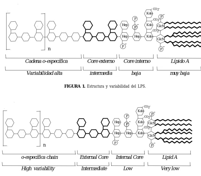
(3) 123. MECHANISMS OF TRANSDUCTION OF LIPOPOLYSACCHARIDE. soactivos de modo que en algunas zonas del lecho vascular hay dilatación, y en otras constricción. El daño endotelial también puede contribuir a la isquemia tisular. Por otra parte, existe taquicardia y en esta fase la presión arterial puede normal o ligeramente reducida 5. 2-. La segunda fase, llamada también hipotensa o hipodinámica, se caracteriza por un gran descenso de la resistencia vascular sistémica y depresión miocárdica, disminuye el volumen de eyección y se dilata el ventrículo izquierdo. La dilatación arteriolar y el incremento de la permeabilidad capilar favorece la hipovolemia que disminuye el retorno venoso, y por tanto, la presión y el gasto cardíaco 3-5.. ESTRUCTURA Desde que se confirmó el papel del LPS como causa principal del shock inducido por bacterias Gram negativas 3,4,6, muchos han sido los estudios que se han realizado para intentar establecer los mecanismos de transducción generados en la respuesta del organismo. Esta endotoxina consiste en un fosfoglicolípido anclado a la membrana bacteriana (lípido A) unido covalentemente a un heteropolisacárido hidrofílico 7, que es la que confiere actividad biológica a la molécula 6,8. El heteropolisacárido comprende dos regiones: la cadena O-específica también llamada antígeno O, formada por unidades repetitivas de oligosacárido; y el core. Éste a su vez se subdivide en core externo (formado por hexosas), mediante el cual se une al antígeno O; y el core interno (formado por heptosas). El lípido A se une a esta porción mediante un residuo llamado KDO (ácido 2-keto-3-deoxioctanoico). La variabilidad del LPS juega un papel importante en cuanto a la capacidad inmunológica y depende de la región que estemos considerando, como se indica en la figura 1 7,8 .. chemia. On the other hand, tachycardia may exist and at this stage the arterial pressure may be normal or slightly reduced 5. 2-. The second stage, also known as hypotense or hypodynamic, is characterised by a marked decrease in systemic vascular resistance and myocardial depression, decreased volume of ejection, and left ventricular dilation. Arteriolar dilation and an increase in capillary permeability favours hypovolemia which decreases venous return, and therefore, pressure and cardiac output 3-5.. STRUCTURE Since the role of LPS was confirmed as being the main cause of shock induced by Gram negative bacteria 3,4,6 , many studies have been carried out in an attempt to establish what the transduction mechanisms generated in the organisms response are. This endotoxin consists of a phosphoglycolipid fixed to the bacterial membrane (lipid A) covalently bonded to a hydrophilic heteropolysaccharide 7, conferring the molecule with its biological activity 6,8 . The heteropolysaccharide is comprised of two regions: The O-specific chain also known as O-antigen, made up of repetitive units of oligosaccaride, and the core. This in turn is subdivided into the external core (made up of hexoses), through which the O-antigen and the internal core (made up of heptoses) are linked. The A lipid is joined to this parcel by means of a residue known as KDO (2-keto-3-deoxyoctanoic acid). The variability of LPS plays an important role with regard to immunological capacity and is dependant upon the region that is being taken into consideration, as indicated in Figure 1 7,8 .. Ars Pharmaceutica, 44:2; 121-139, 2003.
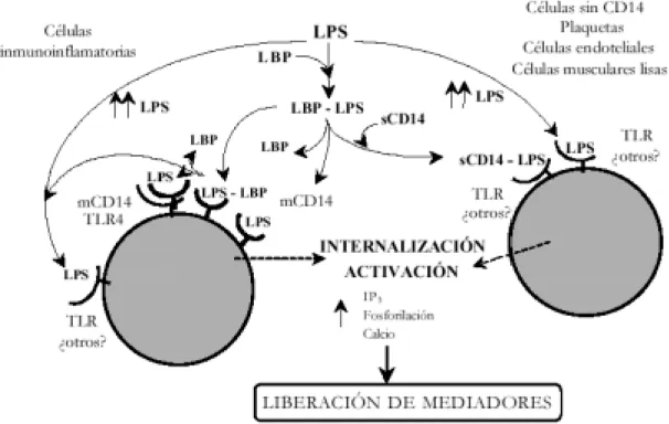
(4) 124. BERMEJO A Y DUARTE J. CO 2 -. Kdo P Hep. P. Hep P. n. Cadena o-específica Variabilidad alta. CO 2 -. -. Kdo. P. -. GlcN CO 2 -. Hep. Kdo. GlcN. P. -. Core externo. Core interno. Lípido A. intermedia. baja. muy baja. FIGURA 1. Estructura y variabilidad del LPS.. CO 2 -. Kdo P Hep Hep P. n. o-específica chain. External Core. High variability. Intermediate. P. CO 2 -. -. Hep. Kdo. P. -. GlcN CO 2 -. Kdo. GlcN. P. Internal Core Low. -. Lipid A Very low. FIGURE 1. Structure and variability of LPS.. MECANISMOS DE TRANSDUCCIÓN. TRANSDUCTION MECHANISMS. Interacciones del lipopolisacárido en el torrente sanguíneo. Lipopolysaccharide interactions in the blood stream. Según se esquematiza en la figura 2, cuando el LPS llega a la sangre formando parte de las bacterias o en forma libre (por ejemplo, procedente de la lisis de microorganismos por los antibióticos) 9, interacciona con algunas de las moléculas que se encuentran en el torrente sanguíneo. Entre estas moléculas están las proteínas bactericidas que incrementan la permeabilidad (BPI), las proteínas catiónicas denominadas CAP 18, CAP 37, y P15A/P15B, las lipoproteínas de alta densidad (HDL), otras lipoproteínas, y proteínas como la albúmina, la hemoglobina (Hb), etc. 3,8. Para dar lugar a la reacción del hospedador tiene que adoptar la llamada conformación. As outlined in Figure 2, when LPS enters into the blood forming part of the bacteria or in a free form (for example, from the lysis of microorganisms through antibiotics) 9, it interacts with some of the molecules that it finds in the blood stream: Among these molecules, there are bactericidal proteins that increase permeability (BPI), cationic proteins known as CAP 18, CAP 37, & P15A/P15B, high density lipoproteins (HDL), other lipoproteins, and other proteins such as albumin, haemoglobin (Hb), etc. 3,8 . In order for the hosts reaction to take place the so called endotoxic conformation must be adopted, which seems to be a monomeric form 10. One of the molecules. Ars Pharmaceutica, 44:2; 121-139, 2003.
(5) MECANISMOS DE TRANSDUCCIÓN DEL LIPOPOLISACÁRIDO. endotóxica, que parece ser que es una forma monomérica 10. Una de las moléculas a las que se une, la proteína de unión al LPS (LBP), con algo menos de afinidad que las lipoproteínas sanguíneas y BPI, forma un complejo con el LPS mediante su unión con el lípido A 11, que al parecer es el inicio de la cascada de reacciones inmunes. La LBP podría actuar catalizando el paso de los agregados de LPS a monómeros y transfiriéndolos a las lipoproteínas 12, a las integrinas β-integrina leucocitaria CD11c/CD18, y preferentemente, al antígeno de diferenciación monocítico CD14 6,8,1315 . Aun así, se ha descubierto que el LPS puede unirse directamente y/o mediante el complejo LPS-CD14, a las selectinas P y L solubles o ancladas en la membrana de plaquetas y células endoteliales 16. También se ha descrito dicha unión a varias proteínas de diversos pesos moleculares, aunque en la mayoría de los casos, su identificación y el papel que poseen en el proceso del SS están aún pendientes de dilucidar 15-17. Entre ellas está la familia de receptores con estructura similar a la de los receptores de respuesta inmune de Drosophila, llamados receptores tipo Toll (Toll like receptors -TLR-). Este grupo de receptores pertenece a la gran superfamilia de los llamados TIR (receptores Toll/IL-1 -interleukina 1-), ya que todos ellos tienen un dominio de gran semejanza estructural y poseen mecanismos de transducción semejantes con mediadores secundarios comunes 18. Por último, el LPS tiene la capacidad de interaccionar con diversos sistemas en el torrente sanguíneo 3,4,19; así, activa los complejos del factor XII de la coagulación-prekalikreína-kininógenos de alto peso molecular 20, por tanto, la vía intrínseca de la coagulación, también la vía extrínseca, la fibrinolisis 21 y las vías clásica y alternativa del complemento 22.. 125. to which it binds, the binding protein to LPS (LBP), with somewhat less affinity than the blood lipoproteins and BPI, forms a complex with LPS by means of a bond with lipid A 11 , which seems to represent the commencement of a cascade of immune reactions. LBP may act by catalysing the flow of the LPS aggregates to monomers and transferring them to the lipoproteins 12, to the integrins, leukocyte β-integrin CD11c/CD18, and preferably, to the antigen of monocytic differentiation CD14 6,8,13-15 . Even so, it has been discovered that LPS may bind directly and/or through the LPS-CD14 complex, to P and L soluble selectins or fixed in the membrane of platelets and endothelial cells 16. This bond to various proteins of various molecular weights has also been described, although in the majority of cases, its identification and the role that they have to play in the SS process is yet to be determined 15-17. Among these is the family of receptors with a similar structure to that of the receptors of the immune response of Drosophila, known as Toll like receptors (TLR). This group of receptors belongs to the great superfamily of the so called TIR (Toll/IL—1—interleukin 1-receptors), given that all have a dominion of great structural similarity and posses similar transduction mechanisms with common secondary mediators 18. Finally, LPS has the capacity to interact with different systems in the blood stream 3,4,19; thus activating factor XII coagulation complexes -prekalikrein kininogens of high molecular weight 20 , and consequently, the coagulation intrinsic pathway, also the extrinsic pathway, the fibrinolysis 21 and the classical and alternative pathways of complement 22 .. Ars Pharmaceutica, 44:2; 121-139, 2003.
(6) 126. BERMEJO A Y DUARTE J.. FIGURA 2. Interacciones del LPS en el torrente sanguíneo y reacciones que genera. (LPS)n: Monómeros de LPS. LBP: Proteína de unión al LPS. sCD14: CD14 soluble. HDL y LDL: Lipoproteínas de alta y baja densidad, respectivamente. CAP: Proteínas catiónicas. Hb: Hemoglobina. C1 y C3b: Factores del complemento.. FIGURE 2. LPS interactions in the blood stream and the reactions that are generated. (LPS)n: Monomers of LPS. LBP: Binding Protein to LPS. sCD14: soluble CD14 . HDL & LDL: High and low density Lipoproteins, respectively. CAP: Cationic Proteins . Hb: Hemoglobin. C1 y C3b: Complement Factors.. Receptores y vías intracelulares activados por el lipopolisacárido. Receptors and intracellular pathways activated by the lipopolysaccharide. El CD14 es uno de los principales responsables de la activación celular inducida la endotoxina, si bien se han postulado mecanismos de activación independientes de dicha molécula. CD14 is one of the main elements responsible for induced cellular activation by endotoxin. Activation mechanisms that are independent of this molecule have been considered to be invol-. Ars Pharmaceutica, 44:2; 121-139, 2003.
(7) 127. MECHANISMS OF TRANSDUCTION OF LIPOPOLYSACCHARIDE. implicados en el SS mediante la integrina CD11/ 18, los receptores TLR y otras moléculas 6,8,15,16 . Éste puede encontrarse de forma soluble en el suero (sCD14) (fundamentalmente liberado por los monocitos), o asociado a la membrana (mCD14) de algunos tipos celulares como monocitos, macrófagos y leucocitos polimorfos nucleares (Figura 3). La forma soluble parece ser que facilita la transferencia a las lipoproteínas 12 y aunque antes se creía que iba asociada al complejo LBP-LPS, puede unirse de forma directa al LPS, y/o mediante el proceso catalítico mediado por la LBP 23. El complejo formado actuaría sobre otras moléculas de membrana en células carentes de CD14 en su superficie (células endoteliales, células musculares lisas, algunas otras células epiteliales). Sin embargo, el LPS puede interaccionar directamente formando complejos con el sCD14, con el mCD14, y cuando está en grandes concentraciones, con los receptores TLR 6,8,13,15,17,24. Dentro de éstos últimos, se pensaba que eran el TLR2 y el TLR4 los que intervenían en la respuesta a la endotoxina 25, pero estudios recientes implican solamente al último de ellos 18,26.. ved in SS by means of the integrin CD11/18, the TLR receptors and other molecules 6,8,15,16. This may be found in soluble form in serum (sCD14) (fundamentally liberated by the monocytes), or associated with the membrane (mCD14) of some types of cells such as monocytes, macrophages and polymorph- nuclear leukocytes (Figure 3). The soluble form seems to facilitate transfer to the lipoproteins 12 and although it was formerly believed to be associated with LBP-LPS, it can bind directly with LPS, and/or through the catalytic process mediated by LBP 23. The complex formed, would act upon other membrane molecules in cells with a lack of CD14 on their surfaces (endothelial cells, smooth muscle cells, some other epithelial cells). However, LPS may interact directly forming complexes with sCD14, mCD14, and when in large concentrations with the TLR receptors 6,8,13,15,17,24. Within the last of these, it was believed that it was TLR2 and TLR4 which intervened in the response to the endotoxin 25 , but recent studies imply that only the latter of these were involved 18,26.. FIGURA 3. Mecanismos de activación celular del LPS.. Ars Pharmaceutica, 44:2; 121-139, 2003.
(8) 128. BERMEJO A Y DUARTE J. FIGURE 3. Mecanisms of cellular activation of LPS.. Una vez unido al CD14, y/o a los distintos receptores, se produciría la cascada de reacciones intracelulares que daría lugar a la activación de diversos tipos de células. Los mecanismos de transducción del LPS están por esclarecer y forman un entramado muy complejo de reacciones cruzadas (Figura 4). Por una parte, puede internalizarse y actuar sobre receptores citosólicos aún desconocidos. Por otro lado, aún se discute si se requieren moléculas intermedias en la membrana para el inicio de las señales 6,15,17.. Once bound to CD14, and/or to the different receptors, the cascade of intracellular reactions would occur, giving rise to the activation of several types of cells. The transduction mechanisms of LPS are yet to be clarified and form a very complex structure of cross reactions (Figure 4). On the one hand, they may be of an internal nature acting upon cytosolic receptors that are not yet known. On the other, whether intermediate molecules in the membrane are required, in order to initiate the signals 6,15,17 , is still a matter under discussion.. Figura 4. Receptores y proteínas celulares implicados en los mecanismos de transducción del LPS. S-P y L: Selectinas P y L. SM: Esfingomielasa. CAK: Kinasa activada por ceramida. PK y PPT: Proteinkinasas y proteinfosfatasas. SrcK: Kinasas Src. pG: Proteínas G. ppG: Pequeñas proteínas G. PLA2 y PLC: Fosfolipasas A2 y C. DAG: Diacilglicerol. IP3 : Inositol 3,4,5-trifosfato. PKC: Proteinkinasa C. JAK-2: Janus kinasa-2. Ars Pharmaceutica, 44:2; 121-139, 2003.
(9) MECANISMOS DE TRANSDUCCIÓN DEL LIPOPOLISACÁRIDO. 129. Figure 4. Receptors and cellular proteins involved in the transduction mecanisms of LPS. S-P & L: Selectins P & L. SM: Sphingomielase. CAK: Kinase activated by ceramide. PK y PPT: Proteinkinases y proteinphosphatases. SrcK: Src Kinases. pG: G Proteins. ppG: Small G proteins. PLA2 & PLC: Phospholipases A2 & C. DAG: Diacilglycerol. IP3: Inositol 3,4,5-triphosphate. PKC: Proteinkinase C. JAK-2: Janus kinase-2.. En diversos estudios in vitro con monocitos, macrófagos y fibroblastos se han implicado proteínas G (pG) y pequeñas proteínas G (ppG) que pueden participar en la activación de tirosinkinasas (TK) 6,27 , la fosfolipasa C (PLC) y A2 (PLA2) 28-30 , así como la calmodulina 31,32 . También se ha atribuido el papel de segundo mensajero a la esfingomielasa (SM), que hidrolizaría la esfingomielina en ceramida, la cual activaría diferentes proteinfosfatasas (PPT) y proteinkinasas (PK), como la PK activada por ceramida (CAK) 33, que podrían intervenir en la señal del LPS activando o inhibiendo diversas enzimas como la PK C (PKC), las fosfolipasas PLC y PLA 2, y la ciclooxigenasa inducible (COX-2) 34,35 . Sin embargo, la vía que más importancia parece tener es la formada por las PK, en la cual están implicadas varios grupos: Las serin-treonin PK A (PKA) y C 36,37 y un gran conjunto de TK 38. Dentro de éstas están las Src kinasas (SrcK): ésta familia de kinasas (familia de TK del sarcomavirus) parece ser que participa en las señales de transducción mediadas por el CD14 desde la membrana hacia el citoplasma, aunque el papel que desempeñan no se ha esclarecido aún. También se ha descrito que pueden ser activadas por la vía de la ceramida 39. Además, se ha hipotetizado según diversos estudios in vitro, que pueden actuar sobre ppG de la familia Ras, por lo que podrían actuar en. Differing in vitro studies with monocytes, macrophages and fibroblasts point to the involvement of proteins G (pG) and small proteins G (ppG) that may participate in the activation of tyrosinkinases (TK) 6,27 , the phospholipase C (PLC) and A 2 (PLA2) 28-30, as well as calmodulin 31,32 . Additionally, the role of a second messenger has been attributed to sphingomielase (SM). This would hydrolyse the sphingomielin in ceramide, which in turn would activate different protein phosphatase (PPT) and proteinkinase (PK), as PK activated by ceramide (CAK) 33, which could be involved in the LPS signal, either by activating or inhibiting differing enzymes such as PK C (PKC), the phospholipase PLC and PLA 2, and the inducible cyclooxigenase (COX-2) 34,35 . However, the most significant pathway seems to be that formed by the PK, in which several groups are involved: Serine-threonine PK A (PKA) y C 36,37 and the large group of TK 38. Forming part of these are the Src kinase (SrcK): This family of kinases (family of TK of sarcoma virus) appears to participate in transduction signals mediated by CD14 from the membrane to the cytoplasm, although the role that they play is yet to be determined. The possibility that they may be activated by the ceramide pathway has also been described 39. Furthermore, according to several in vitro studies, it has been hypothesised they Ars Pharmaceutica, 44:2; 121-139, 2003.
(10) 130. las vías de activación de éstas. Sin embargo, otros estudios eliminan esta posibilidad, por lo que aún existe controversia sobre su actuación en los eventos generados por el LPS 40-43. También las TK janus kinasas (JAK) participan en la activación celular inducida por el LPS, dentro de esta familia parece que la proteína implicada, JAK-2, fosforila al factor de transcripción STAT-1α (proteína transductora de señal y activadora de la transcripción), el cual actuaría sobre diversos genes 44,45 . El conjunto de las llamadas PK activadas por mitógenos, MAP kinasas (MAPK) también participan en gran medida en las señales intracelulares del LPS. Esta gran familia consta de PK que fosforilan restos de serina-treonina y tirosina-treonina. Según diferentes estudios in vitro realizados en macrófagos, otros leucocitos, células endoteliales, células musculares lisas y otros tipos celulares 42,46-49 , existen al menos cuatro subgrupos de MAPK, de los cuales se ha descrito que tres están relacionados con las respuestas inducidas por el LPS. El descubrimiento de la activación de la primera familia de MAPK por el LPS, las kinasas reguladas por señales extracelulares (ERK) , se llevó a cabo por Weinstein et al. 50. Más tarde identificaron la existencia de dos proteínas, la ERK1 y la ERK2 51. Al parecer, la secuencia comienza mediante el efector Ras en la membrana, que interactúa con la molécula Raf-1 52, dicha molécula es fosforilada por una kinasa y así activada, puede fosforilar a la kinasa-1 de la MAPK, llamada también MEK (MKK-1 o MEK-1), que a su vez fosforila las ERK 53 . Los sustratos de estas últimas pueden ser los factores de transcripción ELK-1 y c-Myc entre otros, proteínas citoplasmáticas como la fosfolipasa A2 citosólica (PLA2C), y diferentes PK, como se esquematiza en la figura 5. La fosforilación de la PLA2c daría lugar a la producción de ácido araquidónico, con la consecuente activación de las formas no clásicas de PKC y la producción de eicosanoides 42. Las otras vías relacionadas con las MAPK incluyen el conjunto proteínas que forman la subfamilia de PK del factor de transcripción c-Jun, llamadas JNK (Figura 5). Aunque aún permanecen sin conocerse los primeros iniciadores de esta vía, y algunos elementos intermedios, están implicadas ppG de la familia de las Rho GTPasas, Rac y Cdc42, las cuales son más potentes activadoras de las JNK que las Ars Pharmaceutica, 44:2; 121-139, 2003. BERMEJO A Y DUARTE J.. may act upon the ppG of the Ras family, which may act on their activation pathways. However, other studies have eliminated this possibility. Consequently, controversy about their involvement in the events generated by LPS still exists 40-43 . TK janus kinases (JAK) also participate in LPS induced cellular activation. Within this family, the protein involved, JAK-2, seems to phosphorilize the transcription factor STAT-1α (signal transductor protein and activator of the transcription), which would act upon differing genes 44,45. The so called group of PK activated by the mitogens, MAP kinases (MAPK) also participate to a large degree on the intracellular signals of LPS. This large family consists of PK that phophorilises remains of serine-threonine and tyrosine-threonine. According to different in vitro studies carried out on macrophages, other leukocytes, endothelial cells, smooth muscular cells and other types of cells 42,46-49, at least four subgroups of MAPK exist, of which three have been described as being related to reponses induced by LPS. The discovery of the activation of the first family of MAPK by LPS, the kinases regulated by extracellular signals (ERK), was made by Weinstein et al. 50. Subsequently, the existence of two proteins, ERK1 and ERK2 was identified 51 . Seemingly, the sequence commences through the Ras effector in the membrane which interacts with the Raf-1 molecule52. This molecule is phosphorilised by a by a kinase and thus activated, possibly phosphorilising the kinase-1 of MAPK, also known as MEK (MKK-1 or MEK-1), which in turn phosphorilises the ERK 53. The sustrates of the latter of these may, among others, represent the transcription factors ELK-1 and cMyc, cytoplasmatic proteins, such as phospholipase A2 cytosolic (PLA2C), and different PK, as outlined in Figure 5. The phosphorilisation of PLA2c would give rise to the production of arachidonic acid, with the consequent activation of the non-classical forms of PKC and the production of eicosanoids42. The other pathways associated with MAPK include the group of proteins that make up the PK subfamily of transcription factor c-Jun, known as JNK (Figure 5). Although the first initiators of this pathway and some intermediary elements are still unknown, ppG of the Rho GTpases family, Rac and Cdc42 are involved. These are more powerful activators of.
(11) MECHANISMS OF TRANSDUCTION OF LIPOPOLYSACCHARIDE. proteínas Ras, pero pueden actuar de forma sinérgica con ésta 54. Una vez activadas, Rac y/o Cdc42 se unen y activan la kinasa PAK1 54, se activa la MEKK-1 (implicada en la activación de NF-κB) y se suceden fosforilaciones sucesivas de las MKK4/7 y JNK1/2 que están asociadas a la fosforilación de factores de transcripción, entre ellos c-Jun, AP-1 (proteína activadora 1) y CREB (proteína de unión de elementos de respuesta al adenosín monofosfato cíclico -AMPc-) 42,55-58 . Las p38 MAPK forman la otra familia de este tipo de proteínas implicadas en la activación celular por LPS. El inicio de esta vía permanece también por determinar, algunos estudios implican a ppG de la superfamilia Ras comunes a la activación de las JNK, a las proteínas Rho y a las serin-treoninkinasas activadas por la proteína p21 (PaKs) 18,42,59. Se sabe que luego actúa la MEKK5 sobre las MKK3/6 y éstas fosforilan a las p38 55,59 , siendo la isoforma p38α la más estudiada. Los sustratos de la familia de proteínas p38 MAPK son diversos factores de transcripción, enzimas como la PLA 2c y diversas PK, en algunos casos iguales a las activadas por ERK 42 (Figura 5). Aunque estas vías de transducción estén específicamente reguladas, pueden actuar de modo sinérgico además de establecer una red con efectores comunes y producir las mismas respuestas, como ocurre con los factores de transcripción.. 131. the JNK than the Ras proteins, but may act synergically with them 54. Once activated, Rac and/ or Cdc42 bind together and activate the kinase PAK1 54, MEKK-1 is then activated (involved in the activation of NF-κB) and successive phosphorilisations of MKK4/7 and JNK1/2 occur. These are associated with the phosphorilisation of transcription factors, among which -c-Jun, AP-1 (activating protein 1) and CREB (binding protein of response elements to cyclic adenosine monophosphate -AMPC-) 42,55-58 are to be encountered. The p38 MAPK make up the other family of this type of protein that is involved in cellular activation by LPS. The initiation of this pathway has also yet to be determined. Some studies have implied ppG from the Ras superfamily common to the activation of JNK, to the proteins Rho and to the serine-threoninkinase activated by the protein p21 (PaKs) 18,42,59. MEKK5 is known to act subsequently upon MKK3/6 and these phosphorilise p38 55,59. The isoform p38α has been the most studied example. Of the sustrates of the p38 MAPK protein family, diverse transcription factors are enzymes such as PLA2c, diverse PK, and in some cases are the same as those activated by ERK 42 (Figure 5). Although these transduction pathways are specifically regulated, they may act synergically, in addition to establishing a network of common effectors, producing the same responses, as occurs with other transcription factors.. Ars Pharmaceutica, 44:2; 121-139, 2003.
(12) 132. BERMEJO A Y DUARTE J. FIGURA 5. Cascadas de activación de los receptores TLR y las MAPK e interrelaciones entre ellas.. FIGURE 5. Activation cascades of receptors TLR and the MAPK and interactions between them. Ars Pharmaceutica, 44:2; 121-139, 2003.
(13) MECANISMOS DE TRANSDUCCIÓN DEL LIPOPOLISACÁRIDO. Otras señales de transducción recientemente estudiadas han sido las derivadas de los receptores TLR. Este tipo de receptores forma parte de una gran superfamilia en la que están incluidos los receptores de interleukina 1 (IL-1R) y siguen el mismo patrón de activación (Figura 5). Existen varias proteínas implicadas en esta vía de señalización tras la unión del LPS. Una de ellas es la llamada MD-2, proteína de membrana expresada en macrófagos y otras células, que tiene la capacidad de unirse al TLR4 60. Otra es la proteína yuxtamembranal llamada moesina, aunque aún no se ha determinado cuál es la función de cada una 61. En el citoplasma, se requiere la unión al TLR de la proteína de diferenciación mieloide MyD88 62. Ésta facilita la unión del receptor a su sustrato, la kinasa asociada al IL-1R (IRAK) 63. La IRAK se une a otra proteína adaptadora, el factor asociado al receptor del factor de necrosis tumoral (TRAK-6) 64, la cual se une a la kinasa activada por el factor de crecimiento transformante β (TAK-1) para facilitar la fosforilación de ésta última por IRAK 65. Esta kinasa intermediaria (TAK-1) puede activar la vía de las JNK, aunque no se ha determinado a que nivel actuaría. La proteína asociada a TAK-1 (llamada TAB-1) podría estar implicada, junto con ésta, en la activación de las MAPK tipo JNK y/o p38 18. Se ha postulado también que podría interaccionar directamente con Rac 18 . Según algunos estudios, MyD88, IRAK y TRAF-6 también podrían activar las familias de JNK y p38 18 . Existe la posibilidad de que Rac y MyD88 interaccionen entre sí activando sus respectivas cascadas. Por otra parte, se ha descrito que TRAF-6 puede activar a Rac (aunque aún no se ha dilucidado si lo haría mediante TAK-1). También podría seguir otra secuencia de señales mediante la proteína llamada ECSIT (intermediario evolutivo conservado de señales en la vía Toll), que conduciría a la activación de la MEKK1 18,66 . Por último, parece que tras el intermediario TRAF-6, existe una activación de la proteína Ras, sin haberse podido determinar a través de qué moléculas intermedias se realiza. Al parecer la activación de Ras ocurre con posterioridad a la activación de TRAF-6 y se postula que ésta actúa, directa o indirectamente, sobre las MKK3/6 activadoras de la p38 18. Otro tipo de actuación del LPS estudiado en macrófagos es la ADP-ribosilación de proteínas citosólicas 67. Esta modificación reversible se. 133. Other recently studied transduction signals have been those derived from the TLR receptors. This type of receptor forms part of a large superfamily in which interleukin 1 (IL-1R) receptors have been included. These follow the same activation pattern (Figure 5). Several proteins are involved in this signalling pathway after LPS binding. One of these is known as MD-2, a membrane protein expressed in macrophages and other cells that have the capacity to bind to TLR 460. Another is the yuxtamembranal protein known as moesin. However, the function of each one of these has not been determined 61. In the cytoplasm, binding to the TLR of the differentiation protein myeloid MyD88 is required 62. This facilitates the binding of the receptor to its sustrate, the kinase associated with IL-1R (IRAK) 63. The IRAK is joined to another adapting protein, the associated factor with the receptor of the tumour necrosis factor (TRAK-6) 64, which combines with the kinase, activated by the transformant growth factor ? (TAK-1) to facilitate the phosphorilasation of the latter by IRAK 65. This intermediary kinase (TAK-1) may activate JNK pathways. However, the level at which it would act has not yet been determined. The protein associated with TAK-1 (known as TAB-1) may be involved in conjunction with this, in the activation of MAPK type JNK and/or p38 18. It has also been postulated that it could interact directly with Rac 18. According to some studies, MyD88, IRAK and TRAF-6 may also activate JNK families and p38 18 . The possibility that Rac and MyD88 may interact with each other, activating their respective cascades, also exists. On the other hand, TRAF-6 as an activator of Rac has been described (although whether or not this would occur through TAK-1 has not been clarified). Similarly, another sequence of signals, through the protein known as ECSIT (evolutionary conserved intermediary of signals in the Toll pathway), would lead to the activation of MEKK1 18,66. Finally, it seems that behind the intermediary TRAF-6, an activation of the Ras protein occurs. The intermediate molecules through which this would take place have not yet been determined. Apparently, the activation of Ras is subsequent to the activation of TRAF-6. It has been postulated that this acts, directly or indirectly, upon MKK3/6, the activators of p38 18. Another type of LPS action studied in macrophages is the ADP-ribosilation of cytosolic proArs Pharmaceutica, 44:2; 121-139, 2003.
(14) 134. asocia con la activación celular, ya que constituye un modo de regular la función proteica por parte de las células. A esta ribosilación se le atribuye la activación de proteínas diferentes de las MAPK y se le relaciona con la producción de otras proteínas y ARNm de TNF-α e IL-6 68 . Por otra parte, diferentes estudios concluyen en que la ADP-ribosilación lleva consigo la inhibición de ciertas enzimas, e implican este proceso en la depleción de ATP y la energía celular 69,70. Tal como hemos descrito, el LPS activa una compleja red de proteínas citoplasmáticas que puede dar lugar a la formación de distintos mensajeros secundarios intracelulares. Uno de ellos es el factor nuclear NF-κB, cuya activación puede ser realizada por distintas vías (Figura 4 y 5). Este factor de transcripción está formado por varias subunidades: p50/105, p65 (Re1A), p52/100, c-Rel y Re1B, que se dimerizan para dar lugar a distintas isoformas inducibles por muchos factores 71 . Los diferentes modos en que este factor puede ser activado son múltiples, entre ellos destacamos: 1-. La vía de las MAPK. Numerosos experimentos han demostrado que la MEKK1, intermediario en la activación de las JNK que tiene semejanza estructural con la proteína inductora del factor NF-κB (NIK), es capaz de inducir el NF-αB in vivo e in vitro 42,64. La proteína MEKK1 actuaría mediante la fosforilación de residuos de tirosina del complejo IKK-1/2 o IKKα/β 72,73 de las kinasas del inhibidor (IκBα/β) del NF-κB 74. Las IKK interaccionan con la proteína NIK para fosforilar a las IκB 75. Estas últimas, una vez fosforiladas, se separan del factor nuclear y son degradadas, permitiendo la translocación del NF-κB al núcleo 56,74 . Sin embargo, no sólo la familia JNK conduce a la activación de este factor de transcripción, ya que existe interrelación entre las distintas cascadas de la familia MAPK para la activación del NF-αB 42. 2-. La señalización a través de los receptores TLR también puede conducir a la activación del NF-κB. Otro posible sustrato de la TAK-1, cuya activación aún se discute, podría ser la kinasa inductora del NF-αB (NIK) 26,64. 3-. También se ha estudiado la capacidad de la PKC y otras kinasas como la PKA, la PKG (PK GMPcíclico-dependiente), distintas MAPK específicas, o la PK dependiente de calmodulina, de fosforilar el IκB in vitro, aunque la inhiArs Pharmaceutica, 44:2; 121-139, 2003. BERMEJO A Y DUARTE J.. teins 67 . This reversible modification is associated with cellular activation, given that it constitutes a way of regulating proteic function by the cells. The activation of proteins that are different from MAPK is attributed to this ribosilation and it is related to the production of other proteins and ARNm of TNF-α and IL-6 68. On the other hand, different studies conclude that ADPribosilation carries in itself, the inhibition of certain enzymes and imply this process in the depletion of ATP and cellular energy 69,70. As we have described, LPS activates a complex network of cytoplasmatic proteins that may give rise to the formation of differing secondary intracellular messengers. One of such is the nuclear factor NF-κB, whose activation may be carried out through different pathways (Figure 4 and 5). This transcription factor consists of several sub-units: p50/105, p65 (Re1A), p52/100, c-Rel and Re1B, are dimerised in order to give rise to different isoforms that are inducible by many factors 71. There are many different ways that this factor may be activated, of which we highlight the following: 1-. MAPK pathways. Numerous experiments have demonstrated that MEKK1, intermediary in the activation of JNK, with a similar structure to the factor inducing protein NF-κB (NIK), is capable of inducing NF-αB in vivo and in vitro 42,64 . The protein MEKK1 would act through the phosphorilisation of residues of tyrosine from the complex IKK-1/2 o IKKα/β 72,73 of the kinase of the inhibitor (IκBα/β) del NF-κB 74. The IKK interact with the protein NIK to phosphorilise the IκB 75. The latter of these, once phosphorilised, separate from the nuclear factor and are degraded, allowing the translocation of NF-κB at the nucleus 56,74. However, it is not only the JNK family that leads to the activation of this transcription factor, given that an interrelationship already exists between the different cascades of the MAPK family, in order to activate NF-αB 42 . 2-. Signalling through the TLR receptors may also lead to the activation of NF-κB. Another possible sustrate of TAK-1, whose activation is still a matter of debate, may be the inductor kinase of NF-αB (NIK) 26,64. 3-. The capacity of PKC and other kinases such as PKA, PKG (PK GMPcyclic-dependant), different specific MAPK, or calmodulin depen-.
(15) MECHANISMS OF TRANSDUCTION OF LIPOPOLYSACCHARIDE. bición especifica de estas kinasas no previene la activación del NF-αB en respuesta al LPS 76. 4-. Se ha descrito que la proteína Rac una vez activada, también puede actuar directamente sobre la subunidad p65 del NF-κB y colaborar con otras vías en su activación 77. 5-. Por último, hay experimentos que demuestran la activación de las cascadas ERK y JNK por la CAK de la vía de la ceramida 78, y por lo tanto, la activación de este factor nuclear. El NF-κB es un factor de transcripción implicado en la respuesta de muchos estímulos, entre ellos la IL-1, TNF-α y el LPS. Su importancia radica en que casi todos los genes de los mediadores principales que se inducen en el SS son regulados por dicho factor 79: entre ellos TNF-α, IL-1β, IL-6, IL-8, la COX-2 y la óxido nítrico sintasa inducible (NOSi), donde además se han descrito dos sitios de unión al NF-κB en el gen que la codifica 80-82. El conjunto de mecanismos activados por la interacción del LPS con las células y numerosas moléculas plasmáticas hace que se dispare la producción de diversos mediadores implicados en los procesos de la inflamación y una gran cantidad de hormonas que generan la respuesta del hospedador. Dentro de los mediadores inflamatorios podemos destacar las citokinas TNF-α 83,84 y las interleukinas IL-1 85, IL-6 86 e IL-8 87, el interferón (IFN) 2 o el PAF 88 . De menos importancia tenemos la IL-2 4, y el factor inhibidor de la leucemia 89. Los mediadores antiinflamatorios principalmente implicados en el SS son el factor β transformante del crecimiento (TGF-β) 90 , la IL-4 91, la IL-10 92, la IL-13 93, el antagonista de receptores IL-1R 94, receptores solubles de TNF y esteroides 95. También se ha estudiado la participación de β-endorfinas 96, 97, la lipotropina 96 , y el péptido intestinal vasoactivo 98, aunque aún no se ha determinado su papel. Por lo tanto, considerando que el LPS también tiene la capacidad de activar las vías de la coagulación, la kininogénesis 20, la fibrinolisis 21 y el complemento 22; y que por otra parte induce la producción de moléculas de adhesión como ICAM-1, CD11b/18, VCAM, y las selectinas P, L y E, de metabolitos del ácido araquidónico (leucotrienos, prostaglandinas y tromboxanos) 19,99,100 , radicales libres y especies oxígeno reactivas 101-103 y óxido nítrico 104, 105; es evidente que el shock producido por esta molécula conlleva. 135. dant PK to phosphorilise IκB in vitro have also been studied. However, the specific inhibition of these kinases does not prevent the activation of NF-αB in response to LPS 76. 4-. Once activated the protein Rac has also been described as having the ability to act directly upon the sub-unit p65 of NF-κB and to collaborate with other pathways in its activation 77. 5-. Finally, there are experiments that have demonstrated activation of the ERK and JNK cascades by CAK from the ceramide pathway 78, and consequently the activation of this nuclear factor. NF-κB is a transcription factor involved in the response of many stimuli, among which IL-1, TNF-α and LPS are to be found. Its importance is based on the fact that almost all of the genes of the main mediators that are induced in the SS are regulated by this factor 79: Among which TNF-α, IL-1β, IL-6, IL-8, COX-2 and the inducible nitric oxide sintase (NOSi), where additionally two binding sites to NF-κB in the gene that codifies it, have been described 80-82. The group of mechanisms activated by the interaction of LPS with the cells and numerous plasmatic molecules cause the production of diverse mediators, involved in the processes of inflammation, and a large quantity of hormones that generate the host’s response, to rise dramatically. Within the inflammatory mediators, we may highlight the cytokinase TNF-α 83,84 and the interleukins IL-1 85, IL-6 86 and IL-8 87 and the interferon (IFN) 2 or PAF 88. The IL-2 4, and the inhibitor factor of the leukemia89 are of less importance. The anti-inflammatory mediators that are principally involved in SS are growth factor β transformant (TGF-β) 90, IL-4 91, IL-10 92, IL-13 93 , IL-1R receptor antagonists 94 , soluble receptors of TNF and steroids 95. Similarly, the participation of β-endorphins 96, 97 , lipotropin 96, and the vasoactive intestinal peptide 98 have also been studied but their roles have not been determined. Therefore, considering that on the one hand, LPS also has the capacity to activate coagulation pathways, the kininogenesis 20 , fibrinolysis 21 and the complement 22; and on the other hand, induces the production of molecules of adhesion such as ICAM-1, CD11b/18, VCAM, and the selectins P, L and E, the metabolites of arachidonic acid (leucotriens, prostaglandins and thromboxanes 19,99,100 , free radicals and reactive oxygen species Ars Pharmaceutica, 44:2; 121-139, 2003.
(16) 136. BERMEJO A Y DUARTE J. grandes alteraciones a nivel celular con graves manifestaciones patológicas sistémicas. Además, existe gran interrelación entre las distintas moléculas: muchas de ellas actúan de manera sinérgica en sus mecanismos de acción e inducen la producción de otras potenciando así la cascada de eventos que tienen lugar en el SS y aumentando la magnitud de los efectos del LPS.. 101-103. and nitric oxide 104, 105; it is evident that the shock produced by this molecule entails great alterations at a cellular level with serious systemic pathological manifestations. Furthermore a great interrelationship exists between the different molecules: Many of them act synergically in their action mechanisms and induce the production of others, which thus promotes the cascade of events that take place in the SS and increases the magnitude of the effects of LPS.. BIBLIOGRAFÍA/BIBLIOGRAPHY 1. 2. 3. 4. 5.. Parrillo JE. Mechanisms of septic shock. N Engl J Med 1993; 328: 1471-47. Kilbourn R. Nitric oxide and septic shock. Dis Mon 1997; 43: 281-348. Brandtzaeg P. Significance and pathogenesis of septic shock. Curr Top Microbiol Immunol 1996; 216: 16-37. Shenep JL. Septic shock. Adv Ped Infect Dis 1997; 12: 209-41. Giudici D, Baudo F, Palareti G, Ravizza A, Ridolfi L, D’Angelo A. Antithrombin replacement in patients with sepsis and septic shock. Haematologica 1999; 84: 452-60. 6. Mayeux RP. Pathobiology of lipopolysaccharide. J Tox Environm Health 1997; 51: 415-35. 7. Raetz CRH. Biochemistry of endotoxins. Annu Rev Biochem 1990; 59: 129-70. 8. Rietschel ET, Brade H, Holst O, Brade L, Müller-Loennies S, Mamat U, et al. Bacterial endotoxin: Chemical constitution, biological recognition, host response, and immunological detoxification. Curr Top Microbiol Immunol 1996; 216: 39-81. 9. Hurley JC. Antibiotic-induced release of endotoxin: a reappraisal. Clin Infect Dis 1992; 15: 840-54. 10. Takayama K, Mitchell DH, Din ZZ, Mukerjee P, Li C, Coleman DL. Monomeric Re lipopolysaccharide from Escherichia coli is more active than the aggregated form in the Limulus amoebocyte assay and in inducing Egr-1 mRNA in murine peritoneal macrophages. J Biol Chem 1994; 269: 2241-4. 11. Taylor AH, Heavner G, Nedelman M, Sherris D, Brunt E, Knight D, et al. Lipopolysaccharide (LPS) neutralizing peptides reveal a lipid A binding site of LPS binding protein. J Biol Chem 1995; 270: 17934-8. 12. Wurfel MM, Hailman E, Wright SD. Soluble CD14 acts as a shuttle in the neutralization of lipopolysaccharide (LPS) by LPS-binding protein and reconstituted high density lipoprotein. J Exp Med 1995; 181: 1743-54. 13. Ulevitch R, Tobias P. Receptor-dependent mechanisms of cell stimulation by bacterial endotoxin. Annu Rev Immunol 1995; 13: 437-57. 14. Yu B, Wright SD. Catalitic properties of lipopolysaccharide (LPS) binding protein: Transfer of LPS to soluble CD14. J Biol Chem 1996; 271: 4100-5. 15. Heumann D, Glauser MP, Calandra T. Molecular basis of host-pathogen interaction in septic shock. Curr Opin Microbiol 1998; 1: 49-55. 16. Malhotra R, Priest R, Foster MR, Bird MI. P-selectin binds to bacterial lipopolysaccharide. Eur J Immunol 1998; 28: 983-8. 17. Glauser MP. Pathophysiologic basis of sepsis: considerations for future strategies of intervention. Crit Care Med 2000; 9: S4-8. 18. O’Neill L. The Toll/interleukin-I receptor domain: a molecular switch for inflammation and host defence. Biochem Soc Trans 2000; 28: 557-63. 19. Wolkow PP. Involvement and dual effects of nitric oxide in septic shock. Inflamm Res 1998; 47: 152-66. 20. Pixley RA, De la Cadena R, Page JD, Kaufman N, Wynshock EG, Chang A, et al. The contact system contributes to hypotension but not disseminated intravascular coagulation in lethal bacteremia. In vivo use of a monoclonal anti-factor XII antibody to block contact activation in baboons. J Clin Invest 1993; 91: 6-8. 21. Suffredini AF, Harpel PC, Parrillo JE. Promotion and subsequent inhibition of plasminogen activation after administration of intravenous endotoxin to normal subjects. N Engl J Med 1989; 320: 1165-72. 22. De Boer JP, Creasey AA, Chang A, Roem D, Eerenberg AJ, Hack CE, Taylor FB. Activation of the complement system in baboons challenged with live Escherichia coli: correlation with mortality and evidence for a biphasic activation pattern. Infect Immun 1993; 61: 4293-301. 23. Hailman E, Lichenstein HS, Wurfel MM, Miller DS, Johnson DA, Kelley M, et al. Lipopolysaccharide (LPS)-binding protein accelerates the binding of LPS to CD14. J Exp Med 1994; 179: 269-77. 24. Poltorak A, Ricciardi-Castagnoli P, Citterio S, Beutler B. Physical contact between lipopolysaccharide and toll-like receptor 4 revealed by genetic complementation. Proc Natl Acad Sci USA 2000; 97: 2163-7. 25. Yang RB, Mark MR, Gurney AL, Godowski PJ. Signalling events induced by lipopolysaccharide-activated toll-like receptor 2. J Immunol 1999; 163: 639-43. 26. Akira S. Toll-like receptors: lessons from knockout mice. Biochem Soc Trans 2000; 28: 551-6. Ars Pharmaceutica, 44:2; 121-139, 2003.
(17) MECANISMOS DE TRANSDUCCIÓN DEL LIPOPOLISACÁRIDO. 137. 27. Tanke T, Van de Loo JW, Rhim H, Leventhal PS, Proctor RA, Bertics PJ. Bacterial lipopolysaccharide-stimulated GTPase activity in RAW 264.7 macrophage membranes. Biochem J 1991; 277: 379-85. 28. Chang ZL, Novotney A, Suzuki T. Phospholipase C and A 2 in tumoricidal activation of murine macrophage-like cell lines. FASEB J 1990; 4: A1753. 29. Pruzanski W, Mackensen A, Engelhardt R, Stefanski E, Vadas P. Induction of circulating phospholipase A 2 activity by intravenous infusion of endotoxin in patients with neoplasia. J Immunother 1992; 12: 242-6. 30. Fleming I, Bara AT, Busse R. Calcium signalling and autacoid production in endothelial cells are modulated by changes in tyrosine kinase and phosphatase activity. J Vasc Res 1996; 33: 225-34. 31. Nakano M, Saito S, Nakano Y, Yamasu H, Matsuura M, Shinomiya H. Intracellular protein phosphorylation in murine peritoneal macrophages in response to bacterial lipopolysaccharide (LPS): effects of kinase-inhibitors and LPS-induced tolerance. Immunobiol 1993; 187: 272-82. 32. Mattsson E, Van Dijk H, Van Kessel K, Verhoef J, Fleer A, Rollof J. Intracellular pathways involved in tumor necrosis factor-? release by human monocytes on stimulation with lipopolysaccharide or staphylococcal peptidoglycan are partly similar. J Infect Dis 1996; 173: 212-8. 33. Joseph CK, Wright SD, Bornmann WG, Randolph JT, Kumar ER, Bittmann R, et al. Bacterial lipopolysaccharide has structural similarity to ceramide and stimulates ceramide-activated protein kinase in myeloid cells. J Biol Chem 1994; 269: 17606-10. 34. Hayakawa M, Jayadev S, Tsujimoto M, Hannun YA, Ito J. Role of ceramide in stimulation of the transcription of cytosolic phospolipase A2 and cyclooxigenase 2. Biochem Biophys Res Comm 1996; 220: 681-6. 35. Liu G, Kleine L, Hebert RL. Advances in the signal transduction of ceramide and related sphingolipids. Crit Rev Clin Lab Sci 1999; 36: 511-73. 36. Shapira L, Takashiba S, Champagne C, Amar S, Van Dyke TE. Involvement of protein kinase C and protein tyrosine kinase in lipopolysaccharide-induced TNF? and IL-? production in human monocytes. J Immunol 1994; 153: 1818-24. 37. Kozak W, Klir JJ, Conn CA, Kluger MJ. Attenuation of lipopolysaccharide fever in rats by protein kinase C inhibitors. Am J Physiol 1997; 273: R873-9. 38. Ruetten H, Thiemermann C. Effects of tyrphostins and genistein on the circulatory failure and organ dysfunction caused by endotoxin in the rat: a possible role for protein tyrosine kinase. Br J Pharmacol 1997; 122: 59-70. 39. Knapp KM, English BK. Ceramide-mediated stimulation of inducible nitric oxide synthase (iNOS) and tumor necrosis factor (TNF) accumulation in murine macrophages requires tyrosine kinase activity. Leukoc Biol 2000; 67: 735-41. 40. Kuo ML, Chau YP, Wang JH, Lin PJ. The role of Src kinase in the potentiation by ethanol of cytokine- and endotoxin-mediated nitric oxide synthase expression in rat hepatocytes. Mol Pharmacol 1997; 52: 535-41. 41. Meng F, Lowell C. Lipopolysaccharide (LPS)-induced macrophage activation and signal transduction in the absence of src-family kinases Hck, Fgr, and Lyn. J Exp Med 1997; 185: 1661-7. 42. Downey JS, Han J. Cellular activation mechanisms in septic shock. Front Biosc 1998; 30: D468-76. 43. Li JD, Feng W, Gallup M, Kim JH, Gum J, Kim Y, et al. Activation of NF-?B via a Src-dependent Ras-MAPK-pp90rsk pathway is required for Pseudomonas aeruginosa-induced mucin overproduction in epithelial cells. Proc Natl Acad Sci USA 1998; 95: 5718-23. 44. Nishiya T, Uehara T, Edamatsu H, Kaziro Y, Itoh H, Nomura Y. Activation of Stat1 and subsequent transcription of inducible nitric oxide synthase gene in C6 glioma cells is independent of interferon-?-induced MAPK activation that is mediated by p21 ras. FEBS Lett 1997; 408: 33-8. 45. Nakashima O, Terada Y, Inoshita S, Kuwahara M, Sasaki W, Marumo F. Inducible nitric oxide synthase can be induced in the absence of active nuclear factor –kappaB in rat mesangial cells: involvement of the Janus kinase 2 pathway. J Am Soc Nephrol 1999; 10: 721-9. 46. Arditi M, Zhou J, Torres M, Durden D, Stins M, Kim KS. Lipopolysaccharide stimulates the tyrosine phosphorylation of mitogen-activated protein kinases p44, p42, and p41 in vascular endothelial cells in a soluble CD14-dependent manner. J Immunol 1995; 155: 3994-4003. 47. Schumann RR, Pfeil D, Lamping N, Kirschning C, Scherzinger G, Schlag P, et al. Lipopolysaccharide induces the rapid tyrosine phosphorylation of the mitogen-activated protein kinases erk-1 and p38 in cultured human vascular endothelial cells requiring the presence of soluble CD14. Blood 1996; 87: 2805-14. 48. Pietersma A, Tilly BC, Gaestel M, De Jong N, Lee JC, Foster JF, et al. p38 mitogen activated protein kinase regulates endothelial VCAM-1 expression at the post-transcriptional level. Biochem Biophys Res 1997; 230: 44-8. 49. Baydoun AR, Wileman SM, Wheeler-Jones CPD, Marber MS, Mann GE, Pearson JD, Closs EI. Transmembrane signalling mechanisms regulating expression of cationic aminoacid transporters and inducible nitric oxide synthase in rat vascular smooth muscle cells. Biochem J 1999; 344: 265-72. 50. Weinstein SL, Gold MR, De Franco AL. Bacterial lipopolysaccharide stimulates phosphorylation in macrophages. Proc Natl Acad Sci USA 1991; 88: 4148-52. 51. Weinstein SL, Sanghera JS, Lemke K, De Franco AL, Pelech SL. Bacterial lipopolysaccharide induces tyrosine phosphorylation and activation of mitogen-activated protein kinases in macrophages. J Biol Chem 1992; 267: 14955-62. 52. Reimann T, Buscher D, Hipskind RA, Krautwald S, Lohmann-Matthes M, Baccarini M. Lipopolysaccharide induces activation of the Raf-1/MAP kinase pathway. A putative role for Raf-1 in the induction of the IL-1? and the TNF-? genes. J Immunol 1994; 153: 5740-9. 53. Saklatvala J, Davis W, Guesdon F. Interleukin 1 (IL-1) and tumor necrosis factor (TNF) signal transduction. Phil Trans R Soc London 1996; B 351: 151-7.. Ars Pharmaceutica, 44:2; 121-139, 2003.
(18) 138. BERMEJO A Y DUARTE J.. 54. Kyriakis JM, Avruch J. Sounding the alarm: protein kinase cascades activated by stress and inflammation. J Biol Chem 1996; 271: 13776-80. 55. Derijard B, Raingeaud J, Barrett T, Wu I, Han J, Wlevitch RJ, Davis RJ. Independent human MAP kinase signal transduction pathways defined by MEK and MKK isoforms. Science 1995; 267: 682-5. 56. Chen ZJ, Parent L, Maniatis T. Site-specific phosphorylation of I?Ba by a novel ubiquitation-dependent protein kinase activity. Cell 1996; 84: 853-62. 57. Yao J, Mackman N, Edgington TS, Fan S. Lipopolysaccharide induction of the tumor necrosis factor-a promoter in human monocytic cells: regulation by Egr-1, c-Jun, and NF-?B transcription factors. J Biol Chem 1997; 272: 17795-801. 58. Hecker M, Cattaruza M, Wagner AH. Regulation of inducible nitric oxide synthase gene expression in vascular smooth muscle cells. Gen Pharmacol 1999; 32: 9-16. 59. Zhang S, Han J, Sells MA, Chernoff J, Knaus UG, Ulevitch RJ, et al. Rho family GTPases regulate p38 MAP kinase through the downstream mediator Pak1. J Biol Chem 1995; 270: 23934-6. 60. Shimazu R, Akashi S, Ogata H, Nagai Y, Fukudome K, Miyake K, et al. MD-2, a molecule that confers lipopolysaccharide responsiveness on Toll-like receptor 4. J Exp Med 1999; 189: 1777-82. 61. Beutler B, Poltorak A. Positional cloning of Lps, and the general role of toll-like receptors in the innate immune response. Eur Cytokine Netw 2000; 11: 143-52. 62. Takeuchi O, Takeda K, Hoshino L, Adachi O, Ogawa T, Akira S. Cellular responses to bacterial cell wall components are mediated through MyD88-dependent signaling cascades. Int Immunol 2000; 12: 113-7. 63. Muzio M, Ni J, Feng P, Dixit VM. IRAK (Pelle) family member IRAK-2 and MyD88 as proximal mediators of IL-1 signalling. Science 1997; 278: 1612-5. 64. Malinin NL, Boldin MP, Kovalenko AV, Wallach D. MAP3K-related kinase involved in NF-kappaB induction by TNF, CD95 and IL-1. Nature 1997; 385: 40-4. 65. Ninomiya-Tsuji J, Kishimoto K, Hiyama A, Inoue J, Cao Z, Matsumoto K. The kinase TAK1 can activate the NIK-I kappaB as well as the MAP kinase cascade in the IL-1 signalling pathway. Nature 1999; 398: 252-6. 66. Kopp E, Medzhitov R, Carothers J, Xiao C, Douglas I, Janeway CA, et al. ECSIT is an evolutionarily conserved intermediate in the Toll/IL-1 signal transduction pathway. Genes Dev 1999; 13. 2059-71. 67. Hauschildt SH, Scheipers P, Bessler WG. Lipopolysaccharide-induced change of ADP-ribosylation of a cytosolic protein in bone-marrow-derived macrophages. Biochem J 1994; 297: 17-20. 68. Heine H, Ulmer AJ, Flad HD, Hauschildt S. LPS-induced change of phosphorylation of two cytosolic proteins in human monocytes is prevented by inhibitors of ADP-rybosilation. J Immunol 1995; 155: 4899-908. 69. Molina y Vedia L, McDonald B, Reep B, Brüne B, Di Silvio M, Billiar TR, et al. Nitric-oxide induced S-nitrosylation of glyceraldehyde-3-phosphate dehydrogenase inhibits enzymatic activity and increases endogenous ADP-ribosylation. J Biol Chem 1992; 267: 24929-32. 70. Szabo C. Role of poly(ADP-ribose) synthetase activation in the suppression of cellular energetics in response to nitric oxide and peroxynitrite. Biochem Soc Trans 1997; 25: 919-24. 71. Baeuerle PA, Baltimore D. NF-?B: Ten years after. Cell 1996; 87: 13-20. 72. Di Donato JA, Hayakama M, Rothward DM, Zandi E, Karin M. A cytokine-responsive I?B kinase that activates the transcription factor NF-?B. Nature 1997; 338: 548-54. 73. Zandi E, Rothward DM, Delhase M, Hayakawa M, Karin M. The I?B kinase complex (IKK) contains two kinase subunits, IKK? and IKK?, necessary for I?B phosphorylation and NF-?B activation. Cell 1997; 91: 243-52. 74. Maniatis T. Catalysis by a multiprotein I?B complex. Science 1997; 278: 818-9. 75. Woronicz JD, Gao X, Cao Z, Rothe M, Goeddel DV. I?B kinase-?: NF-?B activation and complex formation with I?B kinase-? and NIK. Science 1997; 278: 866-9. 76. Mukaida N, Ishikawa Y, Ikeda N, Fukioka N, Watanabe S, Kuno K, et al. Novel insight into molecular mechanism of endotoxin shock: biochemical analysis of LPS receptor signalling in a cell-free system targeting NF-?B and regulation of cytokine production/actioin through b2 integrin in vivo. J Leukoc Biol 1996; 59: 145-51. 77. Jefferies CA, O’Neill LA. Rac1 regulates interleukin 1-induced nuclear factor kappaB activation in an inhibitory protein kappaB-alpha-independent manner by enhancing the ability of the p65 subunit to transactivate gene expression. J Biol Chem 2000; 275: 3114-20. 78. Hannin YA. Functions of ceramide in coordinating cellular responses to stress. Nature 1996; 274: 1855-9. 79. Müller JM, Ziegler-Heitbrock HW, Baeuerle PA. Nuclear factor kappa B, a mediator of lipopolysaccharide effects. Immunobiology 1993; 187: 233-56. 80. Xie Q, Kashiwabara Y, Nathan C. Role of transcription factor NF-?B/Rel in induction of nitric oxide. J Biol Chem 1994; 269: 4705-8. 81. Liu SF, Ye X, Malik AB. In vivo inhibition of nuclear factor ?B activation prevents inducible nitric oxide synthase expression and systemic hypotension in a rat model of septic shock. J Immunol 1997; 159: 3976-83. 82. Rao KMK. Molecular mechanisms regulating iNOS expression in various cell types. J Tox Environ Health 2000; 3: 27-58. 83. Waage A, Halstensen A, Espevik T. Association between tumour necrosis factor in serum and fatal outcome in patients with meningococcal disease. Lancet 1987; 1: 355-7. 84. Michie, HR, Manogue, KR, Spriggs, DR, Revhaug, A, O’Dwyer, S, Dinarello, CA, et al. Detection of circulating tumor necrosis factor after endotoxin administration. N Engl J Med 1988; 318: 1481-6.. Ars Pharmaceutica, 44:2; 121-139, 2003.
(19) MECHANISMS OF TRANSDUCTION OF LIPOPOLYSACCHARIDE. 139. 85. Cannon JG, Tompkins RG, Gelfand JA, Michie HR, Standford GG, Van der Meer JWM, et al. Circulating interleukin-1 and tumor necrosis factor in septic shock and experimental endotoxin fever. J Infect Dis 1990; 161: 79-84. 86. Pinsky MR, Vincent JL, Deviere J, Alegre M, Kahn RJ, Dupont E. Serum cytokine levels in human septic shock. Relation to multiple-system organ failure and mortality. Chest 1993; 103: 565-75. 87. Marty C, Misset B, Tamion F, Fitting C, Carlet J, Cavaillon JM. Circulating interleukin-8 concentrations in patients with multiple organ failure of septic and nonseptic origin. Crit Care Med 1994; 22: 673-9. 87. Koltai M, Hosford D, Braquet PG. Platelet-activating factor in septic shock. New Horizons 1993; 1: 87-95. 89. Waring PM, Waring JL, Metcalf D. Circulating leukemia inhibiting factor levels correlate with disease severity in meningococcemia. J Infect Dis 1994; 170: 1224-8. 90. Chantry D, Turner M, Abney E, Feldmann M. Modulation of cytokine production by transforming growth factor-beta. J Immunol 1989; 142: 4295-300. 91. Vannier E, Miller LC, Dinarello CA. Coordinated anti-inflammatory effects of interleukin 4: Interleukin 4 suppresses interleukin 1 production but up-regulates gene expression and synthesis of interleukin 1 receptor antagonist. Proc Natl Acad Sci USA 1992; 89: 4076-80. 92. Derkx B, Marchant A, Goldman M, Bijlmer R, Van Deventer S. High levels of interleukin-10 during the initial phase of fulminant meningococcal septic shock. J Infect Dis 1995; 171: 229-32. 93. Doherty TM, Kastelein R, Menon S, Andrade S, Coffman RL. Modulation of murine macrophage function by IL-13. J Immunol 1993; 151: 7151-60. 94. Dower SK, Fanslow W, Jacobs C, Waugh S, Sims JE, Widmer MB. Interleukin-1 antagonists. Ther Immunol 1994; 1: 113-22. 95. Williams G, Brett P, Giroir MD. Regulation of citokine gene expression: Tumor necrosis factor, interleukin-1, and the emerging biology of cytokine receptors. New Horizons 1995; 3: 276-87. 96. Gurll HJ, Reynolds DG, Holaday JW. Evidence for a role of endorphins in the cardiovascular pathophysiology of primate shock. Crit Care Med 1988; 16: 521-30. 97. Casale TB, Ballas ZK, Kaliner M, Keahey T. The effect of intravenous endotoxin on various host-effector molecules. J Allergy Clin Immunol 1990; 85: 45-51. 98. Revhaug A, Lygren I, Jenssen TF, Giercksky KE, Burhol PG. Vasoactive intestinal peptide in sepsis and shock. Ann NY Acad Sci 1988; 527: 536-45. 99. Schade UF, Engel R, Jacobs D. Differential protective activities of site specific lipoxygenase inhibitors in endotoxic shock and production of tumor necrosis factor. Int J Immunopharmacol 1991; 13: 565-71. 100. Fatehi-Hassanabad Z, Furman BL, Parratt JR. Effect of endotoxin on sympathetic responses in the rat isolated perfused mesenteric bed; involvement of nitric oxide and cyclo-oxygenase products. Br J Pharmacol 1995; 116: 3316-22. 101. Brigham KL. Oxygen radicals – an important mediator of sepsis and septic shock. Klin Wochenschr 1991; 69: 1004-8. 102. Burrel R. Human response to bacterial endotoxin. Circ Shock 1994; 43: 137-53. 103. Mayer AMS. Therapeutic implications of microglia activation by lipopolysaccharide and reactive oxygen species generation in septic shock and central nervous system pathologies: a review. Shock 1998; 58: 377-85. 104. Knowles RG, Merret M, Salter M, Moncada S. Differential induction of brain, lung and liver nitric oxide synthase by endotoxin in the rat. Biochem J 1990; 270: 833-6. 105. Liu SF, Adcock IM, Old RW, Barnes PJ, Evans TW. Lipopolysaccharide treatment in vivo induces widespread tissue expression of inducible nitric oxide synthase mRNA. Biochem Biophys Res Commun 1993; 196: 1208-13.. Ars Pharmaceutica, 44:2; 121-139, 2003.
(20)
Figure


+3


Documento similar
Morphofunctional and Molecular Assessment of Nutritional Status in Head and Neck Cancer Patients Undergoing Systemic Treatment: Role of Inflammasome in Clinical Nutrition..
Expression Pattern of Nitric Oxide Synthase during Development of the Marine Gastropod Mollusc, Crepidula fornicata.. Marta Truchado-Garcia 1,2 , Filomena Caccavale 3 , Cristina
The pathways through the stress influence in IBS include the following: (a) activation of mast cells and the sympathetic nervous system, (b) vagus nerve inhibition on
Inoue, Y., et al., Role of hepatocyte nuclear factor 4alpha in control of blood coagulation factor gene expression.. Safdar, H., et al., Modulation of mouse
17,18 Contrary to graphene, the band gap in ML-MDS separating the valence and conduction bands is naturally large and due to the absence of inversion symmetry in ML-MDS the
HIF-1, hypoxia inducible factor-1; IKKB, inhibitor of nuclear factor kappa-B kinase subunit beta; JNK1, c-Jun N-terminal kinase; LC3, microtubule-associated protein light chain 3;
(G) Representative blots of the expression of pyruvate kinase M2 (PKM2), lactate dehydrogenase A (LDH-A), glyceraldehyde-3-phosphate dehydrogenase (GAPDH) and heat-shock protein
Additionally, the SNS, vascular remodelling and hemodynamic alterations have been compared to those found in spontaneously hypertensive rat (SHR), a rat model of
