Wnt signaling in the nervous system and in Alzheimer's disease
11
0
0
Texto completo
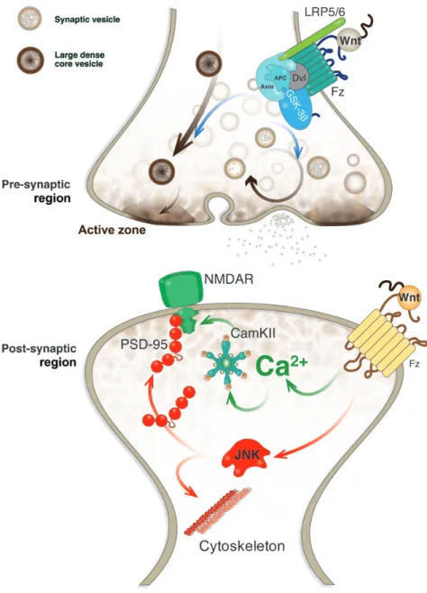
(2) Wnts and neuroprotection. Journal of Molecular Cell Biology. | 65. (Ahmad-Annuar et al., 2006; Cerpa et al., 2008). In addition, by using FM dyes, it was found that Wnt-7a stimulates the endocytosis and recycling of synaptic vesicles (Cerpa et al., 2008) (Figure 2). Wnt-7a also increased the expression and clustering of the a7-nicotinic acetylcholine receptor (a7-nAChR), suggesting that Wnt signaling is able to regulate the clustering of presynaptic receptors (Farias et al., 2007). More recently, a crosstalk between a7-nAChR and Wnt signaling pathway was established (Inestrosa et al., 2013). Nicotine induced the stabilization of b-catenin in a concentration-dependent manner and prevented Ab-induced loss of b-catenin through the a7-nAChR; on the other hand, activation of canonical Wnt signaling induced a7-nAChR expression (Inestrosa et al., 2013). Moreover, analysis of the a7-nAChR promoter indicated that this receptor is a new Wnt target gene (Inestrosa et al., 2013). Interestingly, all Wnt ligands able to modulate presynaptic differentiation activate Wnt/b-catenin signaling pathway to do so, suggesting that components associated with canonical Wnt signaling. cascade are involved in the presynaptic effects of Wnts. On the other hand, one study found that non-canonical ligand Wnt-5a also affects the presynaptic nerve terminals. In fact, Wnt-5a was found to decrease the number of presynaptic terminals (Davis et al., 2008). Although further studies are required to confirm this study, it is possible to suggest that canonical and non-canonical signaling pathways might have promoting and inhibitory effects on presynaptic differentiation, respectively. Electrophysiological recordings on adult rat hippocampal slices demonstrated that Wnt-7a decreases paired pulse facilitation and increases the frequency of mEPSC, indicating that this ligand increases neurotransmitter release in CA3-CA1 synapses (Cerpa et al., 2008) and supporting the promoting effect of canonical Wnt ligands. In contrast, non-canonical Wnt-5a ligand did not show this effect (Cerpa et al., 2008). In addition to Wnt ligands, Fz receptors also have been associated to the presynaptic differentiation (Table 2). Fz1, a welldescribed receptor for Wnt-3a that activates canonical Wnt. Downloaded from http://jmcb.oxfordjournals.org/ at Pontificia Universidad Cat�lica de Chile on May 17, 2016. Figure 1 Wnt signaling pathways. In canonical Wnt/b-catenin signaling pathway, a Wnt ligand binds to Fz receptor and LRP5/6 and activates the protein Dvl. In the absence of Wnt, b-catenin is phosphorylated by GSK-3b in a multiprotein complex with the scaffold protein axin and adenomatous polyposis coli (APC), and phosphorylated b-catenin is ubiquitinated and subsequently degraded by the proteasome. Dvl activation prevents the phosphorylation and degradation of b-catenin, which accumulates in the cytoplasm and translocated into the nucleus where it interacts with the transcription factors TCF/LEF and induces the transcription of Wnt target genes. In non-canonical Wnt/PCP signaling pathway, Dvl activation leads to the activation of small GTPases, such as Rho and Rac, and the downstream JNK pathway. In non-canonical Wnt/Ca2+ signaling pathway, Dvl activation triggers the activation of trimeric G proteins and subsequently PLC that increases DAG and IP3. IP3 induces the release of intracellular Ca2+, inducing the activation of CamKII and calcineurin (CNA) that regulates gene expression through the transcription factor NF-AT..
(3) 66 | Journal of Molecular Cell Biology. Inestrosa and Varela-Nallar. Table 1 Wnt ligands that play a role in the assembly and/or function of central synapses. Wnt ligand Presynaptic Wnt-3a. Wnt-5a Wnt-7a. Wnt-7b. Wnt-5a. Wnt-7a Wnt-7b. Reference. Increased the number of excitatory presynaptic puncta Induced the clustering of the active zone component Bassoon and induced the release of synaptic vesicles Increased the release of presynaptic vesicles and the frequency of miniature excitatory synaptic currents Decreased the number of presynaptic terminals Induced the clustering of synapsin I Increased the clustering and trafficking of a7-nAChRs with the involvement of APC Stimulated the clustering of presynaptic proteins and induced recycling and exocytosis of synaptic vesicles Increased the number of excitatory presynaptic puncta Induced the clustering of presynaptic proteins Increased the clustering of presynaptic proteins and synaptic vesicle recycling Increased the clustering of VGlut1. Davis et al. (2008) Varela-Nallar et al. (2009) Avila et al. (2010) Davis et al. (2008) Hall et al. (2000) Farias et al. (2007) Cerpa et al. (2008) Davis et al. (2008) Sahores et al. (2010) Ahmad-Annuar et al. (2006) Davis et al. (2008). Stimulated dendritic arborization Promoted cortical dendrite growth and dendritic spine formation Increased the synaptic clustering of PSD-95 and the amplitude of fEPSP Increased dendritic spine formation and glutamatergic transmission in cultured hippocampal neurons and hippocampal slices Reduced the depression of synaptic transmission and the reduction of PSD-95 clusters induced by Ab oligomers Induced the recycling of functional GABAA receptor and the amplitude of GABA currents through a postsynaptic mechanism Upregulated synaptic NMDAR currents in rat hippocampal slices and facilitated the induction of LTP The clustering of PSD-95 preceded the increase in synapse formation Increased the density and maturity of dendritic spines through a CamKII-dependent mechanism Increased dendritic branching through activation of Rac and JNK. Wayman et al. (2006) Hiester et al. (2013) Farias et al. (2009) Varela-Nallar et al. (2010) Cerpa et al. (2010) Cuitino et al. (2010) Cerpa et al. (2011) Varela-Nallar et al. (2012) Ciani et al. (2011) Rosso et al. (2005). signaling pathway (Chacon et al., 2008), is located at the synaptic region in hippocampal neurons co-localizing with presynaptic proteins and active synaptic vesicle recycling sites (Varela-Nallar et al., 2009). Overexpression of Fz1 in cultured hippocampal neurons increased the number of clusters of Bassoon, which is a component of the presynaptic active zone involved in the structural organization of neurotransmitter release sites and is recruited early during synapse formation (Zhai et al., 2000). Moreover, treatment with the soluble extracellular cysteine-rich domain (CRD) of Fz1, which is the region of the receptor important for the binding of Wnt proteins and therefore competes with the receptor for the binding of Wnt molecules, decreased Bassoon clustering. These results suggest that Fz1 regulates presynaptic differentiation. Interestingly, treatment with the CRD of Fz2 did not affect the clustering of Bassoon (Varela-Nallar et al., 2009), indicating a receptor specificity for the presynaptic effects of Wnts. All these suggest that the active zone, a structurally relevant presynaptic component, is controlled by canonical Wnt signaling pathway (Figure 2). Wnt signaling also plays relevant roles in the postsynaptic structure (Inestrosa and Arenas, 2010; Oliva et al., 2013) (Table 1). In particular, Wnt-5a that activates non-canonical Wnt signaling cascades in neurons (Farias et al., 2009; Cuitino et al., 2010) modulates postsynaptic assembly and function. In cultured hippocampal neurons, Wnt-5a increased the clustering of the postsynaptic density protein-95 (PSD-95) (Farias et al., 2009), a scaffold protein in the postsynaptic density that contains key molecules involved in the regulation of AMPA and NMDA receptor (NMDAR) targeting and trafficking and regulatory proteins relevant for neurotransmission (Han and Kim, 2008; Sheng and Kim, 2011). This Wnt-5a-induced fast increase in the density of PSD-95 puncta was not associated with the increase in either total level of PSD-95 protein or the clustering of presynaptic proteins, but it was prevented by JNK inhibitors suggesting that Wnt/JNK signaling. pathway is involved (Farias et al., 2009). In addition, Wnt-5a was shown to modulate spine morphogenesis in cultured hippocampal neurons (Varela-Nallar et al., 2010). Time-lapse imaging revealed that Wnt-5a induced both formation of new dendritic spines and an increase in size of preexisting spines (Varela-Nallar et al., 2010). Wnt-7a also increased the density and maturity of dendritic spines through a mechanism involving calcium/calmodulindependent protein kinase II (CamKII) (Ciani et al., 2011). It was shown that Wnt-7a activates CamKII in dendritic spines, while inhibition of this kinase abolished the effect of Wnt-7a on spine growth. This suggests the involvement of Wnt/Ca2+ signaling cascade (Figure 2), which is also supported by the fact that both Wnt-5a and Wnt-7a increase intracellular Ca2+ concentration in neurons (Varela-Nallar et al., 2010; Ciani et al., 2011). Currently, however, little is known regarding the postsynaptic receptors for Wnts (Table 2). In addition, electrophysiological recordings have demonstrated that Wnt ligands exert modulatory effects on glutamatergic neurotransmission. It was observed in hippocampal slices that blockade of Wnt signaling impairs long-term potentiation (LTP), whereas activation of Wnt signaling facilitates LTP (Chen et al., 2006). It was shown that Wnt-5a increases the amplitude of field excitatory postsynaptic potentials (fEPSP) and upregulates synaptic NMDAR currents facilitating the induction of LTP (Cerpa et al., 2011). Interestingly, Wnt-5a triggers a two-step increase in the amplitude of NMDAR responses, which was further investigated by delivery of specific protein kinase inhibitors via the recording pipette. Two known downstream kinases of non-canonical pathway, PKC and JNK (Figure 1), were studied. Inhibition of Ca2+-dependent PKC isoforms prevented the first step of potentiation but did not affect the second step. On the contrary, the slower developing increase in NMDAR currents was blocked by the JNK inhibitors. These data indicated that Wnt-5a induces a fast PKC-dependent potentiation and a slower JNK-dependent potentiation that does not require prior. Downloaded from http://jmcb.oxfordjournals.org/ at Pontificia Universidad Cat�lica de Chile on May 17, 2016. Postsynaptic Wnt-2. Synaptic function described.
(4) Wnts and neuroprotection. Journal of Molecular Cell Biology. | 67. activation of PKC (Cerpa et al., 2011). Importantly, the expression and release of Wnts are regulated by neuronal activity, supporting a role for these ligands in neurotransmission. Wnt-2 expression was increased by activation of NMDAR in hippocampal neurons (Wayman et al., 2006), and an NMDAR-dependent release of Wnt-3a was shown to be induced by tetanic stimulation (Chen et al., 2006). Besides, Wnt signaling also modulates inhibitory synaptic transmission. Specifically, we found that Wnt-5a regulates GABAA receptor-mediated inhibitory currents (Cuitino et al., 2010). Wnt-5a induced the surface expression and maintenance of GABAA receptor in hippocampal neurons, increased the amplitude of GABA-currents through a postsynaptic mechanism, and enhanced the receptor recycling. As observed in Wnt-mediated. dendritic spine growth, these were mediated by activation of CamKII (Cuitino et al., 2010), indicating the activation and involvement of non-canonical Wnt/Ca2+ signaling. Therefore, Wnt signaling cascades may play relevant roles in synaptic plasticity and brain function. The physiological relevance of Wnts to the adult brain was demonstrated by electrophysiological recordings in rat hippocampal slides perfused with the Wnt inhibitors secreted Frizzled-related proteins (sFRPs), which showed that endogenous Wnt ligands modulate glutamatergic neurotransmission (Cerpa et al., 2010, 2011). In addition, treatment of cultured hippocampal neurons with the soluble CRD region of Fz2 receptor, acting as a Wnt signaling inhibitor, decreased spine density, supporting that endogenous Wnt ligands are involved in dendritic spine morphogenesis (Varela-Nallar et al., 2010).. Downloaded from http://jmcb.oxfordjournals.org/ at Pontificia Universidad Cat�lica de Chile on May 17, 2016. Figure 2 Wnt signaling modulates pre- and postsynaptic assembly and function at the glutamatergic synapse. The scheme shows a central synapse. At the presynaptic region, the binding of a canonical Wnt ligand to Fz receptor and co-receptor LRP5/6 triggers the formation of active zones and regulates the synaptic vesicle cycle. At the postsynaptic region, the activation of non-canonical Wnt signaling induces the clustering of PSD-95 and NMDAR, in which both Wnt/JNK and Wnt/Ca2+ play a role..
(5) 68 | Journal of Molecular Cell Biology. memory for the familiar object, indicating that consolidation of object recognition memory depends on canonical Wnt signaling. The effect of Dkk1 was related to an induction of a further increase in Dkk1 protein levels along with a decrease in the levels of phosphorylated glycogen synthase kinase-3b (GSK-3b) that is a key downstream component of the destruction complex in canonical Wnt pathway (Figure 1), which was followed by a decrease in the levels of b-catenin, TCF1, LEF1, Wnt-7a, and the postsynaptic scaffold protein PSD-95, suggesting that the memory impairments induced by Dkk1 is caused by alterations in canonical Wnt signaling pathway (Fortress et al., 2013). This study demonstrated that object learning regulates canonical Wnt signaling that is necessary for hippocampal memory consolidation. During the establishment of the nervous system connectivity in early and late postnatal development, molecules such as the brainderived neurotrophic factor (BDNF), bone morphogenetic protein (BMP), and Wnts act in the nervous system to help newly formed synapses (Lu, 2003; Ciani and Salinas, 2005; Marques, 2005). Recent studies have demonstrated that BDNF is a target gene of the canonical Wnt signaling pathway in glial cells (Yi et al., 2012). A Wnt activator and Wnt-3a both induced BDNF in retina ganglion cells (Seitz et al., 2010; Fragoso et al., 2011), linking BDNF and Wnt signaling pathways (Yi et al., 2012). Figure 3 indicates. Table 2 Wnt receptors at the central synapse. Fz receptor. Evidence of synaptic distribution and/or function. Presynaptic Fz1 Is present in synaptosomes and colocalizes with presynaptic proteins; regulates presynaptic differentiation including the clustering of active zone components Fz3 Co-localizes with the presynaptic vesicular glutamate transporter VGlut1 Co-localizes with the synaptic vesicle protein SV2 Fz5 Is present in synaptosomes and colocalizes with pre- and postsynaptic proteins; mediates the synaptogenic effect of Wnt-7a Is localized in the axonal growth cone and regulates neuronal polarity and axon growth ROR1 Ror1 knockdown decreases presynaptic protein clustering and mediates the presynaptic clustering induced by Wnt-5a ROR2 Ror2 knockdown decreases presynaptic protein clustering and mediates the presynaptic clustering induced by Wnt-5a Postsynaptic Fz4 Co-localizes with PSD-95 Fz5 Regulates dendritic development Fz9 Co-localizes with PSD-95. Reference. Varela-Nallar et al. (2009) Davis et al. (2008) Cerpa et al. (2009) Sahores et al. (2010) Slater et al. (2013) Paganoni et al. (2010) Paganoni et al. (2010) Chen et al. (2006) Slater et al. (2013) Unpublished data. Figure 3 Putative b-catenin binding regions (TCL/LEF) in the promoter and exons of the BDNF gene of human or rodents. In silico detection of TCF/ LEF responsive elements in promoter regions (2 kb) and the entire gene was analysed with the MatInspector Module of the Genomatix Software. The predicted binding sites are indicated as red boxes. The BDNF gene contains several TCF/LEF responsive elements in exons that might be acting as regulatory elements. Transcription starting site and numbering in bp are shown at the top.. Downloaded from http://jmcb.oxfordjournals.org/ at Pontificia Universidad Cat�lica de Chile on May 17, 2016. In vivo, the relevance of Wnt signaling to synaptic connectivity was examined by exposure to enriched environment (EE). Concomitant with an increased number and complexity of large mossy fibre terminals, animals exposed to EE showed increased levels of Wnt-7a/b in CA3 pyramidal neurons (Gogolla et al., 2009). Interestingly, local application of sFRP-1, a Wnt inhibitor, suppressed the effects of EE on synapse number and further reduced synapse numbers in control mice (Gogolla et al., 2009), supporting the importance of Wnt signaling activation. Another in vivo study showed that Wnt-7a/b levels in granule cells of the dentate gyrus were increased after training in the Morris water maze task, and this effect was long-lasting and still observed after 30 days, suggesting a possible association to long-term spatial memory (Tabatadze et al., 2012). In a very recent study that investigated the role of canonical Wnt signaling in hippocampal memory consolidation (Fortress et al., 2013), mice were trained in a hippocampal-dependent object recognition task and immediately after training received a dorsal hippocampal infusion of the Wnt inhibitor Dickkopf-1 (Dkk1), which prevents formation of the Fz/LRP complex and consequent activation of canonical Wnt signaling cascade (Clevers and Nusse, 2012). After 24 h, control mice were able to remember the familiar object explored during training, while mice that received Dkk1 infusion exhibited no. Inestrosa and Varela-Nallar.
(6) Wnts and neuroprotection. Wnt signaling in adult neurogenesis In the adult brain, a continuous generation of new neurons, a process known as neurogenesis, has been reported in a number of mammalian species. This process occurs mainly in the subventricular zone of the lateral ventricles and the subgranular zone (SGZ) of the dentate gyrus of the hippocampus (Gage, 2000; Alvarez-Buylla and Garcia-Verdugo, 2002; Zhao et al., 2008). Adult neurogenesis is highly regulated through intrinsic and extrinsic factors, and many signaling pathways including Notch, Shh, BMPs, and Wnt regulate the maintenance, activation, and fate specification of neural precursor cells (Suh et al., 2009; Ming and Song, 2011). Several studies have shown that canonical Wnt/b-catenin signaling pathway regulates neurogenesis (Varela-Nallar and Inestrosa, 2013). This pathway is active in the SGZ, as determined in the BATGAL Wnt/b-catenin reporter mice (Lie et al., 2005). In that study, it was shown that Wnt-3 was expressed in adult hippocampal astrocytes, and importantly adult hippocampal progenitors (AHPs) expressed key components of canonical Wnt signaling pathway. In vitro experiments revealed that Wnts derived from astrocytes activate Wnt signaling in cultured AHPs and induce their differentiation into neurons (Lie et al., 2005). In vivo, stereotaxic injection of lentiviral vectors expressing Wnt-3a or a mutant Wnt-1 that blocks the activation of Wnt signaling cascade showed that inhibition of this pathway decreases neurogenesis, while stimulating canonical Wnt pathway induces a strong increase in adult hippocampal neurogenesis (Lie et al., 2005). More recently, it was determined that the Wnt inhibitors Dkk1 and sFRP3 negatively regulate neurogenesis. Inducible deletion of Dkk1 in adult central nervous system caused an increase in neurogenesis (Seib et al., 2013), and sFRP3 knockdown in the dentate gyrus through a lentiviral approach increased neural progenitors proliferation (Jang et al., 2013). Interestingly, these factors are regulated under physiological conditions that also regulate neurogenesis (Jang et al., 2013; Seib et al., 2013), suggesting that suppression of Wnt signaling by secreted factors could be a regulatory mechanism to modulate neurogenesis under some stimuli (Varela-Nallar and Inestrosa, 2013). In fact, Wnt signaling pathway has been involved in the regulation of neurogenesis under some physiological conditions. A. | 69. progressive reduction of hippocampal neurogenesis during aging has been observed in different species (Kuhn et al., 1996; Gould et al., 1999; Leuner et al., 2007; Varela-Nallar et al., 2010). This was also evidenced in humans where a reduction of cells expressing the immature neuronal marker DCX was observed with increasing age, suggesting an age-related decline in neurogenesis in the human hippocampus as observed in other species (Knoth et al., 2010). This can be associated to a decline in Wnt signaling, since Wnt-3 level as well as the number of Wnt-3-secreting astrocytes declines with age (Okamoto et al., 2011). More recently, it was reported that Dkk1 was also involved, since the expression of this inhibitor increases with age (Seib et al., 2013). This suggests that Wnt signaling is negatively regulated during aging by secreted factors that may be associated to the decline in neurogenesis. On the other hand, running as a strong inductor of adult neurogenesis in the SGZ (van Praag et al., 1999) modulates the expression of genes involved in Wnt signaling (Stranahan et al., 2010), increases the expression of Wnt-3 in hippocampal astrocytes (Okamoto et al., 2011), and decreases the level of the Wnt inhibitor sFRP-3 in dentate granule neurons (Jang et al., 2013). All these strongly suggest that Wnt signaling pathway is involved in the runningmediated increase in neurogenesis. The mechanism implicated in Wnt-mediated regulation of adult neurogenesis may involve the transcription of Wnt target genes. It was determined that the expression of the transcription factor NeuroD1 is controlled by Wnt/b-catenin signaling activation (Kuwabara et al., 2009). NeuroD1 was shown to be important for the generation of granule cell and olfactory neurons in the embryonic and adult brain (Gao et al., 2009). Interestingly, the promoter of NeuroD1 gene contains overlapping DNA-binding site for Sox2 and TCF/LEF, and therefore, activation of Wnt pathway will induce its expression by removal of Sox2-repression (Kuwabara et al., 2009). Another gene regulated by Wnt/b-catenin pathway that may be involved in neurogenesis is Prox1, which is required for the differentiation and survival of newborn neurons in the hippocampus (Karalay et al., 2011). Therefore, Wnt signaling pathway is relevant to the development of new neurons in the adult brain, the maintenance of the stem cell pool, as well as the differentiation of newborn neurons. Wnt signaling in AD AD is the most common dementia associated with age affecting about 35 million people worldwide. Moreover, it is estimated that by 2050 the number of cases will rise to 100 million people. Therefore, AD is a critical health problem worldwide (Mayeux and Stern, 2012). AD is characterized by a progressive loss of cognitive abilities of which memory and learning are the most affected (Castellani et al., 2010; Ballard et al., 2011) and pathologically by two neuropathological hallmarks: extracellular senile plaques mainly enriched in amyloid-b peptide (Ab) and intracellular neurofibrillary tangles (NFTs) formed by hyperphosphorylated tau protein (Mandelkow and Mandelkow, 2012; Selkoe et al., 2012). Synapse dysfunction appears to be an additional very important feature (Serrano-Pozo et al., 2011; Selkoe et al., 2012). Analysis of AD patient’s brain supports the hypothesis that Ab aggregates are responsible for synaptic failure (Palop and Mucke, 2010).. Downloaded from http://jmcb.oxfordjournals.org/ at Pontificia Universidad Cat�lica de Chile on May 17, 2016. various TCF/LEF elements present in the promoter of BDNF genes of rodents and human. An interesting crosstalk between neurotrophin and Wnt signaling in the regulation of dendritic spine formation has been recently found (Hiester et al., 2013). In cortical neurons, Wnt signaling inhibition disrupted dendritic spine development, reduced dendritic arbor size and complexity, and blocked dendritic spine formation and maturation induced by BDNF. This study showed that BDNF regulates the expression of Wnt-2 and this ligand is sufficient to promote cortical dendrite growth and dendritic spine formation, suggesting that BDNF and Wnt signaling cooperatively regulate dendritic spine formation (Hiester et al., 2013). Altogether, Wnt ligands are important regulators of the synaptic structure and function, and the Wnt cascades are part of the signaling pathways that are regulated by neuronal activity and involved in the regulation of neurotransmission, learning, and memory in adult organisms.. Journal of Molecular Cell Biology.
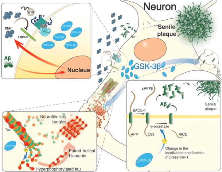
(7) 70 | Journal of Molecular Cell Biology. brains of AD patients carrying presenilin-1 (PS-1)-inherited mutations (Zhang et al., 1998) and Dkk1 that is increased in AD brains or those from transgenic mice as AD models (Caricasole et al., 2004; Rosi et al., 2010). Dkk1 overexpression caused an age-related tau phosphorylation and induced cognitive deficits (Killick et al., 2014). Dkk1 also caused synaptic disassembly at pre- and postsynaptic sites reducing synaptic proteins by a mechanism independent of protein degradation (Purro et al., 2012). On the other hand, the knockdown of Dkk1 prevented the neuronal death and tau phosphorylation induced by Ab (Caricasole et al., 2004) (Figure 4). Also, Ab-induced synaptic loss could be blocked by using an antibody against Dkk1 (Purro et al., 2012). More recently, a new susceptibility factor for late-onset AD called ‘clusterin’ has been identified (Lambert et al., 2009). The knockdown of clusterin protects against Ab neurotoxicity and prevents the induction of Dkk1 by Ab (Killick et al., 2014). By blocking canonical Wnt pathway, Dkk1 activates non-canonical Wnt/JNK signaling cascade, as determined by the increase in c-Jun activity (Killick et al., 2014). These studies support the idea that Ab inducing clusterin/Dkk1/Wnt/JNK pathway could mediate Ab neurotoxicity.. Figure 4 Wnt signaling in different stages of AD progression. In AD, Ab aggregates induce an increased Dkk1 level that blocks the activation of Wnt signaling pathway, which results in higher activity of GSK-3b (top left). The deregulation of Wnt signaling stimulates tau phosphorylation through GSK-3b, inducing the formation of paired helical filaments, which eventually evolves into neurofibrillary tangles (bottom left). Activated GSK-3b also stimulates the amyloidogenic processing of amyloid precursor protein (APP) by b- and g-secretases (bottom right).. Downloaded from http://jmcb.oxfordjournals.org/ at Pontificia Universidad Cat�lica de Chile on May 17, 2016. Currently no cure exists for AD and the exact molecular mechanism leading to its onset is not fully understood. In fact, genetic aberrancies that either cause or increase the risk of AD could be responsible for neuronal degeneration and cognitive dysfunction. Although the majority of AD cases are sporadic, genetic analyses suggest that many genes likely influence the susceptibility to AD (Lambert et al., 2009; Bertram et al., 2010; Bettens et al., 2013). For example, the variant of Wnt signaling pathway co-receptor LRP6 is associated with late-onset of AD and presents low level of Wnt signaling activation (De Ferrari et al., 2007). The allele 4 of apolipoprotein E (apoE4) is considered a risk factor gene for AD and causes inhibition of canonical Wnt signaling in PC12 cells after stimulation with Wnt-7a (Caruso et al., 2006). More than one decade ago, we suggested the strong relationship between AD and Wnt signaling pathway impairments (De Ferrari and Inestrosa, 2000; Inestrosa et al., 2000, 2002; Garrido et al., 2002; De Ferrari et al., 2003) (Figure 4). Different Wnt signaling components are altered in AD (Takashima et al., 1998b; Zhang et al., 1998; Caricasole et al., 2004; Ghanevati and Miller, 2005; De Ferrari et al., 2007), such as b-catenin that is reduced in. Inestrosa and Varela-Nallar.
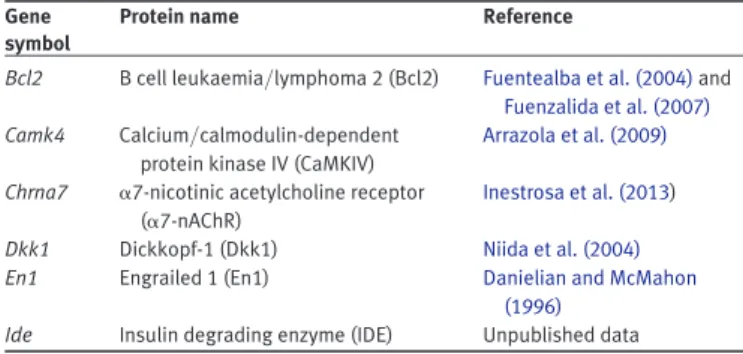
(8) Wnts and neuroprotection. Journal of Molecular Cell Biology. Table 3 TCF/LEF-activated Wnt target genes relevant for neuroprotection. Gene symbol. Protein name. Reference. Bcl2. B cell leukaemia/lymphoma 2 (Bcl2). Camk4. Dkk1 En1. Calcium/calmodulin-dependent protein kinase IV (CaMKIV) a7-nicotinic acetylcholine receptor (a7-nAChR) Dickkopf-1 (Dkk1) Engrailed 1 (En1). Fuentealba et al. (2004) and Fuenzalida et al. (2007) Arrazola et al. (2009). Ide. Insulin degrading enzyme (IDE). Chrna7. Inestrosa et al. (2013) Niida et al. (2004) Danielian and McMahon (1996) Unpublished data. synaptotoxicity (Cerpa et al., 2010). At early stages of the disease, Ab oligomers cause synaptic failure that precedes amyloid plaque deposition and neuronal death (Selkoe et al., 2012). Electrophysiological recordings in hippocampal slices showed that non-canonical ligand Wnt-5a was able to prevent the decrease in the amplitude of fEPSP and EPSCs induced by Ab oligomers (Cerpa et al., 2010). Also, in cultured hippocampal neurons Wnt-5a treatment prevented the decrease in PSD-95 puncta and the synaptic loss induced by Ab (Cerpa et al., 2010), confirming that Wnt-5a prevented the synaptic damage triggered by Ab. A disturbance of cellular energy metabolism increases neuronal dysfunction and the loss of synaptic networks (Kapogiannis and Mattson, 2011; Burns et al., 2013). For instance, deregulation of glucose might alter insulin metabolism and affect the brain energy balance, thus leading to the onset of diabetes and AD (Bosco et al., 2011; Craft et al., 2013). Several reports have suggested a link among AD, insulin metabolism, and Wnt signaling at the GSK-3b level (Cohen and Goedert, 2004). In addition, the transcription factor 7-like 2 (TCF/L2) normally activated by canonical Wnt signaling is a risk factor for the onset of diabetes type 2 (Grant et al., 2006; Cadigan and Waterman, 2012). Canonical Wnt-3a ligand was shown to enhance insulin signaling in vitro and drive mitochondrial biogenesis (Yoon et al., 2010). Recent data from our laboratory indicate the role of non-canonical Wnt-5a in affecting mitochondrial physiology by modulating its dynamics (fusion2fission) (Silva-Alvarez et al., 2013). All these suggest that Wnt signaling acts as neuroprotective factor by regulating the disturbance of energy balance in AD. Concluding remarks This review focuses on the roles of Wnt signaling in synaptic development and function, as well as its neuroprotective effect in AD. Wnts control presynaptic compartment by regulating the clustering of components of the active zones and the recycling of synaptic vesicles, and control postsynaptic compartment by modulating the dendritic spine morphogenesis and the assembly of the postsynaptic apparatus. Moreover, activation of Wnt signaling enhances synaptic plasticity and memory consolidation. Also, Wnt pathway is one of the signaling cascades that regulate the generation of new neurons in the adult brain. Since 1999 when we proposed for the first time an association of the deregulation of Wnt pathway with AD, a number of studies have consistently shown neuroprotective effects of Wnts. Because Wnt activators have shown to rescue synaptic and cognitive impairments, activating Wnt pathway is a feasible therapeutic approach for the treatment of AD. Acknowledgements We thank Cristobal Mejias (Department of Cell and Molecular Biology, P. Catholic University of Chile) for his help in the analysis of the BDNF gene sequence. Funding This work was supported by Grants from FONDECYT (no. 1120156) and the Basal Center of Excellence in Science and Technology (CONICYT-PFB12/2007) to N.C.I. and by a Grant from FONDECYT (no. 11110012) to L.V.-N.. Downloaded from http://jmcb.oxfordjournals.org/ at Pontificia Universidad Cat�lica de Chile on May 17, 2016. One hallmark of AD is the abnormal tau phosphorylation to form intraneuronal NFT (Ballard et al., 2011). Several kinases phosphorylate tau in vitro, while the most relevant kinases involved in tau phosphorylation in vivo are cyclin-dependent kinase 5, extracellular signal-related kinase 2, microtubule affinity-regulating kinase, and GSK-3b (Churcher, 2006; Hooper et al., 2008). In cultured neurons, increased GSK-3b activity was observed after Ab treatment (Takashima et al., 1996, 1998a). Active GSK-3b has been found in AD brains with neurofibrillary changes, with a consequent decrease in b-catenin level and increase in tau hyperphosphorylation (Pei et al., 1999). Moreover, GSK-3b conditional transgenic mice show neurodegeneration and spatial learning deficits (Lucas et al., 2001; Hernandez et al., 2002). Interestingly, it was shown that the phosphorylation of tau antagonizes apoptosis by preventing the phosphorylation and subsequent degradation of b-catenin. Increasing levels of phosphorylated tau were correlated with increased levels of nuclear b-catenin, and b-catenin knockdown antagonized the anti-apoptotic effects of tau (Li et al., 2007), suggesting that b-catenin is a protective element in AD. Several studies have shown that Wnt signaling pathway is neuroprotective against the toxicity of Ab peptide. In cultured hippocampal neurons, decreased level of b-catenin after exposure to Ab aggregates and the neurotoxic effect of Ab were prevented by an incubation with Wnt-3a (De Ferrari et al., 2003; Alvarez et al., 2004). The protective effect of Wnt-3a is mediated by Fz1 receptor in both PC12 cells and hippocampal neurons (Chacon et al., 2008). When Fz1 was overexpressed, a significant increase in Wnt-3ainduced cell survival was observed. On the contrary, knockdown of Fz1 by antisense oligonucleotides reduced neuroprotective effect of Wnt-3a as well as the activation of Wnt/b-catenin signaling (Chacon et al., 2008). In a double transgenic APPswe/ PSEN1DE9 mouse model of AD, in vivo treatment with the GSK-3b inhibitor lithium activated Wnt signaling in the hippocampus with the increase in b-catenin level and inhibition of GSK-3b, and consequently reduced spatial memory impairment, decreased Ab aggregates and oligomers, and decreased astrogliosis (Toledo and Inestrosa, 2010). Thus, impaired canonical Wnt signaling may contribute to the neurodegeneration in AD and the activation of this pathway may be a therapeutic strategy for the treatment of AD (Inestrosa and Arenas, 2010; Inestrosa et al., 2012; Oliva et al., 2013). In addition, several Wnt target genes may mediate the neuroprotective effects of canonical Wnt signaling (Table 3). We have also studied whether non-canonical signaling is able to protect neurons against Ab oligomers, mainly focused on. | 71.
(9) 72 | Journal of Molecular Cell Biology. Conflict of interest: none declared. References. Calmodulin-dependent protein kinase II. Proc. Natl Acad. Sci. USA 108, 10732 – 10737. Clevers, H., and Nusse, R. (2012). Wnt/b-catenin signaling and disease. Cell 149, 1192 –1205. Cohen, P., and Goedert, M. (2004). GSK3 inhibitors: development and therapeutic potential. Nat. Rev. Drug Discov. 3, 479 –487. Craft, S., Cholerton, B., and Baker, L.D. (2013). Insulin and Alzheimer’s disease: untangling the web. J. Alzheimers Dis. 33(Suppl 1), S263 – S275. Cuitino, L., Godoy, J.A., Farias, G.G., et al. (2010). Wnt-5a modulates recycling of functional GABAA receptors on hippocampal neurons. J. Neurosci. 30, 8411 – 8420. Danielian, P.S., and McMahon, A.P. (1996). Engrailed-1 as a target of the Wnt-1 signalling pathway in vertebrate midbrain development. Nature 383, 332 – 334. Davis, E.K., Zou, Y., and Ghosh, A. (2008). Wnts acting through canonical and noncanonical signaling pathways exert opposite effects on hippocampal synapse formation. Neural Dev. 3, 32. De Ferrari, G.V., and Inestrosa, N.C. (2000). Wnt signaling function in Alzheimer’s disease. Brain Res. Brain Res. Rev. 33, 1 –12. De Ferrari, G.V., Chacon, M.A., Barria, M.I., et al. (2003). Activation of Wnt signaling rescues neurodegeneration and behavioral impairments induced by b-amyloid fibrils. Mol. Psychiatry 8, 195 –208. De Ferrari, G.V., Papassotiropoulos, A., Biechele, T., et al. (2007). Common genetic variation within the low-density lipoprotein receptor-related protein 6 and late-onset Alzheimer’s disease. Proc. Natl Acad. Sci. USA 104, 9434 – 9439. Farias, G.G., Valles, A.S., Colombres, M., et al. (2007). Wnt-7a induces presynaptic colocalization of a7-nicotinic acetylcholine receptors and adenomatous polyposis coli in hippocampal neurons. J. Neurosci. 27, 5313– 5325. Farias, G.G., Alfaro, I.E., Cerpa, W., et al. (2009). Wnt-5a/JNK signaling promotes the clustering of PSD-95 in hippocampal neurons. J. Biol. Chem. 284, 15857 – 15866. Fragoso, M.A., Yi, H., Nakamura, R.E., et al. (2011). The Wnt signaling pathway protects retinal ganglion cell 5 (RGC-5) cells from elevated pressure. Cell. Mol. Neurobiol. 31, 163 – 173. Fortress, A.M., Schram, S.L., Tuscher, J.J., et al. (2013). Canonical Wnt signaling is necessary for object recognition memory consolidation. J. Neurosci. 33, 12619 – 12626. Fuentealba, R.A., Farias, G., Scheu, J., et al. (2004). Signal transduction during amyloid-beta-peptide neurotoxicity: role in Alzheimer disease. Brain Res. Brain Res. Rev. 47, 275 – 289. Fuenzalida, K., Quintanilla, R., Ramos, P., et al. (2007). Peroxisome proliferatoractivated receptor gamma up-regulates the Bcl-2 anti-apoptotic protein in neurons and induces mitochondrial stabilization and protection against oxidative stress and apoptosis. J. Biol. Chem. 282, 37006– 37015. Gage, F.H. (2000). Mammalian neural stem cells. Science 287, 1433 – 1438. Gao, Z., Ure, K., Ables, J.L., et al. (2009). Neurod1 is essential for the survival and maturation of adult-born neurons. Nat. Neurosci. 12, 1090 –1092. Garrido, J.L., Godoy, J.A., Alvarez, A., et al. (2002). Protein kinase C inhibits amyloid b peptide neurotoxicity by acting on members of the Wnt pathway. FASEB J. 16, 1982 –1984. Ghanevati, M., and Miller, C.A. (2005). Phospho-b-catenin accumulation in Alzheimer’s disease and in aggresomes attributable to proteasome dysfunction. J. Mol. Neurosci. 25, 79 –94. Gogolla, N., Galimberti, I., Deguchi, Y., et al. (2009). Wnt signaling mediates experience-related regulation of synapse numbers and mossy fiber connectivities in the adult hippocampus. Neuron 62, 510 – 525. Gordon, M.D., and Nusse, R. (2006). Wnt signaling: multiple pathways, multiple receptors, and multiple transcription factors. J. Biol. Chem. 281, 22429 – 22433. Gould, E., Reeves, A.J., Fallah, M., et al. (1999). Hippocampal neurogenesis in adult Old World primates. Proc. Natl Acad. Sci. USA 96, 5263– 5267. Grant, S.F., Thorleifsson, G., Reynisdottir, I., et al. (2006). Variant of transcription factor 7-like 2 (TCF7L2) gene confers risk of type 2 diabetes. Nat. Genet. 38, 320 – 323. Hall, A.C., Lucas, F.R., and Salinas, P.C. (2000). Axonal remodeling and synaptic differentiation in the cerebellum is regulated by WNT-7a signaling. Cell 100, 525 – 535.. Downloaded from http://jmcb.oxfordjournals.org/ at Pontificia Universidad Cat�lica de Chile on May 17, 2016. Ahmad-Annuar, A., Ciani, L., Simeonidis, I., et al. (2006). Signaling across the synapse: a role for Wnt and Dishevelled in presynaptic assembly and neurotransmitter release. J. Cell Biol. 174, 127 – 139. Al-Harthi, L. (2012). Interplay between Wnt/b-catenin signaling and HIV: virologic and biologic consequences in the CNS. J. Neuroimmune Pharmacol. 7, 731 – 739. Alvarez, A.R., Godoy, J.A., Mullendorff, K., et al. (2004). Wnt-3a overcomes b-amyloid toxicity in rat hippocampal neurons. Exp. Cell Res. 297, 186 – 196. Alvarez-Buylla, A., and Garcia-Verdugo, J.M. (2002). Neurogenesis in adult subventricular zone. J. Neurosci. 22, 629 – 634. Anastas, J.N., and Moon, R.T. (2013). WNT signalling pathways as therapeutic targets in cancer. Nat. Rev. Cancer 13, 11 – 26. Arrazola, M.S., Varela-Nallar, L., Colombres, M., et al. (2009). Calcium/ calmodulin-dependent protein kinase type IV is a target gene of the Wnt/ b-catenin signaling pathway. J. Cell. Physiol. 221, 658 – 667. Avila, M.E., Sepulveda, F.J., Burgos, C.F., et al. (2010). Canonical Wnt3a modulates intracellular calcium and enhances excitatory neurotransmission in hippocampal neurons. J. Biol. Chem. 285, 18939 – 18947. Ballard, C., Gauthier, S., Corbett, A., et al. (2011). Alzheimer’s disease. Lancet 377, 1019 – 1031. Bertram, L., Lill, C.M., and Tanzi, R.E. (2010). The genetics of Alzheimer disease: back to the future. Neuron 68, 270 –281. Bettens, K., Sleegers, K., and Van Broeckhoven, C. (2013). Genetic insights in Alzheimer’s disease. Lancet Neurol. 12, 92 – 104. Bosco, D., Fava, A., Plastino, M., et al. (2011). Possible implications of insulin resistance and glucose metabolism in Alzheimer’s disease pathogenesis. J. Cell. Mol. Med. 15, 1807 –1821. Burns, C.M., Chen, K., Kaszniak, A.W., et al. (2013). Higher serum glucose levels are associated with cerebral hypometabolism in Alzheimer regions. Neurology 80, 1557– 1564. Cadigan, K.M., and Waterman, M.L. (2012). TCF/LEFs and Wnt signaling in the nucleus. Cold Spring Harb. Perspect. Biol. 4, a007906. Caricasole, A., Copani, A., Caraci, F., et al. (2004). Induction of Dickkopf-1, a negative modulator of the Wnt pathway, is associated with neuronal degeneration in Alzheimer’s brain. J. Neurosci. 24, 6021– 6027. Caruso, A., Motolese, M., Iacovelli, L., et al. (2006). Inhibition of the canonical Wnt signaling pathway by apolipoprotein E4 in PC12 cells. J. Neurochem. 98, 364 –371. Castellani, R.J., Rolston, R.K., and Smith, M.A. (2010). Alzheimer disease. Dis. Mon. 56, 484 –546. Cerpa, W., Godoy, J.A., Alfaro, I., et al. (2008). Wnt-7a modulates the synaptic vesicle cycle and synaptic transmission in hippocampal neurons. J. Biol. Chem. 283, 5918– 5927. Cerpa, W., Toledo, E.M., Varela-Nallar, L., et al. (2009). The role of Wnt signaling in neuroprotection. Drug News Perspect. 22, 579 – 591. Cerpa, W., Farias, G.G., Godoy, J.A., et al. (2010). Wnt-5a occludes Ab oligomerinduced depression of glutamatergic transmission in hippocampal neurons. Mol. Neurodegener. 5, 3. Cerpa, W., Gambrill, A., Inestrosa, N.C., et al. (2011). Regulation of NMDA-receptor synaptic transmission by Wnt signaling. J. Neurosci. 31, 9466–9471. Chacon, M.A., Varela-Nallar, L., and Inestrosa, N.C. (2008). Frizzled-1 is involved in the neuroprotective effect of Wnt3a against Ab oligomers. J. Cell. Physiol. 217, 215 –227. Chen, J., Park, C.S., and Tang, S.J. (2006). Activity-dependent synaptic Wnt release regulates hippocampal long term potentiation. J. Biol. Chem. 281, 11910 – 11916. Chien, A.J., Conrad, W.H., and Moon, R.T. (2009). A Wnt survival guide: from flies to human disease. J. Invest. Dermatol. 129, 1614 – 1627. Churcher, I. (2006). Tau therapeutic strategies for the treatment of Alzheimer’s disease. Curr. Top. Med. Chem. 6, 579 – 595. Ciani, L., and Salinas, P.C. (2005). WNTs in the vertebrate nervous system: from patterning to neuronal connectivity. Nat. Rev. Neurosci. 6, 351 –362. Ciani, L., Boyle, K.A., Dickins, E., et al. (2011). Wnt7a signaling promotes dendritic spine growth and synaptic strength through Ca2+/. Inestrosa and Varela-Nallar.
(10) Wnts and neuroprotection. | 73. Marques, G. (2005). Morphogens and synaptogenesis in Drosophila. J. Neurobiol. 64, 417– 434. Mayeux, R., and Stern, Y. (2012). Epidemiology of Alzheimer disease. Cold Spring Harb. Perspect. Med. 2, a006239. Ming, G.L., and Song, H. (2011). Adult neurogenesis in the mammalian brain: significant answers and significant questions. Neuron 70, 687 –702. Niida, A., Hiroko, T., Kasai, M., et al. (2004). DKK1, a negative regulator of Wnt signaling, is a target of the beta-catenin/TCF pathway. Oncogene 23, 8520 –8526. Nusse, R., and Varmus, H. (2012). Three decades of Wnts: a personal perspective on how a scientific field developed. EMBO J. 31, 2670– 2684. Okamoto, M., Inoue, K., Iwamura, H., et al. (2011). Reduction in paracrine Wnt3 factors during aging causes impaired adult neurogenesis. FASEB J. 25, 3570 –3582. Oliva, C.A., Vargas, J.Y., and Inestrosa, N.C. (2013). Wnt signaling: role in neural networks and memory. Ageing Res. Rev. 12, 786 – 800. Paganoni, S., Bernstein, J., and Ferreira, A. (2010). Ror1-Ror2 complexes modulate synapse formation in hippocampal neurons. Neuroscience 165, 1261 – 1274. Palop, J.J., and Mucke, L. (2010). Amyloid-b-induced neuronal dysfunction in Alzheimer’s disease: from synapses toward neural networks. Nat. Neurosci. 13, 812 – 818. Pei, J.J., Braak, E., Braak, H., et al. (1999). Distribution of active glycogen synthase kinase 3b (GSK-3b) in brains staged for Alzheimer disease neurofibrillary changes. J. Neuropathol. Exp. Neurol. 58, 1010– 1019. Purro, S.A., Dickins, E.M., and Salinas, P.C. (2012). The secreted Wnt antagonist Dickkopf-1 is required for amyloid b-mediated synaptic loss. J. Neurosci. 32, 3492 – 3498. Rosi, M.C., Luccarini, I., Grossi, C., et al. (2010). Increased Dickkopf-1 expression in transgenic mouse models of neurodegenerative disease. J. Neurochem. 112, 1539 – 1551. Rosso, S.B., and Inestrosa, N.C. (2013). WNT signaling in neuronal maturation and synaptogenesis. Front. Cell. Neurosci. 7, 103. Rosso, S.B., Sussman, D., Wynshaw-Boris, A., et al. (2005). Wnt signaling through Dishevelled, Rac and JNK regulates dendritic development. Nat. Neurosci. 8, 34– 42. Sahores, M., Gibb, A., and Salinas, P.C. (2010). Frizzled-5, a receptor for the synaptic organizer Wnt7a, regulates activity-mediated synaptogenesis. Development 137, 2215– 2225. Seib, D.R., Corsini, N.S., Ellwanger, K., et al. (2013). Loss of dickkopf-1 restores neurogenesis in old age and counteracts cognitive decline. Cell Stem Cell 12, 204 –214. Seitz, R., Hackl, S., Seibuchner, T., et al. (2010). Norrin mediates neuroprotective effects on retinal ganglion cells via activation of the Wnt/ b-catenin signaling pathway and the induction of neuroprotective growth factors in Muller cells. J. Neurosci. 30, 5998 –6010. Selkoe, D., Mandelkow, E., and Holtzman, D. (2012). Deciphering Alzheimer disease. Cold Spring Harb. Perspect. Med. 2, a011460. Serrano-Pozo, A., Frosch, M.P., Masliah, E., et al. (2011). Neuropathological alterations in Alzheimer disease. Cold Spring Harb. Perspect. Med. 1, a006189. Sheng, M., and Kim, E. (2011). The postsynaptic organization of synapses. Cold Spring Harb. Perspect. Biol. 3, a005678. Silva-Alvarez, C., Arrazola, M.S., Godoy, J.A., et al. (2013). Canonical Wnt signaling protects hippocampal neurons from Ab oligomers: role of non-canonical Wnt-5a/Ca2+ in mitochondrial dynamics. Front. Cell. Neurosci. 7, 97. Slater, P.G., Ramirez, V.T., Gonzalez-Billault, C., et al. (2013). Frizzled-5 receptor is involved in neuronal polarity and morphogenesis of hippocampal neurons. PLoS One 8, e78892. Stranahan, A.M., Lee, K., Becker, K., et al. (2010). Hippocampal gene expression patterns underlying the enhancement of memory by running in aged mice. Neurobiol. Aging 31, 1937 –1949. Suh, H., Deng, W., and Gage, F.H. (2009). Signaling in adult neurogenesis. Annu. Rev. Cell Dev. Biol. 25, 253 –275. Tabatadze, N., Tomas, C., McGonigal, R., et al. (2012). Wnt transmembrane signaling and long-term spatial memory. Hippocampus 22, 1228 –1241. Takashima, A., Noguchi, K., Michel, G., et al. (1996). Exposure of rat hippocampal neurons to amyloid b peptide (25 – 35) induces the inactivation of. Downloaded from http://jmcb.oxfordjournals.org/ at Pontificia Universidad Cat�lica de Chile on May 17, 2016. Han, K., and Kim, E. (2008). Synaptic adhesion molecules and PSD-95. Prog. Neurobiol. 84, 263 –283. Hernandez, F., Borrell, J., Guaza, C., et al. (2002). Spatial learning deficit in transgenic mice that conditionally over-express GSK-3b in the brain but do not form tau filaments. J. Neurochem. 83, 1529 –1533. Hiester, B.G., Galati, D.F., Salinas, P.C., et al. (2013). Neurotrophin and Wnt signaling cooperatively regulate dendritic spine formation. Mol. Cell. Neurosci. 56, 115 – 127. Hooper, C., Killick, R., and Lovestone, S. (2008). The GSK3 hypothesis of Alzheimer’s disease. J. Neurochem. 104, 1433 –1439. Inestrosa, N.C., and Arenas, E. (2010). Emerging roles of Wnts in the adult nervous system. Nat. Rev. Neurosci. 11, 77 –86. Inestrosa, N.C., Alvarez, A., Godoy, J., et al. (2000). Acetylcholinesteraseamyloid-b-peptide interaction and Wnt signaling involvement in Ab neurotoxicity. Acta Neurol. Scand. Suppl. 176, 53 – 59. Inestrosa, N., De Ferrari, G.V., Garrido, J.L., et al. (2002). Wnt signaling involvement in b-amyloid-dependent neurodegeneration. Neurochem. Int. 41, 341–344. Inestrosa, N.C., Montecinos-Oliva, C., and Fuenzalida, M. (2012). Wnt signaling: role in Alzheimer disease and schizophrenia. J. Neuroimmune Pharmacol. 7, 788–807. Inestrosa, N.C., Godoy, J.A., Vargas, J.Y., et al. (2013). Nicotine prevents synaptic impairment induced by amyloid-b oligomers through a7-nicotinic acetylcholine receptor activation. Neuromol. Med. 15, 549 –569. Jang, M.H., Bonaguidi, M.A., Kitabatake, Y., et al. (2013). Secreted frizzled-related protein 3 regulates activity-dependent adult hippocampal neurogenesis. Cell Stem Cell 12, 215 – 223. Kapogiannis, D., and Mattson, M.P. (2011). Disrupted energy metabolism and neuronal circuit dysfunction in cognitive impairment and Alzheimer’s disease. Lancet Neurol. 10, 187 – 198. Karalay, O., Doberauer, K., Vadodaria, K.C., et al. (2011). Prospero-related homeobox 1 gene (Prox1) is regulated by canonical Wnt signaling and has a stage-specific role in adult hippocampal neurogenesis. Proc. Natl Acad. Sci. USA 108, 5807 – 5812. Killick, R., Ribe, E.M., Al-Shawi, R., et al. (2014). Clusterin regulates b-amyloid toxicity via Dickopf-1-driven induction of the Wnt-PCP-JNK. Mol. Psychiatry 19, 88 – 98. Knoth, R., Singec, I., Ditter, M., et al. (2010). Murine features of neurogenesis in the human hippocampus across the lifespan from 0 to 1000 years. PLoS One 5, e8809. Kuhn, H.G., Dickinson-Anson, H., and Gage, F.H. (1996). Neurogenesis in the dentate gyrus of the adult rat: age-related decrease of neuronal progenitor proliferation. J. Neurosci. 16, 2027 –2033. Kuwabara, T., Hsieh, J., Muotri, A., et al. (2009). Wnt-mediated activation of NeuroD1 and retro-elements during adult neurogenesis. Nat. Neurosci. 12, 1097 –1105. Lambert, J.C., Heath, S., Even, G., et al. (2009). Genome-wide association study identifies variants at CLU and CR1 associated with Alzheimer’s disease. Nat. Genet. 41, 1094 – 1099. Leuner, B., Kozorovitskiy, Y., Gross, C.G., et al. (2007). Diminished adult neurogenesis in the marmoset brain precedes old age. Proc. Natl Acad. Sci. USA 104, 17169 – 17173. Li, H.L., Wang, H.H., Liu, S.J., et al. (2007). Phosphorylation of tau antagonizes apoptosis by stabilizing b-catenin, a mechanism involved in Alzheimer’s neurodegeneration. Proc. Natl Acad. Sci. USA 104, 3591 –3596. Lie, D.C., Colamarino, S.A., Song, H.J., et al. (2005). Wnt signalling regulates adult hippocampal neurogenesis. Nature 437, 1370 –1375. Logan, C.Y., and Nusse, R. (2004). The Wnt signaling pathway in development and disease. Annu. Rev. Cell Dev. Biol. 20, 781 – 810. Lu, B. (2003). BDNF and activity-dependent synaptic modulation. Learn. Mem. 10, 86 – 98. Lucas, F.R., and Salinas, P.C. (1997). WNT-7a induces axonal remodeling and increases synapsin I levels in cerebellar neurons. Dev. Biol. 192, 31 –44. Lucas, J.J., Hernandez, F., Gomez-Ramos, P., et al. (2001). Decreased nuclear b-catenin, tau hyperphosphorylation and neurodegeneration in GSK-3b conditional transgenic mice. EMBO J. 20, 27 –39. Mandelkow, E.M., and Mandelkow, E. (2012). Biochemistry and cell biology of tau protein in neurofibrillary degeneration. Cold Spring Harb. Perspect. Med. 2, a006247. Marinou, K., Christodoulides, C., Antoniades, C., et al. (2012). Wnt signaling in cardiovascular physiology. Trends Endocrinol. Metab. 23, 628 – 636.. Journal of Molecular Cell Biology.
(11) 74 | Journal of Molecular Cell Biology. Varela-Nallar, L., Alfaro, I.E., Serrano, F.G., et al. (2010). Wingless-type family member 5A (Wnt-5a) stimulates synaptic differentiation and function of glutamatergic synapses. Proc. Natl Acad. Sci. USA 107, 21164 –21169. Varela-Nallar, L., Parodi, J., Farias, G.G., et al. (2012). Wnt-5a is a synaptogenic factor with neuroprotective properties against Ab toxicity. Neurodegener. Dis. 10, 23 – 26. Wayman, G.A., Impey, S., Marks, D., et al. (2006). Activity-dependent dendritic arborization mediated by CaM-kinase I activation and enhanced CREBdependent transcription of Wnt-2. Neuron 50, 897 –909. Yi, H., Hu, J., Qian, J., et al. (2012). Expression of brain-derived neurotrophic factor is regulated by the Wnt signaling pathway. Neuroreport 23, 189 – 194. Yoon, J.C., Ng, A., Kim, B., et al. (2010). Wnt signaling regulates mitochondrial physiology and insulin sensitivity. Genes Dev. 24, 1507– 1518. Zhai, R., Olias, G., Chung, W.J., et al. (2000). Temporal appearance of the presynaptic cytomatrix protein bassoon during synaptogenesis. Mol. Cell. Neurosci. 15, 417 –428. Zhang, Z., Hartmann, H., Do, V.M., et al. (1998). Destabilization of b-catenin by mutations in presenilin-1 potentiates neuronal apoptosis. Nature 395, 698 – 702. Zhao, C., Deng, W., and Gage, F.H. (2008). Mechanisms and functional implications of adult neurogenesis. Cell 132, 645– 660.. Downloaded from http://jmcb.oxfordjournals.org/ at Pontificia Universidad Cat�lica de Chile on May 17, 2016. phosphatidyl inositol-3 kinase and the activation of tau protein kinase I/ glycogen synthase kinase-3b. Neurosci. Lett. 203, 33 – 36. Takashima, A., Honda, T., Yasutake, K., et al. (1998a). Activation of tau protein kinase I/glycogen synthase kinase-3 b by amyloid b peptide (25 –35) enhances phosphorylation of tau in hippocampal neurons. Neurosci. Res. 31, 317 – 323. Takashima, A., Murayama, M., Murayama, O., et al. (1998b). Presenilin 1 associates with glycogen synthase kinase-3 b and its substrate tau. Proc. Natl Acad. Sci. USA 95, 9637– 9641. Toledo, E.M., and Inestrosa, N.C. (2010). Activation of Wnt signaling by lithium and rosiglitazone reduced spatial memory impairment and neurodegeneration in brains of an APPswe/PSEN1DeltaE9 mouse model of Alzheimer’s disease. Mol. Psychiatry 15, 272 – 285. Toledo, E.M., Colombres, M., and Inestrosa, N.C. (2008). Wnt signaling in neuroprotection and stem cell differentiation. Prog. Neurobiol. 86, 281 – 296. van Praag, H., Kempermann, G., and Gage, F.H. (1999). Running increases cell proliferation and neurogenesis in the adult mouse dentate gyrus. Nat. Neurosci. 2, 266 – 270. Varela-Nallar, L., and Inestrosa, N.C. (2013). Wnt signaling in the regulation of adult hippocampal neurogenesis. Front. Cell. Neurosci. 7, 100. Varela-Nallar, L., Grabowski, C.P., Alfaro, I.E., et al. (2009). Role of the Wnt receptor Frizzled-1 in presynaptic differentiation and function. Neural Dev. 4, 41.. Inestrosa and Varela-Nallar.
(12)
Figure

Documento similar
