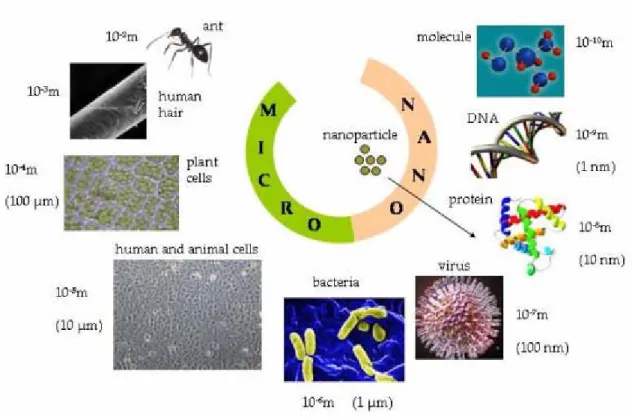
TítuloCellular and molecular effects of metal oxide nanoparticles on human neuronal cells
Texto completo
Figure

Documento similar
In the preparation of this report, the Venice Commission has relied on the comments of its rapporteurs; its recently adopted Report on Respect for Democracy, Human Rights and the Rule
The draft amendments do not operate any more a distinction between different states of emergency; they repeal articles 120, 121and 122 and make it possible for the President to
The characteristic morphology of the ZnO thin films is shown in the top-view FESEM image of Figure 3a, and a magnified cross- sectional image is shown in Figure 3b, which confirms
Mazarío, “Effect of the surface charge on the adsorption capacity of chromium (VI) of iron oxide magnetic nanoparticles prepared by microwave-assisted synthesis,”.
The results shown in this chapter demonstrated that the quality of the sample of pyramidal CdSe NPs pot-deposited on HOPG in terms of NP coverage and polydispersity
The cyclic voltammetry characterization of CdSe NPs with different morphology, size and surface composition reveals that the redox processes depend on the NP
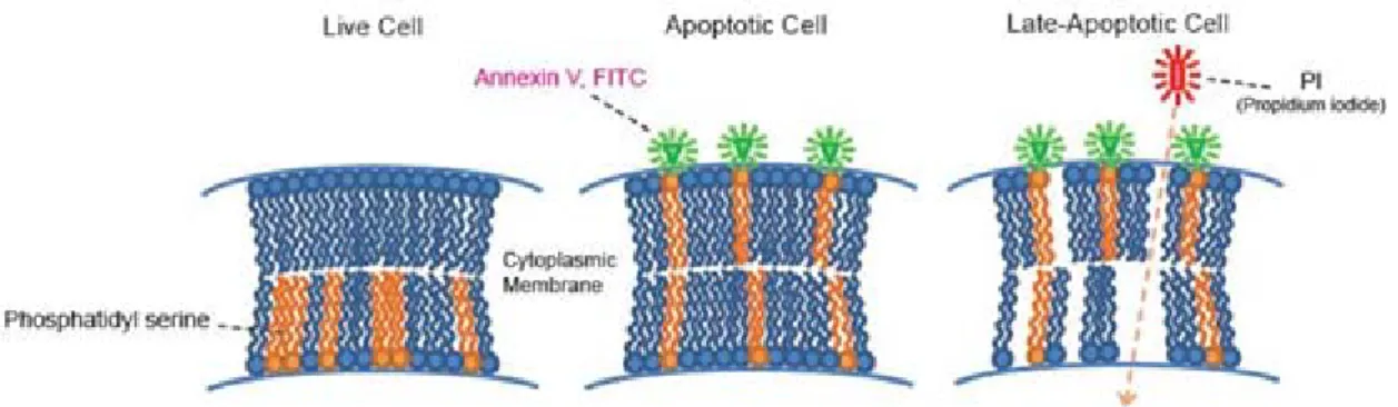
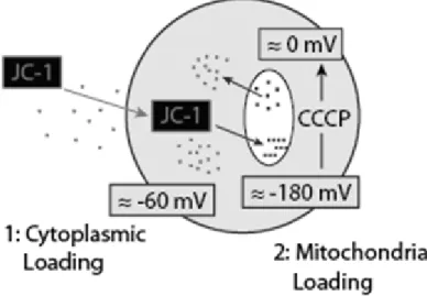
reinhardtii after 1, 24 and 72 h of exposure to SPION as measured by flow cytometry using different fluorochromes: (A) plasma membrane potential, (B) mitochondrial membrane
The aim of this study was to determine, through a preliminary in vitro study on human glioblastoma (A172, LN229), anaplastic glioma (SF268) and neuroblastoma (SK-N-SH) cell lines,
