TítuloCellular and molecular effects of metal oxide nanoparticles on human neuronal cells
189
0
0
Texto completo
(2) Cellular and molecular effects of metal oxide nanoparticles on human neuronal cells Gözde Kılıç. Doctoral thesis 2015 Cellular and Molecular Biology. Thesis supervisors: Blanca Laffon Lage Vanessa Valdiglesias García.
(3) The experimental work presented in this thesis was performed at the: Toxicology laboratory, University of A Coruña, Spain. Genetics laboratory, University of A Coruña, Spain. EPIUnit Institute of Public Health, University of Porto, Porto, Portugal. Department of Environmental Health, Portuguese National Institute of Health, Porto, Portugal. REQUIMTE, Department of Chemistry and Biochemistry, University of Porto, Porto, Portugal. Nanotoxicology Group, Indian Institute of Toxicology Research, Uttar Pradesh, India. Laboratory for Health Protection Research, National Institute for Public Health and the Environment (RIVM), Bilthoven, The Netherlands. This work was supported by Xunta de Galicia (EM 2012/079), MODENA COST Action; TD1204, Pre-doctoral researcher contract from University of A Coruña (May 2012- May 2014) and Inditex-UDC 2014 stay abroad grants for pre-doctoral students.. Publications Scientific papers: 1.. Comparative study on effects of two different types of titanium dioxide nanoparticles on human neuronal cells. Valdiglesias, V; Costa, C.; Sharma, V.; Kiliç, G.; Pásaro, E.; Teixeira, J.P.; Dhawan, A.; Laffon, B. Food and Chemical Toxicology vol. 57 (2013) p.352-361. DOI:10.1016/j.fct.2013.04.010.. 2.. Neuronal cytotoxicity and genotoxicity induced by zinc oxide nanoparticles. Valdiglesias, V.; Costa, C.; Kiliç, G.; Costa, S.; Pásaro, E.; Laffon, B.; Teixeira, J.P. Environment International vol. 55 (2013) p.92-100. DOI:10.1016/j.envint.2013.02.013.. 3.. Effects of iron oxide nanoparticles: Cytotoxicity, genotoxicity, developmental toxicity, and neurotoxicity. Valdiglesias, V.; Costa, C.; Kiliç, G.; Costa, S.; Pásaro, E.; Teixeira, J.P.; Laffon, B. Environmental and Molecular Mutagenesis vol. 56 (2015) p.125-148. DOI:10.1002/em.21909.. 4.. In vitro cytotoxicity of superparamagnetic iron oxide nanoparticles on neuronal and glial cells. Evaluation of nanoparticle interference with viability tests. Costa, C.; Brandão. F.; Bessa, M.J.; Costa, S.; Valdiglesias, V.; Kiliç, G.; Quaresma, P.; Pereira, E.; Pásaro, E.; Laffon, B.; Teixeira , J.P. Journal of Applied Toxicology (2015) (in press).. 5.. In vitro toxicity evaluation of silica-coated iron oxide nanoparticles in human SHSY5Y neuronal cells. Kiliç,G.; Costa,C.; Fernández-Bertólez, N.; Pásaro, E.; Teixeira, J.P.; Laffon,B.; Valdiglesias, V. (under revision). Conference proceedings: 1.. Neurotoxicity assessment of zinc oxide nanoparticles: Cytotoxic and genotoxic effects. Kiliç, G; Costa, C.; Teixeira, J.P.; Pásaro, E.; Valdiglesias, V.; Laffon, B. 49th Congress of the European Societies of Toxicology (EUROTOX). Interlaken, Switzerland, September 01-04, 2013. Toxicology Letters vol. 221 (2013) p.S243-S243. DOI:10.1016/j.toxlet.2013.05.600..
(4) 2.. Cytotoxicity of iron oxide nanoparticles with different coatings on human neuronal cells. Costa, C.; Kiliç, G.; Brandao, F.; Bessa, M.J.; Costa, S.; Pásaro, E.; Valdiglesias, V.; Laffon, B.; Teixeira, J.P. 50th Congress of the European Societies of Toxicology (EUROTOX). Edinburgh, Scotland, September 07-10, 2014.Toxicology Letters vol. 229 (2014) p.S199-S199. DOI:10.1016/j.toxlet.2014.06.673.. Congress Communications: 1.. 4th Croatian Congress of Toxicology (CROTOX 2012) October 02-05 2012, Primosten, Croatia. Oral Presentation. Kiliç, G.; Valdiglesias, V.; Sharma, V.; Teixeira, J.P.; Pásaro, E.; Dhawan, A.; Laffon, B. Cytotoxic and Genotoxic Effects of Titanium Dioxide Nanoparticles on Human Neuronal Cells.. 2.. European Society for Toxicology in vitro International Conference (ESTIV2012) October 16‐19 2012, Lisbon, Portugal. Poster Presentation. Costa, C.; Valdiglesias, V.; Kiliç, G.; Laffon, B; Teixeira, J.P. The cellular response of human neuroblastoma cells to zinc oxide nanoparticles.. 3.. 3rd International Conference on Safe Production and Use of Nanomaterials (NANOSAFE 2012) November 13-15 2012, Grenoble, France. Poster Presentation. Costa, C.; Valdiglesias, V.; Kiliç, G.; Laffon, B; Teixeira, J.P. Cytotoxic and Genotoxic Effects of Zinc Oxide Nanoparticles on Human Neuroblastoma Cells.. 4.. 2nd Encontro Nacional de Nanotoxicologia (E2N 2013) April 02-03 2013, Lisbon, Portugal. Oral Presentation‐in Portuguese. Costa, C.; Valdiglesias, V.; Kiliç, G.; Laffon, B.; Teixeira, J.P. Toxicidade celular de nanopartículas de óxidos metálicos em células de neuroblastoma humano.. 5.. 2nd Working Conditions International Congress (CICOT 2013) September 05-06 2013, Porto, Portugal. Oral presentation. Brandao, F.P.; Costa, C.; Valdiglesias, V.; Kiliç, G.; Pásaro, E.; Laffon, B.; Teixeira, J.P. In vitro cytotoxicity assessment of magnetite nanoparticles on neuronal cells.. 6.. 10th International Comet Assay Workshop (ICAW 2013) September 18‐20 2013, Porto, Portugal. Invited Speaker. Kiliç G.; Costa C.; Teixeira J.P.; Pásaro E.; Laffon B.; Valdiglesias V. Neuronal genotoxicity assessment of iron oxide nanoparticles by comet assay.. 7.. 7th International Nanotoxicology Congress (NANOTOX 2014) April 23‐26 2014, Antalya Turkey. Poster presentation. Costa, C.; Valdiglesias, V.; Kiliç, G.; Costa, S.; Pásaro E; Laffon, B.; Teixeira, J.P. Toxicity of zinc oxide nanoparticles on human neuronal cells.. 8.. 7th International Nanotoxicology Congress (NANOTOX 2014) April 23‐26 2014, Antalya, Turkey. Poster presentation. Costa, C.; Kiliç, G.; Costa, S.; Pásaro E.; Valdiglesias, V.; Laffon B.; Teixeira, J.P. Cytotoxicity of silica coated iron oxide nanoparticles on human neuronal cells.. 9.. 7th International Nanotoxicology Congress (NANOTOX 2014) April 23‐26 2014, Antalya, Turkey. Poster presentation. Kiliç, G.; Costa, C.; Pásaro E.; Laffon, B.; Teixeira, J.P.; Valdiglesias, V. Assessment of DNA Damage induced by Superparamagnetic iron Oxide Nanoparticles on Human Neuronal Cells.. 10. International Conference on Environmental Health (ICEH2014) September 24-26 2014, Porto, Portugal. Invited speaker. Laffon, B.; Kiliç, G.; Castelo, A.; Costa, C.; Costa, S.;.
(5) Teixeira, J.P.; Pásaro, E. ; Laffon ,B. ; Valdiglesias, V. In vitro analysis of neuronal DNA damage induced by magnetite nanoparticles. 11. International Conference on Environmental Health (ICEH2014) September 24‐26 2014, Porto, Portugal. Poster presentation. Costa, C.; Kiliç, G.; Costa, S.; Pásaro, E.; Valdiglesias, V.; Laffon, B. Teixeira, J.P. Neurotoxicity induced by silica coated iron oxide nanoparticles. 12. Nanosafety Forum for Young Scientists October 08‐09 2014, Syracuse, Italy. Oral presentation. Kiliç, G.; Costa, C.; Teixeira, J.P.; Pásaro, E.; Valdiglesias, V.; Laffon, B.; Park, M. Analysis of reactive potential of iron oxide nanoparticles. Conference on Nanomaterials‐Research and Application 13. 6thInternational (NANOCON2014) November 05-07 2014, Brno, Czech Republic. Oral presentation. Kiliç, G.; Fernández-Bertólez, N.; Costa, C.; Costa, S.; Teixeira, J.P.; Pásaro, E.; Laffon, B.; Valdiglesias, V. Toxicity of silica coated iron oxide nanoparticles on SH-SY5Y neuronal cells..
(6) Acknowledgements I would like to express my special appreciation and thanks to my advisors Dr. Blanca Laffon and Dr Vanessa Valdiglesias, you both have been not only tremendous mentors for my research, but also my translator, sometimes my lawyer, and most importantly you have been my family by helping me in a completely different country. I would like to thank you for encouraging my research, my life and supporting me at every step of this journey, and for allowing me to grow as a research scientist and a person. I would also like to thank members of the Genetics Laboratory of University of A Coruña and their leader Prof. Josefina Mendez; members of Department of Environmental Health Department of Portuguese National Institute of Health, Porto, Portugal; especially Dr. João Paulo Teixeira and Dr. Carla Costa for their help and support on this research. I also want to thank Dr. Margriet Park and her team for letting my stay in Netherlands be enjoyable and one of the most remarkable moments of my life, thanks for your brilliant comments and suggestions. I would especially like to thank my fellow friends in Toxicology Laboratory; Maria, Natalia and Aida. All the new hearts that I touched during this PhD journey; friends that I had privilege to meet during scientific congresses and co-workers/friends in RIVM who have been there to support me whenever I needed, and incited me to strive towards my goal via Facebook or Skype. Words cannot express how grateful I am to my Spanish sister Marga. Without her I would not be able to have the courage to follow my dreams and have the strength to tackle all the obstacles. Lastly, special thanks to my family; my brother, my mother, and father for all of the sacrifices that you’ve made on my behalf. Your support for me was what sustained me thus far. Determination in every way!!!.
(7) Annem Nevin ve babam Dr. Ismail KILIÇ´ a Sizin için bütün zaferlerim…. I shall be telling this with a sigh Somewhere ages and ages hence: Two roads diverged in a wood, and I I took the one less travelled by, And that has made all the difference. The Road Not Taken- by ROBERT FROST.
(8)
(9) Abstracts. Abstract The wide scale use of metal oxide nanoparticles (NP) in the world consumer market has resulted in increase in likelihood of exposure to human beings. The potential toxic effects of these NP on mammal cells have been extensively studied. However, studies regarding neurotoxicity and specific effects on neuronal systems are very scarce. Therefore, the aim of this thesis was assessing the toxic potential of titanium dioxide NP (TiO2-NP), zinc oxide NP (ZnONP) and silica-coated iron oxide NP (S-ION) on human SH-SY5Y neuronal cells, to elucidate the possible mechanism of their adverse effects at the cellular and molecular level on the nervous system. After NP characterization, a battery of assays was performed to evaluate the viability, cytotoxicity, genotoxicity and oxidative damage in NP-exposed SH-SY5Y cells. Obtained results showed that internalisation potential of each NP was different: TiO2-NP and ION were effectively internalized in a concentration- and time-dependent manner; nevertheless, ZnO-NP were unable to be internalized by cells. The NP potential to induce toxic effects was material-dependent: TiO2-NP did not reduce the viability; ION presented in general low cytotoxicity, whereas ZnO-NP affected the cells most at the cytotoxicity level. Similar to cytotoxicity, genotoxicity-inducing potential of metal oxide NP was found to be different for each NP. Furthermore, toxicity was found to be mediated by oxidative stress in ZnO-NP and SION exposed cells, but TiO2-NP did not induce such oxidation-related changes. The results obtained in this work contributed to increase the knowledge on the genotoxic and cytotoxic potential of metal oxide NP in general, and specifically on human neuronal cells. Resumen El uso a gran escala de las nanopartículas (NP) de óxidos metálicos en el mercado de consumo mundial se ha extendido notablemente, dando lugar a un incremento en la probabilidad de exposición a las mismas en los seres humanos. Los potenciales efectos tóxicos de estas NP en las células de mamíferos han sido ampliamente estudiados. Sin embargo, los estudios sobre neurotoxicidad, y los efectos específicos en el tejido nervioso son muy escasos. Por tanto, el objetivo de esta tesis ha sido evaluar el potencial tóxico de NP de dióxido de titanio (TiO2), (óxido de zinc) ZnO y óxido de hierro recubiertas de sílice (S-ION) en células neuronales humanas SH-SY5Y, para dilucidar el posible mecanismo de sus efectos adversos a nivel celular y molecular sobre el tejido nervioso humano. Tras la caracterización de las NP, se realizó una batería de ensayos para evaluar la viabilidad, citotoxicidad, genotoxicidad y daño oxidativo en las células SH-SY5Y expuestas a las NP. Los resultados obtenidos mostraron que el potencial de internalización de cada NP es diferente; así las NP de TiO2 y las S-ION fueron eficazmente internalizadas, siendo esta captación dependiente de la concentración y del tiempo de exposición. Por otro lado, las NP de ZnO fueron incapaces de ser internalizadas por las células neuronales. El potencial de las NP analizadas para inducir efectos tóxicos fue dependiente del tipo de material. Las NP de TiO2 no redujeron la viabilidad y las S-ION presentaron en general.
(10) Gözde KILIÇ. baja citotoxicidad, mientras que las NP de ZnO mostraron un alto nivel de citotoxicidad. Al igual que en el caso de la citotoxicidad, el potencial genotóxico inducido por las NP de óxidos metálicos resultó diferente para cada una de ellas. Además, la toxicidad encontrada en células expuestas a NP de ZnO y a S-ION estuvo mediada por estrés oxidativo; sin embargo, para las NP de TiO2 tales cambios no se relacionaron con la oxidación. Los resultados obtenidos en este trabajo contribuyen en general a incrementar el conocimiento sobre el potencial genotóxico y citotóxico de las NP de óxidos metálicos, específicamente sobre las células neuronales humanas. Resumo O uso a gran escala das nanopartículas de óxidos metálicos (NP) no mercado de consumo mundial estendeuse notablemente, dando lugar a un incremento na probabilidade de exposición ás mesmas nos seres humanos. Os potenciais efectos tóxicos destas NP nas células de mamíferos foron ampliamente estudados. Con todo, os estudos sobre neurotoxicidade, e os efectos específicos no tecido nervoso son moi escasos. Polo tanto, o obxectivo desta tese ten sido avaliar o potencial tóxico de NP de dióxido de titanio (TiO2), óxido de zinc (ZnO) e óxido de ferro recubertas de sílice (S-ION) en células neuronais humanas SH-SY5Y, para dilucidar o posible mecanismo dos seus efectos adversos a nivel celular e molecular, no tecido nervioso humano. Logo da caracterización das NP, realizouse unha batería de ensaios para avaliar a viabilidade, citotoxicidade, xenotoxicidade e dano oxidativo nas células SH-SY5Y expostas ás NP. Os resultados obtidos mostraron que o potencial de internalización de todos os NP é diferente; así as NP de TiO2 e as S-foron eficazmente internalizadas, sendo esta captación dependente da concentración e do tempo de exposición. Doutra banda, as NP de ZnO foron incapaces de ser internalizadas polas células neuronais. O potencial das NP analizadas para inducir efectos tóxicos foi dependente do tipo de material. As NP de TiO2 non reduciron a viabilidade, e as S-ION presentaron en xeral baixa citotoxicidade, mentres que as NP de ZnO mostraron un alto nivel de citotoxicidade. Do mesmo xeito que no caso da citotoxicidade, o potencial xenotóxico inducido polas NP de óxidos metálicos resultou diferente para cada unha de elas. Ademais, a toxicidade atopada en células expostas a NP-ZnO e a S-ION estivo mediada por estrés oxidativo; sen embargo, para as NP-TiO2 tales cambios non se realacionaron coa oxidación. Os resultados obtidos neste traballo contribúen en xeral, a aumentar o coñecemento sobre o potencial xenotóxico e citotóxico das NP de óxidos metálicos, específicamente sobre as células neuronais humanas..
(11) Extended Summary. Extended Summary in Spanish-Resumen Amplio La nanotecnología se puede definir como la investigación científica y el desarrollo tecnológico que permiten entender, a nivel atómico y molecular, todos los fenómenos que ocurren a escala nanométrica, con el fin de utilizar este conocimiento para crear estructuras, materiales, dispositivos y sistemas de complejidad creciente que posean nuevas propiedades y realicen nuevas funciones debido al pequeño tamaño de sus componentes. La nanotecnología se está desarrollando en estos momentos a un ritmo frenético en un número creciente de laboratorios de entidades públicas y privadas de todo el mundo, y en los últimos años ha crecido considerablemente la producción y empleo de nuevos nanomateriales en la industria para muy diversas aplicaciones. Los nanomateriales son, por definición, materiales cuyo tamaño oscila, en alguna de sus tres dimensiones, entre 1 y 100nm, confiriéndoles este pequeño tamaño propiedades únicas y diferentes de las del mismo material a mayor escala, debido fundamentalmente a la mayor área superficial, lo que les proporciona una reactividad muy superior. A medida que el tamaño de partícula desciende, su área superficial se incrementa, lo que causa que una mayor proporción de átomos o moléculas se encuentren en la superficie y no en el interior del material. Este aumento del área superficial determina por tanto muy probablemente a un incremento en la actividad biológica. Según el número de dimensiones que tengan en la nanoescala, estos materiales se clasifican en nanosuperficies (solo una dimensión), nanohilos o nanotubos (dos dimensiones) y nanopartículas (NP) (las tres dimensiones), siendo estas últimas las más frecuentemente utilizadas. Los nanomateriales pueden ser orgánicos o inorgánicos, y su usos potenciales incluyen ahorro de energía para vehículos, desarrollo de energías renovables, disminución de la contaminación, filtración del agua, materiales de construcción, aplicaciones cosméticas (maquillajes, cremas de protección solar, etc.), textiles, electrónica, pinturas y tintas, entre otras. Asimismo, se han desarrollado aplicaciones biomédicas de las NP para liberación de medicamentos, terapias farmacológicas, prótesis, técnicas de imagen in vivo, diagnóstico in vitro e implantes activos. En concreto, en los últimos años se ha concentrado una gran atención en la utilización de NP para el tratamiento y diagnóstico de enfermedades neurológicas, tales como las enfermedades de Parkinson o Alzheimer, la esclerosis múltiple, neoplasias del sistema nervioso o enfermedades neurodegenerativas visuales. Actualmente hay más de 1.500 productos comercializados con usos muy diversos que contienen nanomateriales. Junto con el rápido crecimiento de la industria de la nanotecnología se produce la expansión de la cantidad y tipos de nanomateriales manufacturados, lo que da lugar a exposiciones ocupacionales potencialmente elevadas, y a exposición de la población general a través de la utilización de los productos que las contienen y de sus productos de desecho. Es por.
(12) Gözde KILIÇ. ello que, desde la primera década de este siglo, existe una creciente preocupación por los potenciales efectos adversos para la salud humana de la exposición a nanomateriales. Numerosos autores muestran su acuerdo en que la progresiva presencia en el mercado de productos que contienen nanomateriales constituye un riesgo potencial emergente, principalmente debido a que los posibles efectos tóxicos de estos nanomateriales no han sido todavía caracterizados, y pueden diferir notablemente de los propios del material de que están compuestos cuando se encuentra en una escala mayor. La reducción en el tamaño proporciona una mayor biodisponibilidad, que se acompaña de un incremento en la capacidad de los nanomateriales para acceder a los sistemas biológicos. Además, las partículas y materiales en el rango nanométrico pueden causar daños debido a su elevada reactividad. Los organismos vivos están compuestos de células cuyo tamaño es habitualmente de 10-100µm. Sin embargo, los componentes celulares son mucho más pequeños, y las proteínas, el ADN y otras biomoléculas son incluso menores, encontrándose en un rango de tan solo 5-50nm. Esta sencilla comparación de tamaños da una idea de que los nanomateriales pueden resultar útiles para acceder a la maquinaria celular con objetivos diversos, pero también pueden interaccionar fácilmente con cualquier componente celular para causar efectos tóxicos. En general, el comportamiento de los nanomateriales se podría resumir de la siguiente manera: las partículas en el rango nanométrico pueden introducirse en el cuerpo humano a través de las vías inhalatoria, transdérmica, oral o parenteral. Entonces los nanomateriales entran en contacto inmediato con un conjunto de proteínas y células característico de ese ambiente, y tratan de disminuir su energía superficial adsorbiendo sobre su superficie biomoléculas circundantes incluyendo proteínas, lípidos, sacáridos y ácidos nucleicos. Los nanomateriales absorbidos pueden ser transportados a órganos diversos incluyendo cerebro, corazón, hígado, riñones, bazo, médula ósea y el sistema nervioso. Finalmente, pueden atravesar las membranas celulares y penetrar en el citoplasma de las células que forman el órgano y residir allí por un período incierto de tiempo, antes de afectar a otros órganos o ser excretados fuera del organismo. Desde el citoplasma pueden también penetrar en los compartimentos subcelulares, tales como el núcleo y las mitocondrias. Esto hace a las NP singularmente adecuadas para usos terapéuticos y de diagnóstico, pero también motiva que órganos diana, como el sistema nervioso central, sean vulnerables a los potenciales efectos adversos. Aunque la translocación de NP al cerebro es posible y ha sido ya estudiada bajo diferentes condiciones experimentales, la relevancia para la salud de estas situaciones todavía no está en absoluto clara. Por tanto, se hace necesaria la evaluación de los efectos tóxicos que las NP puedan ocasionar en las células neuronales, junto con una descripción detallada de sus posibles mecanismos de actuación. El objetivo general de esta tesis consistió en evaluar el potencial tóxico de una serie de NP de óxidos metálicos (concretamente de dióxido de titanio, óxido de zinc y óxido de hierro).
(13) Extended Summary. sobre células neuronales humanas (SH-SY5Y). Para ello se aplicó un conjunto completo de metodologías celulares y moleculares in vitro, a fin de caracterizar los potenciales efectos adversos de las NP evaluadas e identificar sus mecanismos de acción subyacentes. Así, esta Tesis se divide en tres capítulos principales, cada uno de ellos dedicado un tipo diferente de NP evaluada. Entre todos los nanomateriales manufacturados, las NP de dióxido de titanio (NP-TiO2) son los que se produjeron industrialmente de forma más temprana y, de acuerdo con la National Nanotechnology Initiative de Estados Unidos, son los de mayor fabricación actualmente en el mundo. En la vida diaria estas NP se pueden encontrar en casi todas partes: en pinturas, revestimientos, plásticos, papeles, tintas, medicamentos, productos farmacéuticos, cosméticos y productos de higiene personal. Incluso se han utilizado como pigmento para blanquear la leche desnatada, y se han aprobado como aditivo alimentario colorante y para ser utilizados en envases alimentarios por la Food and Drug Administration de Estados Unidos (FDA). El rápido incremento en el número de productos de uso cotidiano que contienen NP-TiO2 en se acompaña de una creciente preocupación acerca de su seguridad para las personas y el medio ambiente. Está bien documentado el hecho de que las NP-TiO2 pueden acceder al sistema nervioso, bien cruzando la barrera hematoencefálica o bien mediante transporte axonal directo a través del nervio olfatorio. Sin embargo, son todavía escasos los estudios sobre la potencial neurotoxicidad y efectos sobre el sistema nervioso de estas NP, y están esencialmente limitados a células u organismos animales. Así, en el capítulo 3 de esta Tesis se investigó si las NP-TiO2 podrían causar efectos adversos en las células neuronales, elucidando los posibles mecanismos moleculares. Además, mediante la comparación de dos tipos de NP-TiO2 con dos estructuras cristalinas diferentes (anatasa pura y anatasa:rutilo 80:20), exploramos la posible influencia de esta característica sobre su interacción con las células neuronales bajo diversas condiciones in vitro. Tras la caracterización físico-química de las NP, se realizó una batería de ensayos para evaluar viabilidad celular, citotoxicidad, genotoxicidad y daño oxidativo en células SH-SY5Y expuestas a NP-TiO2. Aunque estas NP fueron internalizadas eficazmente por las células neuronales, no redujeron la viabilidad celular aunque sí causaron alteraciones del ciclo celular dependientes de la dosis y apoptosis por la vía intrínseca. Los ensayos de genotoxicidad mostraron resultados positivos en el ensayo del cometa y el test de micronúcleos (MN), pero resultados negativos en el ensayo H2AX, sugiriendo que las NP-TiO2 inducen genotoxicidad en las células neuronales no relacionada con la producción de roturas de cadena doble. Además, todos estos efectos no se asociaron con la producción de estrés oxidativo, ya que no se detectaron incrementos en el daño oxidativo en el ADN o descensos en la tasa glutatión reducido/glutatión oxidado. Los resultados obtenidos revelaron que el comportamiento de ambos tipos de NP resultó en gran medida comparable; los resultados de gentoxicidad fueron.
(14) Gözde KILIÇ. similares, pero las NP de anatasa pura fueron internalizadas por las células con mayor efectividad y mostraron mayor potencial para inducir citotoxicidad, en concreto alteraciones del ciclo celular. Las observaciones realizadas en este estudio reiteran la preocupación acerca de la seguridad de las NP-TiO2 que se utilizan en productos de consumo habitual. Las NP de óxido de zinc (NP-ZnO) son también unas de las NP de óxidos metálicos más abundantemente utilizadas en bienes de consumo y aplicaciones biomédicas, debido a sus propiedades específicas como son la transparencia, su alto punto isoeléctrico, la biocompatibilidad y la eficiencia fotocatalítica. Estas NP se emplean ampliamente en una variedad de productos, incluyendo cosméticos, pasta de dientes, protectores solares, rellenos en materiales médicos, textiles, pinturas de pared, y otros materiales de construcción, y pueden ser también utilizadas en la remediación ambiental para la eliminación o degradación de los contaminantes en el agua o el aire. Como resultado de todos estos usos, la exposición humana a estas NP es altamente frecuente. Aunque la toxicidad de las NP-ZnO se ha estudiado ampliamente y se ha demostrado que afectan a muchos tipos diferentes de células y sistemas animales, los datos toxicológicos acerca de las NP-ZnO en el sistema nervioso son significativamente escasos, especialmente en lo que se refiere a las células y tejidos nerviosos humanos. Por lo tanto, en el capítulo 4 de esta tesis, se investigaron los efectos citotóxicos y genotóxicos de las NP-ZnO en las células neuronales humanas SH-SY5Y, bajo diferentes condiciones de exposición. Los resultados obtenidos por citometría de flujo mostraron que las NP-ZnO no entran en las células neuronales, pero su presencia en el medio indujo descensos significativos de la viabilidad celular dependientes de la dosis a partir de 25µg/ml, a todos los tiempos de tratamiento testados. La exposición a NP-ZnO causó importantes alteraciones en el ciclo celular, incluyendo detención mitótica, e inducción de apoptosis apenas relacionada con descenso en el potencial de membrana mitocondrial, indicando que este tipo de muerte celular se produjo principalmente por una ruta independiente de la mitocondria. Los resultados también mostraron que las NP-ZnO causaron genotoxicidad en las células neuronales, esencialmente daño primario en el ADN, evaluado mediante el ensayo del cometa, y fosforilación de la histona H2AX desde las 3h de exposición, e inducción de MN tras 6h de exposición. Asimismo, estas NP produjeron incremento en el daño oxidativo en el ADN en todas las condiciones testadas. Sin embargo, no se observaron alteraciones en la integridad de la membrana citoplasmática en presencia de las ZnO-NP, aunque no se puede descartar la posibilidad de que las NP interaccionen con algún componente de la membrana sin alterar su integridad. A fin de determinar si los iones de zinc pudiesen ser los responsables de los efectos tóxicos observados para las ZnO-NP, se evaluó la liberación de estos iones a partir de las NP..
(15) Extended Summary. Los resultados mostraron efectivamente la liberación de iones hasta niveles de 0.3mM. Sin embargo, estos niveles de iones no causaron descenso en la viabilidad celular, sugiriendo que no son los causantes de los efectos tóxicos descritos, al menos en lo que se refiere a la disminución en la viabilidad de las células neuronales. Sin embargo, no se puede descartar por completo que puedan jugar un papel relevante en otros tipos de daño celular. Dado que en este estudio se han demostrado efectos neurotóxicos de la NP-ZnO a diferentes niveles, aunque su mecanismo de acción todavía requiere de mayor clarificación, se precisa realizar investigaciones adicionales para desarrollar las necesarias precauciones de seguridad y límites de exposición para las personas que trabajan en la producción y manufactura de materiales que contengan estas NP, y para aquellas que estén expuestas a ellas cuando se implementan en diferentes aplicaciones comerciales, industriales y médicas. Las NP de óxido de hierro (ION) han ido ganando progresivo interés desde el nacimiento de la nanotecnología gracias a sus características únicas. Debido a sus propiedades magnéticas, las ION tienen un inmenso potencial en una amplia variedad de aplicaciones en diversos campos de la biotecnología y la biomedicina, tanto para diagnóstico como para terapia, tales como para agentes de contraste en imágenes por resonancia magnética, como vehículos para la administración de fármacos y genes, y como mediadores térmicos para terapia anticancerosa; de hecho algunas de ellas han sido ya aprobadas para su uso en clínica. Aparte de estas aplicaciones principales, las ION están siendo intensamente exploradas en neuromedicina dado que tienen la capacidad de atravesar la barrera hematoencefálica. Esta capacidad las hace muy adecuadas para ser utilizadas como herramientas terapéuticas y de diagnóstico prometedoras para tumores malignos del sistema nervioso. Teniendo en cuenta que los óxidos de hierro se producen naturalmente en forma de cristales de tamaño nanométrico en la corteza terrestre, y que las ION ya se han utilizado en aplicaciones clínicas, podría parecer que no existe riesgo subyacente para la salud asociado con estas NP. Aunque algunos estudios en la literatura han demostrado que las ION son menos tóxicas que otros tipos de NP metálicas, son aún escasos los estudios sistemáticos acerca de sus efectos sobre el sistema nervioso humano, y sus resultados han sido hasta la fecha inconsistentes. Por lo tanto, teniendo en cuenta los usos relevantes y prometedoras aplicaciones de las ION en el campo de la neuromedicina, resulta necesario evaluar en profundidad sus potenciales efectos nocivos sobre las células neuronales. Las ION desnudas (sin cubierta superficial) tienden a formar aglomerados que se vuelven inestables con el tiempo. Pueden ser fácilmente atrapadas por las células del sistema inmune como materiales extraños, lo que supone que no pueden llegar al objetivo deseado, y son además químicamente muy activas y fácilmente oxidadas, lo que resulta frecuentemente en la pérdida de magnetismo y dispensabilidad. Para resolver estos problemas, la superficie de las NP puede ser modificada mediante el recubrimiento con una serie de materiales con diferentes.
(16) Gözde KILIÇ. propósitos, y la funcionalización de la superficie puede desempeñar un papel clave no sólo en la regulación de la penetración a través de la membrana celular, sino también interfiriendo con la actividad de las células. Sin embargo, la evaluación de los posibles efectos nocivos de ION recubiertas superficialmente con productos varios en diferentes líneas de células neuronales muestran hasta la fecha resultados contradictorios. Entre todas las posibles modificaciones superficiales para las ION, la cubierta de sílice (SiO2) tiene varias ventajas que la hacen especialmente adecuada para ser empleada con fines médicos. Por ejemplo, las ION recubiertas de sílice (S-ION) presentan carga negativa al pH de la sangre, lo que ayuda a evitar la formación de agregados en los fluidos corporales, y su matriz transparente permite el paso eficiente de la luz de excitación y emisión, lo que representa una propiedad relevante para el diagnóstico por imagen. Sin embargo, su potencial toxicidad sobre el sistema nervioso no se ha abordado en profundidad. Por lo tanto, el objetivo del capítulo 5 de esta Tesis consistió en evaluar la toxicidad de las S-ION en las células neuronales humanas SH-SY5Y. Para ello se analizaron como parámetros de citotoxicidad las alteraciones en el ciclo celular, la muerte celular por apoptosis o necrosis y la integridad de la membrana plasmática. Se determinó también la genotoxicidad mediante la evaluación de los niveles de fosforilación de la histona H2AX (ensayo γH2AX), el test de MN, y el ensayo de cometa. Además, se analizó la producción de estrés oxidativo mediante la detección de la producción de especies reactivas de oxígeno (ROS), del nivel de glutatión y del daño oxidativo en el ADN. De forma complementaria, se evaluaron asimismo los posibles efectos de las S-ION sobre la reparación del daño en el ADN por medio de ensayo de competencia de reparación del ADN. Los resultados de los experimentos mostraron que, a pesar de ser internalizadas eficazmente por las células neuronales, las S-ION presentan en general baja citotoxicidad; aunque se obtuvieron resultados positivos en algunas condiciones experimentales, la viabilidad celular se mantuvo por encima del 80% en todos los casos. Los ensayos de determinación de hierro en el medio de cultivo manifestaron que, a pesar de estar recubiertas por sílice, estas NP liberan iones de hierro de forma dependiente de la dosis y del tiempo. En la evaluación de citotoxicidad se observó que las S-ION no producen cambios en la integridad de la membrana plasmática, y solamente inducen alteraciones del ciclo celular o muerte celular por apoptosis cuando se testan a las dosis más elevadas o el tiempo de exposición más prolongado. Respecto a la genotoxicidad, el tratamiento de las células neuronales con S-ION indujo daño primario en el ADN dependiente de la dosis pero que, según los resultados negativos obtenidos en el test de MN y el ensayo γH2AX, no está relacionado con la producción de roturas de doble cadena del ADN o pérdidas cromosómicas. A pesar de que las S-ION no fueron.
(17) Extended Summary. capaces de producir ROS en ausencia de contacto con células, se encontró incremento en la producción de estas especies reactivas en las células SH-SY5Y tras la exposición a las NP, resultando además en descenso del glutatión y aumento del daño oxidativo en el ADN. Según los resultados obtenidos en el ensayo de competencia de reparación del ADN, en el que se induce un daño en el material genético utilizando peróxido de hidrógeno y luego se permite un cierto tiempo para su reparación, los mecanismos de reparación no resultaron afectados por las S-ION en las células cuando la exposición a las NP se realiza antes de la inducción del daño o durante el periodo de reparación. No obstante, cuando la exposición a las S-ION se produce simultáneamente con el peróxido de hidrógeno, el periodo de reparación de 30 minutos no resulta suficiente para reparar el daño provocado. En suma, los resultados experimentales obtenidos en esta Tesis contribuyen a incrementar el conocimiento sobre el potencial citotóxico y genotóxico de las NP de óxidos metálicos en general, y específicamente en las células neuronales humanas. Los resultados obtenidos también destacan la necesidad evaluar las posibles interacciones de las NP con los sistemas vivos, con el fin de revelar sus efectos a diferentes niveles. En concreto, cuando los nanomateriales son diseñados para estar en contacto directo con el ser humano, o incluso para ser introducidos en el interior de su organismo (como es en el caso de las aplicaciones biomédicas o alimentarias), resulta de crucial importancia determinar si estos nuevos materiales son suficientemente seguros; en otras palabras, que para que la sociedad se beneficie plenamente de la nanotecnología la relación beneficio-riesgo ha de ser favorable, de forma que las enormes posibilidades de esta ciencia emergente puedan ser explotadas convenientemente sin riesgos para la salud, o con riesgos mínimos..
(18)
(19) Index. LIST OF ABBREVIATIONS. I. LIST OF FIGURES. V. LIST OF TABLES. XI. 1. GENERAL INTRODUCTION 1.1. Nanotoxicology. 1 3. 1.1.1. A brief history of nanotechnology. 3. 1.1.2. Terminology. 5. 1.1.3. Physicochemical properties of nanomaterials. 7. 1.1.4. Exposure and biodistribution. 10. 1.1.5. Potential adverse effects. 12. 1.2. Methods for Understanding the Interaction between Nanoparticles and Cells 1.2.1. Characterization. 14 14. 1.2.1.1. Size and dispersion. 15. 1.2.1.2. Surface properties. 17. 1.2.2. Cellular interaction with nanoparticles. 19. 1.2.2.1. Cellular uptake of nanoparticles. 19. 1.2.2.2. In vitro cytotoxicity and cell death. 20. 1.2.2.3. In vitro genotoxicity. 26. 1.2.2.4. Generation of reactive oxygen species. 30. 2. OBJECTIVES AND OUTLINE OF THE THESIS. 35. 3. EFFECTS OF TITANIUM DIOXIDE NANOPARTICLES WITH TWO DIFFERENT CRYSTALLINE STRUCTURE ON HUMAN NEURONAL CELLS. 39. 3.1. Introduction. 41. 3.2. Materials and Methods. 45. 3.2.1. Nanoparticles: preparation and characterization. 45. 3.2.2. Cell culture and treatments. 46. 3.2.3. Cellular viability. 46. 3.2.4. Cellular uptake. 47. 3.2.5. Cell cycle. 48. 3.2.6. Apoptosis. 48. 3.2.6.1. Annexin V-FITC/PI staining. 48. 3.2.6.2. Mitochondrial membrane potential analysis. 49. 3.2.7. Genotoxicity. 49. 3.2.7.1. Micronucleus evaluation by flow cytometry. 49. 3.2.7.2. γH2AX assay. 49. 3.2.7.3. Comet assay. 50. 3.2.8. Oxidative damage. 51.
(20) Gözde KILIÇ 3.2.8.1. Oxidative DNA damage. 51. 3.2.8.2. Glutathione determination. 51. 3.2.9. Statistical analysis 3.3. Results and Discussion. 52 52. 3.3.1. Nanoparticle characterization. 53. 3.3.2. Cellular viability. 55. 3.3.3. Uptake. 56. 3.3.4. Cell cycle. 57. 3.3.5. Apoptosis. 58. 3.3.6. Genotoxicity. 60. 3.3.7. Oxidative damage. 63. 3.4. Conclusions. 66. 4. NEURONAL CYTOXICITY AND GENOTOXICITY INDUCED BY ZINC OXIDE NANOPARTICLES. 67. 4.1. Introduction. 69. 4.2. Materials and Methods. 71. 4.2.1. Nanoparticles: preparation and characterization. 71. 4.2.2. Cell culture and treatments. 71. 4.2.3. Cellular viability. 72. 4.2.4. Cellular uptake. 72. 4.2.5. Cell cycle. 72. 4.2.6. Apoptosis. 72. 4.2.6.1. Annexin V-PI staining. 72. 4.2.6.2. Mitochondrial membrane potential analysis. 72. 4.2.7. Genotoxicity. 73. 4.2.7.1. Micronucleus evaluation by flow cytometry. 73. 4.2.7.2. γH2AX assay. 73. 4.2.8. Comet assay. 73. 4.2.9. Oxidative DNA damage. 73. 4.2.10. Membrane integrity. 73. 4.2.11. Dissolved zinc concentrations in cell culture medium. 74. 4.2.12. Cytotoxic effects of Zinc(II)ions.. 74. 4.2.13. Statistical analysis. 74. 4.3. Results and Discussion. 75. 4.3.1. Nanoparticle characterization. 75. 4.3.2. Cellular viability. 76. 4.3.3. Cellular uptake. 77. 4.3.4. Cell cycle. 78. 4.3.5. Apoptosis. 79. 4.3.6. Genotoxicity. 80. 4.3.7. Oxidative DNA damage. 83.
(21) Index 4.3.8. Membrane integrity. 84. 4.3.9. Cytotoxic effects of Zinc(II)ions. 85. 4.4. Conclusions. 88. 5. NEURONAL CYTOTOXICITY AND GENOTOXICITY INDUCED BY IRON OXIDE NANOPARTICLES. 91. 5.1. Introduction. 93. 5.2. Materials and Methods. 97. 5.2.1. Nanoparticles: preparation and characterization. 97. 5.2.2. Dissolved iron concentrations in the cell culture medium. 98. 5.2.3. Cell culture and treatments. 98. 5.2.4. Cellular viability. 98. 5.2.5. Cellular uptake. 98. 5.2.6. Cell cycle. 98. 5.2.7. Apoptosis and necrosis. 99. 5.2.8. Membrane integrity. 99. 5.2.9. Genotoxicity. 99. 5.2.9.1. Micronuclei evaluation by flow cytometry. 99. 5.2.9.2. γH2AX assay. 99. 5.2.9.3. Comet assay. 99. 5.2.10. Oxidative stress 5.2.10.1. DCFH Assay. 99 99. 5.2.10.2. Glutathione determination. 101. 5.2.10.3. Oxidative DNA damage. 101. 5.2.11. DNA repair competence assay. 101. 5.2.12. Statistical analysis. 101. 5.3. Results and Discussion. 102. 5.3.1. Nanoparticle characterization. 102. 5.3.2. Cellular viability. 104. 5.3.3. Dissolved iron concentrations in the cell culture medium. 105. 5.3.4. Cellular uptake. 106. 5.3.5. Cell cycle. 107. 5.3.6. Apoptosis and necrosis. 108. 5.3.7. Membrane integrity. 110. 5.3.8. Genotoxicity. 110. 5.3.9. Oxidative stress. 113. 5.3.10. DNA repair competence assay. 117. 5.4. Conclusions. 118. 6. CONCLUDING REMARKS. 121. 7. REFERENCES. 125.
(22)
(23) List of Abbreviations. List of Abbreviations 8-oxoG %tDNA α-Fe2O3 γ-Fe2O3 Δψm μm μM µl AFM APTMS ATP BBB BER BET BLM BSI CAE Campt CM-H2DCFDA CNS DAPI DCF DCFH-DA DEM DLS DMSA DMSO DNA DSB DTNB EC ENDOIII ENM EPA FAAS FBS FCM FDA Fe2+ Fe3+ Fe3O4 FeO FPG FSC GR GSH GSSG h. 8-oxoguanine percentage of DNA in the comet tail hematite maghemite Changes in mitochondrial membrane potential micrometre micromolar microliter atomic force microscopy (3-aminopropyl)trimethoxysilane adenosine triphosphate blood -brain barrier base excision repair Brunauer-Emmett-Teller bleomycin British Standards Institution constant analyser energy camptothecin 5-(and-6)-chloromethyl-2',7'-dichlorodihydrofluorescein central nervous system 4,6-diamidino-2-fenilindol 2’,7’-dichlorodihydrofluorescein 2',7'-dichlorodihydrofluorescein diacetate diethyl maleate dynamic light scattering dimercaptosuccinic acid dimethylsulfoxide deoxyribonucleic acid DNA double strand breaks 5,5'-dithiobis-(2-nitrobenzoic acid) European Commission endonuclease III engineered nanomaterials Environmental Protection Agency flame atomic absorption spectroscopy foetal bovine serum flow cytometry United States Food and Drug Administration ferrous ion ferric ion magnetite wüstite formamidopyrimidine DNA-glycosylase forward scatter glutathione reductase enzyme glutathione oxidized glutathione hours. I. diacetate,. acetyl.
(24) Gözde KILIÇ HRP IARC ICP-MS INT ION ISO JC-1 LDH LMA M3-PALS mA. horseradish peroxidase International Agency for Research on Cancer inductively coupled plasma mass spectrometry iodonitrotetrazolium iron oxide nanoparticles International Standards Organization 5,5′,6,6′-tetrachloro-1,1′,3,3′-tetraethylbenzimidazolylcarbocyanine iodide lactate dehydrogenase low-melting-point agarose mixed mode measurement phase analysis light scattering milliampere. MMC MMP. mitomycin-C mitochondrial membrane potential Micronucleus. MN MPTP MRI MTT mV NAD NIOSH nm NMA NP NRU OECD OGG1 OH• PBS PI PS Rhod123 RNA ROS SCM SiO2 S-ION. mitochondrial permeability transition pore magnetic resonance imaging 3-(4,5-dimethylthiazol-2-yl)-2,5-diphenyltetrazolium bromide millivolt nicotinamide adenine dinucleotide National Institute for Occupational Safety and Health nanometre normal melting point agarose nanoparticles neutral red uptake Organization for Economic Co-operation and Development 8-oxoguanine DNA glycosylase hydroxyl radicals phosphate buffer solution propidium iodide phosphatidylserine rhodamine 123 ribonucleic acid reactive oxygen species scanning capacitance microscope silica silica-coated ION. siRNA. small interfering RNA. SSA SSB SSC STM TEM TEOS TiO2-D TiO2-NP TiO2-S TMRM TNB UV W. sulfosalicylic acid DNA single strand breaks side scatter scanning tunnelling microscope transmission electron microscope tetraethyl-orthosilicate TiO2-NP in anatase:rutile (80:20) structure (purchased from Degussa) Titanium dioxide nanoparticles TiO2-NP in pure anatase structure (purchased from Sigma) tetramethylrhodamine methyl 5-thio-2-nitrobenzoic acid ultraviolet watt. II.
(25) List of Abbreviations WHO XPS ZnO-NP. World Health Organization Photoelectron spectroscopy Zinc oxide nanoparticles. III.
(26)
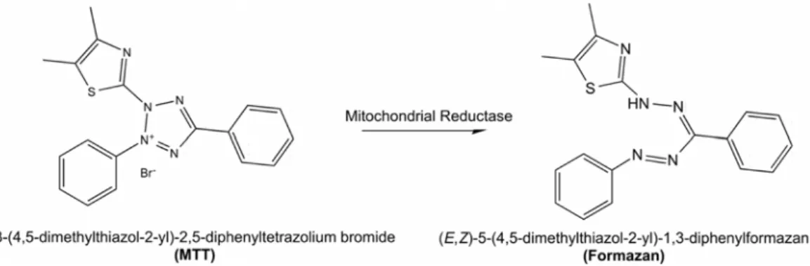
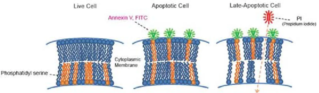
(27) List of Figures. List of Figures Figure 1.1. Number of publications from Thomson Reuters Web of ScienceTM database on ‘nanotoxicity’ or ‘nanotoxicology’ topics. Data for year 2015 is up to the end of May..... 5 Figure 1.2. Nanoscale showing nanomaterials compared to biological components, with indication of 'nano' and 'micro' sizes (from Wibke Busch, 2010). ...................................... 6 Figure 1.3. Nanoparticles tend to agglomerate in physiological fluids but may also build stable aggregates. Agglomerates and aggregates are characterised by the hydrodynamic diameter that can differ significantly from the primary particle diameter (from Jiang et al., 2008). .................................................................................................................................. 7 Figure 1.4. For the same quantity of material, the decrease in particle size means a high increase. in. surface. area. (from. www.uwgb.edu/dutchs/GRAPHIC0/GEOMORPH/SurfaceVol0.gif)................................ 8 Figure 1.5. Summary of possible mechanisms of NP for entering cells and cellular compartments. NP may actively be taken up by cells via phagocytosis (A), macropinocytosis (B), clathrin-mediated endocytosis (C), clathrin- and caveolaeindependent endocytosis (D), caveolae-mediated endocytosis (E) or by passive diffusion (F) (from Mühlfeld et al., 2008)........................................................................................ 11 Figure 1.6. Schematic illustration of mechanisms of action of ENM (from Dusinska et al., 2011). ................................................................................................................................ 13 Figure 1.7. Principle of DLS technique. Smaller particles tend to diffuse more rapidly than larger particles at a given temperature and viscosity, and the hydrodynamic diameter is calculated by experimentally determining the fluctuations of the scattered light. ............ 16 Figure 1.8. Analysing NP uptake in cells by flow cytometry. (A) Light scattering by a cell having no association with NP;(B) NP adhered to the cell surface leading to increase in both FSC and SSC; (C) NP internalization by the cell leading to increase only in SSC (from Sharma, 2011). ........................................................................................................ 20 Figure 1.9. MTT reduction in live cells by mitochondrial reductase results in the formation of insoluble. formazan. (from. http://www.intechopen.com/source/html/41784/media/image4_w.jpg) ........................... 21 Figure. 1.10.. LDH. assay. detection. mechanism. (from. tools.lifetechnologies.com/content/sfs/gallery/high/88953-001-LDH-Cytotox.jpg). ....... 22 Figure. 1.11.. The. principle. of. apoptosis. detection. by. annexin. V/PI. (from. www.dojindo.com/store/p/847-Annexin-V-FITC-Apoptosis-Detection-Kit.html). ......... 23. V.
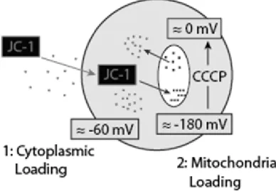
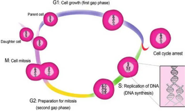
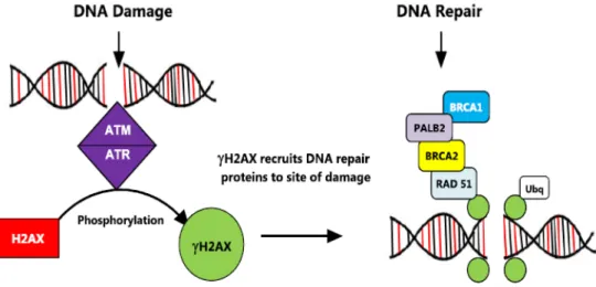
(28) Gözde KILIÇ. Figure 1.12. The principle of apoptosis detection with the fluorescent cationic dye JC-1 (from http://www.nexcelom.com/Applications/Apoptosis.html)................................................ 24 Figure 1.13. Phases of the cell cycle (from Mahmoudi et al., 2011). ......................................... 25 Figure 1.14. Scheme of the standard comet assay (from Azqueta and Collins, 2006). .............. 27 Figure 1.15. Images of cells subjected to comet assay. A) not damaged cell, B) moderately damaged cell, C) intensely damaged cell. ......................................................................... 28 Figure 1.16. Scheme of H2AX phosphorylation process and its consequences in DNA damage response. (from. http://www.amsbio.com/news/whitepaper/gamma_H2AX_white_paper.pdf). ................ 29 Figure 1.17. Representation of MN formation in cells undergoing nuclear division (from Fenech et al., 2011). .......................................................................................................... 30 Figure. 3.1.. TiO2. rutile. crystalline. structure. (from. www.chemtube3d.com/solidstate/_rutile%28final%29.htm) ........................................... 41 Figure. 3.2.. TiO2. anatase. crystalline. structure. (from. http://www.chemtube3d.com/solidstate/_anatase%28final%29.htm). ............................. 42 Figure 3.3. Size distribution of TiO2-S (a) and TiO2-D (b) in water, and TiO2-S (c) and TiO2-D (d) in cell culture medium. ................................................................................................ 54 Figure 3.4. MTT assay (a-b) and NRU assay (c-d) results on neuronal cells treated with TiO2-S or TiO2-D. The relative cell viability in the exposed cells was expressed as a percentage relative to the untreated control cells. ............................................................................... 56 Figure 3.5. Flow cytometry analysis of TiO2-S (a) or TiO2-D (b) uptake by SH-SY5Y cells. *P<0.05, significant difference with regard to the corresponding negative control. ........ 56 Figure 3.6. Cell-cycle analysis after treatment of SH-SY5Y cells with TiO2-S (a, b) or TiO2-D (c, d) for 3h (a, c) or 6h (b, d). PC: positive control. *P<0.05, **P<0.01, significant difference with regard to the negative control. ................................................................. 58 Figure 3.7.Genotoxicity in neuronal cells treated with TiO2-S (a) or TiO2-D (b) by MN evaluation. PC: positive control. *P<0.05, **P<0.01, significant difference with regard to the corresponding negative control. .................................................................................. 61 Figure 3.8. Genotoxicity in neuronal cells treated with TiO2-S (a) or TiO2-D (b) by γH2AX assay. PC: positive control. **P<0.01, significant difference with regard to the corresponding negative control. ........................................................................................ 61 Figure 3.9. Genotoxicity in neuronal cells treated with TiO2-S (a) or TiO2–D (b) by comet assay. PC: positive control. *P<0.05, **P<0.01, significant difference with regard to the corresponding negative control. ........................................................................................ 62. VI.
(29) List of Figures. Figure 3.10. Results of OGG1-modified comet assay in neuronal cells treated with TiO2–S for 3h (a) or for 24h (b), and with TiO2–D for 3h (c), or for 24h (d). PC: positive control. *P<0.05, significant difference with regard to the corresponding buffer. ........................ 64 Figure 3.11. Ratio of reduced to oxidised glutathione in neuronal cells treated with TiO2-S (a) or TiO2-D (b). PC: positive control. .................................................................................. 64 Figure 4.1. The hexagonal wurtzite structure model of ZnO. The tetrahedral coordination of ZnO is shown; O atoms are the larger red spheres while the Zn atoms are the smaller yellow spheres (from http://pixshark.com/wurtzite-structure.htm). ................................. 70 Figure 4.2.Characterization of ZnO-NP: (a) hydrodynamic diameter and (b) zeta potential in water; (c) hydrodynamic diameter and (d) zeta potential in culture medium. .................. 76 Figure 4.3. Results from MTT assay (a) and NRU assay (b) on viability of neuronal cells treated with ZnO-NP. Values inside the rectangle are statistically different from the corresponding controls. ..................................................................................................... 77 Figure 4.4. Uptake of ZnO-NP by SH-SY5Y cells as analysed by FCM. PC: positive control. *P<0.05, significant difference with regard to the corresponding negative control. ........ 78 Figure 4.5. Results of cell cycle analysis in neuronal cells treated with ZnO-NP for 3h (a) or 6h (b) (percentage of cells in each phase). *P<0.05; **P<0.01; significant difference with regard to the corresponding negative control. PC: positive control. ................................. 79 Figure 4.6. Results of MN evaluation in SH-SY5Y cells treated with ZnO-NP. *P<0.05, **P<0.01, significant difference with regard to the corresponding negative control. PC: positive control.................................................................................................................. 81 Figure 4.7. Results of γH2AX evaluation in SH-SY5Y cells treated with ZnO-NP. *P<0.05, **P<0.01, significant difference with regard to the corresponding negative control. PC: positive control.................................................................................................................. 82 Figure 4.8 Results of comet assay in SH-SY5Y cells treated with ZnO-NP. *P<0.05, **P<0.01, significant difference with regard to the corresponding negative control. PC: positive control. .............................................................................................................................. 82 Figure 4.9. Results of OGG1-modified comet assay in neuronal cells treated with ZnO-NP for 3h (a) or 6h (b). *P<0.05, **P<0.01, significant difference with regard to the corresponding buffer. PC: positive control. ...................................................................... 84 Figure 4.10.Results of membrane integrity assessment (LDH assay) in SH-SY5Y cells exposed to ZnO-NP. PC: positive control. ...................................................................................... 85 Figure 4.11. Analysis of Zn (II) ions released from ZnO-NP in cell culture medium. .............. 87. VII.
(30) Gözde KILIÇ. Figure 4.12. Cytotoxicity of Zinc (II) ions: MTT assay in SH-SY5Y cells treated with ZnSO4. **P<0.01, significant difference with regard to the negative control. .............................. 88 Figure 5.1. Cellular toxicity induced by ION (from Singh et al., 2010). ................................... 95 Figure 5.2. Characterization of S-ION: (a) TEM microphotograph, (b) distribution of hydrodynamic diameter in cell culture medium. ............................................................ 103 Figure 5.3. Viability of SH-SY5Y cells after exposure to S-ION assessed by MTT assay (a) and NRU assay (b). Values were normalized considering negative control as 100%. PC: positive control. *P<0.05, significant difference with regard to the corresponding negative control............................................................................................................... 104 Figure 5.4. Iron ion release from S-ION. ................................................................................. 106 Figure 5.5. Cellular uptake of S-ION as analysed by FCM. **P<0.01, significant difference with regard to the corresponding control. ....................................................................... 106 Figure 5.6. Cell cycle analysis of SHSY-5Y cells after exposure to S-ION: (a) 3h treatment; (b) 24h treatment. PC: positive control. **P<0.01, significant difference with regard to the corresponding negative control. ...................................................................................... 107 Figure 5.7. Apoptosis (subG1 region) in neuronal cells treated with S-ION. PC: positive control. *P<0.05, significant difference with regard to the corresponding negative control. ...... 109 Figure 5.8. Apoptosis and necrosis induction by S-ION for 3h (a) and 24h (b) evaluated by annexinV/PI staining. PC: positive control. *P<0.05, **P<0.01, significant difference with regard to the corresponding negative control.......................................................... 109 Figure 5.9. LDH leakage analysis after S-ION exposure. PC: positive control. ...................... 110 Figure 5.10. Results of γH2AX analysis on neuronal cells treated with S-ION. PC: positive control. **P<0.01, significant difference with regard to the corresponding negative control. ............................................................................................................................ 111 Figure 5.11. Results of MN evaluation in SH-SY5Y cells treated with S-ION. PC: positive control. *P<0.05, significant difference with regard to the negative control.................. 111 Figure 5.12. Results of comet assay in neuronal cells treated with S-ION. PC: positive control. *P<0.05; **P<0.01, significant difference with regard to the corresponding negative control. ............................................................................................................................ 111 Figure 5.13. Results of the acellular DCFH assay with S-ION, expressed as %fluorescence fold increase. PC: positive control. ........................................................................................ 113 Figure 5.14. Results of the DCFH assay in neuronal cells treated with S-ION, expressed as % fold increase compared to control. PC: positive control. **P<0.01, significant difference with regard to the corresponding negative control.......................................................... 115. VIII.
(31) List of Figures. Figure 5.15. Ratio of reduced to oxidised glutathione in neuronal cells treated with S-ION. PC: positive control. **P<0.01, significant difference with regard to the corresponding negative control............................................................................................................... 115 Figure 5.16. Results of OGG1-modified comet assay in neuronal cells treated with S-ION for 3h (a) and 24h (b). PC: positive control. *P<0.05, **P<0.01, significant difference with regard to the corresponding buffer. ................................................................................. 115 Figure 5.17. Effects of S-ION on repair of H2O2-induced DNA damage in neuronal cells. Incubation with S-ION was carried out either before H2O2 treatment (phase A), simultaneously (phase B), or during the repair period (phase C). *P<0.05, **P<0.01, significant difference with regard to the same treatment before repair; #P<0.05, significant difference with regard to the control. .............................................................................. 117. IX.
(32)
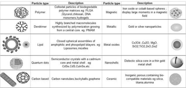
(33) List of Tables. List of Tables Table 1.1. Types of ENM classified by their structure and composition (adapted from (Dawidczyk et al., 2014; Tang et al., 2009). .................................................................... 9 Table 3.1. Characterization of TiO2-NP. .................................................................................... 54 Table 3.2. Apoptotic cell rates (%) after TiO2-NP exposure ...................................................... 58 Table 4.1. Physicochemical characterization of ZnO-NP .......................................................... 76 Table 5.1. Physicochemical description of S-ION. .................................................................. 103. XI.
(34)
(35) 1.. GENERAL INTRODUCTION.
(36)
(37) General Introduction. 1.1. NANOTOXICOLOGY 1.1.1. A brief history of nanotechnology Nanotechnology is the term given to those areas of science and engineering where nanometre scale (approximately 1-100nm) is utilized in the design, characterization, production and application of materials, structures, devices and systems (Blaunstein and Linkov, 2010). Study of the nanoscale phenomena is not a new field since scientists have been working in nanoscience for many decades in several areas of technology and medicine (Pitkethly, 2004). For example Pasteur’s work with spoilage bacteria, measurable on the micrometre (μm) scale (1μm = 1000nm), and Watson and Crick’s discovery of the DNA structure (a molecule of DNA is about 2.5nm wide) can be considered nanoscience (Institute of Medicine (US) Food Forum, 2009). However, interest in nanomaterials, in particular in engineered nanomaterials (ENM), i.e., those not naturally produced but manmade, has increased significantly over the past several decades. ENM are intentionally developed materials that have at least one dimension of 1–100 nm and exhibit novel properties compared to the larger scale form of a material of the same composition (Majuru and Oyewumi, 2009). The concept of nanotechnology was firstly introduced by the American physicist Richard Feynman in his lecture ‘There's plenty of room at the bottom’ at an American Physical Society meeting in Caltech on December 29, 1959 (Fitzpatrick et al., 2014). Dr. Feynman’s talk has been viewed as the first academic talk where he issued an invitation to physicists of his generation to enter a new field of physics: the field of nanotechnology (Ando and Kumar, 2010). Although he did not use the term ‘nanotechnology’, he described a process by which the ability to manipulate individual atoms and molecules might be developed (Feynman, 1960). A Japanese student called Norio Taniguchi was the first person in using the term ‘nanotechnology’ in a 1974 conference, where he stated ‘nano-technology mainly consists of the processing, separation, consolidation, and deformation of materials by one atom or one molecule’ (Taniguchi, 1974). However, nanotechnology did not develop into a field until the 1980s, when Eric Drexler, scientist at the Massachusetts Institute of Technology, who was unaware of Taniguchi's prior use of the term, published his first paper on nanotechnology in 1981, entitled ‘Molecular engineering: an approach to the development of general capabilities for molecular manipulation’ in the journal Proceedings of the National Academy of Sciences. (Ranjit and Klabunde, 2007). In this article, Drexler discussed the possibility of molecular manufacturing as a process of fabricating objects with specific atomic specifications, using designed protein molecules (Drexler, 1981). Drexler took these concepts and expanded the. 3.
(38) Gözde KILIÇ. study of their potential in a book entitled ‘Engines of creation: the coming era of nanotechnology’ (Drexler, 1996). Later, due to the publicity generated by Drexler’s work, scientists from all over the world began to have a vested interest in the field of nanotechnology. Just as Watson and Crick’s discovery of the structure of DNA led to a biotechnology revolution, Drexler created interest in the field but also outlined a nanotech revolution, and researchers around the world have brought nanotechnology to a more realistic and attainable level (Santamaria, 2012). Nanotechnology and nanoscience got a boost in the early 1980s with the availability of tools that allow scientists to see things that they were not able to see in the past, with the development of microscopic technologies such as scanning tunnelling microscope (STM) (Binnig and Rohrer, 1986) and atomic force microscopy (AFM). While nanotechnology came into existence through Feynman’s and then Drexler’s vision of molecular manufacturing, the field has evolved into research in several fields, including chemistry, materials science, medicine, toxicology, ecology, and industrial hygiene by multidisciplinary attempts with combination of chemistry, materials science, molecular biology, and molecular engineering (Oberdörster et al., 2007). The past two decades have witnessed tremendous progress in nanotechnology. Not only nanomaterials became an important part of our everyday lives as components of cosmetics, polymers, sporting goods, sensors, gasoline additives, or materials for medical diagnostics, but also they moved into our research labs to narrow down the concerns about their possible toxicity and influence on organisms caused by long-term exposure. Beginning in the early 2000s, concerns about the potential human and environmental health effects of nanomaterials were being expressed by many scientists, regulators, and nongovernmental agencies, because particles and materials in the nanosize range may pose toxicological hazards due to their enhanced reactivity (Santamaria, 2012). It was not until very recently that this topic began to gain increased interest and, as seen in Figure 1.1, the number of scientific articles published on ‘nanotoxicity’ or ‘nanotoxicology’, progressively increased; before 2005 it was almost negligible. An understanding of the toxicological profiles of ENM is necessary in order to ensure that these materials are safe for use and are developed responsibly, with optimization of benefits and minimization of risks. However, the development and production of ENM are increasing faster than their toxicological information is being acquired. The uncertainty and the lack of information on possible adverse effects of ENM have been taken into consideration by many organizations worldwide such as the US Environmental Protection Agency (EPA), the World Health Organization (WHO), the US National Institute for Occupational Safety and Health (NIOSH), the European Commission (EC) and the Organization for Economic Co-operation and Development (OECD). Many official documents have been published by these organizations. 4.
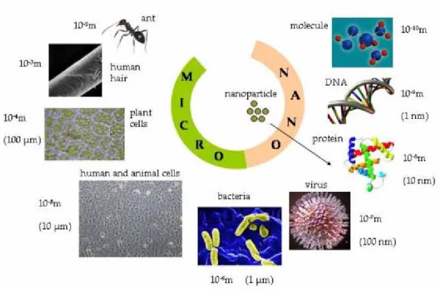
(39) General Introduction. addressing the need of dedicated research on appropriate methodological assays for assessing ENM toxicity (Colognato et al., 2012).. Figure 1.1. Number of publications from Thomson Reuters Web of ScienceTM database on ‘nanotoxicity’ or ‘nanotoxicology’ topics. Data for year 2015 is up to the end of May.. 1.1.2. Terminology Many committees and reports were set up worldwide by various institutions, to define terms in the context of nanotechnology [(Department for Environment, Food and Rural Affairs of the United Kingdom Government, 2007), (European Commission Joint Research Centre, 2011), US (National Institute for Occupational Safety and Health (NIOSH, 2006),US National Nanotechnology Initiative (NNI, 2007)] which might give a false impression of a not yet achieved consensus. Thus, this section will be devoted to define such important terms. The prefix ‘nano’, derived from the Greek ‘nanos’ which means ‘dwarf’, is becoming increasingly common in scientific literature (Goncalves et al., 2011). Popularly, ‘nano’ is used as an adjective to describe objects, systems, or phenomena with characteristics arising from nanometre-scale structure (Buzea et al., 2007). The term ‘nanomaterial’ refers to a material, i.e., functionally specific matter, which, owing to its nanometric structure, has a modified chemical or physical property (or combination of properties) that is improved, adapted, or new compared with the bulk material of the same composition (Bréchignac et al., 2010). The British Standards Institution (BSI, 2011) makes the more precise definition of a nanomaterial as being either a nanoparticle (in the sense of a nanoobject), or a nanostructured material whose dimensions exceed the nanometric scale (1nm to 100nm). The nanometre is a metric unit of length, and denotes one milliardth of a metre or 109. m.. 5.
(40) Gözde KILIÇ. The technical committee of the International Standards Organization (ISO) devoted to nanotechnologies defines nanoparticles (NP) as ‘particles with a nominal diameter (such as geometric, aerodynamic, mobility, projected-area or otherwise) smaller than about 100 nm (ISO/TS 27687). For comparison purposes, the width of an average hair is 100,000 nm, human blood cells are 2,000 to 5,000nm long, and a strand of DNA has a diameter of 2.5nm (Naahidi et al., 2013) (Figure 1.2). These simple size comparisons give an idea of using NP as very small probes that would allow us to spy at the cellular machinery without introducing too much interference. Hence, NP possess the ability to imitate and interact with biological systems at the same scale of their parts; this is the explanation what has driven the burst of nanobiotechnology research recently. Materials engineered to such a small scale are often referred to as engineered nanomaterials (ENM), which can take on unique optical, magnetic, electrical, and other properties (NIH, 2014). In the context of this document, to avoid the confusion, the terms ENM and NP will be used interchangeably.. Figure 1.2. Nanoscale showing nanomaterials compared to biological components, with indication of 'nano' and 'micro' sizes (from Wibke Busch, 2010).. Nanotoxicology was proposed as a new branch of toxicology to address the adverse health effects caused by nanomaterials (Oberdörster et al., 2005). Toxicology, as it is defined by the Society of Toxicology, is the study of the adverse effects of chemical, physical or biological agents on living organisms and the ecosystem, including the prevention and improvement of such adverse effects (SOT, 2005). When this definition of toxicology is combined to nanomaterials, nanotoxicology could be described as the study of the adverse effects of ENM on. 6.
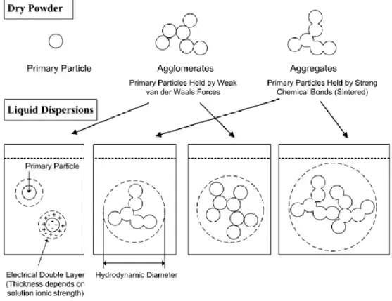
(41) General Introduction. living organisms and the ecosystems, including the prevention and amelioration of such adverse effects (Oberdörster, 2010). For many reasons related to their behaviour and environment, e.g., during their fabrication, nanomaterials rarely occur in free form, i.e., isolated from one another (Ju-Nam and Lead, 2008; S. Isfort and Rochnia, 2009). They tend to group together into more or less stable but disordered clusters, some dimensions of which may be significantly longer than 100 nm. The term ‘primary particle’ is also used to designate the elements making up a cluster. According to ISO/TR27628 document, there are two types of clusters: when primary particles adhere to one another by weak physical bonds, e.g., Van der Waals forces, it is called agglomerate. If the cluster consists of primary particles connected by strong chemical bonds (covalent bonds), or they have partially fused together, the cluster is referred to as an aggregate (Witschger, 2011) (Figure 1.3).. Figure 1.3. Nanoparticles tend to agglomerate in physiological fluids but may also build stable aggregates. Agglomerates and aggregates are characterised by the hydrodynamic diameter that can differ significantly from the primary particle diameter (from Jiang et al., 2008).. 1.1.3. Physicochemical properties of nanomaterials Advances in nanotechnology in both scientific research and application areas are likely to significantly benefit society and economy (Oberdörster et al., 2007). By some estimates, nanotechnology promises to far exceed the impact of the Industrial Revolution and is projected. 7.
(42) Gözde KILIÇ. to become a three billion dollars market by 2015. The reason for ENM to be that popular is mainly because of their unique physical and chemical properties, which give them a great potential for industrial and biomedical applications (Nel et al., 2006). The unusual physicochemical properties of ENM, such as their small size, surface structure, solubility, shape, and aggregation (Sutariya and Pathak, 2015) are also the reason for requirement of an interdisciplinary approach to study these materials as potential toxic agents, involving multiple aspects ranging from physics and chemistry to biology and medicine (Fubini et al., 2007). Nanosize is one of the main physicochemical features that make ENM different from their larger equivalents (De Jong and Borm, 2008). As size decreases, the mobility, potential transport, and availability of the particle in the environment increase. This mobility could translate to passive transport across cellular membranes. The NP that cross membranes will then be able to interact with macromolecules such as proteins, DNA, or RNA. Also, as a direct consequence of their small size, NP have a very large surface area with regard to the same mass of bigger particles. This may be one of the reasons why NP are generally considered more toxic than larger particles of the same material. In other words, while the size of a particle decreases, its surface area increases, which allows a greater proportion of its atoms or molecules to be exposed on the surface rather than the interior of the material (Figure 1.4). The increase in surface area determines the potential number of reactive groups on the particle surface and therefore it is strongly possible that biological activity might increase.. Figure 1.4. For the same quantity of material, the decrease in particle size means a high increase in surface area (from www.uwgb.edu/dutchs/GRAPHIC0/GEOMORPH/SurfaceVol0.gif). Another very important factor that may contribute to the behaviour and biological responses of ENM is the state of dispersion in a particulate system, which refers to the relative. 8.
Figure
+7

Documento similar
The aim of this study was to determine, through a preliminary in vitro study on human glioblastoma (A172, LN229), anaplastic glioma (SF268) and neuroblastoma (SK-N-SH) cell lines,
In the preparation of this report, the Venice Commission has relied on the comments of its rapporteurs; its recently adopted Report on Respect for Democracy, Human Rights and the Rule
The draft amendments do not operate any more a distinction between different states of emergency; they repeal articles 120, 121and 122 and make it possible for the President to
The characteristic morphology of the ZnO thin films is shown in the top-view FESEM image of Figure 3a, and a magnified cross- sectional image is shown in Figure 3b, which confirms
Mazarío, “Effect of the surface charge on the adsorption capacity of chromium (VI) of iron oxide magnetic nanoparticles prepared by microwave-assisted synthesis,”.
The results shown in this chapter demonstrated that the quality of the sample of pyramidal CdSe NPs pot-deposited on HOPG in terms of NP coverage and polydispersity
The cyclic voltammetry characterization of CdSe NPs with different morphology, size and surface composition reveals that the redox processes depend on the NP
reinhardtii after 1, 24 and 72 h of exposure to SPION as measured by flow cytometry using different fluorochromes: (A) plasma membrane potential, (B) mitochondrial membrane
