Metabolic fate of ellagitannins from tropical highland blackberry (Rubus Adenotrichos) and its relation with gut microbiota ecology
143
0
0
Texto completo
(2) 2. SupAgro Ecole doctorale Sciences des Procédés – Sciences des Aliments Unité Mixte de Recherche (UMR) Spécialité : Biotechnologie, microbiologie Présentée par M CRISTINA GARCIA MUÑOZ A. BIOCONVERSION DES ELLAGITANNINS DE LA MURE TROPICALE DE MONTAGNE (Rubus Adenotrichos) ET RELATION AVEC L’ÉCOLOGIE DU MICROBIOME INTESTINAL Soutenue le 12 Décembre 2013 devant le jury composé de M. Olivier DANGLES, Université d'Avignon, INRA Mme Joëlle QUETIN-LECLERCQ, Université Catholique de Louvain M. Lars Ove, DRAGSTED Université de Copenhagen Mme Christèle HUMBLOT, Institut de recherche pour le développement, IRD M. Jean-Christophe MEILE, La recherche agronomique pour le développement, CIRAD M. Fabrice VAILANT La recherche agronomique pour le développement, CIRAD-CITA. Professeur. Président du jury. Professeur. Rapporteur. Professeur. Rapporteur. Chercheur. Examinateur. Chercheur. Examinateur. Chercheur. Directeur de Thèse. You created this PDF from an application that is not licensed to print to novaPDF printer (http://www.novapdf.com).
(3) 3. DEDICATION. I dedicate this thesis to my mother Maria Yolanda Muñoz who taught me the moral values based on honesty, compassion, courage, and forgiveness, to become an integral person in both personal and professional life.. You created this PDF from an application that is not licensed to print to novaPDF printer (http://www.novapdf.com).
(4) 4. ACKNOWLEDGEMENTS The project has been conducted at the Research Centre in Natural Products CIPRONA and at the National Centre of Food technology CITA belong to University of Costa Rica, and also at the Centre of Agricultural Research for Development, CIRAD. The project has been sponsored by the Centre of Agricultural Research for Development and Colombian Corporation of Agricultural Research, CORPOICA. I am deeply appreciative of their efforts and I sincerely acknowledge their wholehearted cooperation. I am grateful to The Almighty God for giving me the strength, perseverance, courage and patience to achieve this goal. I would like to thanks first and foremost to my thesis advisor Dr. Fabrice Vaillant researcher from CIRAD and teacher at the University of Costa Rica for this opportunity to explore and learn about new fields of the science and emerging technologies. Fabrice’s enthusiasm for research and his great interest in my work was encouraging. My immense gratitude is due to the dissertation committee for their advices, comments, thoughtful questions and comments. They were valued greatly. I am greatly indebted to CIPRONA in head of Dr. Rosaura Romero, not only for open the doors of her latest technology laboratories when all others were closed, but also for the warm atmosphere I experienced. I also enjoyed the very pleasant work environment offered by the CITA, in head of Dr. Carmela Velazquez and the professional and multivariate work environment proper of the CIRAD, in the unit of QUALISUD in head of Dr Max Reynes where is possible to count with the support of researchers from diverse scientific areas. My deepest and immense gratitude is due to Lic. Lorena Hernández. Her broad expertise and her patience in introducing me to the exciting and complex world of LC-MS are warmly remembered. Her friendliness made my work and life more pleasant. I will never forgot her invaluable help when I was sick and especially when for a week I was not able to walk, she picked me up and took me to the lab and back home; no matter the difficulties this could bring her. I will always remember her strength, energy, enthusiasm and. You created this PDF from an application that is not licensed to print to novaPDF printer (http://www.novapdf.com).
(5) 5 compromise with her work; but even more remarkable, her altruism, an example of life that deserve to be followed. I also express my gratitude to Dr Ana Mercedes Pérez for her constant encouragement, advice and suggestions. This project would not have been possible without her invaluable support especially in logistics topics. I was also privileged to be supervised by Dr. Jean Christophe Meile. His expertise, support, encouragement, sincere and valuable guidance introducing me into the wonderful field of molecular biology will be always remembered. My sincere thanks to Dr. Max Reynes, Pierre Brat, Didier Montet and Gerard Loiseau, their interests in my progress and their insightful comments throughout the process have been invaluable. I am sincere grateful to Dr. Christian Mertz for his advice, friendly conversation and insightful comments throughout the project This thesis would have remained a dream had it not been for my superiors at CORPOICA, especially Dr. Hugo García, Gustavo García and Diego Aristizabal; who giving me the opportunity, means, and time to come true this dream. I also must thank to laboratory assistants Henry Velasquez from School of Food Technology and Dennis Camareno from School of Microbiology from University of Costa Rica for their invaluable and unconditional help just when I need it. Special thanks to Mr. Miguel Urruela who tirelessly signed the entry permits for all the weekends and holidays at CITA facilities, during my stay in Costa Rica.. My warmest thanks are owed to Dr Jean Michel Roger and Dr. Nathalie Gorretta, whose contribution in the final stages of this work was invaluable and for their patience and enthusiasm introducing me to the interesting world of chemometrics. My deepest gratitude is due to all the technician and administrative staff Marcy González, Vanny Mora, Alejandra Aguero, Marvin Soto, Randall Cordero, Marielos Torres, Mariela Boniche, Martin Loira, Marta Avila, Deisylia Alpizar from CITA, for their skillful technical. assistance, enthusiasm and friendliness made my work much more pleasant.. You created this PDF from an application that is not licensed to print to novaPDF printer (http://www.novapdf.com).
(6) 6 I owe my deepest gratitude to technician Juan Carlos Brenes and Diego Zuñiga for their invaluable help, friendless and constant encouragement. I am grateful to technicians who shared their memories and experiences, especially to Bernard Lyan, Adrian Servant, Pascalin Alter, Gill Moran and Isabell Metayer. I also express my appreciation to Dr. Alice Perez, Dr. Guiselle Tamayo, Alicia Hernández, and fellow student Sebastian de la Osa for their friendly conversation and encouragement throughout my study and research. I also thanks to Mrs Marie Pierre Obede, Mrs Chantal Canales, Miss Karla Salazar, and Mrs Guisselle Casante for their help in administrative issues. I wish to thank to Mrs Martine Barraud, for her valuable and kind assistance in all the issues concerning to the Ecole doctorale. My gratitude is due to Mrs Katia Gómez, and Seidy Vargas who provided me with a delicious and enough coffee to keep me awake and active along the 12200 hours of work. I am sincerely grateful also to my friends Carolina Castro, Ana Maria Garavito, Mónica Espíndola, Miguel Sabogal, Miguel Llaín, Rodrigo Buitrago, Angela López, Jose María Martínez, Teruya Satokichi, Elazar Fallik for their permanent encouragement. My heartfelt thanks go to my parents, for their unceasing encouragement and support for. their unconditional love and support and for teaching me how work without rest. I take this opportunity to record my sincere and warm thanks to my brothers Guillermo, Juan, German, Fernando and their partners and children for their constant support and friendship. They have given me the drive and discipline to tackle any task with enthusiasm and determination.. My gratitude is also due to all who, directly or indirectly, helped me to achieve this important goal.. You created this PDF from an application that is not licensed to print to novaPDF printer (http://www.novapdf.com).
(7) 7. ABSTRACT Consumption of dietary ellagitannins (ETs) could be associated mainly with prevention of cardiovascular diseases and regulation of hormone-dependent cancers. Nonetheless, ETs are not bioavailable as such; therefore, after being partially converted into ellagic acid (EA) in the upper gastrointestinal (GI) tract, they undergo sequential bioconversion in the colon by gut microbiota into urolithins, a more bioavailable and bioactive group of molecules that persist up to 4 days at relatively high concentrations in urine. Variability of urolithin excretion in urine is high and three main groups, “no or low urolithin excreters,” “predominantly UA derivatives excreters” and “predominantly UB derivatives excreters,” were observed on a cohort of 26 healthy volunteers. These categories were also unambiguously observed following the total excretion of main ETs’ metabolites over a 4 day period after ingesting one shot of juice, and at different periods of time along one year. Although relatively high inter- and intra-individual variabilities were observed, individuals preserved their status during various intervention periods with different amounts of ETs ingested. UPLC-PDA and ESI-Q-TOF/MS1 and MS2 allowed the tentative assignment of an identity to 15 other ETs metabolites in urine, but this profiling did not allow the discrimination of any other compounds aside from UA or UB derivatives. In-vitro fermentation of ETs and EA with fecal stools showed a specific metabolic pathway ending in the production of UA. Nonetheless, metabolites excreted in-vivo are much more complex, highlighting strong interactions between host excretory system and composition of gut microbiota. Hepatic recirculation and additional bioconversion of Phase II metabolites in the colon may explain predominant excretion of UB in some volunteers. Microbiota ecology assessed by PCR-Denaturing Gradient Gel Electrophoresis (DGGE) fingerprint method allowed the association of some microorganism species to higher capacity of bioconversion of dietary ETs into urolithins. Keywords: Ellagitannins, blackberry, urolithin, colonic metabolites, ETs degradation patterns, gut microbiota, gastrointestinal tract,. You created this PDF from an application that is not licensed to print to novaPDF printer (http://www.novapdf.com).
(8) 8. RESUME La consommation d’aliments riches en ellagitannins (ETs) pourrait être associée principalement à la prévention des maladies cardiovasculaires et la régulation des cancers hormono-dépendants. Néanmoins, les ETs ne sont pas biodisponibles en tant que tel et, après avoir été partiellement transformés en acide ellagique (EA) dans le tractus gastrointestinal (GI) supérieur, ils sont métabolisés dans le côlon par la flore intestinale en urolithines, un groupe de molécules plus biodisponibles et bioactives qui peuvent persister jusqu'à 4 jours à des concentrations relativement élevées dans le plasma et l'urine. La variabilité de l'excrétion des urolithines dans l'urine est importante et à partir d’un échantillon de population de 26 volontaires sains, trois groupes principaux d’individus ont pu être distingués : "faible ou non-excréteur d’urolithin », « Excréteur prédominant d’UA et dérivés» et « Excréteur prédominant d’UB et dérivés»". Ces groupes ont également été observés en considérant la cinétique totale d’excrétion sur une période de 4 jours après ingestion du jus et à des périodes différentes tout au long d'une année. Bien que les variabilités inter-et intra-individuelles soient relativement élevées, les individus conservent leur statut au cours des différentes périodes d'intervention même en modifiant les quantités d'ETs ingérées. L’analyse par UPLC-PDA/ESI-Q-TOF/MS2 a permis d’attribuer hypothétiquement une identité à 15 autres métabolites d’ETs dans l'urine, mais le profilage métabolomique n’a pas permis de discriminer d’autres composés exceptés les dérivés d’UA ou d’UB. La fermentation in-vitro des ETs et EA, par les matières fécales a montré une voie métabolique spécifique qui débouche sur la production d’UA. Néanmoins, les métabolites excrétés in vivo sont beaucoup plus complexes ce qui met en évidence de fortes interactions entre le système excréteur de l'hôte et la composition du microbiote intestinal. La recirculation hépatique suivie par une re-conversion des métabolites de phase II dans le côlon permettrait d’expliquer l’excrétion d’UB chez certains volontaires. L’écologie spécifique de la flore intestinale évaluée par la méthode des empreintes PCR-DGGE a permis d’identifier quelques microorganismes associés à une plus grande capacité de bioconversion des ETs en urolithins. You created this PDF from an application that is not licensed to print to novaPDF printer (http://www.novapdf.com).
(9) 9 TABLE OF CONTENTS. DEDICATION ................................................................................................................. 3 ACKNOWLEDGEMENTS ............................................................................................... 4 ABSTRACT ...................................................................................................................... 7 RESUME ........................................................................................................................... 8 LIST OF PUBLICATIONS.............................................................................................. 12 LIST OF TABLES ........................................................................................................... 13 LIST OF FIGURES ......................................................................................................... 15 LIST OF ABBREVIATIONS .......................................................................................... 19 1. INTRODUCTION .................................................................................................... 21. 2. LITERATURE REVIEW.......................................................................................... 24 2.1. ELLAGITANNINS ........................................................................................... 24. 2.1.1. STRUCTURE ............................................................................................. 24. 2.1.2. OCCURRENCE ......................................................................................... 25. 2.1.3. ELLAGITANNINS BIOAVAILABILITY ................................................. 27. 2.1.4. POTENTIAL IMPACT ON HEALTH ........................................................ 31. 2.1.5. TOXICITY OF DIETARY ELLAGITANNINS.......................................... 38. 2.2. GUT MICROBIOTA AND BIOCONVERSION OF DIETARY COMPOUNDS... .......................................................................................................................... 38. 3. MATERIALS AND METHODS .............................................................................. 44 3.1. REAGENTS AND MATERIALS ...................................................................... 44. 3.1.1. CHEMICALS ............................................................................................. 44. 3.1.2. GROWTH MEDIUM ................................................................................. 44. You created this PDF from an application that is not licensed to print to novaPDF printer (http://www.novapdf.com).
(10) 10 3.1.3 3.2. BLACKBERRY JUICE, BBJ ..................................................................... 44. ANALYTICAL METHODS .............................................................................. 45. 3.2.1. SAMPLE PREPARATION......................................................................... 45. 3.2.2. ANALYSIS. PERFORMED. FOR. THE. IDENTIFICATION. AND. QUANTIFICATION OF ETs AND THEIR METABOLITES .................................. 46. 4. 3.2.3. CLINICAL STUDIES WITH HEALTHY VOLUNTEERS ........................ 60. 3.2.4. IN-VITRO FERMENTATION WITH STOOLS ......................................... 62. 3.2.5. PROFILING OF INTESTINAL MICROBIOTA ........................................ 65. 3.2.6. MULTIVARIATE ANALYSIS OF DGGE PROFILES .............................. 76. METABOLIC FATE OF ELLAGITANNIN’S COLONIC DERIVATIVES ............. 79 4.1. Identification and quantification of Ellagitannins in blackberry juice ................. 79. 4.2. IDENTIFICATION OR ANNOTATION OF ETs METABOLITES PRESENT IN. URINE AFTER INGESTION OF BBJ ......................................................................... 79 4.2.1. IDENTIFICATION OF UAG AND UBG IN URINES ............................... 80. 4.2.2. ANNOTATION OF OTHER ETS METABOLITES FOUND IN URINE... 82. 4.3. DIVERSITY OF URINARY EXCRETION PATTERNS IN HUMANS ............ 85. 4.3.1. INTER-INDIVIDUAL VARIABILITY OF URINARY EXCRETION OF. UROLITHIN OVER A 4-DAY PERIOD .................................................................. 86 4.3.2. KINETIC EXCRETION OF UROLITHIN A AND UROLITHIN B ........... 89. 4.3.3. INTRA-INDIVIDUAL VARIABILITY OF URINARY EXCRETION OF. UROLITHINS .......................................................................................................... 92 4.4. IN-VITRO SIMULATION OF COLONIC FERMENTATION ......................... 94. 4.4.1. IDENTIFICATION. OF. ETs. METABOLITES. PRESENT. IN. FERMENTATION BROTH ..................................................................................... 94 4.5. VARIABILITY OF STOOLS' ABILITY TO PRODUCE ETs METABOLITES 98. You created this PDF from an application that is not licensed to print to novaPDF printer (http://www.novapdf.com).
(11) 11 4.5.1. EFFECT OF SUBSTRATE IN THE PRODUCTION OF UROLITHINS ... 99. 4.5.2. SUGGESTED BIOCONVERSION PATHWAY OF ETs TO UROLITHIN. IN-VITRO .............................................................................................................. 101 4.6 5. POTENTIAL INTERACTION BETWEEN MICROBIOTA AND HOST ....... 102. MICROBIOTA ECOLOGY AND ABILITY TO PRODUCE UROLITHINS ......... 105 5.1. RESEARCH OF CORRELATION BETWEEN GUT ECOLOGY AND IN-VIVO. ABILITY TO EXCRETE UROLITHINS ................................................................... 105 5.1.1. PCR-DGGE PROFILING OF THE 26 VOLUNTEERS............................ 105. 5.1.2. RELATION WITH UROLITHIN EXCRETERS STATUS DETERMINED. ON A URINE SPOT ............................................................................................... 109 5.2. RELATION BETWEEN MICROBIOTA AND IN-VITRO BIOCONVERSION. OF UROLITHIN ........................................................................................................ 115 5.3. IDENTIFICATION OF FECAL BACTERIA INVOLVED IN UROLITHIN. METABOLISM ......................................................................................................... 118 5.4. EFFECTS OF ETs INGESTION ON MICROFLORA PROFILE .................... 120. 6. CONCLUSIONS .................................................................................................... 124. 7. BIBLIOGRAPHY ................................................................................................... 128. You created this PDF from an application that is not licensed to print to novaPDF printer (http://www.novapdf.com).
(12) 12. LIST OF PUBLICATIONS Results from this study have been published or will be published like scientific papers: Garcia-Muñoz, Cristina and Vaillant Fabrice. Metabolic Fate of Ellagitannins: Implications for Health, and Research Perspectives for Innovative Functional Foods. Critical Reviews in Food Science and Nutrition Garcia-Muñoz, Cristina; Hernandez Lorena, Pérez, Ana M, Vaillant Fabrice. Diversity of urinary excretion patterns of primary ellagitannin colonic metabolites after ingestion of tropical highland blackberry (Rubus adenotrichos) juice, submitted and accepted by Food Research International. Garcia-Muñoz, Cristina, Meile Jean Christophe, Hernandez, Lorena, Vaillant, Fabrice Microbiota ecology affects urolithin production during in-vitro simulation of colonic fermentation of blackberry juice. Garcia-Muñoz, Cristina; Hernandez Lorena, Pérez, Ana M., Vaillant Fabrice Colonic microflora and the bioconversion of ellagitannins from tropical highland blackberries in to bioactive molecules. International Conference on Nutrigenomics - INCON 2012 “GeneDiet Interaction for Personalized Health and Disease Prevention" San José, Costa Rica. October, 1fst to 4th, 2012 Garcia-Muñoz, Cristina; Hernandez Lorena, Mertz, Christian, Vaillant Fabrice Approaching to metabolic pathway of urolithin production from blackberry juice. International Conference on Nutrigenomics - INCON 2012 “Gene-Diet Interaction for Personalized Health and Disease Prevention" San José, Costa Rica. October, 1fst to 4th, 2012.. You created this PDF from an application that is not licensed to print to novaPDF printer (http://www.novapdf.com).
(13) 13. LIST OF TABLES Table 2.1 Major ellagitannins food sources ...................................................................... 26 Table 2.2 Content of main phenolic compounds in blackberries (R. adenotrichos) at three maturity stages. [31]......................................................................................................... 27 Table 2.3 Contents of main phenolic compounds in blackberries (R. adenotrichos) at three maturity stages.[35].......................................................................................................... 28 Table 2.4 Recent prospective clinical studies of consumption of food rich in ellagitannins 33 Table 2.5 Summary of intervention studies with animals to evidence health benefits of dietary ETs ...................................................................................................................... 34 Table 2.6 Summary of some recent results obtained on cell-models by ETs, EA and urolithins.......................................................................................................................... 35 Table 2.7 Main studies on bioconversion of dietary compounds into bioactive colonic metabolites [130] ............................................................................................................. 41 Table 3.1 Relative efficiency of electrospray ionization of common chemical functional groups (Fiehn Laboratory, UC Davis) .............................................................................. 53 Table 3.2 PCR primers targeted for 16S rRNA gene, without GCClamp [197] ................. 67 Table 3.3 Encoding for the four bases and for ambiguous positions in DNA sequences .... 75 Table 4.1 Contents of ellagitannins in blackberry (Rubus adenotrichos) juice .................. 79 Table 4.2 UPLC-DAD/ESI(+)-Q-TOF/MS and MS2 analyses of ellagitannins colonic metabolites found in urines of volunteers ......................................................................... 83 Table 4.3 UPLC /ESI(+)-Q-TOF/MS profiling of the main ETs metabolites excreted in urine during the 4 days after ingestion of blackberry juice for nine individuals (letters) and for different intervention periods (numbered in chronological order) ................................ 89 Table 4.4 Total excretion of urolithins UA and UB and percentage of conversion in relation to ingested ETs ................................................................................................................ 91 Table 4.5 Chromatographic and mass spectra characteristic of ellagitannins metabolites found in broth culture incubation of EA and BBJ inoculated with fecal stools from urolithin excreters .......................................................................................................................... 95. You created this PDF from an application that is not licensed to print to novaPDF printer (http://www.novapdf.com).
(14) 14 Table 4.6 Main ETs metabolites released along the degradation of ETs.a) EA, b) UM5, c) Iso UD1, d) Iso UD2, e) Iso UD3. .................................................................................... 96 Table 4.7 Main ETs metabolites released along the degradation of ETs. a)Iso UC1, b) Iso UC2, c) Iso UC3 d) UA.................................................................................................... 97 Table 5.1 Concentration of urolithin A and B derivatives in a urine spot collected at 51±5 h, after ingestion of one shot of 250 ml of blackberry juice. ............................................... 109 Table 5.2 EA and UR Index of stools from 9 individuals ................................................ 116 Table 5.3 Recapitulation of retardation factors (Rf) of the bands found as discriminative in the different trials ........................................................................................................... 118 Table 5.4 Results of the sequence similarities of the excised bacterial bands from different DGGE gels .................................................................................................................... 119. You created this PDF from an application that is not licensed to print to novaPDF printer (http://www.novapdf.com).
(15) 15. LIST OF FIGURES Figure 2.1: A. Puncalagin; B. Lambertianin C10, and the HHDP group. [3] ..................... 25 Figure 2.2 Phylogenetic tree of the microbial diversity in the human gastrointestinal tract, based on the 16S rDNA bacterial sequence data [2] ......................................................... 39 Figure 3.1 HPLC-MS and B) UPLC-MS analysis of mouse urine in a 3-dimensional map to observe the differences on the information generated [1] .................................................. 47 Figure 3.2 Comparing speed, sensitivity and resolution in HPLC and UPLC analysis [2, 3] ........................................................................................................................................ 47 Figure 3.3 Calibration curve of EA, for quantification of ellagitannins by HPLC, following the method proposed by Mertz, 2007................................................................................ 50 Figure 3.4 UV chromatogram of blackberry (Rubus adenotrichos) juice. ......................... 50 Figure 3.5 Calibration curve of EA, UA and UB for quantification of ellagitannins in BBJ, and ETs metabolites in urine and broth culture incubation. ............................................... 51 Figure 3.6 Basic information provided by UPLC: UV chromatogram-Area under the curve and the UV spectra for m/z+ 213.0552 ion is illustrated ................................................... 51 Figure 3.7 Electrospray ionization principles[190] ........................................................... 53 Figure 3.8 Illustration of a mass spectrometer MS V-Mode and each one of their parts, along with UPLC that become a powerful tool for metabolite analysis ............................. 56 Figure 3.9 Basic information obtained from MS: A. Total ion chromatogram (TIC) B. extracted ion chromatogram m/z and C. Exact mass measured spectrum and elemental composition report ........................................................................................................... 58 Figure 3.10 Scheme of the clinical test followed to approach ETs metabolism. ............... 61 Figure 3.11 Layout of study design, different steps followed ............................................ 62 Figure 3.12 Scheme of in vitro test followed to identify microbial metabolites and potential microbial species involved in ETs degradation ................................................................. 64 Figure 3.13 PCR process. 1. Denaturing, 2. Annealing, 3. Extension, 4. Cycles [202] ...... 66 Figure 3.14 DGGE principles: voltage, gradient, and direction of moving ........................ 68 Figure 3.15 Spatial distribution of microbes at a selected site in the gastrointestinal tract [1] ........................................................................................................................................ 69. You created this PDF from an application that is not licensed to print to novaPDF printer (http://www.novapdf.com).
(16) 16 Figure 3.16 Example of the calibration of the retardation factor (Rf) between lanes and two different gels .................................................................................................................... 73 Figure 3.17 Process followed by Phoretix 1D® to obtain aligned bands between lanes and gels and Excel file with band % corresponding to each retardation time. .......................... 74 Figure 3.18 Example of DNA sequences provided by GATC BIOTECH ......................... 76 Figure 4.1 Example of UV chromatogram (at 278 nm) of a urine sample from a volunteer collected 3 days after ingestion of blackberry juice, including peaks and UV spectra of glucuronide forms of urolithin A (UAG) and B (UBG). ................................................... 80 Figure 4.2 MS2 Fragmentation of urolithin A and urolithin B ........................................... 81 Figure 4.3 MS2 fragmentation of urolithin A glucuronide ................................................. 81 Figure 4.4 Example of MS2 fragmentation pattern of Ellagic Acid, urolithins: UM5, UD UC, dimethyl elagic acid glucuronide (DMEAG), methyl urolithin A (MUA) and dimethylurolithin C (DMUC), and tentative prediction of some fragment ions ............................... 84 Figure 4.5 Concentration of primary urolithin A and B derivatives (aglycone and glucuronides forms) in a urine spot, from 26 volunteers, collected at 51±5 h after ingestion of 250 mL of blackberry juice diluted (50:50) with water. ................................................ 85 Figure 4.6 Total conversion of ETs to UA and UB derivatives (aglycone and glucuronide determined by UPLC/DAD) excreted in urine over 4 days after ingestion of 250-mL of tropical blackberry juice for 9 individuals (C,D,F,G,H,I,O,R,W) at different interventions. ........................................................................................................................................ 87 Figure 4.7 Urolithin UA and UB detected in every one of the samples of urine excreted for 5 individuals up to seven days after they ingested a 250-ml of BBJ. ................................. 90 Figure 4.8 Excretion kinetics of UA(on the right) and UB(on the left) derivatives for four individuals (C,G,H, and I numbered in chronological order) performed at different periods of time around one year.................................................................................................... 93 Figure 4.9 Example of UV chromatogram (at 278 nm) from a fermentation broth (EA as substrate, 120 h fermentation, stool 96 h inoculate from individual G............................... 95 Figure 4.10 Primary ellagitannins metabolites assessed after 24, 72 and 120 h of batch anaerobic incubations of blackberry juice (A) and ellagic acid (B) with fecal stools donated by nine volunteers (A, B,C,D,E,F,G,H,I), previously qualified as urinary ......................... 99. You created this PDF from an application that is not licensed to print to novaPDF printer (http://www.novapdf.com).
(17) 17 Figure 4.11 Suggested metabolite pathway occurring during incubation of pure ellagic acid with active fecal stool from urolithin excreter ................................................................ 102 Figure 4.12 Schematic representation of proposed metabolic pathways of the excretion of the main metabolites identified in human urine after ingestion of tropical highland blackberry juice ............................................................................................................. 104 Figure 5.1 Hierarchical cluster analysis of the 26 PCR-DGGE profile PCR-DGGE fingerprint corresponding to amplicons of the V3 region of 16S rDNA of faecal bacteria from stool (Letter and number of lane corresponds respectively to volunteers identity and number of repetition; Ctrl is a control lane with lactobacillus plantarum and E.Coli) ..... 107 Figure 5.2 Complete linkage cluster analyses of 1-r (1 minus the Pearson coefficient between lanes) (Letter and number of lane corresponds respectively to volunteers identity and number of repetition; Ctrl is a control lane with L. plantarum and E.Coli) ............... 108 Figure 5.3 Principal Component Analysis (PCA) of PCR-DGGE profile for 26 volunteers and “urolithin excreters status” (N for “No urolithin excreters”, UA for “Predominant UA excreters” and UB for “Predominant UB excreters”. ...................................................... 110 Figure 5.4 Score scatter plot of 26 individuals (plus some repetition) for PLS-DA regression of PCR-DGGE bands “X” taking as response (“Y”) the urolithin excreters status (R2Xcum=0.146, R2Ycum=0.68, Q2cum=0.49, CV-ANOVA<0.05). ............................ 111 Figure 5.5: Loading plot of the different bands from PCR-DGGE analysis “X” for PLS-DA regression of PCR-DGGE bands “X” taking as response (“Y”) the urolithin excreters status (R2Xcum=0.146, R2Ycum=0.68, Q2cum=0.49, CV-ANOVA p-Value =0.003) (Size of “X” variable ................................................................................................................... 112 Figure 5.6 PLS-DA regression coefficients with confidence intervals for the three different groups, for each bands of the PCR-DGGE ..................................................................... 113 Figure 5.7 PLS variable importance for projection for each bands of the PCR-DGGE with confidence intervals (R2Xcum=0.0525, R2Ycum=0.6, Q2cum=0.39, CV-ANOVA for UA P-value =0.12, for UB 0.038 and for total urolithin 0.048 )............................................. 114 Figure 5.8 PLS regression coefficients for the “UB predominant excreters” group. ........ 115 Figure 5.9 Scatter plot (A) and Loading plot (B) of PCR-DGGE bands “X” (I) and regression coefficients with confidence interval (C) obtained by OPLS taking as response (“Y”), bioconversion ability index of ellagitannins into ellagic acid (EA index) (C1) and. You created this PDF from an application that is not licensed to print to novaPDF printer (http://www.novapdf.com).
(18) 18 into urolithin (UR index) (C2) (R2Xcum=0.232, R2Ycum=0.93, Q2cum=0.60, CV-ANOVA pvalue for EA index and UR index P-value 0.07 and 0.04 respectively) (In graph I, size of loading plot is proportional to “X” variable importance for projection of the model) ...... 117 Figure 5.10 Percentage of molar conversion of EA or ETs (EA equ.) into primary ETmetabolites, when blackberry or ellagic acid (EA) are fermented 120 h with stools from one individual (individual G) collected at 48h and 96h after consumption of 250 ml of blackberry ...................................................................................................................... 122 Figure 5.11 DGGE gel of PCR products of variable V3 region from 16S rDNA of fecal samples from 3 individuals (I , F and C) that donated fecal stool samples at 0, 48, 96 or 120 hours after ingesting one shot of 250-ml of BBJ. ..................................................... 123. You created this PDF from an application that is not licensed to print to novaPDF printer (http://www.novapdf.com).
(19) 19. LIST OF ABBREVIATIONS. BBJ:. Blackberry juice. BEH.. Ethylene bridged hybrid. DAD:. Diode Array Detector. DGGE:. Denaturing Gel Gradient Electrophoresis. DMEA:. Dimethyl ellagic acid. DMSO:. Dimethyl sulfoxide. DMUC:. Dimethyl Urolithin C. EA:. Ellagic Acid. EAME:. Ellagic acid Methyl Ether. ELSD. Evaporative light scattering detector (ELSD),. ESI.. Electro Spray Ionization. ET:. Ellagitannins. HPLC. High pressure liquid chromatography. kPa. Kilo Pascal. LC. Liquid Chromatography. LCMS. Liquid Chromatography Mass spectrometry. M.A.S.L. Meters above sea level. MeOH. Methanol. MS:. Mass Spectrometer. MUA:. Methyl Urolithin A. PCA:. Principal Component Analysis. PCR:. Polymerase chain reaction. PDA.. Photo Diode Array. PLS DA.. Principal Least Square Discriminant Analysis. Q-TOF.. Quadrupole Time Of Flight. Si-OH. Silanols group. SPE. Solid phase extraction. SPEC. Solid phase extraction chromatography. UA:. Urolithin A. You created this PDF from an application that is not licensed to print to novaPDF printer (http://www.novapdf.com).
(20) 20 UAG:. Urolithin A Glucuronide. UAS:. Urolithin A Sulfate. UB:. Urolithin B. UBG:. Urolithin B Glucuronide. UBS:. Urolithin B sulfate. UC:. Urolithin C. UCG:. Urolithin C Glucuronide. UCM:. Urolithin C methyl ether. UD:. Urolithin D. UDM:. Methyl urolithin D. UM5:. Urolithin M5. UPLC :. Ultra Performance Liquid Chromatography. You created this PDF from an application that is not licensed to print to novaPDF printer (http://www.novapdf.com).
(21) 21. 1. INTRODUCTION. Fruits, vegetables, and nuts are important components of a healthy diet, as they are rich sources of vitamins, minerals, dietary fiber, and many other phytochemicals that are important to prevent diseases and ensure health. Numerous epidemiological studies have shown that regular consumption of these food groups help prevent major chronic degenerative diseases such as cardiovascular diseases and certain types of cancer [4]. Most of them show biological activity, following different mechanisms of action such as antioxidant activity, modulation of detoxification enzymes, stimulation of the immune system, reduction of platelet aggregation, alteration of cholesterol metabolism, modulation of steroid hormone concentrations and hormone metabolism, blood pressure reduction, and antibacterial and antiviral activity [5]. According to the World Health Organization (WHO) [6], about 14% of gastrointestinal cancer deaths, 11% of ischemic heart deaths, and 9% of stroke deaths are caused by low intake of fruits and vegetables. Ellagitannins (ETs), a type of plant polyphenol found in berries, pomegranates, pecans, walnuts, wood-aged wine and derived processed foodstuffs [7-13] could be an active protagonist in some chronic disease prevention and in their nutritional management. Nonetheless, this class of polyphenols with very high antioxidant capacity in-vitro has been largely neglected, from the nutritional point of view due to their large and complex chemical structure, and their ability to bind protein that limits their bioavailability. However, they have recently attracted particular interest among scientists because of the high potential of biological activity of their colonic derivatives. Actually, as ETs are not absorbed as such, they could be partially hydrolyzed into ellagic acid (EA), either in the stomach or in the jejunum; and both ETs and EA can reach the colon where they can also exert local benefits within the gastrointestinal tract [14]. However ETs and ellagic acid can also be used as substrates by gut bacteria and be converted into smaller and more bioavailable compounds called urolithins [15, 16] that have important bioactivity and are able to exert systemic effects, given the time they remain in the circulation system and the possibility to reach target organs and tissues. It has been. You created this PDF from an application that is not licensed to print to novaPDF printer (http://www.novapdf.com).
(22) 22 stated that urolithins may exert chemopreventive, anti inflammatory effects on the gastrointestinal tract, prostate, and breast cancer and they can also prevent cardiovascular diseases. However, variability in their ability to transform ellagitannins into urolithins has been found in human beings. This has led to classify some volunteers as high or low urolithin producers [17]. Accordingly, some consumers may not be able to obtain all the health benefits from the consumption ETs rich food as the urolithins may not reach sufficient physiological concentrations. To date, there are still many gaps to understand the metabolic fate of ellagitannins. Studies about the inter and intra- individual variability of urolithin production are scarce. The role of microbiota is not completely elucidated and even less the interaction between host and colonic bacteria. The current knowledge on gut bacteria and their role in metabolic pathways involved in polyphenol bio-conversions is still limited. Today, the identification of bacteria species responsible of the transformation of ellagitannins into urolithins is still unknown. The aim of this study is to tackle these main challenges and, with the help of a multidisciplinary approach, unravel the metabolic fate of ETs and the interaction with gut microbiota. First we are going to evidence the inter- and intra-variability of urolithin excretion pattern on a cohort of 26 healthy volunteers. This research includes the identification, and in some cases the annotation, of different urolithin compounds excreted in urine. Diversity of excretion pattern will be studied, not only on urine spot, but also for longer periods of up to 7 days after blackberry juice ingestion. Afterwards, the bio-conversion of ETs into urolithin will be simulated in-vitro to try to evidence the ability of gut microbiota to perform this task. From the analysis of these results, we will suggest a bioconversion pathway and discuss the possible interaction between microbiota and the host to explain the excretion patterns observed. In the second chapter, we will try to evidence how gut microbiota composition could influence the ability of the host to produce urolithins. With the help of recently developed culture-independent techniques, we will profile the intestinal microbiota of the volunteers to find common patterns according to the ability to produce urolithin, both in-vivo and in-vitro. This method will create for the first time a tentative identification of the species involved in the bioconversion of ellagitannins.. You created this PDF from an application that is not licensed to print to novaPDF printer (http://www.novapdf.com).
(23) 23 To achieve these objectives, the study required the articulation of different analytical tools, from different scientific areas, such as biochemistry, microbiology, molecular biology, nutrition and multivariate statistics. Multiple experimental approaches were also tested, with in-vivo studies and in-vitro simulation. Additionally, this research was held in different geographic sites, within the framework of a strong collaboration between the Colombian Corporation of Agricultural Research, CORPOICA, the National Center for Food Science and Technology, CITA, the Center for Natural Products Research, CIPRONA from University of Costa Rica and the International Centre in Agricultural Research for Development (CIRAD) in France. Tropical highland blackberry is a very important local crop in mountain areas with depressions, in Latin-America from south Mexico to the Andean “altiplano”, where large communities depend on this crop for survival [18]. This work pays a tribute to these isolated and quiet peasant families that grow a food product that could contribute to a healthier diet for the stressed urban population in developed and developing countries.. You created this PDF from an application that is not licensed to print to novaPDF printer (http://www.novapdf.com).
(24) 24. 2 2.1. LITERATURE REVIEW. ELLAGITANNINS. Plant polyphenols (PP), are secondary metabolites found in fruits, vegetables and derived products composed of aromatic rings with hydroxyl groups bonded. They have several functions in the prevention of degenerative diseases, and are one of the major antioxidants in our diet. [19]. PP are divided in three groups: hydrolysable tannins, condensed tannins, and floratannins, the smallest group [20]. Tannins in higher plants are classified as hydrolysable or condensed. The former are readily hydrolyzed by acids, bases, hot water, or certain enzymes (tannase) [7]. Hydrolysable tannins are divided in two subgroups, gallotannins and ellagitannins, which form the largest group of tannins. Both of them are synthesized from galloyl glucoses, and pentagalloyl glucose [21]. Different aspects of ellagitannins have been studied, such as synthesis, chemical composition and structure, bioavailability, metabolic pathways and their effects on health. 2.1.1 STRUCTURE Ellagitannins, ET contain two or more neighboring galloyl groups that by oxidation form hexahydroxydiphenoyl (HHDP) units [22]. These HHDP groups are esterified to a sugar, usually glucose (Lei et al., 2001; Lee et al., 2005), and form the largest group of tannins due to diverse possibilities of bonding HHDP residues with the glucose moiety, and their ability to form dimeric and oligomeric derivatives [7]. Depending on the food source ellagitannins exhibit important structural diversity. Punicalagins (C48H28O30) and punicalins (C34H22O22) are the main ETs found in pomegranates [23] and are the most studied ETs so far. Sanguiin H6 (C82H54O52), sanguiin H10 (C68H48O44), and lambertianin C (C123H80O78) are the major ETs found in berries [7, 8, 24, 25]. Pedunculagin (C34H24O22) is the major ellagitanin found in walnuts, while the isomers vescalagin (C41H26O26) and castalagin (C41H26O26) predominate in oak-aged wine [26, 27]. According to the glucose core, ETs can. You created this PDF from an application that is not licensed to print to novaPDF printer (http://www.novapdf.com).
(25) 25 be classified into acyclic and cyclic glucose. Vescalagin, castalagin, castavaloninic acid are examples of the first group, while casuarictin, pedunculagin, lambertianin C, Sanguiin H6 are representative ETs of cyclic glucose group [28]. Figure 2.1 Whatever the structural complexity of ETs, they all share a common core—the HHDP— even if the number of monomer residues varies according to ET structure. Furthermore, ellagic acid (EA) and direct derivatives are also found in free form in most ET-rich food products [12, 29, 30], especially berries and nuts [7]. When ETs are exposed to acids or strong bases, ester bonds are hydrolyzed and the HHDP acid spontaneously rearranged into the water-insoluble dilactone, EA [7], as a consequence, the units of ellagic acid that can be released per mol of each ET, can be estimated. For instance, each mol of punicalagins Figure 2,1 A, has only one HHDP group and then releases only one mol EA. Sanguiin H6 that comprises four HHDP groups, renders four mol of EA; while Lambertianin C, Figure 2.1 B that contains six HHDP groups for each mol, can liberates six EA-mol.. HHDP group. Figure 2.1: A. Puncalagin; B. Lambertianin C10, and the HHDP group. [3]. 2.1.2 OCCURRENCE The content of ETs in plants is expressed usually as EA equivalents, which include ETs such as, free EA, and EA derivatives which can be found in free form in most ET-rich food products [12, 29, 30]. Among the major sources of ETs are trees and fruits, such as oak (Quercus sp.) sweetgum (Liquidambar styraciXua), linden (Tilia sp.), aile (Alnus sp.), eucalyptus species, chestnut (Castanea dentata), pomegranate, guava, strawberry,. You created this PDF from an application that is not licensed to print to novaPDF printer (http://www.novapdf.com).
(26) 26 raspberry, blackberry, pistachio, mango, hazelnut, walnut, pepper, plum, apricot, peach, black raisin, red raisin, currant, tea, grape, wines, and aged brandies in oak casks [31-33] but the most important dietary ellagitannins sources are berries and pomegranate, Table 2.1. [34] Table 2.1 Major ellagitannins food sources Source. Total ellagitannins. Pomegranate juice. 1500-1900 mg/L. Raspberry. 2630 – 3300 mg /kg fresh weight. Raspberry jam. 760 mg/kg fresh weight. Strawberry. 770-850 mg/kg fresh weight. Strawberry jam. 240 mg/1 kg fresh weight. Cloudberry. 3150 mg/kg fresh weight. Blackberry. 1500-2000 mg/kg fresh weight. Muscadine grape juice. 8 – 84 mg/L. Muscadine grape wine. 2 – 65 mg/L. Oak aged red wine. 9.4 mg/L. Whiskey. 1-2 mg / L. Cognac. 31-55 mg/L. BERRIES From the botanical point of view berry is a fruit produced from a single ovary, such as grapes and blueberries, however commercially some aggregate fruits such as blackberries and raspberries are consider as berries and they are amply known and consumed fruits, in fresh and processed forms, such as jams and yogurts [7]. They are also known for their high amount of polyphenols, [35, 36]. However ETs content is affected by internal factors such as variety and ripeness, and external factors, especially those related to climate (temperature, rain), cultural practice (molding, mulching, irrigation, fertilizing), among others. [7, 32, 35-39].. You created this PDF from an application that is not licensed to print to novaPDF printer (http://www.novapdf.com).
(27) 27 Rubus adenotrichos and Rubus glaucus varieties grown in the tropical highlands of Central and South America and their production have increased to 25% of world blackberry production in 2005 [18, 36]. These varieties are characterized by a higher acidity, whereby they are used mainly by juice industries for blends [18, 36]. Even though casuaricitin, potentillin, pedunculagin and lambertianin D and ET tetramer have been identified in some varieties of blackberries [7], the main phenolic compounds identified in these two blackberry varieties were ellagitannins (Sanguiin H6 and Lambertianin C) and anthocyanins as depicted in Table 2.2, while Table 2.3 shows the effect of ripeness in polyphenol content [32]. Table 2.2 Contents of main phenolic compounds in Rubus adenotrichos (R.A) and Rubus glaucus (R.G) in mg per 100 g of dry matter[32] R.A. R. G. Cyanidine-3-glucoside. 680+20. 380. Cyanidine-3-rutinoside. nd. 630+20. Cyanidine-3-malonyl glucoside. 40+3. nd. Lambertiannin C. 598+20. 520+30. Sanguiin H-6. 420+17. 2450+100. Compound Anthocyanins. Ellagitannins. Ellagic acid free and conjugated Ellagic Acid pentosides. 13,5+0,2. 43,8+0,2. Ellagic Acid. 2,0+0,1. n.d. Ellagic Acid glucoside. 8,4+0,3. 33,4+0,1. Methyl Pentoside Ellagic Acid. 7,5+0,2. 2,0+0,1. Ellagic Acid derivatives. n.d. 4,3+0,3. 2.1.3 ELLAGITANNINS BIOAVAILABILITY Bioavailability could be defined as the fraction of an ingested nutrient or compound that reaches the systemic circulation, and the specific sites or target tissues where it can exert its biological action; it involves dissolution and absorption, distribution to and disposition in target tissues, metabolism and excretion. [15, 40]. You created this PDF from an application that is not licensed to print to novaPDF printer (http://www.novapdf.com).
(28) 28 Table 2.3 Contents of main phenolic compounds by HPLC in blackberries (R. adenotrichos) at different maturity stages [36]. Grade 1. Grade 2. Grade 3. Ellagitannins (mg EA equivalents /g) Lambertianin C. 12.0 ± 1.0. 11.0 ± 1.0. 8.0 ± 0.4. Sanguiin H-6. 10.0 ± 0.5. 9.0 ± 0.4. 6.6 ± 0.3. 1.03 ± 0.03. 3.10 ± 0.20. 8.30 ± 0.30. 0.111 ± 0.002. 0.391 ± 0.005. 0.660 ± 0.040. 0.231 ± 0.004. 0.137 ± 0.002. Anthocyanins (mg Cy-3-glc equivalents /g) Cyanidin 3-glucoside Cyanidin 3-(6’malonyl) glucoside. Flavonols (mg quercetin equivalents /g) 0.298 ± 0.002. Ellagic acid derivatives (mg EA equivalents /g). Total (mmol /g). 0.300 ± 0.003. 0.216 ± 0.004. 0.202 ± 0.004. 77.0 ± 5.0. 74.0 ± 2.0. 69.5 ± 0.3. There are many factors affecting the bioavailability of dietary polyphenols in humans: external factors such as environmental aspects, food processing related factors, (i.e thermal treatments; homogenization), food matrix factors, interaction with other compounds such as proteins, chemical structure of polyphenols, concentration, and host factors like intestinal transit or colonic microbiota [41]. Increasing evidence proposes that gut microbiota is one of the most important factors contributing to the host response towards nutrients [42]. The large and complex chemical structure characteristic of ETs limits their bioavailability, [43-48]. ETs have never been reported in the human systemic circulation system or in urine, even after consumption of high amounts of dietary ETs [14, 24, 46, 47]. Ellagitannins have been detected in plasma in only one case study of laboratory rats subjected to a prolonged diet of ET-containing foods [49]. Despite dietary ETs are not bioavailable, they have been found in relatively low concentrations in the gastrointestinal (GI) tract, including feces [17, 50, 51]. In human subjects who have undergone ileostomy, only 23% of the ellagitannin sanguiin H6, ingested by consuming raspberries reaches the ileum (final section of the small intestine), whereas recovery of free EA in ileum fluids increased 2.5 times [52]. This has demonstrated that ETs are partially degraded into EA before reaching this region of the GI. You created this PDF from an application that is not licensed to print to novaPDF printer (http://www.novapdf.com).
(29) 29 tract. Many studies [14, 43, 44, 49, 50, 52-54] have shown that ETs are partially hydrolyzed in the upper GI tract to EA. However EA, is poorly absorbed due to its low water solubility, its tendency to form insoluble complexes with calcium and magnesium, and to bind to intestinal epithelium [31, 47, 55]. In addition, in the colon and in fecal matter, ETs and EA are found only at relatively low concentrations, which suggest they can be degraded by intestinal microbials during intestinal transit [48, 49, 52, 56]. Other studies show ETs can be hydrolyzed by brush border membrane-bound glucosidases, by gut bacterial -glucosidases in the lower small intestine and colon, or by the physiological conditions of the small intestine (mild alkaline pH, 7.0–7.3) to release free EA, which is absorbed and further delactonized. EA is absorbed and rapidly methyl conjugated by the action of the enzyme COMT (catechol O-methyl transferase) producing EA monomethyl ether and dimethyl ethers. In a second step, conjugation with glucuronic acid yielded the corresponding dimethyl-EA glucuronide which was the most abundant metabolite detected in vitro test. [53, 57]. On the other hand, residual ellagitannins that reach the colon are metabolized first into EA by microbial enzymes—tannin-hydrolase and lactonase—which, respectively, cleave the galloyl-glucose residue from the HHDP group and induce enzyme-catalyzed lactonization. Afterwards, farther down the GI tract, other microbial derivatives—the polyhydroxylated dibenzopyranones—are synthesized from EA. Microbial enzymes catalyze the opening of the lactone ring of EA and progressive dehydroxylation. to. yield. urolithins.. Dehydroxylation. of. the. polyhydroxylated. dibenzopyranones by microbial hydrolases first produces urolithin D (3,4,8,9-tetrahydroxydibenzopyranone), then, urolithin C (3,8,9-trihydroxy-dibenzopyranone), urolithin A (3,8dihydroxy-dibenzopyranone) and finally, the smallest, the more lipophilic and more available dibenzopyranone metabolite urolithin B (3-monohydroxy-dibenzopyranone), is released [9]. Other microbial derivatives may also be metabolized such as 3,8,9,10tetrahydroxy-dibenzopyranone, isourolithins, and methyl derivatives. The extent of hydroxyl removal from urolithins and the variety of metabolites produced depend on the time of exposure to gut microbiota; the composition of microbiota; and, eventually, the type of dehydroxylase enzyme involved [58]. The progressive dehydroxylation from penta- to mono-hydroxy-dibenzopyranone are linked to an increase. You created this PDF from an application that is not licensed to print to novaPDF printer (http://www.novapdf.com).
(30) 30 in lipophilicity and adsorption ability [9, 59]. These metabolites are absorbed passively through the epithelium where they can be conjugated in gut epithelium or undergo a phase II metabolism (methylation, glucuronidation and sulphation) in the liver to render metabolites more soluble that may to facilitate their excretion by urine or secretion in bile. Glucuronidation utilizes a glucuronil group, a very common metabolite supplied by glucose contain in foods, for which urolithins are preferably excreted through conjugation with glucuronic acid under action of UGTs UDP-glucuronosyltransferases. Glucuronides are often eliminated in the bile, to undergo enterohepatic circulation remaining more time in the body, up to 56 hours, which increases the possibility to exert any systemic effect. The easy absorption, the long residence time, and the concentrations reached for those colonic metabolites in the plasma are the main features that support the possible bioactivity related to ingestion of ellagitannins. In slaughtered Iberian pigs fed with acorns, urolithins start to appear in the jejunum [9], and 31 microbial ET-derived metabolites were identified in the plasma, urine, and bile, corresponding mostly to glycosylated and sulfated methyl glucuronides, glucuronidated conjugates of urolithins, and EA methyl ether glucuronides. In the case of humans, urolithins are probably synthesized in the large intestine, as healthy volunteers with ileostomies do not produce urolithins [60, 61]. In healthy humans, the maximum concentration of ETs metabolites is reached between 48 and 72 h, after ingestion of dietary ETs, but the persistence of urolithin derivatives in plasma and urine lasts until the fifth day [43], due to the enterohepatic recirculation [9, 54]. However, urolithin concentration in both plasma and urine after ingestion of dietary ETs varies considerably between individuals and depends on the assay conditions (i.e., amount of ET-rich food ingested, concentration and type of ETs, and composition of food matrix) [62]. In some healthy humans, urolithin A was found in plasma at concentrations as high as 18,6 µmol/L after the third and fourth day of pomegranate juice consumption [43], others reported an average of 1 µmol/L whereas other healthy human subjects were not able to release urolithins. In another study performed with 10 volunteers, urine was collected between 7 and 48 h after ET ingestion. 50% of the subjects excreted relatively high concentrations of urolithins, whereas others excreted very low amounts. In fact, one subject did not produce any urolithins during the study period [60]. These observations led to the concept of “high-. You created this PDF from an application that is not licensed to print to novaPDF printer (http://www.novapdf.com).
(31) 31 urolithin producer” and “low-urolithin producer”. More than genetic factors, the high variability of physiological responses to dietary ETs was attributed to differences in gut microbiota ecology [17, 63, 64]. In vitro anaerobic incubations of EA with fecal suspensions showed that, depending on the case, 80% of EA could be converted into urolithins, while others either did not produce urolithins or only traces after 72-h incubations [61]. In addition, variations between fecal suspensions were not only quantitative but also qualitative, as the profiles of conversion of EA to urolithins D, C, A, and B and other derivatives were markedly different [56, 61]. 2.1.4 POTENTIAL IMPACT ON HEALTH 2.1.4.1 Reported Effect of Ellagitannins and EA. Ellagitannins have been largely neglected in relation to their health benefits due to the high protein precipitation capacity associated to proanthocyanidins and gallotannins, in in vitro studies, but ETs have only a moderate one (e.g., Haslam et al., 1992; Kilkowski & Gross, 1999[65]. Regarding antioxidant activity ETs show the highest activity comparing to galloylglucoses, gallotannins and proanthocyanidins (condensed tannins) [21]. Despite their high antioxidant capacity [29, 66-69] ETs health benefits have been associated to other important biological properties, such as anticancer [70-72], antiatherogenic,. anti-thrombotic,. anti-inflammatory,. anti-angiogenic. effects,. [73],. antihepatoxic, antibacterial and anti-HIV replication activities as mentioned in previous reviews [7]. Furthermore the hydrolytic derivatives of EA has also been extensively studied and associated to health benefits such as antioxidative, anti-inflammatory, anti-hyperlipidaemic and anticarcinogenic activities in a wide range of assays, both in vitro and in vivo [37, 53, 74-88], among other biological activities. However, given the metabolic studies that have shown the poor bioavailability of ETs and EA, it has been proposed that these compounds are able to exert only local health benefits in the colon, whereas the urolithins are the compounds that exert the most import health benefits such as prevention of cardiovascular diseases, anti inflammatory effects, chemoprevention of colorectal cancer, prostate and breast cancer under different mechanism of action. Tables 2.4, 2.5, 2.6, show the most. You created this PDF from an application that is not licensed to print to novaPDF printer (http://www.novapdf.com).
(32) 32 important results from intervention trials at clinical, animal and cell level, in which the health benefits attributed to ETs, EA and urolithins were evaluated. 2.1.4.2 Reported Effect of Urolithins. Cellular models suggest that urolithins are active at physiological concentrations (4-18 M) against chronic degenerative diseases; and health benefits have been proven on animal and cellular model studies. Table 2.5, and 2.6 2.1.4.2.1 Chemoprevention of Prostate and Breast Cancers. Cell models have shown urolithins as having mainly cancer chemoprevention effects, more specifically on prostate, breast, and colon cancers (Table 2.6). A major property of urolithins comprises their estrogenic (capacity to induce proliferation of MCF-7) and antiestrogenic (capacity to prevent or diminish the proliferation of MCF-7 cells in the presence of 1 pM estradiol) activities in binding estrogenic receptors, ER and ER. This may be due to the type and number of biophores in their molecular structures [89], giving them a structural analogy to estrogens [45]. Urolithin A, the main urolithin found in the circulatory system, was shown to have significant affinity for estrogen receptors, especially for ER. Urolithin B, the second metabolite of importance, showed lower but relatively high affinity for the estrogen receptors ER and ER [45, 50] than endogenous estrogen. As a consequence, urolithins strongly inhibit the growth of both androgen-dependent and androgen-independent human prostate-cancer cell lines (CaP). Urolithins appeared to inhibit the activation of the nuclear factor kappa B, which trigger the inhibition of subcellular pathways of inflammation, in CaP [23].. You created this PDF from an application that is not licensed to print to novaPDF printer (http://www.novapdf.com).
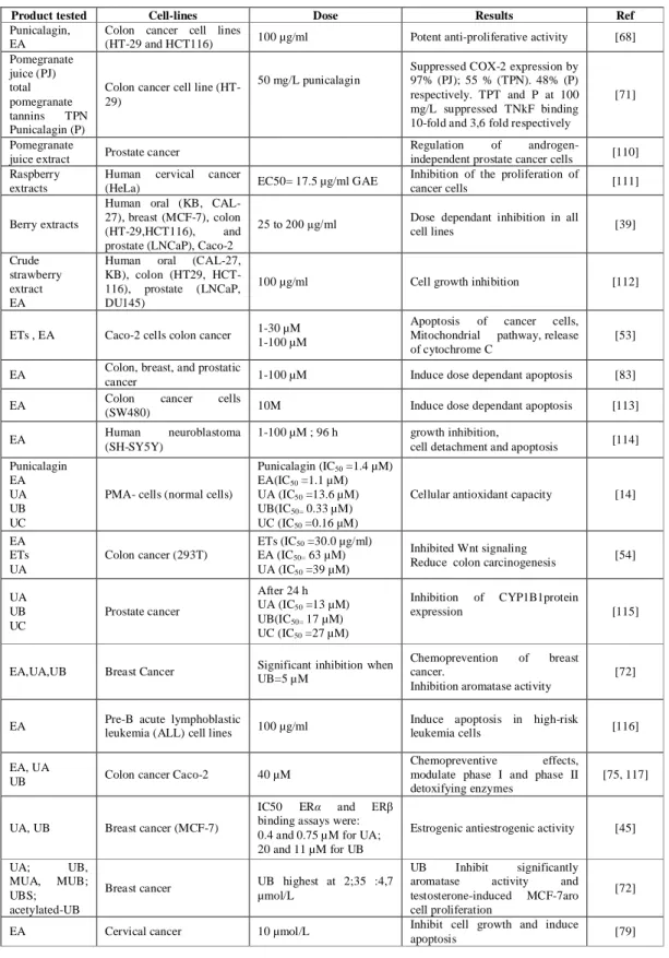
(33) 33. Table 2.4 Recent prospective clinical studies of consumption of food rich in ellagitannins Ets Source Pomegranate juice. ETs intake. Daily juice Healthy(10) supplementation. Pomegranate juice 50 ml daily. POMx capsules. Status (Subjects number). 1 and 3 years. Healthy (10) non-insulin dependent 3 month diabetes mellitus (10). 1 capsule/days Overweight (410-850 mg GAE) (64/32). Pomegranate juice 230 ml daily. Duration intervention. 4 weeks. patients with prostate 54 months cancer (24). Commercial 2*400mg in a pomegranate extract single Healthy (11) in capsule administration. 24 h. 5 weeks. Biomarker affected. Mains biomarker not affected. Ref. Decrease in arotid intima-media thickness (IMT). [90]. Reduction of cellular peroxides (by 71%), and increased glutathion levels (by 141%). Decreased the extent of Ox-LDL cellular uptake (by 39%). [91]. Glucose, BUN, creatinine, Increase in plasma antioxidant status lipids, insulin, c-peptide, paraoxonase-1, or electrolytes or liver enzymes (AST or ALT). [69]. Significant reduction of specific antigen. [92]. prostate. Generation of reactive oxygen species + 32% ORAC plasma antioxidant (ROS), inflammation marker interleukin-6 activity at 30 min (IL-6). [46]. No benefit. Serobiochemical and haematological, urinary 8-iso-PGF(2 alpha), respiratory function variables and clinical symptoms of chronic obstructive pulmonary disease. [93]. No changes in cardiac medications, blood sugar, hemoglobin A1c, weight, or blood pressure. [94]. Pomegranate juice 400 ml juice. Healthy. Pomegranate juice 240 ml/day. patients with CHD and myocardial 3 month ischemia. Stress-induced ischemia decreased. Pomegranate juice 50 ml/day. hypertensive patients 2 weeks. 36% decrement in serum angiotensin converting enzyme (ACE) activity and a 5% reduction in systolic blood pressure. [95]. Mixed nuts. 30g/day. subjects with 12 weeks metabolic syndrome. Increased excretion of serotonin metabolites. [96]. Strawberry. 250 g daily. healthy. Lipid peroxidation lag time increased DNA strand breaks in lymphocytes, and by 20% activity of phase II enzymes.. [97]. 3 weeks. 33. You created this PDF from an application that is not licensed to print to novaPDF printer (http://www.novapdf.com).
(34) 34 Table 2.5 Summary of intervention studies with animals to evidence health benefits of dietary ETs Product ingested. Objective. Dose used. Results. Ref. -Urolithins inhibited the growth of both androgen-dependent and androgen-independent prostate cancer cell lines, with equ. IC50 values lower than EA. -EA and UA decreased prostate cancer xenograft size, tumor vessel density in 4 weeks. Pomegranate extract. DU145, LNCaP, 22Rv1 LNCAP-AR cells Human 0.3 mg prostate cancer cells punicalagin/mice (LNCaP) injected subcutaneously into mice. Raspberry juice. Heart health. Inhibited cardiac and aortic production of superoxide anion Equ. 275 ml by a 70 kg and increased hepatic glutathione peroxidase. lower plasma human triglyceride level.. [99]. Blackberry extract Antioxidant. 2.68mg EA eq kg−1 body Reduced thiobarbituric acid reactive substance levels and weight Wistar rat increased glutathione levels in the liver, kidney and brain. [100]. Inhibited aortic lipid deposition by 79–96% and triggered in Equivalent to 275 ml by a 70 reduced activity of hepatic antikg human oxidant enzymes after 12 weeks. [101]. Berry juice and tea atherosclerosis berry hamsters Black raspberries Anti-cancer extract freeze-dried berries. Esophagus cancer. 45 g per day. and. colon. Purified Geranniin from Geranuuim Plasma antioxidant level thunbergii Inhibition of N- NMBAinduced esophageal Lyophylized black tumorigenesis in the rat raspberries, LBRs during initiation and post initiation of carcinogenesis. [98]. Inhibit esophageal tumorigenesis. [102]. Inhibition of rodent esophagus cancer by 30-60% and of the 0.25-0.5 mg/kg body weight colon by up- to 80% Reduction of levels of carcinogen-induced DNA damage.. [103]. Increase plasma ORAC antioxidant value after 6 hours of ingestion. [104]. 20 mg/rat. 0.25 mg/kg, weekly for 15 Reduced tumor multiplicity (39 and 49%, respectively). weeks and throughout a 30- Inhibit adduct formation (64%) after NMBA administration week bioassay, at 0.50 mg/kg At 25 weeks, both 5 and 10% LBRs significantly reduced 0.25 mg/kg, three times per tumor incidence (54 and 46%, respectively), tumor week for 5 weeks multiplicity (62 and 43%, respectively), proliferation rates, and preneoplastic lesion development. [105]. EA. mitochondrial damage in beta adrenergic agonist Protective effect of EA against mitochondrial damage in EA (7.5-15 mg/kg)/ 10 days induced myocardial myocardial infarction infarction. EA. Apoptosis. 60 mg/kg body weight p.o. Prevention of P13K-AKT activation in 15 weeks every day. [106]. EA. Anti-inflammatory properties. 4 mg/kg rat. [77]. 300 nmol Inhibitory effect on EA, robinetin, tumorigenecity of B[a]P myricetin 7,8-diol-9,10-epoxide-2 45, 90, 180 nmol EA on mouse skin in the 200, 400, 800 newborn mouse. robinetin, myricetin. Pomegranate extract urolithin A. and Colon inflammation. Significantly reduce paw edema. [85]. 59- 66% Reduction in the number of skin tumors; after 15 and 20 weeks of promotion with 12-0-tetradecanoylphorbol13-acetate [76] Avoid the formation of tumors in animals killed at 9-11 nmol months later 44 — 75% inhibition in the- number of diol-epoxide-induced pulmonary tumors per mouse Both PE and UROA decreased inflammation markers (iNOS, cycloxygenase-2, PTGES and PGE2 in colonic mucosa) and modulated favorably the gut microbiota, (increase in. 250 mg PE/Kg day 50 mg UA/Kg day. [107]. lactobacilli and bifidobacteria). Orally / intraperitoneal PG: 0.8 mg/mouse/dose Inhibition of 50% of tumor volume 2 weeks after 5days / week UA: 0.3 mg/mouse/dose. Pomegranate extract Urolithin A. Inhibition of tumor xenograft (LAPC-4) grow in immune compromised SCID mice. Urolithin A. Anti-inflammatory effect carrageenan-induced paw 5 mg/mice edema in mice. Reduction of paw edema after one hour of administration, increase of ORAC plasma level. [108]. oral. [109]. 34. You created this PDF from an application that is not licensed to print to novaPDF printer (http://www.novapdf.com).
(35) 35. Table 2.6 Summary of some recent results obtained on cell-models by ETs, EA and urolithins Product tested Punicalagin, EA Pomegranate juice (PJ) total pomegranate tannins TPN Punicalagin (P) Pomegranate juice extract Raspberry extracts. Cell-lines Colon cancer cell lines (HT-29 and HCT116). Colon cancer cell line (HT29). Crude strawberry extract EA ETs , EA. Caco-2 cells colon cancer. EA EA EA Punicalagin EA UA UB UC EA ETs UA. 50 mg/L punicalagin. Prostate cancer Human cervical cancer (HeLa) Human oral (KB, CAL27), breast (MCF-7), colon (HT-29,HCT116), and prostate (LNCaP), Caco-2 Human oral (CAL-27, KB), colon (HT29, HCT116), prostate (LNCaP, DU145). Berry extracts. Dose 100 µg/ml. Colon, breast, and prostatic cancer Colon cancer cells (SW480) Human neuroblastoma (SH-SY5Y). PMA- cells (normal cells). Colon cancer (293T). EC50= 17.5 µg/ml GAE. Results. Ref. Potent anti-proliferative activity. [68]. Suppressed COX-2 expression by 97% (PJ); 55 % (TPN). 48% (P) respectively. TPT and P at 100 mg/L suppressed TNkF binding 10-fold and 3,6 fold respectively. [71]. Regulation of androgenindependent prostate cancer cells Inhibition of the proliferation of cancer cells. [110] [111]. 25 to 200 µg/ml. Dose dependant inhibition in all cell lines. [39]. 100 µg/ml. Cell growth inhibition. [112]. 1-30 µM 1-100 µM. Apoptosis of cancer cells, Mitochondrial pathway, release of cytochrome C. [53]. 1-100 µM. Induce dose dependant apoptosis. [83]. 10M. Induce dose dependant apoptosis. [113]. 1-100 µM ; 96 h. growth inhibition, cell detachment and apoptosis. [114]. Cellular antioxidant capacity. [14]. Inhibited Wnt signaling Reduce colon carcinogenesis. [54]. Punicalagin (IC50 =1.4 µM) EA(IC50 =1.1 µM) UA (IC50 =13.6 µM) UB(IC50= 0.33 µM) UC (IC50 =0.16 µM) ETs (IC50 =30.0 µg/ml) EA (IC50= 63 µM) UA (IC50 =39 µM). UA UB UC. Prostate cancer. After 24 h UA (IC50 =13 µM) UB(IC50= 17 µM) UC (IC50 =27 µM). EA,UA,UB. Breast Cancer. Significant inhibition when UB=5 µM. Chemoprevention of breast cancer. Inhibition aromatase activity. [72]. EA. Pre-B acute lymphoblastic leukemia (ALL) cell lines. 100 µg/ml. Induce apoptosis in high-risk leukemia cells. [116]. EA, UA UB. Colon cancer Caco-2. 40 µM. Chemopreventive effects, modulate phase I and phase II detoxifying enzymes. [75, 117]. UA, UB. Breast cancer (MCF-7). IC50 ERα and ERβ binding assays were: 0.4 and 0.75 µM for UA; 20 and 11 µM for UB. Estrogenic antiestrogenic activity. [45]. UA; UB, MUA, MUB; UBS; acetylated-UB. Breast cancer. UB highest at 2;35 :4,7 µmol/L. EA. Cervical cancer. 10 µmol/L. Inhibition of expression. CYP1B1protein. UB Inhibit significantly aromatase activity and testosterone-induced MCF-7aro cell proliferation Inhibit cell growth and induce apoptosis. [115]. [72]. [79]. 35. You created this PDF from an application that is not licensed to print to novaPDF printer (http://www.novapdf.com).
(36) 36 Urolithins were also found to inhibit activity of the enzyme CYP1B1. This inhibition may affect the three stages of prostate cancer development. Urolithins therefore, appear to display a dual mode of action—inhibiting the activity and/or expression of the enzyme. Urolithin A and B inhibit CYP1 E ROD activity by inhibiting both the protein expression and the activity of CYP1B1; and thus decreasing the incidence of prostate cancer [118]. In addition, if used as an adjuvant during chemotherapy, these microbial metabolites appear to decrease the resistance to the drug mediated by CYP1B1. Urolithin A glucuronide and, in a much lesser amount, dimethyl ellagic acid were reported as accumulating in human prostate tissues after subjects had consumed pomegranate juice [50]. Subjects who had received the primary treatment of surgery or radiation also demonstrated significant decrease in prostate-specific antigens (PSA) after consuming pomegranate juice [23, 92] All these findings, along with animal studies [98, 108] (Table 2.5), justify the high interest in ET-rich diets as preventing prostate cancer. Less information has been published so far on the urolithins’ role in reducing risks of breast cancer, although they have been shown in vitro to significantly enhance the apoptosis of breast-cancer cell lines [45, 71, 72] (Table 2.6). 2.1.4.2.2 Chemoprevention of Colorectal Cancer. Cell models have shown that urolithins induce dose-dependent apoptosis of colon-cancer cells [54, 75, 117]. Urolithins A and B appeared to modulate phases I and II detoxifying enzymes in Caco-2 cells [117]. Both phase enzymes enhance the detoxification of carcinogenic compounds. Another study proposes an additional pathway by modulating the expression levels of multiple genes in the epithelial cells lining the colon. This pathway might be used by EA and urolithins A and B, thereby exerting an effect towards reducing risks of colon cancer [75]. This study suggested the urolithins’ antiproliferative activity may be due to the modulation of gene expression, including the MAPK/ERK pathway signaling. Urolithin A can induce cell cycle arrest, and modulate key cellular processes associated with colon cancer development, such as MAPK signaling in vitro. Additionally, urolithin A was shown to inhibit Wnt signaling with IC50 at even lower concentrations than for EA [54].. 36. You created this PDF from an application that is not licensed to print to novaPDF printer (http://www.novapdf.com).
Figure
![Table 2.2 Contents of main phenolic compounds in Rubus adenotrichos (R.A) and Rubus glaucus (R.G) in mg per 100 g of dry matter[32]](https://thumb-us.123doks.com/thumbv2/123dok_es/7132023.415179/27.918.193.717.450.811/table-contents-phenolic-compounds-adenotrichos-rubus-glaucus-matter.webp)

![Figure 2.2 Phylogenetic tree of the microbial diversity in the human gastrointestinal tract, based on the 16S rDNA bacterial sequence data [2]](https://thumb-us.123doks.com/thumbv2/123dok_es/7132023.415179/39.918.251.627.597.855/figure-phylogenetic-microbial-diversity-human-gastrointestinal-bacterial-sequence.webp)
+7
![Table 2.7 Main studies on bioconversion of dietary compounds into bioactive colonic metabolites [131]](https://thumb-us.123doks.com/thumbv2/123dok_es/7132023.415179/41.918.151.800.489.696/table-studies-bioconversion-dietary-compounds-bioactive-colonic-metabolites.webp)
![Figure 3.2 Comparing speed, sensitivity and resolution in HPLC and UPLC analysis [2, 3]](https://thumb-us.123doks.com/thumbv2/123dok_es/7132023.415179/47.918.244.679.703.962/figure-comparing-speed-sensitivity-resolution-hplc-uplc-analysis.webp)

![Table 3.2 PCR primers targeted for 16S rRNA gene, without GCClamp [198]](https://thumb-us.123doks.com/thumbv2/123dok_es/7132023.415179/67.918.183.738.231.453/table-pcr-primers-targeted-s-rrna-gene-gcclamp.webp)
Documento similar
Specifically, we discuss the modulation of CKD-MBD by uremic toxins of bacterial origin, the impact of dietary phosphate and phosphate binders on the gut microbiota, the
(C) Phenotype of human B-cells, monocytes, pDCs, and cDCs from healthy controls (HCs) and inflammatory bowel disease (IBD) patients based on the basal expression of CCR2, CD40,
The Effects of Exercise Interventions on Executive Functions in Children and Adolescents with Autism Spectrum Disorder: A Systematic Review and Meta-analysis.. A systematic review
The aim of the present work was to identify microbial signatures linked to immunity traits and to characterize the contribution of host‑genome and gut microbiota to
Because gut microbiota composition may also play a key role in controlling the physiological and neurophysiological mechanisms involved in the weaning stress response, we
Conversely, a very significant reduction of virus in serum, nasal viral shedding and clinical signs were observed when pigs transplanted with warthog feces were
Role of gut microbiota in chronic low-grade inflammation as potential driver for atherosclerotic cardiovascular disease: A systematic review of human studies.. A systematic review
The following topics are reviewed: (a) intestinal barrier function and microbiota; (b) leaky-gut, gut microbiota relationship and liver disease; (c) current main strategies to
