Molecular, microbiological and clinical
characterization of Clostridium difficile isolates
from tertiary care hospitals in Colombia
Clara Lina Salazar1, Catalina Reyes2, Santiago Atehortua3, Patricia Sierra4, Margarita Marı´a Correa5, Daniel Paredes-Sabja6, Emma Best7, Warren N. Fawley7, Mark Wilcox7, A´ ngel Gonza´lez2*
1 Research Group in Anaerobic Bacteria (GIBA), School of Microbiology, Universidad de Antioquia, Medellı´n,
Colombia, 2 Basic and Applied Microbiology Research Group (MICROBA), School of Microbiology, Universidad de Antioquia, Medellı´n, Colombia, 3 San Vicente Fundacio´n University Hospital, Medellı´n, Colombia, 4 Clı´nica Leo´n XIII, IPS Universitaria, Universidad de Antioquia, Medellı´n, Colombia, 5 Molecular Microbiology Group, School of Microbiology, Universidad de Antioquia, Medellı´n, Colombia, 6 Microbiota-Host Interactions and Clostridia Research Group, Departamento de Ciencias Biolo´gicas, at Universidad Andres Bello, Santiago, Chile, 7 Departament of Microbiology, Leeds Teaching Hospitals NHS Trust, Leeds, United Kingdom
Abstract
In Colombia, the epidemiology and circulating genotypes of Clostridium difficile have not yet been described. Therefore, we molecularly characterized clinical isolates of C.difficile from patients with suspicion of C.difficile infection (CDI) in three tertiary care hospitals. C.difficile was isolated from stool samples by culture, the presence of A/B toxins were detected by enzyme immunoassay, cytotoxicity was tested by cell culture and the antimicrobial suscepti-bility determined. After DNA extraction, tcdA, tcdB and binary toxin (CDTa/CDTb) genes were detected by PCR, and PCR-ribotyping performed. From a total of 913 stool samples collected during 2013–2014, 775 were included in the study. The frequency of A/B toxins-positive samples was 9.7% (75/775). A total of 143 isolates of C.difficile were recovered from culture, 110 (76.9%) produced cytotoxic effect in cell culture, 100 (69.9%) were tcdA +/tcdB+, 11 (7.7%) tcdA-/tcdB+, 32 (22.4%) tcdA-/tcdB- and 25 (17.5%) CDTa+/CDTb+. From 37 ribotypes identified, ribotypes 591 (20%), 106 (9%) and 002 (7.9%) were the most prevalent; only one isolate corresponded to ribotype 027, four to ribotype 078 and four were new ribotypes (794,795, 804,805). All isolates were susceptible to vancomycin and metroni-dazole, while 85% and 7.7% were resistant to clindamycin and moxifloxacin, respectively. By multivariate analysis, significant risk factors associated to CDI were, staying in orthope-dic service, exposure to third-generation cephalosporins and staying in an ICU before CDI symptoms; moreover, steroids showed to be a protector factor. These results revealed new C. difficile ribotypes and a high diversity profile circulating in Colombia different from those reported in America and European countries.
a1111111111
Citation: Salazar CL, Reyes C, Atehortua S, Sierra
P, Correa MM, Paredes-Sabja D, et al. (2017) Molecular, microbiological and clinical characterization of Clostridium difficile isolates from tertiary care hospitals in Colombia. PLoS ONE 12(9): e0184689.https://doi.org/10.1371/journal. pone.0184689
Editor: Gayatri Vedantam, University of Arizona,
UNITED STATES
Received: May 11, 2017
Accepted: August 29, 2017
Published: September 13, 2017
Copyright:©2017 Salazar et al. This is an open access article distributed under the terms of the
Creative Commons Attribution License, which permits unrestricted use, distribution, and reproduction in any medium, provided the original author and source are credited.
Data Availability Statement: All relevant data are
within the paper.
Funding: This work was supported by
Introduction
Clostridium difficile, a common causal agent of diarrhea in hospitalized patients, is associated
with substantial morbidity and mortality [1]. The clinical manifestations ofC.difficileinfection
(CDI) range from mild, self-limiting diarrhea to fulminant colitis that include
pseudomembra-nous colitis and toxic megacolon [2]. CDI is a hospital-acquired infection; but lately,C.difficile
has been -associated with diarrhea in the community [2]. Several antibiotic classes have been
associated with the development of CDI, including clindamycin, cephalosporins and
fluoro-quinolones [3,4]. Oral metronidazole is usually used to treat mild to moderate CDI, and
van-comycin, severe infection [5]; although, resistance to vancomycin is not yet a major issue,
reduced susceptibility to this antibiotic has been reported [6–8].
Clostridium difficileproduces two major toxins, toxin A and toxin B that are encoded by the
tcdAandtcdBgenes, respectively, and are responsible for the pathogenicity of this bacterium.
In addition, some isolates produce a binary toxin, which has been associated with the severity
of the disease [9,10]. Various laboratory tests are available to diagnose CDI, they include
isola-tion of toxigenicC.difficileby culture, detection of A and B toxins by immunoassays, and
molecular assays to detect toxin genes in stool samples [11–13].
PCR ribotyping is a molecular tool employed for the global analysis ofC.difficilerelated
vir-ulent strains based on a reference library. Thus, various ribotypes have been described in Europe and United States; nonetheless, the most prevalent ribotypes are 027, 001/072 and 014/
020 [1]. Particularly, in recent years, the virulent or epidemic strain NAP1/PCR ribotype 027
and to a lasser extent ribotype 078 have been associated with an increase on the incidence and
mortality rates in North America and European countries [2]. In Latin America, some CDI
cases have documented and the NAP1/027 ribotype was reported in Costa Rica, Panama,
Me´xico, Chile and Colombia [14–19]. Other ribotypes including 014, 106, 010, 020, 133 and
233 have been registered in Brazil, but 027/078 have not yet been detected [20–22]. Although,
ribotye NAP1/027 has been reported in Colombia [23], the diversity of circulating genotypes
remains to be described. Therefore, the aim of the present work was to characterize at the
molecular and microbiological levelC.difficileisolates, and to analyse the clinical
characteris-tics of patients with CDI from three high compelxity hospitals in Medellı´n, Colombia.
Materials and methods
Patients and study design
A prospective cross-sectional study was conducted during January 2013 to December 2014 in three tertiary care hospitals in Medellı´n, Colombia. Hospital A had 731 beds, hospital B 650 beds and hospital C 653 beds. All patients with suspicion of CDI and those who received at least one dose of antimicrobial therapy six weeks before the symptoms started, were involved in the study. A CDI case was defined as a patient with clinical suspicion of the disease, and
with a stool positive forC.difficileA/B toxins. CDI was classified as mild, moderate, severe or
complicated according to guidelines by the American College of Gastroenterology (ACG)
[24]. Clinical and demographic data were obtained from medical records, after patients have
read and signed an informed consent.
Ethics statement
This work was approved by the Human Research Ethics Committee of Universidad de Antio-quia (Comite´ de Bioe´tica, Sede de Investigacio´n Universitaria, CBEIH- SIU, approval number 12-35-458).
funders had no role in study design, analysis and interpretation of experiments, decision to publish, or preparation of the manuscript.
Competing interests: The authors have declared
Toxin detection, C. difficile culture and identification on stool samples
Samples were processed immediately after arrival at the Lab or refrigerated for no more than
72 h before being processed. Stool samples were initially tested for freeC.difficleA/B toxins
using one or two enzyme immunoassays (EI) ImmunoCard toxin A&B (Meridian Bioscience,
Cincinnati, OH) or MiniVidasC.difficileToxin A/B assay (bioMerieux, Marcy I´Etoile,
France). All samples were inoculated on cycloserine-cefoxitin-fructose-taurocholate agar
(TCCFA) [25], and incubated at 37˚C for 48 h in an anaerobic chamber (90% N2, 5%CO2, 5%
H2). FromC.difficilepositive TCCFA, a single colony was subcultured on blood agar and then
identified on the basis of colony morphology, fluorescence under UV light, L-proline amino-peptidase production and confirmed using the API Rapid ID 32A system (BioMe´rieux Inc.,
Durhan, NC, USA). Isolates not clearly confirmed asC.difficilewere subjected to matrix-
assis-ted laser desorption ionization–time of flight mass spectrometry (MALDI-TOF MS).C.
diffi-cileisolates were stored at -70˚C in brain-heart infusion (BHI) broth (Becton Dickinson,
Franklin Lakes, NJ, USA) supplemented with 20% glycerol, for further analysis.
Antimicrobial susceptibility testing
Antimicrobial susceptibility to metronidazole and vancomycin was determined by minimum inhibitory concentration (MIC) using the standard agar dilution method, following guidelines
by the Clinical and Laboratory Standards Institute (CLSI) [26], and susceptibility to
clindamy-cin and moxifloxaclindamy-cin, were tested by the epsilometric method (Etest BioMe´rieux, Marcy
l’Etoile, France). To perform the agar dilution method,C.dificilleisolates were grown on
Bru-cella agar (Oxoid Ltd., Basingstoke, UK) previously supplemented with 5μg hemin, 1μg
Vita-min K1 per mL and 5% v/v laked blood sheep, and mixed with the antimicrobial agent solution; for the case of the epsilometric method, we used the same Brucella agar but instead of laked we used defibrinated blood sheep. Susceptible or resistant microorganisms used as
con-trols were,B.fragilisATCC 25285 andC.difficileATCC 700057.
Cytotoxicity assay
For cytotoxicity assays, a colony ofC.difficilewas inoculated into BHI broth and incubated
under anaerobic conditions for 48 h at 37˚C. After centrifugation, 100μl of each culture
super-natant was added to a confluent monolayer of Vero cells in 96-well plate containing Dulbecco’s Modified Eagle’s Medium (DMEM, HyClone, Thermo Scientific), supplemented with 10%
fetal bovine serum (FBS, HyClone), for a final concentration of 5x10³cells/well. Cells were
cul-tured at 37˚C, 5% CO2and examined to evaluate the cytopathic effect at 24 and 48 hours.
Posi-tivity was confirmed by neutralization using a polyclonal goat antiserum anti-C.difficiletoxins
A/B (TechLab1, Blacksburg, VA) [27].
Detection of C. difficile toxin genes
DNA was extracted from cultures grown in BHI broth, incubated overnight in anaerobic con-ditions, using the DNA isolation kit (MoBio Laboratories Inc., Carlsbad, CA) and following
the manufacturer instructions. ThetcdA (toxin A) andtcdB (toxin B) genes were determined
using the NK2/NK3 and NK104/NK105 primers respectively, to amplify a 252-bp fragment for
tcdA and 203-bp fragment fortcdB[28]. The binary toxins genes were detected using thecdt
A-pos/cdtArev andcdtBpos/cdtBrev primers that amplify a 375-bp fragment for thecdtAgene
and 510-bp fragment forcdtB, and following previously described protocols [29,30].
Condi-tions for amplification were: 35 cycles of denaturation at 95˚C for 45s (tcdAandtcdB) and
and 56.4˚C for 60s (cdtB) and extension at 72˚C for 60s. PCR products were visualized in 1.5% to 2% agarose gels stained with ethidium bromide.
PCR ribotyping
The PCR ribotyping was performed using a high-resolution capillary gel-based electrophoresis after amplification of the 16S–23S intergenic spacer, following protocols of Department of
Microbiology, Leeds Teaching Hospitals NHS Trust, Leeds, UK [31]. Briefly, PCR products
were analyzed using an ABI-PRISM 313xl automated sequencer and fragment analysis system, a 16 capillary 36 cm array with POP-7 separation matrix (Life Technologies, Paisley, UK) and a GeneScan 600 LIZ as an internal marker. Bio- Numerics v.7.1 software (Applied Maths, Sint-Martens-Latem, Belgium) was used to import the fluorescent signals and GeneMapper v.4.0 software (Applied Biosystems, Life Technologies, Grand Island, NY) was used to size the
frag-ments [31]. Cluster analysis of PCR ribotype band profiles was performed using the DICE
sim-ilarity coefficient with relationships represented in a UPGMA dendrogram within Bio-Numerics v.7.1 software. Ribotypes were identified by comparison of the band profiles with
those in the reference UK library (Clostridium difficileNetwork for England and Northern
Ireland).
Statistical analysis
Questionnaire data were stored in a Microsoft Access database. Statistical analysis was per-formed using the SPSS v. 23.0 statistic software package (IBM-SPSS Inc, Armonk, NY) and Stata v.13.0 software (StataCorp LP, College Station, TX). Data were described using absolute and relative frequencies, and media and standard deviation for categorical and numerical vari-ables, respectively. Group comparisons for categorical and numerical variable types were per-formed using chi-square and Mann Whitney U tests, respectively. Associations between clinical variables and CDI were analyzed using multivariate logistic regression analysis;
vari-ables with aP<0.25 in the bivariate analysis were considered in the logistic stepwise regression
performed with the Hosmer-Lemeshow criteria, to find the best model that fit the data with
the lowest Bayesian information criterion (BIC), and those withP<0.05 were considered
sig-nificant in the final model.
Results
Frequency of CDI according to positive free toxin in stool samples
A total of 913 stool samples from patients with suspicion of CDI were analyzed. One hundred and thirty-eight (15.1%) samples were excluded because they did not meet the antibiotics usage critera or were duplicated. From a total of 775 samples analyzed, 487 (62.9%) were from hospital A, 190 (24.5%) from hospital B and 98 (12.6%) from hospital C. The global frequency of free positive toxin in stool samples was 9.7% (75/775). The frequency of CDI in each hospi-tal was 7.0%, 17.9% and 7.1% for hospihospi-tals A, B and C, respectively.
Demographic and clinical characteristics of patients with suspicion of
CDI according to positive stool toxin
Several clinical characteristics and laboratory data from patients with (CDI) or without
(non-CDI) stool toxin were compared in a bivariade analysis (Tables1and2). The median age of
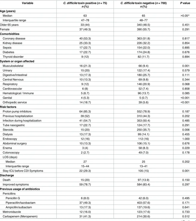
Table 1. Demographic and clinical characteristics of those patients with and without stool toxin.
Variable C. difficile toxin positive (n = 75)
n (%)
C. difficile toxin negative (n = 700) n (%)
P value
Age (years)
Median 63 65 >0.05*
Interquartile range 47–78 48–77
Older 65 years 33 (44) 340 (48.5) 0.451
Female 37 (49.3) 390 (55.7) 0.291
Comorbidities
Coronary disease 40 (53.3) 363 (51.8) 0.817
Kidney disease 25 (33.3) 226 (32.2) 0.854
Sepsis 17 (22.7) 154 (22.0) 0.895
Diabetes 17 (22.7) 174 (24.8) 0.676
Thyroid disorder 9 (12) 82 (11.7) 0.894
System or organ affected
Musculoskeletal 16 (21.3) 66 (9.4) 0.001
Urinary 15 (20) 122 (17.4) 0.579
Digestive/Intestinal 13 (17.3) 180 (25.7) 0.111
Central Nervous 10 (13.3) 69 (9.8) 0.344
Respiratory 9 (12) 146 (20.9) 0.068
Cardiovascular 6 (8) 52 (7.4) 0.858
Hematological / Immune 5 (6.7) 96 (13.7) 0.085
Genital 4 (5.3) 5 (0.7) <0.001
Orthopedic service 14 (18.7) 39 (5.6) <0.001
Risk factors
Proton pump inhibitors 64 (85.3) 552 (78.9) 0.187
Previous hospitalization 39 (52) 310 (44.3) 0.202
Infection during hospitalization 41 (54.7) 353 (50.4) 0.485
Tube nasogastric 17 (22.7) 124 (17.7) 0.291
Steroids 15 (20) 250 (35.7) 0.006
Dialysis 13 (17.3) 99 (14.1) 0.455
Endoscopy 12 (16) 112 (16) 1.000
Abdominal surgery 10 (13.3) 106 (15.1) 0.676
Enema 3 (4) 58 (8.3) 0.229
Colonoscopy 2 (2.7) 49 (7.0) 0.178
LOS (days)
Median 27 25 0.202
Interquartile range 15–44 13–41
Stay ICU before CDI Symptoms 22 (29.3) 105 (15) 0.001
Discharge
Death 15 (20) 97 (13.9) 0.150
Improved symptoms 59 (78.7) 584 (83.4) 0.297
Previous usage of antibiotics
Penicillins
Penicillin G 6 (8.0) 42 (6.0) 0.495
Piperacillin/tazobactam 37 (49.3) 403 (57.6) 0.171
Ampicillin/sulbactam 13 (17.3) 137 (19.6) 0.641
Metronidazole 12 (16.0) 123 (17.6) 0.733
Carbapenem (Meropenem) 31 (41.3) 214 (30.6) 0.512
median of length of stay (LOS) at the hospital was 27 days (IQR 15–44 days) for CDI patients versus 25 days (IQR 13–41) for those non-CDI individuals. Of note, a significant difference was observed between CDI patients staying in ICU before presenting the symptoms as
com-pared to those negative for theC.difficiletoxin in stools (29.3% versus 15%,P= 0.001).
Furthermore, analyses of comorbidities, systems or organs affected, risk factors, death or
improvement of the CDI patients as well as previous administration of antibiotics (Table 1),
showed significant differences on those CDI patients who have affected the musculoskeletal (21.3% versus 9.4%) and genital (5.3% versus 0.7%) systems or those who assisted to orthope-dic service (18.7% versus 5.6%), as compared to those non-CDI patients. Among the risk
Table 1. (Continued)
Variable C. difficile toxin positive (n = 75)
n (%)
C. difficile toxin negative (n = 700) n (%)
P value
Cephalosporin
1st Generation 8 (10.7) 59 (8.4) 0.057
3rd generation 16 (21.3) 51 (7.3) <0.001
4th generation 8 (10.7) 53 (7.6) 0.344
Glycopeptides (vancomycin) 21 (28) 169 (24.1) 0.461
Aminoglycosides 9 (12.0) 74 (10.6) 0.704
Macrolides (Clarithromycin) 5 (6.6) 106 (15.1) 0.046
Lincosamides (Clindamycin) 8 (10.7) 60 (8.6) 0.542
Fluoroquinolones (Ciprofloxacin) 15 (20) 148 (21.1) 0.817
Oxazolidinones (Linezolid) 6 (8.0) 85 (12.1) 0.290
CDI, C. difficile infection; LOS, length of stay; *Mann-Whitney test
https://doi.org/10.1371/journal.pone.0184689.t001
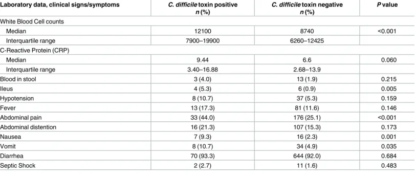
Table 2. Laboratory data, clinical signs and symptoms of those patients with and without stool toxin.
Laboratory data, clinical signs/symptoms C. difficile toxin positive n (%)
C. difficile toxin negative n (%)
P value
White Blood Cell counts
Median 12100 8740 <0.001
Interquartile range 7900–19900 6260–12425
C-Reactive Protein (CRP)
Median 9.44 6.6 0.060
Interquartile range 3.40–16.88 2.68–13.9
Blood in stool 3 (4.0) 13 (1.9) 0.215
Ileus 4 (5.3) 6 (0.9) 0.005
Hypotension 8 (10.7) 37 (5.3) 0.159
Fever 13 (17.3) 81 (11.6) 0.146
Abdominal pain 33 (44.0) 176 (25.1) <0.001
Abdominal distention 16 (21.3) 107 (15.3) 0.173
Nausea 7 (9.3) 16 (2.3) 0.001
Vomit 8 (10.7) 34 (4.9) 0.035
Diarrhea 70 (93.3) 644 (92.0) 0.684
Septic Shock 2 (2.7) 11 (1.6) 0.483
CDI, C. difficile infection
factors analyzed, only steroid usage showed a significant difference between CDI and
non-CDI patients (20% versus 35.7%,P= 0.006).
The most frequently administered antibiotics during the six week period prior symptoms or toxin test were, piperacillin/tazobactam administered to 440 (56.7%) patients, followed by meropenem: 245 (31.6%), vancomycin: 190 (24.5%), ciprofloxacin: 163 (21.0%), ampicillin/ sulbactam: 150 (19.3%), metronidazole: 135 (17.4%), clarithromycin: 111 (14.3%), linezolid: 91 (11.7%) and others, at less than 10% each. Among the different antibiotics used, only third generation cephalosporin and macrolides showed a significant difference between CDI and
non-CDI patients (Table 1).
Regarding the severity of the diseases, 58 patients had mild-moderate CDI; from these 30 (51.7%) patients received only oral metronidazole and 6 (10.3%) received both oral metronida-zole and vancomycin. Six patients had severe disease and two severe complicated, and nine patients were not classified. A total of 69 (92%) of CDI patients recieved therapy for this dis-ease, while five patients were discharged and one died before starting the specific treatment. About the laboratory data and clinical signs and symptoms, CDI patients showed significant differences in white blood cell counts, ileus, abdominal pain, nausea and vomit, in comparison
with those patients with negativeC.difficiletoxin (Table 2).
In addition, 51% of the patients (394/775) had a concomitant infection; of these, 143 (36.3%) had urinary tract and 97 (24.6%) bloodstream infections. The most frequent
patho-gens isolated from patients with these infections wereEscherichia coliandStaphylococcus
aureus, respectively (data not shown). Moreover, from 644 patients with diarrhea, 8 (1.2%)
had other pathogens as possible cause of diarrhea; one of them hadSalmonella enterica, two
Strongyloides stercolarisand 5Entamoeba histolytica.
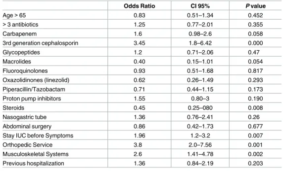
The bivariate analysis to compare CDI versus non-CDI patients conduced to identify vari-ables associated with the development of CDI, showed that staying in an orthopedic service (OR 3.8, 95% CI 2.0–7.56) or ICU (OR 3.45, 95% CI 1.8–6.42), administration of third-genera-tion cephalosporins (OR 1.98, 95% CI 1.2–3.2), and affecthird-genera-tion of the musculoskeletal system (OR 2.6, 95% CI 1.41–4.78) before CDI symptoms, were factors associated with CDI.
Interest-ingly, steroid usage was associated as a protector factor (OR 0.45, 95% CI 025–080) (Table 3).
A multivariate logistic regresision model was performed using a stepwise selection
includ-ing variables that showed significant outcomes or aPvalue<0.25 in the bivariate analysis; this
analysis showed that staying in an orthopedic service (OR 3.97, 95% CI 1.98–7.93), exposure to third-generation cephalosporins (OR 3.91, 95% CI 2.06–7.46), staying in ICU before CDI symptoms (OR 2.01, 95% CI 1.20–3.36), remained as significant risk factors associated with CDI, and as described above, steroid usage (OR 0.45, 95% CI 0.22–0.75) appears to be a
protec-tive factor for CDI (Table 4).
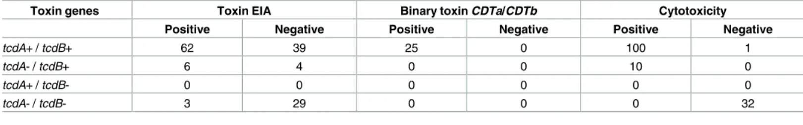
Detection of C. difficile toxin genes
A total of 143C.difficileisolates were obtained by culture. From these, 71 corresponded to
patients with positiveC.difficiletoxin, and from these, 62 (87.3%) harbored thetcdA/tcdB
genes, six (8.5%) weretcdA-/tcdB+ and 3 (4.2%) were negative for these toxin genes (Table 5).
The remaining 72C.difficileisolates corresponded to non-CDI patients. From these 72
iso-lates, 39 (54.2%) weretcdA+/tcdB+ and four (5.6%)tcdA-/tcdB+ and 29 (40.2%) were negative
for these genes. Only 25 isolates were positive for the binary toxin (CDTa/CDTb) (Table 5);
Clostridium difficile clinical isolates induce a cytotoxic effect on Vero cells
In the cytotoxicity assays, 110 from the 143C.difficileisolates produced a cytotoxic effect in
cell culture; from these, 100 (90.1%) weretcdA+/tcdB+ and 10 (9.9%)tcdA-/tcdB+. The
remaining 33 isolates did not show cytotoxic effect, and 32 (97%) of them were negative for
toxin genes, and only one (3%) harbored these genes (tcdA+/tcdB+) (Table 5). In addition,
from the 110 cytotoxic isolates, 67 (60.9%) were from CDI patients, while 43 (39.1%) were from non-CDI patients. From the 33 isolates with a negative cytotoxic effect, 29 (87.9%) were from non-CDI and four (12.1%) from CDI patients (data not shown).
Diversity of C. difficile PCR ribotypes
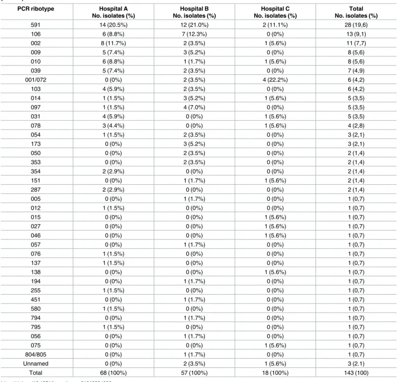
A total of 37C.difficileribotypes were identified, 3 isolates produced unidentified ribotypes
(unnamed). Among the 12 most frequent ribotypes (Fig 1), ribotype 591 was the most
preva-lent (19.6%,n= 28), followed by ribotypes 106 (13%,n= 13), 002 (7.7%,n= 11), 009 (5.6%,
n= 8) and 010 (5.6%,n= 8). Remarkably only four isolates corresponded to ribotype 078
(2.8%) and one to ribotype 027 (0.7%). Further, four new ribotypes were detected, named 794, 795, 804 and 805.
Table 4. Multivariate analysis of Clostridium difficile infection risk factors.
Factor Adjusted Odds Ratio CI 95% P value
Third-generation cephalosporin 3.91 2.06–7.46 0.000
Stay in ICU before CDI symptoms 2.01 1.20–3.36 0.000
Steroids 0.45 0.22–0.75 0.004
Orthopedic Service 3.97 1.98 7.93 0.000
https://doi.org/10.1371/journal.pone.0184689.t004
Table 3. Results of bivariate analysis to compare CDI versus those non-CDI patients.
Odds Ratio CI 95% P value
Age>65 0.83 0.51–1.34 0.452
>3 antibiotics 1.25 0.77–2.01 0.355
Carbapenem 1.6 0.98–2.6 0.058
3rd generation cephalosporin 3.45 1.8–6.42 0.000
Glycopeptides 1.2 0.71–2.06 0.47
Macrolides 0.40 0.15–1.01 0.054
Fluoroquinolones 0.93 0.51–1.68 0.817
Oxazolidinones (linezolid) 0.62 0.26–1.49 0.293
Piperacillin/Tazobactam 0.71 0.44–1.15 0.173
Proton pump inhibitors 1.55 0.80–3 0.190
Steroids 0.45 0.25–080 0.008
Nasogastric tube 1.36 0.76–2.41 0.26
Abdominal surgery 0.86 0.42–1.73 0.677
Stay IUC before Symptoms 1.96 1.2–3.2 0.007
Orthopedic Service 3.8 2.0–7.56 0.001
Musculoskeletal Systems 2.6 1.41–4.78 0.002
Previous hospitalization 1.36 0.84–2.19 0.203
CDI, C. difficile infection; ICU, Intensive Care Unit.
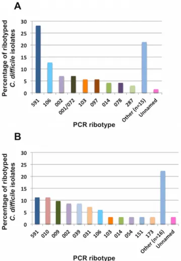
The most prevalent ribotypes from CDI patients were, 591 (28%,n= 20), 106 (12.6%,
n= 9), 002 (7%,n= 5), 001/072 (7%,n= 5), 103 (5.6%,n= 4) and 097 (5.6%,n= 4) (Fig 2A),
while those from non-CDI patients were, 591 (11.1%,n= 8), 010 (11.1%,n= 8), 009 (9.7%,
n= 7), 002 (8.3%,n= 6), 039 (8.3%,n= 6) and 031 (6.9%,n= 5) (Fig 2B).
A total of 68 isolates were from hospital A, 57 from hospital B and 18 from hospital C (Table 6). The most prevalent ribotypes in hospital A were 591 (20.5%,n= 14) and 002 (11.7%,
n= 8), 591 (21%,n= 12) and 106 (12.3%,n= 7) in hospital B, and 001/072 (22.2%,n= 4) and
591 (11.1%,n= 2) in hospital C.
Antibiotic susceptibility of C. difficle isolates
In the antimicrobial susceptibility assays, the 143 isolates (100%) were susceptible to vancomy-cin and metronidazole, while 11 (7.7%) and 126 (86.1%) isolates were resistant to moxifloxavancomy-cin and clindamycin, respectively. The minimum inhibitory concentrations (MIC50 and MIC90)
Table 5. Distribution of toxin genes profiles, toxin EIA and cytotoxicity in culture of C. difficile isolates (n = 143).
Toxin genes Toxin EIA Binary toxin CDTa/CDTb Cytotoxicity
Positive Negative Positive Negative Positive Negative
tcdA+ / tcdB+ 62 39 25 0 100 1
tcdA- / tcdB+ 6 4 0 0 10 0
tcdA+ / tcdB- 0 0 0 0 0 0
tcdA- / tcdB- 3 29 0 0 0 32
EIA, Enzyme immunoassay
https://doi.org/10.1371/journal.pone.0184689.t005
Fig 1. Distribution of the most commonly isolated C.difficile PCR Ribotypes from all C. difficile islates in three tertiary care hosiptals in Medellı´n, Colombia, 2013–2014 (n = 143). Other column includes the
for vancomycin, metronidazole, moxifloxacin and clindamycin were 0.5–1.0μg/mL, 0.25–
0.5μg/mL, 1.5–2.0μg/mL and 8.0–256 g/mL, respectively. Breakpoints to characterize isolates
as susceptible or resistant were as follow: metronidazole (susceptible8μg/mL, resistant
32μg/mL), clindamycin, moxifloxacin and vancomycin (susceptible2μg/mL, resistant
8μg/mL). Intermediate breakpoints were considered as resistant. Notoriously, all isolates
(100%) of the most prevalent ribotype 591, were resistant to clindamycin, while only four (14.3%) were resistant to moxifloxacin. Interstinlgy, the only isolate detected of ribotype 027 was susceptible to all of the antibiotics tested (data not shown).
Discussion
Little is known about the epidemiology of CDI in Colombia. Therefore, in this study a cohort of patients with suspicion of CDI was evaluated to report for the first time the diversity of cir-culating ribotypes, and new ribotypes are described.
Fig 2. Distribution of the most commonly isolated C.difficile PCR Ribotypes from (A) cases of CDI (71 isolates) and (B) patients with negative C. difficile toxin (72 isolates) in three tertiary care hosiptals in Medellı´n, Colombia, 2013–2014 (n = 143). Other column for CDI cases includes the ribotypes 005, 009, 010,
012, 015, 027, 031,039, 046, 050, 054, 057, 056, 075, 076, 137, 138, 151, 173, 194, 255, 353, 354, 451, 580, 794, 795, 804/805; while other column for negative C. difficile toxin includes the ribotypes 001/072, 005, 012, 015, 027, 046, 050, 056, 057, 075, 076, 078, 097, 137, 138, 194, 255, 287, 353, 354, 451, 580, 794, 795, 804/ 805.
Most of the risk factors frequently reported in several studies for the development CDI
include, number and type of antibiotics, patients older than 65 years [32,33], length of stay
(LOS) at the hospital [34,35], nasogastric tube insertion [36], and various comorbidities or
pre-existing conditions [23,34,37,38]; nonetheless, in this study, these variables did not show
sig-nificant associations.
The only two studies previously conducted in Colombia reported as CDI associated factors, age over 65 years, proton pump inhibitors (PPIs) usage, previous administration of third-gen-eration cephalosporins and staying in ICU before CDI symptoms; and the comorbidities
asso-ciated with CDI were diabetes mellitus and leukaemia [23]. In concurrence with these studies,
in the present study the multivariate regression model performed indicated that staying in ICU before CDI symptoms, previous administration of third-generation cephalosporins and staying in an orthopedic service were associated factors. For instance, patients at the orthope-dic service, subjected to surgery and to extend antibiotic therapies, are most likely to develop CDI; also steroids were a protector factor. Similarly, a previous study conducted in a geriatric hospital as well reported that steroids were more frequently used in patients without CDI in
comparison with those eldery patients who developed CDI [37]. These results contrast with
those of investigations that indicate that steroids are potentially useful markers for CDI
mor-tality prediction [38,39]. Moreover, in a mouse model it was demostrated that
immunosup-pressive drugs, such steroids, increase the severity of CDI by alteration of the gut microbiota
[40]. These controversial results indicate the need of conducting additional studies to
deter-mine the effect of steroids on the gut microbiota and inflammatory response influencing the severity of CDI.
Also in this study and in agreement with various reports, no significant association of CDI development with PPIs or histaminic receptor-blocking gastric acid suppressors was observed; however, other studies have shown contrasting results indicating an association of PPIs with a
decreased risk for CDI development in those who received antibiotics [41–43].
Several antibiotics have been associated with CDI development in hospitals, these include cephalosporins, clindamycin, penicillins, metronidazole, vancomycin and fluoroquinolones
[3,4]. From these, cephalosporins are the most frequently antibiotics implicated in CDI
because of their widely effect on gut microbiota [10]. Noteworthy, in this study, cephalosporins
were administered to 25% of the patients. First-generation cephalosporins were regularly used as the prophylactic treatment previous to a surgery. An association was found between
third-generation cephalosporins and CDI, similar to previously reported [3,4].
In the present study the frequency of CDI cases was 9.7%; this frequency is lower than the
one observed in others studies [1,44]. Thus, a total of 75 from 775 patients were classified as
CDI; nonetheles, 143 isolates were obtained by culture. From these isolates, 71 were from CDI
and 72 from non-CDI patients. From the 71 CDI isolates, 68 (95.7%) harbored thetcdB+ and
3 (4.3%) were negative for the toxin genes. From the 72 isolates of non-CDI patients, 43
(59.7%) harbored thetcdB+ and 29 (40.3%) were negative for the toxin genes. Interestingly,
from the 111 isolates harboring thetcdB+ genes (68 from CDI and 43 from non-CDI cases),
110 (99.1%) produced toxin in culture; this suggests that perhaps some patients classified as non-CDI were realy CDI cases. Other studies have reported that toxin detection alone or in
combination with other methods show a sensitivity that ranges from 67.3% to 84.3% [45,46].
Our results clearly indicate that the combination of two or more diagnostic methods should be
implemented in an diagnostic algortim in order to improve CDI diagnosis [33]. In addition,
degradation of the toxin in the stool samples during the preanalytic phase due to a delayed pro-cesses or samples arriving later to the laboratory could not be ruled out; therefore, other factors could also be considered, i.e. the proportion of false negative microbiological test which may reach 14% after 1 day, and up to 45% after 3 days of processing the clinical samples,
indepen-dently of the detection method used [47].
The diversity ofC.difficileis commonly evaluated in countries of Europe and United States.
Although epidemiology of this infection is changing, the ribotype 027 continues as one of the
most prevalent and increasing ribotype in these localities [1,6]. In addition ribotypes such as
ribotypes were decteted, ribotype 591 being the most prevalent, followed by 106, 002, 009 and 010. Of note, only one isolate corresponded to the ribotype 027, four to ribotype 078 and four new ribotypes were reported, named 794, 795, 804 and 805. Interestingly, the most prevalent ribotype, 591, is found at low frequency in Europe and North America, while the ribotypes 106
and 002 were considered epidemic in the United Kingdom during the last decade [48].
Although associations between ribotypes and clinical characteristics or risk factors were not observed, ribotype distribution between CDI and non-CDI patients varied. The most prevalent ribotypes among CDI patients were 591, (28%), 106 (12.6%), 002 (7%), 001/072 (7%), 103 (5.6%) and 097 (5.6%), while non-CDI patients showed ribotypes 591 (11.1%), 010 (11.1%), 009 (9.7%), 002 (8.3%), 039 (8.3%) and 031 (6.9%). These results showed that ribotypes 010 and 031 were only found in non-CDI patients while ribotype 001/072 only in CDI. Of note,
ribotype 010 has been reported as non-toxigenic [49], fact that could explain why this ribotype
is found only in this group of non-CDI patients. Therefore, further studies should be con-ducted to stablish associations between CDI and specific ribotypes.
In this study, allC.difficileisolates were susceptible to metronidazole and vancomycin,
including the virulent ribotypes 027 and 001/0072 that in other studies have been associated
with resistance to various antimicrobials [2,50]. Of note, 87.7% of the isolates were resistant to
clindamycin and 7.7% to moxifloxacin, results that contrast with those from the Pan-European longitudinal surveillance of antibiotic study which reported resistance of 39.9% and 49.6% to
moxifloxacin and clindamycin, respectively [50].
Conclusions
In Colombia this is the first study onC.difficileribotype diversity. Results indicate that the
epi-demiology of CDI, its associated risk factors and ribotype distribution differ with regards to what is reported for the United States and Europe. Finally, the results serve to emphasize the importance of establishing diagnostic algorithms, adapt international guidelines to local
condi-tions and implement a surveillence program to monitor the epidemiology ofC.difficilein
Colombia.
Acknowledgments
We thank Vanesa Cienfuegos for her support in statistcal analysis.
Author Contributions
Conceptualization: Clara Lina Salazar, Margarita Marı´a Correa, Daniel Paredes-Sabja, Mark
Wilcox, A´ ngel Gonza´lez.
Data curation: Clara Lina Salazar, Catalina Reyes, Santiago Atehortua, Patricia Sierra, Emma
Best, Warren N. Fawley.
Formal analysis: Clara Lina Salazar, Daniel Paredes-Sabja, Emma Best, Warren N. Fawley,
Mark Wilcox, A´ ngel Gonza´lez.
Funding acquisition: A´ ngel Gonza´lez.
Investigation: Clara Lina Salazar, Catalina Reyes, Santiago Atehortua, Patricia Sierra,
Marga-rita Marı´a Correa, Daniel Paredes-Sabja, Emma Best, Warren N. Fawley, Mark Wilcox,
A´ ngel Gonza´lez.
Methodology: Clara Lina Salazar, Catalina Reyes, Santiago Atehortua, Patricia Sierra,
Project administration: A´ ngel Gonza´lez.
Resources: Daniel Paredes-Sabja, A´ ngel Gonza´lez.
Supervision: A´ ngel Gonza´lez.
Validation: Clara Lina Salazar, Catalina Reyes, A´ ngel Gonza´lez.
Writing – original draft: Clara Lina Salazar, Margarita Marı´a Correa, Daniel Paredes-Sabja,
A´ ngel Gonza´lez.
Writing – review & editing: Clara Lina Salazar, Daniel Paredes-Sabja, A´ ngel Gonza´lez.
References
1. Davies KA, Ashwin H, Longshaw CM, Burns DA, Davis GL, Wilcox MH (2016) Diversity of Clostridium
difficil e PCR ribotypes in Europe: results from the European, multicentre, prospective, biannual,
point-prevalence study of Clostridium difficile infection in hospitalised patients with diarrhoea (EUCLID), 2012 and 2013. Eur Surveill 29:1–11.
2. Smits WK, Lyras D, Lacy DB, Wilcox MH, Kuijper EJ (2016) Clostridium difficile infection. Nat Rev Dis Primers 2:16020.https://doi.org/10.1038/nrdp.2016.20PMID:27158839
3. Slimings C, Riley TV (2014) Antibiotics and hospital-acquired Clostridium difficile infection: Update of systematic review and meta-analysis. J Antimicrob Chemther 69:881–891.
4. Hensgens MPM, Goorhuis A, Dekkers OM, Kuijper EJ (2012) Time interval of increased risk for
Clos-tridium difficile infection after exposure to antibiotics. J Antimicrob Chemther 67:742–748.
5. Marston HD, Dixon DM, Knisely JM, Palmore TN, Fauci AS (2016) Antimicrobial resistance. JAMA 316:1193–1204.https://doi.org/10.1001/jama.2016.11764PMID:27654605
6. Tickler IA, Goering R V., Whitmore JD, Lynn ANW, Persing DH, Tenover FC (2014) Strain types and antimicrobial resistance patterns of Clostridium difficile isolates from the United States, 2011 to 2013. Antimicrob Agents Chemother 58:4214–4218.https://doi.org/10.1128/AAC.02775-13PMID:24752264
7. Dong D, Zhang L, Chen X, Jiang C, Yu B, Wang X, et al. (2013) Antimicrobial susceptibility and resis-tance mechanisms of clinical Clostridium difficile from a Chinese tertiary hospital. Int J Antimicrob Agents 41:80–84.https://doi.org/10.1016/j.ijantimicag.2012.08.011PMID:23148985
8. Hecht DW, Galang MA, Sambol SP, Osmolski JR, Johnson S, Gerding DN (2007) In vitro activities of 15 antimicrobial agents against 110 toxigenic Clostridium difficile clinical isolates collected from 1983 to 2004. Antimicrob Agents Chemother 51:2716–2719.https://doi.org/10.1128/AAC.01623-06PMID: 17517836
9. Elliott B, Reed R, Chang BJ, Riley TV (2009) Bacteremia with a large clostridial toxin-negative, binary toxin-positive strain of Clostridium difficile. Anaerobe 15:249–251.https://doi.org/10.1016/j.anaerobe. 2009.08.006PMID:19723585
10. Kuehne SA, Cartman ST, Minton NP (2011) Both, toxin A and toxin B, are important in Clostridium
diffi-cile infection. Gut Microbes 2:711–713.
11. Halstead DC, Abid J, Sloan L, Meza D, Ramsey-Walker D, Hata DJ (2016) A multi-laboratory compari-son of two molecular methods for the detection of toxigenic Clostridium difficile. J Infect Dev Ctries 10:62–67.https://doi.org/10.3855/jidc.6634PMID:26829538
12. Wilcox MH, Chalmers JD, Nord CE, Freeman J, Bouza E (2016) Role of cephalosporins in the era of
Clostridium difficile infection. J Antimicrob Chemother 72:1–18.https://doi.org/10.1093/jac/dkw385 PMID:27659735
13. Wilkins TD, Lyerly DM (2003) Clostridium difficile Testing after 20 Years, Still Challenging. J Clin Micro-biol 41:531–534.https://doi.org/10.1128/JCM.41.2.531-534.2003PMID:12574241
14. Lopez-Urena D, Quesada-Gomez C, Miranda E, Fonseca M, Rodrı´guez-Cavallini E (2014) Spread of epidemic Clostridium difficile NAP1/027 in Latin America: Case reports in panama. J Med Microbiol 63:322–324.https://doi.org/10.1099/jmm.0.066399-0PMID:24287669
15. Legaria MC, Lumelsky G, Rosetti S (2003) Clostridium difficile-associated diarrhea from a general hos-pital in Argentina. Anaerobe 9:113–116.https://doi.org/10.1016/S1075-9964(03)00088-XPMID: 16887697
17. Garcia C, Samalvides F, Vidal M, Gotuzzo E, Dupont HL (2007) Epidemiology of Clostridium difficile-associated diarrhea in a Peruvian tertiary care hospital. Am J Trop Med Hyg 77:802–805. PMID: 17984329
18. Balassiano IT, Yates EA, Domingues RMCP, Ferreira EO (2012) Clostridium difficile: A problem of con-cern in developed countries and still a mystery in Latin America. J Med Microbiol 61:169–179.https:// doi.org/10.1099/jmm.0.037077-0PMID:22116982
19. Becerra MG, Ospina S, Atehortu´a SL, Berbesi DY (2011) Factores de riesgo para la infeccio´n por
Clos-tridium difficile [Risk factors for ClosClos-tridium difficile infection] Infectio 15:220–226.
20. de Almeida Monteiro A, Pires RN, Persson S, Filho EMR, Pasqualotto AC (2014) A search for
Clostrid-ium difficile ribotypes 027 and 078 in Brazil. Braz J Infect Dis 18:672–674.https://doi.org/10.1016/j.bjid. 2014.08.004PMID:25307680
21. Balassiano IT, Miranda KR, Boente RF, Pauer H, Oliveira ICM, Santos-Filho J, et al. (2009) Characteri-zation of Clostridium difficile strains isolated from immunosuppressed inpatients in a hospital in Rio de Janeiro, Brazil. Anaerobe 15:61–64.https://doi.org/10.1016/j.anaerobe.2008.12.007PMID:19154793
22. Balassiano IT, Dos Santos-Filho J, Vital-Brazil JM, Noue´r SA, Souza CRC, Brazier JS, et al. (2011) Detection of cross-infection associated to a Brazilian PCR-ribotype of Clostridium difficile in a university hospital in Rio de Janeiro, Brazil. Antonie van Leeuwenhoek, Int J General Mol Microbiol 99:249–255.
23. Oñate-Gutierrez J, Villegas M, Correa A (2016) Prevalencia y factores relacionados con la infeccio´n por
Clostridium difficile en un centro hospitalario de alta complejidad en Cali (Colombia) [Prevalence and
factors related to Clostridium difficile infection in a highly complex hospital center in Cali (Colombia)]. Infectio 21:9–14
24. Surawicz CM, Brandt LJ, Binion DG, Ananthakrishnan AN, Curry SR, Gilligan PH, et al. (2013). Guide-lines for diagnosis, treatment, and prevention of Clostridium difficile infections. Am J Gastroenterol 108:478–498.https://doi.org/10.1038/ajg.2013.4PMID:23439232
25. Foster NF, Riley TV (2012) Improved recovery of Clostridium difficile spores with the incorporation of synthetic taurocholate in cycloserine-cefoxitin-fructose agar (CCFA). Pathology 44:354–356.https:// doi.org/10.1097/PAT.0b013e328353a235PMID:22531346
26. Clinical and Laboratory Standards Institute (2013) M100-S23. Performance standards for antimicrobial susceptibility testing: 23th informational supplement, 23th ed. Clinical and Laboratory Standards Insti-tute, Wayne, Pennsylvania.http://reflab.yums.ac.ir/uploads/clsi_m100-s23-2013.pdf
27. Herna´ndez-rocha C, Barra-carrasco J, A´ lvarez-lobos M (2013) Prospective comparison of a commer-cial multiplex real-time polymerase chain reaction and an enzyme immunoassay with toxigenic culture in the diagnosis of Clostridium difficile–associated infections. Diagn Microbiol Infect Dis 75:361–365. https://doi.org/10.1016/j.diagmicrobio.2012.12.010PMID:23415540
28. Kato N, Ou C-Y, Kato H, Bartley SL, Brown VK, Dowell VR, et al. (1991) Identification of Toxigenic
Clos-tridium dijficile by the Polymerase Chain Reaction. J Clin Microbiol 29:33–37. PMID:1993763
29. Terhes G, Urba´n E, So´ki J, Hamid KA, Nagy E (2004) Community-acquired Clostridium difficile diarrhea caused by binary toxin, toxin A, and toxin B gene-positive isolates in Hungary. J Clin Microbiol
42:4316–4318.https://doi.org/10.1128/JCM.42.9.4316-4318.2004PMID:15365032
30. Chia JH, Lai HC, Su LH, Kuo AJ, Wu TL (2013) Molecular Epidemiology of Clostridium difficile at a Med-ical Center in Taiwan: Persistence of GenetMed-ically Clustering of A-B+ Isolates and Increase of A+B+ Iso-lates. PLoS One 8:e75471.https://doi.org/10.1371/journal.pone.0075471PMID:24116048
31. Fawley WN, Knetsch CW, MacCannell DR, Harmanus C, Du T, Mulvey MR, et al. (2015) Development and validation of an internationally-standardized, high-resolution capillary gel-based electrophoresis PCR-ribotyping protocol for Clostridium difficile. PLoS One 10:e0118150.https://doi.org/10.1371/ journal.pone.0118150PMID:25679978
32. Crabtree T, Aitchison D, Meyers BF, Tymkew H, Smith JR, Guthrie TJ, et al. (2007) Clostridium difficile in cardiac surgery: risk factors and impact on postoperative outcome. Ann Thorac Surg 83:1396–1402. https://doi.org/10.1016/j.athoracsur.2006.10.067PMID:17383346
33. Fehe´r C, Mensa J (2016) A Comparison of Current Guidelines of Five International Societies on
Clos-tridium difficile Infection Management. Infect Dis Ther 5:207–230. https://doi.org/10.1007/s40121-016-0122-1PMID:27470257
34. Foster NF, Collins DA, Ditchburn SL, Duncan CN, van Schalkwyk JW, Golledge CL, et al. (2014) Epide-miology of Clostridium difficile infection in two tertiary-care hospitals in Perth, Western Australia: A cross-sectional study. New Microbes New Infect 2:64–71.https://doi.org/10.1002/nmi2.43PMID: 25356346
36. Wijarnpreecha K, Sornprom S, Thongprayoon C, Phatharacharukul P, Cheungpasitporn W, Nakkala K (2016) The risk of Clostridium difficile associated diarrhea in nasogastric tube insertion: A systematic review and meta-analysis. Dig Liver Dis 48:468–472.https://doi.org/10.1016/j.dld.2016.01.012PMID: 26905926
37. Leibovitz A, Yarovoy A, Sharshar N, Buckman Z, Mizrahi EH, Lubart E (2016) Clostridium difficile-asso-ciated disease: A primary clinical evaluation of elderly patients in a geriatric hospital. A J Infect Control 44:1158–1160.
38. Dudukgian H, Sie E, Gonzalez-Ruiz C, Etzioni DA, Kaiser AM (2010) C. difficile colitis-predictors of fatal outcome. J Gastrointes Surg 14:315–322.
39. Bloomfield MG, Sherwin JC, Gkrania-Klotsas E (2012) Risk factors for mortality in Clostridium difficile infection in the general hospital population: A systematic review. J Hosp Infect 82:1–12.https://doi.org/ 10.1016/j.jhin.2012.05.008PMID:22727824
40. Kim HB, Wang Y, Sun X (2016) A detrimental role of immunosuppressive drug, dexamethasone, during
Clostridium difficile infection in association with a gastrointestinal microbial shift. J Microbiol Biotechnol
26:567–571.https://doi.org/10.4014/jmb.1512.12017PMID:26809802
41. Tleyjeh IM, Bin Abdulhak AA, Riaz M, Alasmari FA, Garbati MA, AlGhamdi M, et al. (2012) Association between Proton Pump Inhibitor Therapy and Clostridium difficile Infection: A Contemporary Systematic Review and Meta-Analysis. PLoS One 7:e50836.https://doi.org/10.1371/journal.pone.0050836PMID: 23236397
42. Dial S, Alrasadi K, Manoukin C, Huang A, Menzies D (2004) Risk of Clostridium difficile diaarhea among hospital inpatients prescribed proton pump inhibitors: Cohort and case-control studies. CMAJ 171:33–38.https://doi.org/10.1503/cmaj.1040876PMID:15238493
43. Faleck DM, Salmasian H, Furuya EY, Larson EL, Abrams JA, Freedberg DE (2016) Proton pump inhibi-tors do not affect risk for Clostridium difficile infection in the intensive care unit. Am J Gastroenterol 111:1641–1648.https://doi.org/10.1038/ajg.2016.343PMID:27575714
44. Magill SS, Edwards JR, Bamberg W, Beldavs ZG, Dumyati G, Kainer MA, et al. (2014) Multistate Point-Prevalence Survey of Health Care—Associated Infections. N Engl J Med 370:1198–1208.https://doi. org/10.1056/NEJMoa1306801PMID:24670166
45. Ashwin H, Longshaw C, Ashwin H, Davies KA, Davis GL, Lee F, et al. (2014) Optimised diagnosis of Clostridium difficile infection; is there still room for improvement? Results of a European point preva-lence study of C. difficile infection (EUCLID) Poster presented at 24th European Congress of Clinical Microbiology and Infectious Diseases (ECCMID).
46. Eastwood K, Else P, Charlett A, Wilcox M (2009) Comparison of nine commercially available
Clostrid-ium difficile toxin detection assays, a real-time PCR assay for C. difficile tcdB, and a glutamate
dehydro-genase detection assay to cytotoxin testing and cytotoxigenic culture methods. J Clin Microbiol 47:3211–3217.https://doi.org/10.1128/JCM.01082-09PMID:19710274
47. Sunkesula VCK, Kundrapu S, Muganda C, Sethi AK, Donskey CJ (2013) Does empirical Clostridium
dif-ficile infection (CDI) therapy result in false-negative CDI diagnostic test results? Clin Infect Dis 57:494–
500.https://doi.org/10.1093/cid/cit286PMID:23645849
48. Freeman J, Bauer MP, Baines SD, Corver J, Fawley WN, Goorhuis B, et al. (2010) The changing epide-miology of Clostridium difficile infections. Clin Microbiol Rev 23:529–549.https://doi.org/10.1128/CMR. 00082-09PMID:20610822
49. Freeman J, Vernon J, Morris K, Nicholson S, Todhunter S, Longshaw C, et al. (2015) Pan-European longitudinal surveillance of antibiotic resistance among prevalent Clostridium difficile ribotypes. Clin Microbiol Infect 21:248.e9–248.e16.