Presence of Streptococcus dentisani in the dental plaque of children from different Colombian cities
7
0
0
Texto completo
(2) ANGARITA‐DÍAZ. 1. |. 185. ET AL.. I N T RO D U CT I O N. (Mineoka et al., 2008), Prevotella intermedia, and Prevotella nigrescens (Zhang et al., 2017) in periodontal disease; or Fusobacterium nucleatum. The oral cavity includes several niches, such as the surfaces of teeth and. in halitosis (Lee, Mak, & Newsome, 2004). In contrast, healthy mouth. cheeks, periodontal pockets, tongue, saliva, gingival sulcus, and soft and. conditions are associated with other species with interesting character-. hard palates, among others. Each region of the mouth has its own char-. istics to control the dysbiosis. Streptococcus salivarius K12 (Burton et al.,. acteristics, with unique microenvironments that allow the establish-. 2013), the Streptococcus strain designated A12 (Huang et al., 2016), and. ment of the oral microbiome, where bacteria predominate with over. Streptococcus dentisani (López‐López, Camelo‐Castillo, Ferrer, Simon‐. 700 different species (Kilian et al., 2016). The literature describes the. Soro, & Mira, 2017) are some examples of the beneficial bacteria within. composition of the oral microbiome, analyzes its roles in healthy and. the oral cavity that inhibit the pathobionts, restoring the microbial eco-. unhealthy mouths, and infers the interactions between the oral. logical balance. In Colombia, there are no reports available on the deter-. microbiome and its host (Jiang, Gao, Jin, & Lo, 2016; Kilian et al.,. mination of beneficial bacteria with oral probiotic characteristics and. 2016; Simón‐Soro, & Mira, 2015; Zaura, Nicu, Krom, & Keijser, 2014).. the possible host conditions that determine their presence.. After birth, a newborn acquires a wide variety of microorganisms.. S. dentisani belongs to the Mitis group and has been isolated from. Only part of them are able to colonize the individual, which can influence. the dental plaque of caries‐free Spanish individuals (Camelo‐Castillo,. the subsequent colonization by other microorganisms (Sampaio‐Maia &. Benítez‐Páez, Belda‐Ferre, Cabrera‐Rubio, & Mira, 2014). This coccus‐. Monteiro‐Silva, 2014). The initial colonization process is dominated by. shaped bacterium grows in colonies of approximately 1.5 mm in diame-. Streptococcus, which make up to 80% of microorganisms of the biofilm. ter, is a facultative anaerobe, and has an optimum pH of 7, although it. (Kreth, Merritt, & Qi, 2009), followed by Actinomyces and other bacteria.. resists moderately acidic conditions (Camelo‐Castillo, Benítez‐Páez,. During maturation of the microbiome, environmental conditions, such as. Belda‐Ferre, Cabrera‐Rubio, & Mira, 2014). One of its beneficial fea-. oxygen concentration, pH, diet, salivary flow, hormonal changes, and oral. tures is the production of bacteriocins, which inhibit the growth of car-. hygiene habits, among others, modify the microbial community (Jiang,. iogenic bacteria, such as S. mutans and Streptococcus sobrinus; another is. Gao, Jin, & Lo, 2016; Kilian et al., 2016; Sampaio‐Maia & Monteiro‐Silva,. its buffering capacity through the production of ammonium from argi-. 2014; Simón‐Soro, & Mira, 2015; Zaura, Nicu, Krom, & Keijser, 2014).. nine (López‐López, Camelo‐Castillo, Ferrer, Simon‐Soro, & Mira,. Advances in molecular biology have allowed the development of. 2017). Consequently, Spanish researchers have proposed this bacte-. methods favoring the knowledge of the diversity and composition of. rium as a probiotic to promote oral health. The purpose of this pilot. the oral microbiome, the understanding of the dynamics and establish-. study is to determine the presence of S. dentisani in the dental plaque. ment of microorganisms, and their roles in health and disease (Benn,. of children from different Colombian cities (Bogotá, Medellín, Pasto,. Heng, Broadbent, & Thomson, 2018). Real‐time quantitative polymer-. and Villavicencio) and whether the presence of this bacterium is related. ase chain reaction (qPCR) allows simultaneous amplification and quan-. to oral health and other conditions, such as origin, oral hygiene, food. tification of the amplicons using a fluorescent dye. This dye or. and carbohydrate intake, or the use of fluoride products.. fluorophore binds only to double‐stranded DNA after each amplification cycle; therefore, the fluorescence intensity reflects the number of DNA amplicons generated. The point at which fluorescence inten-. 2. |. MATERIALS AND METHODS. sity increases above the detection threshold corresponds proportionally to the initial number of molecules of the sample DNA template.. This exploratory study involved a convenience sample including 100. This point is called the quantification cycle (Cq) and allows the abso-. children divided into the following groups: caries‐free children (with-. lute amount of target DNA to be determined according to the con-. out the presence of caries and restorations International Caries Detec-. structed calibration curve. This technique is highly accurate and. tion and Assessment System [ICDAS] 0 or Decayed Missing Filled. sensitive for the quantification of individual bacterial species as long. Teeth index [DMFT] 0), children with ICDAS 1 or 2 (early caries),. as appropriate and specific primers are used (Kralik P, & Ricchi, 2017).. and children with ICDAS >3 (moderate and extensive caries). Samples. Different bacterial species are associated with mouth diseases,. were collected in four different cities of Colombia (Table 1). The inclu-. such as Streptococcus mutans, Lactobacillus (Becker, Paster, & Leys,. sion criterion was an age between 6 and 12 years, and the exclusion. 2002) and Scardovia wiggsiae (Kressirer et al., 2017) in caries;. criteria were having basic systemic pathologies, having received antibi-. Porphyromonas gingivalis, Treponema denticola, Tannerella forsythia. otics in the last 3 months, and having teeth brushed at least 8 hr prior. TABLE 1. Characteristics of the cities where samples were collected. City. Location. Altitude (masl). Average temperature (°C). Bogotá, capital of Colombia and of Cundinamarca Department. Central Colombia in the Eastern of the Andes Mountains. 2,625. 14. Pasto, capital of Nariño Department. Southwest of Colombia in the center of the Andes Mountains. 2,527. 12. Medellín, capital of Antioquia Department. Northwestern Colombia in the center of the Andes Mountains. 1,479. 24. Villavicencio, capital of Meta Department. Eastern central Colombia in the foothills of the Eastern Andes of Colombia. 467. 27. Note. masl: meters above sea level. Source: Official City Website..
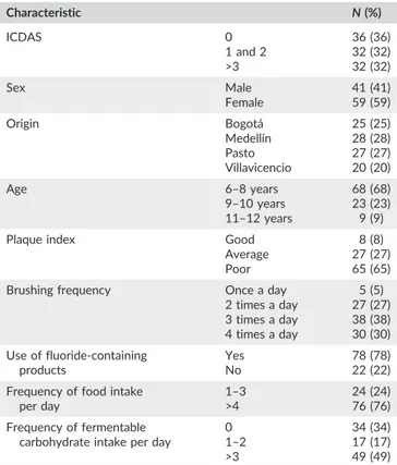
(3) 186. ANGARITA‐DÍAZ. ET AL.. to sample collection. Before starting the study, informed consent was. reference strain of S. dentisani 7446 was used as a positive control,. requested from the parents of the children and also a questionnaire. and water was used as a negative control to detect potential contami-. that requested personal information (geographic origin, sex, and age),. nation. Prior to quantification, the technique was standardized, and. diet information (frequency of food intake per day and frequency of. the calibration curve was constructed according to López‐López et al.. fermentable carbohydrates intake per day), and oral hygiene habits. (2017). The concentration of S. dentisani in each sample was calculated. (brushing frequency and use of fluoride‐containing products). This. by comparing the Cq value obtained to the standard curve. This curve. study was approved by the ethics subcommittee of the Universidad. was generated using serial dilutions of DNA extracted from the refer-. Cooperativa de Colombia, Villavicencio (21102015).. ence strain S. dentisani 7746. The Cq value obtained from dental plaque samples was replaced into the standard equation and expressed as an. 2.1 | International Caries Detection and Assessment System. absolute number. The samples were normalized to determine the num-. For the diagnosis and selection of children, the ICDAS was used to. 2.4. ber of S. dentisani cells/ng of DNA present in the sample.. Statistical analysis. |. determine carious activity. The dentists who made the diagnosis and collected the samples were calibrated in the ICDAS system,. For statistical analyses, SPSS 25.0 software was used. To determine. obtaining satisfactory inter‐ and extra‐examiner reproducibility. whether there were significant differences between the groups of each. (Kappa value ≥0.7).. variable studied, and according to the normality of the data, nonparametric tests were performed, including the Mann–Whitney U (sex,. 2.2. |. use of fluoride‐containing products, and frequency of food intake). Sample collection. and the Kruskal–Wallis H tests (oral health status of children [ICDAS], origin, age, plaque index, brushing frequency, and fermentable carbohy-. Prior to sample collection, the dental plaque index was determined using the modified Silness and Löe index (Mombelli, Van Oosten,. drate intake per day).. Schürch, & Lang, 1987). Subsequently, supragingival plaque was collected with a sterile curette, scraping over tooth surfaces (buccal, lin-. 3. |. RESULTS. gual, and occlusal) in Quadrants 1 and 3 for temporary and permanent dentition, without touching the gums. Samples were collected in. One hundred dental plaque samples from children divided into three. microtubes containing 200 μl of sterile saline solution, to which the cor-. groups were evaluated: ICDAS 0 with 36 samples analyzed, ICDAS 1. responding code was assigned. The samples were kept frozen at −20°C. and 2 with 32 samples, and ICDAS >3 with 32 samples. Table 2 shows. until molecular analysis.. the distribution according to each characteristic evaluated.. 2.3. |. TABLE 2. DNA extraction and qPCR. Characteristics of the children in the study. Characteristic. For DNA extraction, 200 μl of each sample was used, to which 5 μl. ICDAS. 0 1 and 2 >3. 36 (36) 32 (32) 32 (32). Sex. Male Female. 41 (41) 59 (59). Origin. Bogotá Medellín Pasto Villavicencio. 25 28 27 20. Age. 6–8 years 9–10 years 11–12 years. 68 (68) 23 (23) 9 (9). Plaque index. Good Average Poor. 8 (8) 27 (27) 65 (65). Brushing frequency. Once a day 2 times a day 3 times a day 4 times a day. 5 27 38 30. Use of fluoride‐containing products. Yes No. 78 (78) 22 (22). Frequency of food intake per day. 1–3 >4. 24 (24) 76 (76). Frequency of fermentable carbohydrate intake per day. 0 1–2 >3. 34 (34) 17 (17) 49 (49). of lysozyme from chicken egg white (10 mg/ml, Sigma‐Aldrich, Dorset, UK). The samples were then incubated at 37°C for half an hour, after which the extraction was continued according to the protocol of the High Pure PCR Template Preparation Kit (Roche Diagnostics GmbH, Mannheim, Baden‐Württemberg, Germany), following the manufacturer's recommendations. DNA was quantified with a Nanodrop 2000c spectrophotometer (Thermo Scientific, Wilmington, Delaware, USA), and samples with an adequate quantity and quality of DNA were selected for further analyses. For qPCR, the CFX96 Touch™ Real‐Time PCR Detection System (Bio‐Rad, Berkely, California, USA) was used with the FastStart Universal SYBR Green Master (Rox) Kit (Roche Diagnostics GmbH, Mannheim, Baden‐ Württemberg, Germany) using specific primers for S. dentisani, with a 25‐μl reaction volume. The specific primers used match with the gene encoding carbamate kinase (CkSdF5′‐GTAACCAACCGCCCAG AAGG‐3′ and CkSdR5′‐CCGCTTTCGGACTCGATCA‐3′; Mombelli, Van Oosten, Schürch, & Lang, 1987). Primers specificity was confirmed by the absence of amplification by qPCR from S. mutans, S. sobrinus, Streptococcus sanguinis, S. salivarius, Streptococcus mitis, Streptococcus. infantis,. and. Streptococcus. oralis. (López‐López,. Camelo‐Castillo, Ferrer, Simon‐Soro, & Mira, 2017). DNA of the. N (%). Grouped by age according to stages of mixed dentition.. (25) (28) (27) (20). (5) (27) (38) (30).
(4) ANGARITA‐DÍAZ. 187. ET AL.. 3.1 | Identification and quantification of S. dentisani in dental plaque samples. and 2.29, respectively. No statistically significant differences were. All analyzed samples were positive for S. dentisani, with a median of 0.46. the groups related to characteristics such as sex, origin, age, plaque. cells/ng DNA (interquartile range [IQR] 0.13–1.02), a minimum value of. index, oral hygiene, and eating habits, statistically significant differ-. found between caries‐free children and those with caries (Table 3). When comparing the quantification of S. dentisani according to. 0.001 cells/ng DNA, and a maximum of 7.20 cells/ng DNA. In caries‐free. ences were found in the origin of children (P < 0.01), the use of. children, a median of 0.45 cells/ng DNA (IQR 0.14–1.23) was found,. fluoride‐containing products (P < 0.01), and the frequency of food. with minimum and maximum values of 0.002 and 7.20, respectively.. intake (P < 0.05). Samples from children from Bogotá, Pasto, and. In children with ICDAS 1 and 2, the median was 0.49 cells/ng DNA. Medellín had medians of 0.69 cells/ng DNA (IQR 0.33–1.16), 1.04. (IQR 0.11–0.97), with a minimum value of 0.003 and a maximum of. cells/ng DNA (IQR 0.23–1.76), and 0.43 cells/ng DNA (IQR 0.17–. 6.39; and in children with ICDAS >3, the median was 0.35 cells/ng. 0.89), respectively; the median of the Villavicencio samples was 0.11. DNA (IQR 0.12–1.07), with minimum and maximum values of 0.01. cells/ng DNA (IQR 0.007–0.30).. TABLE 3. Analysis of the quantification of Streptococcus dentisani, with respect to the characteristics studied. Characteristic. Median. IQR. Range (minimum‐maximum). P‐value. 0.14–1.23. 0.002–7.20. 0.98c. ICDAS 0. 0.45. 1 and 2. 0.49. 0.11–0.97. 0.003–6.39. >3. 0.35. 0.12–1.07. 0.001–2.29. Caries‐free. 0.45. 0.14–1.23. 0.002–7.20. With caries. 0.46. 0.12–0.99. 0.003–6.39. Male. 0.42. (0.13–1.23). 0.001–6.39. Female. 0.47. (0.13–0.95). 0.003–7.20. Bogotá. 0.69. 0.33–1.16. 0.05–2.29. Medellín. 0.43. 0.17–0.89. 0.005–7.20. Pasto. 1.04. 0.23–1.76. 0.001–6.39. Villavicencio. 0.11. 0.007–0.30. 0.001–0.72. 0.35. 0.14–1.03. 0.003–6.39. Oral condition 0.91d. Sex 0.95d. Origin 0.00b,c. Agea 6–8 years 9–10 years. 0.88. 0.13–1.23. 0.002–7.20. 11–12 years. 0.40. 0.081–7.23. 0.002–0.95. Good. 0.52. 0.13–0.87. 0.08–1.63. Average. 0.28. 0.05–0.99. 0.003–1.75. Poor. 0.58. 0.15–1.06. 0.003–7.20. 0.63. 0.1–0.97. 0.08–1.08. 0.27c. Plaque index 0.34c. Brushing frequency Once a day 2 times a day. 0.22. 0.05–0.72. 0.001–4.17. 3 or more times a day. 0.67. 0.18–1.26. 0.005–6.39. Yes. 0.38. 0.11–0.90. 0.002–7.20. No. 1.12. 0.25–1.77. 0.03–6.39. 1–3. 1.04. 0.19–1.72. 0.009–6.39. >3. 0.40. 0.12–0.89. 0.002–7.20. 0.035–1.57. 0.003–1.63. 0.07c. Use of fluoride‐containing products 0.007b,d. Frequency of food intake per day 0.039b,d. Frequency of fermentable carbohydrate intake per day 0. 0.10. 1–2. 0.57. 0.26–1.13. 0.004–7.20. >3. 0.40. 0.13–0.98. 0.002–2.29. 0.30c. a Age‐grouped according to stages of mixed dentition. bAccording to the statistical analysis, these groups showed significant differences (P < 0.05). cKruskal‐ Wallis test. dMann‐Whitney U test..
(5) 188. ANGARITA‐DÍAZ. Children whose parents did not report the use of fluoride products had a significantly higher median of S. dentisani (1.12 cells/ng. ET AL.. nutrient supplements, and pH in microbial diversity and quantification (Wake et al., 2016).. DNA, IQR 0.03–6.39) than those that used these products regularly. Therefore, the lifestyle can influence in the concentration of. (0.38 cells/ng DNA, IQR 0.002–7.20). Similarly, children who had a. S. dentisani in the children. In this study, one condition that signifi-. lower food intake (Jiang et al., 2016; Kilian et al., 2016; Zaura, Nicu,. cantly influenced the quantification of S. dentisani is the amount of. Krom, & Keijser, 2014) presented higher levels than those with higher. daily food intake. Children with a maximum frequency of food intake. intakes (1.04 cells/ng DNA, IQR 0.009–6.39 vs. 0.40 cells/ng DNA,. of 3 showed significantly higher quantification of S. dentisani than. IQR 0.002–7.20, respectively; Table 3).. children with higher food intake. This difference could occur due to changes in the oral environment after food intake, resulting in differences in the multiplication and permanence of S. dentisani. One. 4. |. DISCUSSION. example would be the decrease in salivary pH. Because the optimum pH range for this bacterium is between 7 and 7.5, its growth would. In the present pilot study, S. dentisani was detected in the dental. be expected to decrease as conditions reached an acidic pH. How-. plaque samples of all the children evaluated, with a median of 0.46. ever, in this study, no significant quantification differences in. cells/ng DNA (IQR 0.13–1.02), without finding significant differences. S. dentisani were detected in children with higher or lower frequen-. between caries‐free children and those with some caries index level.. cies of fermentable carbohydrate intake, which are the type of carbo-. One of the possible reasons for not finding a significant difference. hydrates that have the greatest effect in terms of pH reduction;. between the two groups could be the sample size and the large. however, it has been shown that a high dietary content of starches. inter‐individual variation in bacterial load. It should also be noted that. or fruits also reduces plaque pH (Moynihan, 2005) and the repeated. there are other factors involved in the presence and density of. acidic challenge imposed by multiple meals has been proposed to. microorganisms.. represent an important selective pressure against nonacidophilic. One of these factors is the great diversity of microorganisms and. microorganisms (Rosier, Marsh, & Mira, 2018).. interactions involved in disease or health processes, where it is not. Another aspect that may influence the quantification of. only one or a few microorganisms generating one state or the other.. S. dentisani in this variable is that after food intake, some microorgan-. Currently, with advances in molecular techniques, it has been possible. isms are metabolically activated, and through different interactions. to identify many microorganisms involved in different processes, such. during biofilm formation (Benítez‐Páez, Belda‐Ferre, Simón‐Soro, &. as caries. Simón‐Soro, Guillen‐Navarro, and Mira (2014) used tran-. Mira, 2014), they may influence the presence of S. dentisani. This influ-. scriptomics to identify the bacterial composition in caries lesions in. ence is mediated through the competition of nutrients and/or space or. different individuals (n = 13) and stages of disease progression. The. by the production of substances toxic to bacteria, such as bacteriocins.. authors found that the microbiota of carious lesions is highly complex. This finding could indicate that diet is influencing the presence of. because it is polymicrobial and because it is organized in consortiums. this bacterium and, therefore, the relationship with the children's place. that vary between individuals and even between caries lesions (Simón‐. of origin, because the altitude of the city (Villavicencio) that was home. Soro, Guillen‐Navarro, and Mira 2014.. to the children with the lowest levels of S. dentisani is very different from. Similarly, other studies have shown that under healthy conditions,. those of the other cities. Altitude can determine the type of food that is. many bacteria are involved in beneficial processes, such as the produc-. grown and therefore more frequently consumed. This possible relation-. tion of ammonium for pH stabilization. Nascimiento et al., (2009), when. ship with diet could be influenced by the endogenous nutritional envi-. using qPCR to quantify alkaline‐producing bacteria from arginine. ronment (saliva, tissues, crevicular fluid, microbial metabolites, etc.). (S. sanguinis and Streptococcus gordonii) and from urea (S. salivarius and. through systemic circulation. In an exploratory study involving. Actinomyces naeslundii), in patients with caries and caries‐free patients,. metagenomic sequencing of 16S ribosomal RNA, Kato et al. (2017). found no significant differences in the proportions of S. sanguinis,. reported an association between intake of a specific nutrient (saturated. S. gordonii, and A. naeslundii between the two groups, despite having. fat acids, vitamin C, and glycemic load) and microbial diversity. Saturated. found a higher ammonium production in the dental plaque of caries‐. acid was correlated with relative abundance of Betaproteobacteria and. free patients. These findings suggested the presence and intervention. Fusobacteria, vitamin C exhibited positive correlations with abundance. of other ammonium‐producing bacteria that contributed to the process. in fusobacteria class and Leptotrichiaceae and Lachnospiraceae families,. (Nascimiento, Gordan, Garvan, Browngardt, & Burne RA, 2009).. and finally, the glycemic load was positively correlated with. Another factor that could have conditioned the results of this. Lactobacillaceae abundance (Kato, Vasquez, & Moyerbrailean, 2017).. study involves the particular physicochemical conditions of each hab-. In Africa, an important difference was detected on salivary. itat in the oral cavity interacting with the characteristics of each host,. microbiome composition between agricultural groups from Sierra. such as diet and oral hygiene habits. In this study, significant variations. Leona and Congo, and a former hunter‐gatherer group from Uganda. were detected in the quantification of S. dentisani among individuals of. (Batwa). This difference could be explained by the protein‐rich diet. the same group. The quantification range in caries‐free children was. of the Batwa, being approximately half of hunted animal meat, in con-. 0.002–7.20 cells/ng DNA, whereas that of children with caries was. trast to the predominantly agriculture diet of both Sierra Leona and. 0.003–6.39 cells/ng DNA. Wake et al. 2016, in an in situ study on. Congo, which have similar lifestyle and diet although they are geo-. dental biofilm formation at different time intervals, demonstrated the. graphically distant (Nasidze, Li, Schroeder, Creasey, Li, & Stoneking,. fundamental roles of certain conditions, such as oxygen pressure,. 2011). In addition to diet, other cultural condition such as hygiene.
(6) ANGARITA‐DÍAZ. 189. ET AL.. and environmental exposure may influence in geography variation, but. ORCID. studies are required in oral microbiome.. María P. Angarita‐Díaz. https://orcid.org/0000-0002-5435-3456. Finally, children whose parents did not report the use of fluoride‐ containing products showed a significantly higher quantification of. RE FE RE NC ES. S. dentisani than those who used the products. This finding could indi-. Becker, M. R., Paster, B. J., Leys, E. J., Moeschberger, M. L., Kenyon, S. G., Galvin, J. L., … Griffen, A. L. (2002). Molecular analysis of bacterial species associated with childhood caries. Journal of Clinical Microbiology, 40, 1001–1009. https://doi.org/10.1128/JCM.40.3.1001‐1009.2002. cate an inhibitory effect of fluoride on this bacterium. Few studies have analyzed the effect of fluoride within the diversity of the human oral microbiome, and the results indicate only a minimal effect. However, these studies did not control for the use of water and fluoride products. A study conducted in mice by Yasuda et al., (2017) compared the effects of water and fluoride products and found a selective impact on oral microbiota, especially acidogenic bacteria (Yasuda et al., 2017). Fluoride has been shown to affect the metabolism of S. mutans and Streptococcus sanguis because this ion binds to the active site of the glycolytic enzyme enolase or forms metal‐fluoride complexes (AlF4−), which inhibit proton translocation by F‐ATPases by limiting the phosphate required in enzymatic reactions. Fluoride also disrupts the ion gradient across the bacterial membrane (Kato, Vasquez, & Moyerbrailean G, 2017). Although S. dentisani is not an acidogenic bacterium, fluoride could have an effect on its metabolism or on tooth adhesion. In 2013, Loskill et al. performed an in vitro study with the bacteria S. mutans, S. oralis, and Staphylococcus carnosus to determine adhesion to hydroxyapatite surfaces treated with a fluoride solution and showed low bacterial adhesion strength by force spectroscopy (Loskill et al., 2013). In conclusion, the presence of S. dentisani was identified and quantified using the qPCR technique in dental plaque samples of children from four Colombian cities. The study found no significant differences in the quantification of bacteria in caries‐free children with respect to those classified with some caries index level. This could be due to the large variability in S. dentisani levels with a limited sample size or to a lack of association with the oral health parameters studied. Regarding other variables studied in the quantification of this bacterium, a possible relationship was found between the use of fluoride‐containing products, origin, and the frequency in daily food intake. In the future, a potential relationship between the different factors studied in the present work with other potentially beneficial (e.g., ammonia producers) or cariogenic (e.g., acid producers) organisms should be investigated. The design of new in vitro or in vivo experiments to confirm the possible relationships between S. dentisani and variables such as the use of fluoride‐containing products and the frequencies of food intake could determine the ideal conditions for using this bacterium as a probiotic. Additionally, understanding this relationship would add knowledge of the behavior of this bacterium as part of the oral microbiome and its possible interactions in the oral cavity. ACKNOWLEDGEMEN TS The authors thank the Comité Nacional para el Desarrollo de la Investigación (CONADI) of the Universidad Cooperative de Colombia for financing the study.. CONF LICT OF INT E RE ST The authors have no conflicts of interest to declare.. Benítez‐Páez, A., Belda‐Ferre, P., Simón‐Soro, A., & Mira, A. (2014). Microbiota diversity and gene expression dynamics in human oral biofilms. BMC Genomics, 15, 311. https://doi.org/10.1186/1471‐2164‐15‐311 Benn, A. M. L., Heng, N. C. K., Broadbent, J. M., & Thomson, W. M. (2018). Studying the human oral microbiome: Challenges and the evolution of solutions. Australian Dental Journal, 63, 14–24. https://doi.org/ 10.1111/adj.12565 Burton, J. P., Drummond, B. K., Chilcott, C. N., Tagg, J. R., Thomson, W. M., Hale, J. D. F., & Wescombe, P. A. (2013). Influence of the probiotic Streptococcus salivarius strain M18 on indices of dental health in children: A randomized double‐blind, placebo‐controlled trial. Journal of Medical Microbiology, 62, 875–884. https://doi.org/10.1099/jmm.0.056663‐0 Camelo‐Castillo, A., Benítez‐Páez, A., Belda‐Ferre, P., Cabrera‐Rubio, R., & Mira, A. (2014). Streptococcus dentisani sp. nov., a novel member of the mitis group. International Journal of Systematic and Evolutionary Microbiology, 64, 60–65. https://doi.org/10.1099/ijs.0.054098‐0 Huang, X., Palmer, S. R., Ahn, S. J., Richards, V. P., Williams, M. L., Nascimento, M. M., & Burne, R. A. (2016). A highly arginolytic Streptococcus species that potently antagonizes Streptococcus mutans. Applied and Environmental Microbiology, 82, 2187–2201. https://doi.org/ 10.1128/AEM.03887‐15 Jiang, S., Gao, X., Jin, L., & Lo, E. C. (2016). Salivary microbiome diversity in caries‐free and caries‐affected children. International Journal of Molecular Sciences, 17, E1978. Kato, I., Vasquez, A., & Moyerbrailean, G. (2017). Nutritional correlates of human oral microbiome. Journal of the American College of Nutrition, 36, 88–98. https://doi.org/10.1080/07315724.2016.1185386 Kilian, M., Chapple, I. L. C., Hannig, M., Marsh, P. D., Meuric, V., Pedersen, A. M. L., … Zaura, E. (2016). The oral microbiome—An update for oral healthcare professionals. British Dental Journal, 221, 657–666. https://doi.org/10.1038/sj.bdj.2016.865 Kralik, P., & Ricchi, M. (2017). A basic guide to real time PCR in microbial diagnostics: Definitions, parameters, and everything. Frontiers in Microbiology, 8, 108. Kressirer, C. A., Smith, D. J., King, W. F., Dobeck, J. M., Starr, J. R., & Tanner, A. C. R. (2017). Scardovia wiggsiae and its potential role as a caries pathogen. Journal of Oral Biosciences, 59, 135–141. https://doi.org/ 10.1016/j.job.2017.05.002 Kreth, J., Merritt, J., & Qi, F. (2009). Bacterial and host interactions of oral streptococci. DNA and Cell Biology, 28, 397–403. https://doi.org/ 10.1089/dna.2009.0868 Lee, P. P., Mak, W. Y., & Newsome, P. (2004). The aetiology and treatment of oral halitosis: An update. Hong Kong Medical Journal, 10, 414–418. López‐López, A., Camelo‐Castillo, A., Ferrer, M. D., Simon‐Soro, A., & Mira, A. (2017). Health‐associated niche inhabitants as oral probiotics: The case of Streptococcus dentisani. Frontiers in Microbiology, 8, 379. Loskill, P., Zeitz, C., Grandthyll, S., Thewes, N., Müller, F., Bischoff, M., … Jacobs, K. (2013). Reduced adhesion of oral bacteria on hydroxyapatite by fluoride treatment. Langmuir, 29, 5528–5533. https://doi.org/10.1021/la4008558 Mineoka, T., Awano, S., Rikimaru, T., Kurata, H., Yoshida, A., Ansai, T., & Takehara, T. (2008). Site‐specific development of periodontal disease is associated with increased levels of Porphyromonas gingivalis, Treponema denticola, and Tannerella forsythia in subgingival plaque. Journal of Periodontology, 79, 670–676. https://doi.org/10.1902/jop.2008.070398 Mombelli, A., Van Oosten, M. A. C., Schürch, E., & Lang, N. P. (1987). The microbiota associated with successful or failing osseointegrated titanium implants. Oral Microbiology and Immunology, 2, 145–151..
(7) 190. ANGARITA‐DÍAZ. ET AL.. Moynihan, P. J. (2005). The role of diet and nutrition in the etiology and prevention of oral diseases. Bulletin of the World Health Organization, 83, 694–699.. Wake, N., Asahi, Y., Noiri, Y., et al. (2016). Temporal dynamics of bacterial microbiota in the human oral cavity determined using an in situ model of dental biofilms. NPJ Biofilms Microbiomes, 10, 16018.. Nascimiento, M. M., Gordan, V. V., Garvan, C. W., Browngardt, C. M., & Burne, R. A. (2009). Correlations of oral bacterial arginine and urea catabolism with caries experience. Oral Microbiology and Immunology, 24, 89–95. https://doi.org/10.1111/j.1399‐302X.2008.00477.x. Yasuda, K., Hsu, T., Gallini, C. A., et al. (2017). Fluoride depletes acidogenic taxa in oral but not gut microbial communities in mice. mSystems, 2, e00047–e00017.. Nasidze, I., Li, J., Schroeder, R., Creasey, J. L., Li, M., & Stoneking, M. (2011). High diversity of the saliva microbiome in Batwa Pygmies. PLoS One, 6, 23352. Rosier, B. T., Marsh, P. D., & Mira, A. (2018). Resilience of the oral microbiota in health: Mechanisms that prevent dysbiosis. Journal of Dental Research, 97, 371–380. https://doi.org/10.1177/0022034517742139 Sampaio‐Maia, B., & Monteiro‐Silva, F. (2014). Acquisition and maturation of oral microbiome throughout childhood: An update. Dental Research Journal, 11, 291–301.. Zaura, E., Nicu, E. A., Krom, B. P., & Keijser, B. J. F. (2014). Acquiring and maintaining a normal oral microbiome: Current perspective. Frontiers in Cellular and Infection Microbiology, 4, 85. Zhang, Y., Zhen, M., Zhan, Y., Song, Y., Zhang, Q., & Wang, J. (2017). Population‐genomic insights into variation in Prevotella intermedia and Prevotella nigrescens isolates and its association with periodontal disease. Frontiers in Cellular and Infection Microbiology, 7, 409. https:// doi.org/10.3389/fcimb.2017.00409. Simón‐Soro, A., Guillen‐Navarro, M., & Mira, A. (2014). Metatranscriptomics reveals overall active bacterial composition in caries lesions. Journal of Oral Microbiology, 6, 25443. https://doi.org/ 10.3402/jom.v6.25443. How to cite this article: Angarita‐Díaz MP, Díaz JA, Tupaz. Simón‐Soro, A., & Mira, A. (2015). Solving the etiology of dental caries. Trends in Microbiology, 23, 76–82. https://doi.org/10.1016/j. tim.2014.10.010. Dent Res. 2019;5:184–190. https://doi.org/10.1002/cre2.158. HA, et al. Presence of Streptococcus dentisani in the dental plaque of children from different Colombian cities. Clin Exp.
(8)
Figure


Documento similar
In addition to two learning modes that are articulated in the literature (learning through incident handling practices, and post-incident reflection), the chapter
The development of the research started with a first phase of rigorous observation of films produced in the delimited period (1964-2006), through literature review of direct
In the “big picture” perspective of the recent years that we have described in Brazil, Spain, Portugal and Puerto Rico there are some similarities and important differences,
Figure 3 and Figure 4 present the Nyquist diagrams of low carbon steel AISI 1018 registered in the model acetic solution, in the presence of kerosene (hydrocarbon), through
The objective of the present study was to identify the effect of unmet dental treatment needs and socioe- conomic and sociodemographic variables on the patterns of
by the presence of fibrous cap discontinuity with a communication between the lumen and the inner core of a plaque or with a cavity formation within the plaque. B, Plaque erosion
For instance, the best overall accuracy of bagging in Breast with 20% noise is achieved using a 10% sampling ratio: The test error goes from 4.1% when no noise is injected to 3.5%
In particular, different versions of the training set for the base learners can be used, as in bagging (bootstrap sampling of training data), class-switching (noise injection in
