Arsenic speciation in edible alga samples by microwave-assisted extraction and
high performance liquid chromatography coupled to atomic fluorescence
spectrometry
S. García-Salgado, MA Quijano*, M.M. Bonilla
Departamento de Ingeniería Civil: Tecnología Hidráulica y Energética, Escuela Universitaria de Ingeniería Técnica de Obras Públicas, Universidad Politécnica de Madrid, Alfonso XII3 y 5,28014 Madrid, Spain
A B S T R A C T
Keywords: Arsenic speciation
Microwave-assisted extraction Edible algae
Inductively coupled plasma atomic emission spectrometry
High performance liquid chromatography Hydride generation-atomic fluorescence spectrometry
Twelve commercially available edible marine algae from France, Japan and Spain and the certified reference material (CRM) NIES No. 9 Sargassum fulvellum were analyzed for total arsenic and arsenic species. Total arsenic concentrations were determined by inductively coupled plasma atomic emission spectrometry (ICP-AES) after microwave digestion and ranged from 23 to 126 (xgg-1. Arsenic species in alga samples were extracted with deionized water by microwave-assisted extraction and showed extraction efficiencies from 49 to 98%, in terms of total arsenic. The presence of eleven arsenic species was studied by high performance liquid chromatography-ultraviolet photo-oxidation-hydride genera-tion atomic-fluorescence spectrometry (HPLC-(UV)-HG-AFS) developed methods, using both anion and cation exchange chromatography. Glycerol and phosphate sugars were found in all alga samples analyzed, at concentrations between 0.11 and 22 (xgg-1, whereas sulfonate and sulfate sugars were only detected in three of them (0.6-7.2 (xgg-1). Regarding arsenic toxic species, low concentration levels of dimethy-larsinic acid (DMA) (<0.9 (xgg-1) and generally high arsenate (As(V)) concentrations (up to 77 (xgg-1) were found in most of the algae studied. The results obtained are of interest to highlight the need to per-form speciation analysis and to introduce appropriate legislation to limit toxic arsenic species content in these food products.
1. Introduction
Arsenic occurs in seawater mainly as inorganic arsenic (arsen-ite, As(III), and arsenate, As(V)) at low |jLgL_1 level [1]. However, some marine organisms accumulate arsenic; thus, they often have much higher arsenic concentrations than in the surrounding envi-ronment, typically in the range of 10-100 |Jtgg_1 [2], even though arsenic has been reported to be mostly biotransformed into less toxic organic arsenic compounds [3]. Arsenobetaine (AsB) has been the major arsenic species found in marine fauna, while in algae the most frequently occurring arsenic species are arsenosugars, especially the derivatives of dimethylarsonylribosides commonly named glycerol, phosphate, sulfonate and sulfate sugars [4-8], whose structures are shown in Fig. 1. Besides these species, others like monomethylarsonic acid (MMA), dimethylarsinic acid (DMA), trimethylarsine oxide (TMAO), tetramethylarsonium ion (TETRA), arsenocholine (AsC) and some inorganic arsenic (As(III) and As(V)) can be found in marine organisms as well [9-12].
Toxicity studies of arsenic have shown that different forms exhibit different toxicities, thus inorganic arsenic species are more toxic than organic compounds and toxicity generally decreases with increasing degree of methylation, with the exception of TETRA species [13]. Arsenosugars are assumed to be much less toxic than inorganic arsenic species [14], although their toxicity has not been well assessed [15]. However, studies based on the presence of arsenic metabolites in humans point to indirect toxicity resulting from the ingestion of algae containing arsenosugars, due to their biotransformation in toxic arsenicals [16]. Since arsenic species have different toxicity, information about the distribution of the species within algae is necessary for assessing the risk associated with the entry of arsenic into the wildlife and human food chains. Therefore, there is considerable interest in developing analytical methodologies for arsenic speciation in food products. This inter-est is accentuated in the case of algae, because it is well known that they contribute substantial amounts of arsenic to the human diet, and their consumption is nowadays increasing due to their prop-erties as food additives, nutritional values (high contents of iodine, minerals and vitamins) and suggested medical applications [17].
HO OH
R = O H (Glycerol sugar, Gly-sug) OPC^CE^CHOHCHjOH (Phosphate sugar, P04-sug)
SO3" (Sulfonate sugar, SO^-sug) OSO3" (Sulfate sugar, S04-sug)
Fig. 1. Structures of the four most common arsenosugars found in algae.
mass spectrometry (HPLC-ICP-MS) is the most frequently used hyphenated technique for arsenic speciation [5-7,11,12,21]. How-ever, the hydride generation-atomic fluorescence spectrometry (HG-AFS) coupled to HPLC represents a suitable alternative to this technique [22,23]. HG-AFS has been reported to be simi-lar to inductively coupled plasma mass spectrometry (ICP-MS) regarding sensitivity and linear calibration range, although it has some advantages for arsenic speciation analysis, such as simplic-ity, lower acquisition and running costs [24-26]. However, the destruction of the organic part of arsenic species is required to determine them by HG-AFS, as they do not generate volatile hydrides or because the generation efficiency is very low [27]. Photo-oxidation to convert these species into inorganic arsenic is achieved after chromatographic separation by using a strong oxidant (K2S2O8) in basic media and ultraviolet (UV) radiation emitted from a low pressure mercury lamp. Regarding arsenic species separation, ion-exchange chromatography is preferred for coupling to HG-AFS detection, as the separation process is more reproducible and less prone to sample matrix interferences than ion-paring chromatography [22]. On the other hand, both anion and cation exchange chromatography is needed for arsenic spe-ciation in biological samples, due to the different ionic character of aqueous soluble arsenic species. The mobile phases commonly used are phosphate and pyridine solutions, for anion and cation exchange, respectively. HPLC-(UV)-HG-AFS have been success-fully applied to a great variety of samples. For example, Slejkovec et al. [28] analyzed six CRMs of biological origin (dogfish mus-cle and liver, lobster hepatopancreas, oyster tissue, brown algae and scallop) by HPLC-(UV)-HG-AFS, using both anion and cation exchange chromatography methods. They identified AsB, DMA, TETRA, AsC, TMAO, As(V), MMA and two arsenosugars, together with four unidentifiable compounds (one in brown algae, one in dogfish muscle and liver and lobster hepatopancreas, and two in scallop). In 2002, Sánchez-Rodas et al. [29] applied this same hyphenated technique for the analysis of aqueous extracts of oys-ters. These authors concluded that it is an alternative technique to liquid chromatography-electrospray-mass spectrometry (LC-ES-MS) forthe detection of arsenosugars in crude extracts, since signal response in HPLC-(UV)-HG-AFS is little influenced by the sam-ple matrix, whereas LC-ES-MS analysis usually requires previous sample clean-up steps. Schaeffer et al. [30] determined twelve arsenic compounds in mussels, anchovies, seabreams, sea bass and sardines by HPLC-(UV)-HG-AFS, using both anion and cation exchange methods. AsB was detected as the major compound in all the samples, with trace amounts of As(III), DMA and AsC, whereas arsenosugars were detected only in mussel samples. Slejkovec et al. [31] applied it for the determination of arsenic species in ten dif-ferent marine algae (red, green and brown) from the littoral zone along the Adriatic Sea coast of Slovenia. They found that arseno-sugars were the most abundant arsenicals in most of the analyzed
algae, although they also found AsB, As(III), As(V) and DMA, As(V) being the major species in three alga samples (Ceranium sp., Polisy-phonia sp., Cystoseira barbata). In 2009, Geng et al. [32] compared the performance of the combination of solvent extraction and HPLC-(UV)-HG-AFS with the combination of alkaline digestion and cryogenic trap-hydride generation-atomic absorption spec-trometry (CT-HG-AAS), for arsenic speciation in marine product samples. They analyzed six CRMs of marine animal samples and four seaweed samples, and concluded that results obtained by both methodologies are comparable, because the concentrations of arsenobetaine obtained in CRMs from the extraction-HPLC method were very consistent with those of trimethylated arsenic species measured by the digestion-CT method. In the case of seaweed sam-ples, the amounts of dimethylated arsenic species measured by the digestion-CT method were approximately equal to the sum of the amounts of DMA and three arsenosugars determined by the extraction-HPLC method.
The aims of this work consisted on: (1) the development of arsenic speciation analysis methods based on the coupling of high performance liquid chromatography (anion and cation exchange) with ultraviolet photo-oxidation-hydride generation-atomic fluo-rescence spectrometry (HPLC-(UV)-HG-AFS); (2) the application of the developed methods to carry out the identification and quan-tification of arsenic species present in the water extracts, obtained by microwave-assisted extraction, of commercially available edible alga samples.
2. Materials and methods
2.1. Instrumentation
Microwave assisted extraction was conducted using a MARS 5 microwave oven (CEM, Matthews, NC, USA), with Pyrex extraction vessels. The temperature was monitored in a control vessel by an armored fiber-optic temperature control probe.
An ETHOS 1 closed vessel microwave digestion system (Mile-stone S.r.l., Sorisole, BG, Italy), with built-in ATC-400-CE automatic temperature control and employing TFM-Teflon microwave ves-sels, was used for alga samples digestion.
Total arsenic determination was performed with an induc-tively coupled plasma atomic emission spectrometer (ICP-AES) model Liberty Series II Axial Sequential (Varian Australia, Mulgrave, Australia).
Other equipment used included a Binder GmbH ED53 drying oven (Tuttlingen, Germany), used to dry the samples; a Retsch MM 301 mixer mill (Haan, Germany) with tungsten carbide grinding jars and balls, used to grind the samples; and an Eppendorf 5804 R centrifuge (Hamburg, Germany), used for the centrifugation of the alga extracts.
2.2. Reagents and standard solutions
All solutions were prepared from analytical reagent grade chemicals using deionized water obtained from a Millipore water purification system (Elix®, Molsheim, France).
As(III) (994±2mgL-1) and As(V) (1000 mgL"1) stock standard solutions were purchased from Fluka (Sigma-Aldrich, Stein-heim, Germany) and Merck (Darmstadt, Germany), respectively. 1000mgL-1 standard solutions of MMA, DMA, AsB, TETRA and TMAO were prepared by dissolving the appropriate amounts of CH3As03Na2 (MMA) purchased from Supelco (Bellefonte, PA, USA); C2H6AsNa02-3H20 (DMA) from Fluka; AsC5Hn02 (AsB) fromTCLC (Tri Chemical Laboratories Inc., Yamanashi, Japan); and A s C ^ ^ B r (TETRA) and AsC3H90 (TMAO) from Argus Chemicals srl (Vernio, PO, Italy). All the stock solutions were kept at 4°C, and further diluted solutions for the analysis were prepared daily. Yttrium stock standard solution (lOOOmgL-1), purchased from Fluka, was used as an internal standard for ICP-AES total arsenic analysis. The As(V) stock standard solution was used for both total As calibration in ICP-AES and As(V) species calibration in HPLC-(UV)-HG-AFS.
An aliquot freeze-dried extract of Fucus serratus, kindly donated by Prof. Kevin A. Francesconi (Department of Analytical Chem-istry, Institute of ChemChem-istry, Karl-Franzens University Graz, Graz, Austria) [33], containing the four common arsenosugars (glycerol (Gly-sug), phosphate (P04-sug), sulfonate (S03-sug) and sulfate (S04-sug) sugars), was used to assign the arsenosugar peaks in the chromatograms and for their concentration estimation in the alga extracts. The extract was dissolved in 4.0 mL of deionized water, the concentration of the four above mentioned arsenosugars being 25, 21.5,155 and 100 (xg \rl of arsenic per species, respectively.
The 5, 20 and lOOmmolL-1 phosphate buffers used as mobile phases for anion exchange chromatographic method were pre-pared by dissolving appropriate amounts of K2HPO4 and KH2PO4 (Scharlau, Barcelona, Spain) and mixing the solutions until pH 9.0 was reached. A 2.5mmolL~1 pyridine solution (Sigma-Aldrich) at pH 2.65 adjusted with hydrochloric acid (Scharlau) was used for cation exchange chromatographic method. The mobile phases were filtered through a 0.45 |jim Magna nylon filters (Osmonics Inc., Minnetonka, MN, USA) and degassed before use by ultrasonic shak-ing. Furthermore, the alga extracts were filtered through 0.22 |jim nylon syringe filters (Osmonics) before their injection in the HPLC system.
Sodium borohydride solution 1.4% (w/v) was prepared by dis-solving NaBH4 powder (Aldrich, Milwaukee, WI, USA) in deionized water and stabilizing it with 0.1 mol \rl sodium hydroxide (Fluka).
It was prepared daily and filtered before use. Hydrochloric acid solution (8.0 mol \rl) was prepared by dilution of HC1 37%
(Schar-lau). Both solutions were used in the hydride generation step. Potassium peroxodisulfate solutions 2 and 4% (w/v), stabilized in 2 and 4% (w/v) sodium hydroxide, respectively, used as oxidiz-ing agents in the photo-oxidation step, were prepared daily from K2S2O8 (Scharlau) and NaOH and filtered before use.
Nitric acid (69.5%) and hydrogen peroxide (35%), both from Scharlau, were used to digest the samples.
2.3. Alga samples
Edible alga samples from Spain and Japan were purchased at local markets as dried products, ground to a fine powder in a
tungsten carbide disc mill and kept into pre-cleaned polyethy-lene containers at room temperature for subsequent analyses. The samples include four Atlantic algae harvested by the local manu-facturer in Galician coast (Northwest Spain): Himanthalia elongata (Sea spaghetti), Laminaria ochroleuca (Kombu), Undaria pinnati-flda (Wakame) and Porphyra umbilicalis (Nori). Furthermore, the
edible alga samples from Japan: Eisenia arbórea (Árame), Hizikia fusiformis (Hijiki), L. ochroleuca (Kombu), Li. pinnatiflda (Wakame)
and P. umbilicalis (Nori) were also analyzed, together with three alga samples from France (Fucus vesiculosus (Fucus), L ochroleuca (Kombu) and Laminaria digitata (Laminaria)), donated by the Centre d'Etude et de Valorisation des Algues (Pleubian, France).
The reference material NIES No. 9, from the National Institute for Environmental Studies (Tsukuba, Ibaraki, Japan) and certified for total arsenic (115±9|jLgg_1), was a lyophilized Sargasso material
(Sargassum fulvellum) and was used for quality control purposes. 2.4. Experimental
2.4.1. Sample treatment
Total arsenic concentration in alga samples was determined by ICP-AES after microwave digestion with nitric acid and hydrogen peroxide, following a method previously developed for total arsenic determination in plant samples [34].
Arsenic species were extracted in deionized water (8mL per 200 mg of alga sample) following a previously developed microwave-assisted extraction method [10]. The extraction pro-cess (heating at 90°C for 5 min) was repeated three times. The extracts were centrifuged at 14,000 x g for 10min and the three supernatants were mixed, diluted up to 25 mL with deionized water and centrifuged again. Total arsenic concentration extracted was determined by ICP-AES, using the experimental conditions previ-ously optimized [34]. Prior to the chromatographic analysis, final alga extracts were filtered through 0.22 |jim nylon syringe filters.
2.4.2. Chromatographic separation and detection
The identification and quantification of arsenic species in alga extracts was carried out by HPLC-(UV)-HG-AFS. The separation of arsenic compounds was performed by two chromatographic meth-ods, based on anion and cation exchange chromatography. The chromatographic separation of As(III), As(V), MMA, DMA and four arsenosugars (Gly-sug, P04-sug, S03-sug and S04-sug) was car-ried out by injecting solutions containing the eight compounds onto the anion exchange column (Hamilton PRP-X100) and ini-tially eluting with 5mmolL_ 1 phosphate buffer of pH 9.0, at a flow rate of l.OmLmin- 1. After 1 min, the mobile phase was changed to 20mmolL~1 phosphate buffer of pH 9.0, using a linear con-centration gradient over a 0.1-min period. Finally, after 5 min, the mobile phase was changed to lOOmmolL-1 phosphate buffer of pH 9.0, using the same linear concentration gradient. The eluted arsenic species were conducted to an on line photo-oxidation sys-tem, equipped with a UV lamp and a 2% (w/v) potassium persulfate solution in 2% (w/v) NaOH, at a flow rate of 0.5 mLmin- 1. Arsenic species were detected by HG-AFS, using the operation conditions given in Table 1.
The separation of As(III), As(V), AsB, TETRA and TMAO was per-formed on a Hamilton PRP-X200 cation exchange column and using pyridine 2.5mmolL~1 at pH 2.65 (adjusted with HC1) as mobile phase, at a flow rate of l.OmLmin- 1. The eluted arsenic species were detected by (UV)-HG-AFS, using a 4% (w/v) potassium per-sulfate solution in 4% (w/v) NaOH. The operation conditions are given in Table 1.
Table 1
Experimental conditions for arsenic speciation by HPLC-(UV)-HG-AFS.
HPLC
Anion exchange (concentration gradient): Column
Mobile phase
Cation exchange (isocratic): Column
Mobile phase
Flow rate (mL min- 1) Injection volume (|xL)
On line UV lamp
UVlamp
UV cracking frequency (Hz) UV cracking voltage (V) K2S208(%,w/v):
K2S2O8 flow rate (mLmin- 1)
Hydride generation
HCltmolL-1)
HC1 flow rate (mLmin^1) NaBH4 (%, w/v)
NaBH4 flow rate (mL min- 1) Ar(GL separator) flow rate (mLmin- 1) Air(drying) flow rate (Lmin- 1)
AFS instrument
Detector
Wavelength (nm) Primary current (mA) Boost current (mA) Gain
Hamilton PRP-X100 Phosphate buffer 5, 20 and lOOmmolL-'.pHaO
Hamilton PRP-X200 Pyridine 2.5 mmol L_1, pH 2.65 (HC1)
1.0 100
10.570 UV Cracker (PS Analytical)
50 230
2a
4b
0.5
8.0 1.4 1.4 1.4 250 2.5
Millennium Excalibur(PS Analytical)
193.7 27.5 35.0 10 and 100
For the anion exchange method. For the cation exchange method.
identified due to the increase in their areas when arsenic species standard solutions were added, except for arsenosugars, which were identified by their retention time in the F. serratus extract solu-tion and alga sample extracts. The instrument drift was checked by injection of a standard solution every two samples injections.
2.4.3. Stability of arsenic spedes during the extraction procedure Hijiki (H. fusiformis) sample was spiked with a mixture of the three arsenic species (AsB, TETRA and TMAO). A portion of 200 mg of the alga was weighed and 1 mL of stock solution con-taining lOmgL- 1 (as arsenic) of each one of the three species was added. The mixture was remained overnight. The spiked sample was processed by the extraction procedure previously described and arsenic species were analyzed by cation exchange HPLC-(UV)-HG-AFS.
3. Results and discussion
3.1. Determination of total arsenic in alga samples
Total arsenic concentrations in alga samples were determined by ICP-AES. Table 2 shows the results obtained in the total arsenic determination, expressed in terms of dry weight (dw), for the twelve alga samples analyzed (10 brown algae and 2 red algae). The accuracy of the method was assessed by using the CRM NIES No. 9, S.fulvellum (Sargasso). The result obtained (109 ± 2 |jigg_1) showed the absence of significant differences, at the 95% confi-dence level, between the As concentration found and the certified value (115±9|jLgg_1). Therefore, the analytical method used has proven to be suitable for total arsenic determination in alga sam-ples. The total arsenic concentrations found in alga samples varied over a wide range (23-126 |jigg_1), which is in agreement with those reported in the bibliography for edible algae (8-150|jLgg_1)
Table 2
Concentrations expressed as |xgg_ 1 on dry mass (mean±SD, n = 3) of total arsenic found in commercially available edible marine alga samples after microwave diges-tion and determinadiges-tion by ICP-AES.
Division Species Common name Origin Total As
Phaeophyta (brown algae)
Eisenia arbórea Árame Japan 26 ± 1
Rhodophyta (red algae)
Fucus vesiculosus Fucus Himanthalia elongata Sea spaghetti Hizikia fusiformis Hijiki Laminaria ochroleuca Kombu
Laminaria digitata Laminaria lindaría pínnatifida Wakame
Porphyra umbílícalís Nori
France Spain Japan France Japan Spain France Japan Spain Japan
Spain
47 ± 2 24 ± 2 98 ± 5 42 ± 5 97 ± 6 46 ± 4 126 ± 5 37 ± 2 37 ± 1 23 ± 3
34 ± 3
[35-37]. The highest arsenic concentrations were found in the brown algae Laminaria from France (126 |jLgg_1), followed by Hijiki and Kombu algae from Japan (both about 100|jLgg_1), whereas the lowest arsenic concentration levels were found in the red alga Nori from Japan (23 (jigg-1), and the brown algae Sea spaghetti from Spain (24 |jLgg_1) and Arame from Japan (26 |jLgg_1). From the results, total arsenic contents in Phaeophytes (brown algae) were generally higher than in Rhodophytes (red algae), which is in agree-ment with several authors that reported a relationship between arsenic content and algal division, so that brown > red > green [36,38].
Regarding the comparison between the results obtained for the same alga species from different origins, similar arsenic con-centration levels were found between the two Wakame samples (37 |jigg_1, for both Japanese and Spanish algae), as well as for the two Nori algae (23 and 34|jigg_ 1, for the samples from Japan and Spain, respectively). However, in the three Kombu samples, it was observed that algae from France and Spain have similar arsenic levels, whereas the sample from Japan presented a concentration approximately two times higher. From these results, a relationship between the arsenic concentration levels and the alga species or their origin cannot be established. Algae absorb arsenic directly from sea water and there are some factors influencing the arsenic levels in these marine organisms, such as the light intensity, tur-bidity, temperature, depth, salinity and nutrient uptake [21]. These factors may contribute to the differences observed in total arsenic concentration levels in the algae studied.
3.2. Arsenic species determination by HPLC-(UV)-HG-AFS
The choice of strong ion-exchange HPLC for arsenic species separation was based in our previous experience in the arsenic speciation field [9,10,39], as well as in the consulted bibliography [21,31,32].
4 0
-c
>
Time (min)
c
>
60-
50-
40-
30-
20-{
• i '
T
D |
11 1
111
i ' i
i
í(4)
;¡
* i
H|%
i J i j ,
6 S Time (min)
10 12 14
Fig. 2. (a) Chromatogram obtained by anion exchange HPLC-(UV)-HG-AFS (gain 10) for a mixture of As(III), As(V), DMA and MMA (25 |xg L-1 As) and Gly, P04, S03 and S04-sug (25, 21.5,155 and lOOixgL-1 As, respectively): (1) Gly-sug, (2) As(III), (3) DMA, (4) P04-sug, (5) MMA, (6) S03-sug, (7) As(V), and (8) S04-sug. (*) AsB (25 |xg L"1 As), (b) Chromatogram obtained by cation exchange HPLC-(UV)-HG-AFS (gain 10) for a mixture of As(III), As(V), MMA, DMA, AsB, TETRA and TMAO (25 |xg L-1 As): (1) As(III) + MMA, (2) As(V) + DMA, (3) AsB, (6) TETRA and (7) TMAO. Chromatogram for Fucus serratus extract (dashed line): (2) P04-sug, (3) S04-sug, (4) S03-sug, and (5) Gly-sug (21.5,100,155 and 25|xgL-1 As, respectively).
coeluted with the arsenic species MMA, whereas the species As(V) coeluted with the arsenic species DMA, which can be differed by the anion exchange method. Regarding arsenosugars, the injection of an extract from the seaweed F. serratus [33] (Fig. 2b, dashed line) allowed us to verify that at the pH used in this method (2.65), these arsenic species behave like cations, so they are retained in the column [6]. The species PÜ4-sug and SÜ4-sug coeluted with the species As(V) and AsB, respectively, whereas SÜ3-sug and Gly-sug eluted between the species AsB and TETRA. In conclusion, both anion and cation exchange chromatographic methods are needed for the secure identification of the eleven arsenic species stud-ied, especially when cationic arsenic species are present together with inorganic and arsenosugars species, although the AsB con-centration could only be estimated by subtracting the SÜ4-sug concentration (determined by the anion exchange method) if both species are present in the sample.
Arsenic species determination was carried out by HG-AFS. It is generally accepted that organic arsenic species do not form hydrides, or form them with low hydride generation efficiencies [27]. Therefore, the introduction of a decomposition step before
the hydride generation is necessary. In this study, an on line photo-oxidation was carried out using a UV lamp and a solution of potassium persulfate, prepared in sodium hydroxide. According to Slejkovec et al. [28], the detector response depends on the degree of decomposition in the UV photo-oxidation step. Therefore, the effect of the potassium persulfate concentration was evaluated from 1 to 4% (w/v). A concentration of 2% (w/v) was chosen for the oxida-tion of organic arsenic species when they were separated by the anion exchange chromatographic method, due to the better sig-nal/noise ratio obtained, whereas a higher concentration (4% (w/v)) was needed when the cation exchange method was used. This fact can possibly be due to a part of the persulfate is consumed by the organic mobile phase used (pyridine), which is also oxidized dur-ing the photo-oxidation step. Hydride generation and AFS detection were achieved according to the experimental conditions shown in Table 1.
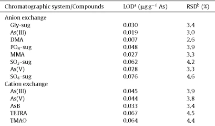
The analytical characteristics were evaluated for the two meth-ods used. The precision, evaluated as relative standard deviation, was calculated from five independent replicates of a standard solu-tion containing 10 |jig L_1 of arsenic per species, whereas the LODs were calculated as three times the standard deviation obtained from ten replicates of the lowest standard used in the calibra-tion curve, which was of 1 |jig L_1 of arsenic per species, except for arsenosugars, due to the absence of standard solutions of these species. In this case, %RSD and LODs were estimated in the anion exchange method, using different dilutions of the F. serratus extract solution (see Table 3 footnote). The results obtained, for a detector gain of 100, are shown in Table 3.
3.3. Arsenic speciation in alga samples
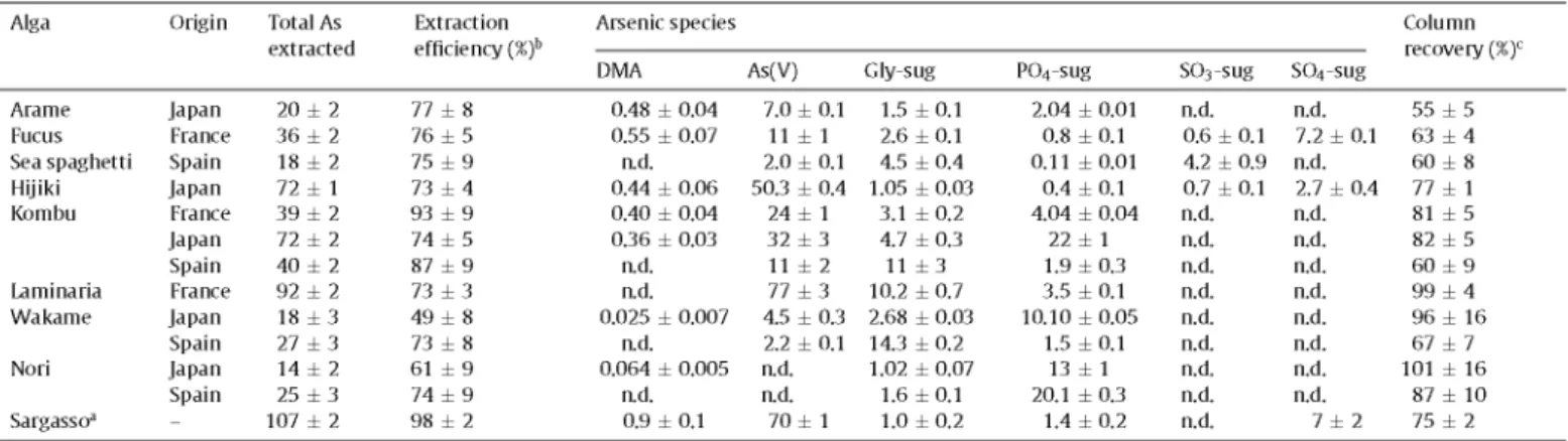
Table 4 shows the total arsenic extracted in deionized water by the microwave extraction method for the twelve alga samples studied, as well as for the CRM NIES No. 9, S. fulvellum (Sargasso material). Extraction efficiencies, calculated as the ratio of total arsenic in the water extract to total arsenic in the algae, are also shown in Table 4 and ranged from 49 (for Wakame from Japan) to 98% (for Sargasso material). Most of the algae analyzed present a high percentage of arsenic compounds soluble in water, which was higher than 73% for 11 of the 13 samples analyzed. The results obtained are in agreement with the wide ranges of extraction
Table 3
Limits of detection, expressed as ixgg-1 of arsenic, and relative standard deviation, expressed as percentage, for arsenic species studied by anion and cation exchange HPLC-(UV)-HG-AFS, for a detector gain of 100.
Chromatographic system/Compounds LODa (ixgg-1 As) RSDb {%)
Anion exchange Gly-sug As(III) DMA P04-sug MMA S03-sug As(V) S04-sug Cation exchange
As(III) As(V) AsB TETRA TMAO
0.030 0.019 0.007 0.048 0.027 0.062 0.028 0.076
0.045 0.044 0.033 0.067 0.064
3.4 3.0 2.6 3.9 3.3 4.2 3.3 4.6
3.9 3.8 3.4 4.5 4.4
a Calculated for a standard solution containing 1 |xg L-1 of As per species, except for arsenosugars (estimated by dilution of the F. serratus extract solution containing 2.5, 2.15,1.5 and l.OixgL-1 of As for Gly, P04, S03 andS04-sug, respectively).
efficiencies in water reported depending on the particular algae type [10,19]. On the other hand, the arsenic fraction not extracted with water could correspond to arsenic bound to compounds such as lipids and might account for up to 50% of total arsenic in algae [40].
Arsenic speciation studies were carried out by HPLC-(UV)-HG-AFS, by the two chromatographic methods developed. The lack of peak eluting in the dead volume of the chromatograms obtained by the anion exchange chromatographic method showed the absence of cationic arsenic species in the water alga extracts. This fact was confirmed by cation exchange chromatography. Therefore, the anion exchange method was appropriate to perform arsenic speciation analysis in the alga samples studied. Fig. 3 shows, as examples, the chromatograms obtained by the anion exchange method for the water extracts of the red alga Nori from Japan (Fig. 3a, at gain 10 for AFS detector), the brown algae Fucus from France (Fig. 3b, gain 10) and Hijiki from Japan (Fig. 3c, gain 100), and the NIES No. 9 Sargasso material (Fig. 3d, gain 100). Arsenic compounds identified in the edible alga samples studied comprised DMA, As(V) and the four arsenosugars (Gly-sug, P04-sug, SÜ3-sug and S04-sug), depending on the kind of algae analyzed. The arsenic species As(III), MMA, AsB, TMAO and TETRA were not detected in any alga sample studied, because either no chromatographic peak matched the corresponding retention time (for As(III) and MMA) or was found in the dead volume of the anion exchange method (for AsB, TMAO and TETRA). Species quantification by HPLC-(UV)-HG-AFS was carried out by external calibration and the standard additions method, except for arsenosugars due the lack of standard solutions of these species. For the rest of the arsenic species studied, no matrix effects were found due to the absence of significant differences, at the 95% confidence level, between the calibration slopes obtained by both calibration methods. Arsenosugar concentrations were estimated by external calibration, both by using two different dilutions of the E serratus extract solution (diluted 2 and 4 times) and by calibra-tion against the nearest neighboring peak of a standard As species, which were DMA for Gly-sug and P04-sug, MMA for S03-sug, and As(V) for S04-sug. The results of species quantification are shown in Table 4.
From the results, arsenate was found in all alga samples studied (2-80 |jig g_ 1), with the exception of both Nori samples, and repre-sents between 8 and 84% of total As extracted. Therefore, the red algae Nori are the only alga samples between those studied where arsenate was not detected (Fig. 3a), which is in agreement with
Wei et al. [8], who did not detect As(V) in the H20/MeOH extracts of different Porphyra algae originating from China. However, a rela-tionship between the alga division (brown or red) and the presence of arsenate could not be established since some authors reported its presence in families different from Porphyra, such as Laurenda sp., Ceramium sp. and Polisyphonia sp., belonging to Rhodophyta alga division [7,31 ]. On the other hand, DMA was identified in eight of the alga samples studied, but always in low concentration lev-els and below 3% of the extracted As. It is remarkable that DMA was found in the five alga samples from Japan, although it was not detected or quantified in any of the four samples from Spain. Regarding arsenosugars, Gly-sug and P04-sug were found in all the analyzed alga samples, whereas S03-sug and S04-sug were detected only in three samples. Gly-sug concentrations ranged from 1 to 14|jLgg_1 (1-53% of total As extracted), being the pre-dominant species in the Wakame sample from Spain. In the case of P04-sug, concentrations ranged from 0.1 to 22 (jigg-1 (0.6-93% of total As extracted) and it is the major species in the Wakame sam-ple from Japan, as well as in both Nori samsam-ples. S03-sug was only detected in Fucus (Fig. 3b), Sea spaghetti and Hijiki (Fig. 3c) sam-ples, at concentration levels between 0.6 and 4.2uJgg~1 (1-23% of total As extracted), whereas S04-sug was only detected in Fucus (7.2 |jLgg_1) and Hijiki (2.7 (jigg-1) samples (Fig. 3b and c, respec-tively), representing 4 and 20% of total As extracted, respectively. Therefore, it is remarkable that arsenosugars were the only arsenic species that could be detected in the water extracts of Nori from Spain, P04-sug being the major species, followed by Gly-sug, as it was reported by Wei et al. [8], although DMA was also detected in Nori fromjapan. In brown algae, toxic arsenic species (low concen-tration levels of DMA and considerably high concenconcen-tration levels of As(V)) were detected together with arsenosugars.
In arsenic speciation studies, mass balance between total arsenic concentration and the total arsenic extracted provides an esti-mation of the extraction yield. For quality assessment, column recovery must also be established to guarantee the suitability of the chromatographic separation. With this aim, the ratios of the sum of the concentrations of the species eluted from the chromatographic column with the total arsenic concentration in the extract injected into the column were calculated. The values obtained for column recoveries are also shown in Table 4 and ranged between 55 and 101%, depending on the alga species and their origin. Low column recoveries could indicate the presence of species different from those studied, which cannot be detected with the chromatographic separations used, such as arsenic species present at concentration
Table 4 Quantitative HPLC-(UV)-Alga Arame Fucus
results for arsenic species in alga samples and in the NIES No. HG-AFS, and total arsenic extracted determined by ICP-AES.
Sea spaghetti Hijiki Kombu Laminaria Wakame Nori Sargasso3 Origin Japan France Spain Japan France Japan Spain France Japan Spain Japan Spain -Total As extracted
20 ± 2 36 ± 2 18 ± 2 72 ± 1 39 ± 2 72 ± 2 40 ± 2 92 ± 2 18 ± 3 27 ± 3 14 ± 2 25 ± 3 107 ± 2
Extraction efficiency (%)b
77 ± 8 76 ± 5 75 ± 9 73 ± 4 93 ± 9 74 ± 5 87 ± 9 73 ± 3 49 ± 8 73 ± 8 61 ± 9 74 ± 9 98 ± 2
Arsenic species
DMA
0.48 ± 0.04 0.55 ± 0.07
a d . 0.44 ± 0.06 0.40 ± 0.04 0.36 ± 0.03
a d . a d . 0.025 ± 0.007
a d . 0.064 ± 0.005
a d . 0.9 ± 0.1
9 expressed
As(V)
7.0 ± 0.1 11 ± 1 2.0 ± 0.1 50.3 ± 0.4 24 ± 1 32 ± 3 11 ± 2 77 ± 3 4.5 ± 0.3 2.2 ± 0.1 a d . a d .
70 ± 1
as ixgg-1 of
Gly-sug
1.5 ± 0 . 1 2.6 ± 0.1 4.5 ± 0.4 1.05 ± 0.03
3.1 ± 0.2 4.7 ± 0.3 11 ± 3 10.2 ± 0.7 2.68 ± 0.03 14.3 ± 0.2 1.02 ± 0.07
1.6 ± 0 . 1 1.0 ± 0.2
arsenic on dry
P04-sug
2.04 ± 0.01 0.8 ± 0.1 0.11 ± 0 . 0 1
0.4 ± 0.1 4.04 ± 0.04
22 ± 1 1.9 ± 0.3 3.5 ± 0.1 10.10 ± 0.05
1.5 ± 0 . 1 13 ± 1 20.1 ± 0.3
1.4 ± 0.2
mass (mean:
S03-sug
a d . 0.6 ± 0.1 4.2 ± 0.9 0.7 ± 0.1 a d . a d . a d . a d . a d . a d . a d . a d . a d .
tSD, n = 3) by anion exchange
S04-sug
a d . 7.2 ± 0.1 a d . 2.7 ± 0.4 a d . a d . a d . a d . a d . a d . a d . a d .
7 ± 2
Column recovery (%)c
55 ± 5 63 ± 4 60 ± 8 77 ± 1 81 ± 5 82 ± 5 60 ± 9 99 ± 4 96 ± 16 67 ± 7 101 ± 16
87 ± 10 75 ± 2
1 Sargassum fulvellum (CRM NIES No. 9).
b Calculated as the ratio between the total As extracted concentration and the total As in digested samples (Table 2). c Calculated as the ratio between the sum of As species concentrations and the total As concentration in the extract,
a wo c íooo-i
b
>
=
f
90
80
70
60
50
40
30
20
10
10 12 Time (min)
10 12
Time (min)
Fig. 3. Chromatograms obtained by anion exchange HPLC-(UV)-HG-AFS for the water extracts of (a) Nori alga from Japan (gain 10), (b) Fucus from France (gain 10), (c) Hijiki
from Japan (gain 100) and (d) the NIES No. 9 Sargasso material (gain 100).
levels lower than the LOD of the developed methods or arsenic species that are not able to elute from the analytical column, for example macromolecules such as arsenic bound to water soluble proteins [41].
The comparison between arsenic species found in the same alga species, from different origins, is interesting (Table 4). In both Nori samples, a similar pattern can be observed, since PC>4-sug was the major species (80 and 93% of total As extracted in Nori from Spain andjapan, respectively) and As(V) was not detected. However, DMA was found only in Nori from Japan, but at a very low concentra-tion level (0.064 |jigg_1), despite its lower total As (Table 2) and total As extracted concentrations (Table 4). High column recover-ies were found for both samples (101 and 87% for Nori from Japan and Spain, respectively). Therefore, most of the arsenic extracted was identified as species in Nori samples.
On the other hand, different patterns were observed for Kombu and Wakame samples from different origins. In the case of Wakame, very different extraction efficiencies were found (Table 4), despite the same total As concentration (Table 2). Regarding arsenic species, As(V) and two arsenosugars (Gly-sug and P04-sug) were detected in both samples, but DMA was again only detected in the alga from Japan. The most remarkable difference is about the pre-dominant species, which were Gly-sug in Wakame from Spain (53% of total As extracted) and PC>4-sug in Wakame from Japan (56% of total As extracted). As(V) was found at a lower concentration level in Wakame from Spain than in Wakame from Japan, accounting for
8 and 25% of total As extracted, respectively. Column recovery was quantitative in Wakame from Japan (96%), but it was considerably lower in the sample from Spain (67%). This fact could be related with the higher extraction efficiency found in the alga from Spain, since the sum of arsenic concentrations found as species were sim-ilar for both samples (about 18 |Jtgg_1 of arsenic). Therefore, part of the water soluble arsenic species in Wakame from Spain could not be identified with the method used.
was not reflected in a considerable increase in As(V) concentration, but in PÜ4-sug concentration, which was about five and ten times higher than in Kombu from France and Spain, respectively. On the other hand, column recoveries were higher for the samples from France and Japan (about 80%) than for the sample from Spain (60%). In conclusion, the differences found between the same alga species from different origins seem to be more pronounced for brown alga than red alga samples, and are more noticeable regarding arsenic species concentration levels than the species identified.
Arsenic speciation analysis of Sargasso material (Fig. 3d, Table 4) showed the predominant presence of As(V) (65% of the extracted As), which is in agreement with previous studies [10,21,42]. Fur-thermore, DMA and three arsenosugars (Gly-sug, PÜ4-sug and SÜ4-sug) were detected and quantified in this study. Ruiz-Chancho et al. [42] and Llorente-Mirandes et al. [21] also reported the pres-ence in this CRM of S03-sug, in a concentration of approximately 2 |jLgg_1. However, this arsenosugar was not detected in the present study. On the other hand, the column recovery found was 75%, so a part of the arsenic extracted was not identified as species.
Arsenic in alga samples studied is present under different chem-ical forms, with different toxicities. Therefore, from the results obtained, total arsenic content is not a useful parameter in the assessment of the toxicological implications derived from the con-sumption of edible algae. In Spanish legislation they are included in the canned vegetables group, for which the maximum established limit of total arsenic is 1 |Jtgg_1 dw [43]. However, this restric-tive legislation for total arsenic is not appropriate for algae, since they are primary arsenic accumulators in the marine environment, as well as an important stage of arsenic metabolism through the food chain. Therefore, the determination of toxic arsenic species, especially inorganic arsenic, is necessary. At this regard, arsenic concentration present in edible algae is limited in a few countries on the basis of the inorganic arsenic concentration levels. Thus, reg-ulations in France and the United States has maximum permissible limits of 3 |Jtgg_1 dw inorganic arsenic [44], whereas in Australia and New Zealand the limit is lower (1 |Jtgg_1 dw) [45]. Nori sam-ples (red algae) did not exceed these values. However, they were exceeded by all the brown alga samples analyzed, although Sea spaghetti and Wakame from Spain did not exceed the maximum limit of 3 |Jtgg_1 dw admitted by the regulations in France and the USA. Therefore, most of the alga samples analyzed could not be sold in these countries. It was reported that some algae from differ-ent origins were withdrawn from the French market because their inorganic arsenic concentration was higher than 3 |Jtgg_1 dw [46]. The absence of legislation on this point in other countries means that they are likely to receive products that are denied entrance into other markets owing to legislation. From the point of view of health considerations, the absence of data concerning the consumption of algae in Western countries makes it difficult to calculate intake levels, and most estimates are based on the consumption of the population of Japan, with a daily average consumption of brown algae of 2-3 g dw and a maximum consumption of 12g dw [14]. For Hijiki and Laminaria, taking into account the mean concen-tration consumption (3gday_ 1) and the arsenate concentration reported in Table 4, the intake of inorganic arsenic would be 151 and 231 iJigday-1, respectively, values close to or slightly higher than the tolerable daily intake (TDI) of 150(jLg inorganic arsenicday- 1 for an adult weighing 70 kg established by the World Health Orga-nization (WHO) [47]. Therefore, these algae could be considered a health risk.
3.4. Stability of arsenic spedes during the extraction procedure In any elemental speciation study, it is important to investigate whether the individual species are altered during any step of the method in order to confirm the reliability of the proposed analytical
method. For this purpose, reference materials certified for arsenic species of interest would be ideal. However, the certified reference materials frequently used in arsenic speciation studies in alga sam-ples have no certified values for the arsenic compounds. Therefore, spiking of alga material was used to assess the stability of arsenic species during the extraction procedure. In a previous work [10], spiking studies showed that As(III), As(V), MMA and DMA remained stable during the extraction procedure, since between 93 and 115% of the spiked arsenic species were recovered. In the present study, the stability of AsB, TETRA and TMAO were assessed to confirm that their absence in alga samples was not due to possible trans-formations between arsenic species during the extraction process. Recoveries obtained were 95 ±6%, 98 ±5% and 103 ±5%, for AsB, TETRA and TMAO, respectively. Therefore, the absence of signifi-cant losses or transformations of arsenic species evaluated during the sample treatment could be confirmed. It was not possible to perform spiking studies of arsenosugar species, due to the absence of standard solutions which led us to the addition of appreciable concentration levels of these species on alga samples. However, since the four arsenosugars could be detected in alga samples and the rest of the arsenic species studied remaining stable during the sample treatment, it can be considered that these arsenic species are likely to remain stable as well. This is in agreement with Tukai et al. [7] and Wei et al. [8], who reported that arsenosugars were stable during 5 min at 70 °C and a short-term heating at 100°C, respectively.
4. Conclusions
The results obtained in the present study contribute to increase the existing data on the presence of arsenic compounds in marine alga samples, especially those used for human consumption. Taking into account the considerably high arsenate concentration levels found in brown edible algae, the development of robust and reliable arsenic speciation methods is of interest to highlight the need to introduce appropriate harmonized legislation to limit the inorganic arsenic content in these food products.
Acknowledgements
This work was financially supported by the Universidad Politéc-nica de Madrid (project 188/Q105815-102) and Ministerio de Educación y Ciencia (project CTM2007-66432). The authors thank Dr. Kevin A. Francesconi for the kind donation of the Fucus ser-ratus extract, as well as the Centre d'Etude et de Valorisation des Algues (Pleubian, France) for the kind donation of Fucus vesiculosus, Laminaria ochroleuca and Laminaria digitata.
References
[11 J.C. Ng, Environ. Chem. 2 (2005) 146-160.
[2] J.S. Edmonds, K.A. Francesconi, in: P.J. Craig (Ed.), Organometallic Compounds in the Environment, second ed., Wiley, New York, 2003, pp. 195-222. [3] M. Leermakers, W. Baeyens, M. De Gieter, B. Smedts, C. Mee it, H.C. De Bisschop,
R. Morabito, P. Quevauviller, TrAC Trends Anal. Chem. 25 (2006) 1-10. [41 K.A. Francesconi, J.S. Edmonds, Adv. Inorg. Chem. 44 (1997) 147-189. [5[ D. Kuehnelt, K.J. Irgolic, W. Goessler, Appl. Organomet. Chem. 15 (2001)
445-456.
[6] G. Raber, K.A. Francesconi, K.J. Irgolic, W. Goessler, Fresen. J. Anal. Chem. 367 (2000)181-188.
[7] R. Tukai, W.A. Maher, I.J. McNaught, M.J. Ellwood, Anal. Chim. Acta 457 (2002) 173-185.
[8[ C. Wei, W. Li, C. Zhang, M. Van Hulle, R. Cornelis, X. Zhang, J. Agrie. Food Chem. 51(2003)5176-5182.
[9[ S. García Salgado, M.A. Quijano Nieto, M.M. Bonilla Simón, Talanta 68 (2006) 1522-1527.
[10] S. García Salgado, M.A. Quijano Nieto, M.M. Bonilla Simón, J. Chromatogr. A 1129(2006)54-60.
12] J.J. Sloth, E.H. Larsen, K. Julshamn, J. Anal. Atom. Spectrom. 18 (2003) 452-459. [31 13] V.K. Sharma, M. Sohn, Environ. Int. 35 (2009) 743-759.
14] T. Sakurai, T. Kaise, T. Ochi, T. Saitoh, C. Matsubara, Toxicology 122 (3) (1997) [32 205-212.
15[ J- Feldmann, E.M. Krupp, Anal. Bioanal. Chem. 399 (2011) 1735-1741. [33 16] K.A. Francesconi, R. Tanggaard, C.J. McKenzie, W. Goessler, Clin. Chem. 48 (1)
(2002)92-101. [34 17[ C Niegel, F.M. Matysik, Anal. Chim. Acta 657 (2010) 83-99.
18[ Z. Gong, X. Lu, M. Ma, C. Watt, X.C. Le, Talanta 58 (2002) 77-96. [35 19] S. McSheehy, J. Szpunar, R. Morabito, Ph. Quevauviller.TrAC Trends Anal. Chem.
22(2003)191-209. [36 20[ K.A. Francesconi, D. Kuehnelt, Analyst 129 (2004) 373-395.
21] T. Llorente-Mirandes, M.J. Ruiz-Chancho, M. Barbero, R. Rubio, J.F. López- [37 Sánchez, Chemosphere 81 (2010) 867-875.
22[ D. Sánchez-Rodas, W.T. Corns, B. Chen, P.B. Stockwelll, J. Anal. Atom. Spectrom. [38
25(2010)933-946. [39 23[ Y.W. Chen, N. Belzile, Anal. Chim. Acta 671 (2010) 9-26.
24] J.L. Gómez-Ariza, D. Sánchez-Rodas, R. Beltrán, W. Corns, P. Stockwell, Appl. [40
Organomet. Chem. 12 (1998) 439-447. [41 25[ JX. Gómez-Ariza, D. Sánchez-Rodas, I. Giráldez, E. Morales, Talanta 51 (2000) [42
257-268.
26[ A.L. Lindberg, W. Goessler, M. Grander, B. Nermell, M. Mahter, Toxicol. Lett. 168 [43 (3) (2007)310-318.
27[ E. Schmeisser, W. Goessler, M. Kienzl, K.A. Francesconi, Anal. Chem. 76 (2004) [44
418-423. [45 28[ Z. Slejkovec, J.T. van Elteren, A.R. Byrne, Talanta 49 (1999) 619-627.
29] D. Sánchez-Rodas, A. Geiszinger,J.L. Gómez-Ariza, K.A. Francesconi, Analyst 127 [46
(2002) 60-65. [47 [30[ R. Schaeffer, C. Soeroes, I. Ipolyi, P. Fodor, N.S. Thomaidis, Anal. Chim. Acta 547
(2005)109-118.
Z. Slejkovec, E. Kápolna, I. Ipolyi, J.T. van Elteren, Chemosphere 63 (2006) 1098-1105.
W. Geng, R. Komine, T. Ohta, T. Nakajima, H. Takanashi, A. Ohki, Talanta 79 (2009) 369-375.
A.D. Madsen, W. Goessler, S.N. Pedersen, K.A. Francesconi, J. Anal. Atom. Spec-trom. 15 (2000) 657-662.
S. García-Salgado, D. García-Casillas, M.A. Quijano-Nieto, M.M. Bonilla-Simón, Water Air Soil Pollut. (2011), doi: 10.1007/sl 1270-011-0882-x.
R. Domínguez-González, A. Moreda-Piñeiro, A. Barrera, P. Bermejo-Barrera, Talanta 66 (2005) 937-942.
C. Almela, M. Jesus Clemente, D. Velez, R. Montoro, Food Chem. Toxicol. 44 (2006)1901-1908.
C. García-Sartal, V. Romarís-Hortas, M.C. Barciela Alonso, A. Moreda-Piñeiro, R. Domínguez-González, P. Bermejo-Barrera, Microchem. J. 98 (2011) 91-96. M. Morita, Y. Shibata, Appl. Organomet. Chem. 4 (1990) 181-190.
S. García Salgado, M.A. Quijano Nieto, M.M. Bonilla Simón, Talanta 75 (2008) 897-903.
K.A. Francesconi, Appl. Organomet. Chem. 17 (2003) 682-683. T. Rezanka, K. Sigler, Phytochemistry 69 (2008) 585-606.
M.J. Ruiz-Chancho, J.F. Lopez-Sanchez, R. Rubio, J. Appl. Phycol. 22 (2010) 465-472.
Real Decreto, 2420/78, de junio, por el que se aprueba la Reglamentación técnico-sanitaria para la elaboración y venta de conservas vegetables. S. Mabeau, J. Fleurence, Trends Food Sci. Technol. 4 (1993) 103-107. Australian New Zealand Food Authority (ANZFA), Food Standards Code, Issue 41,1997.