Therapeutic Effect of Human Adipose
Tissue-Derived Mesenchymal Stem Cells in Experimental
Corneal Failure Due to Limbal Stem Cell Niche
Damage
S
ARAG
ALINDO,
a,bJ
OSE´M. H
ERRERAS,
a,bM
ARINAL
O´ PEZ-P
ANIAGUA,
a,bE
STHERR
EY,
a,bA
NA DE LAM
ATA,
a,bM
AR´ı
AP
LATA-C
ORDERO,
a,bM
ARGARITAC
ALONGE,
a,bT
ERESAN
IETO-M
IGUELa,b Key Words.Adipose tissue-derived mesenchymal stem cells•Limbal stem cell deficiency•Corneal failure•Ocular surface failure•Mesenchymal stem cell transplantation• In vivo experimental eye models
A
BSTRACTLimbal stem cells are responsible for the continuous renewal of the corneal epithelium. The destruction or dysfunction of these stem cells or their niche induces limbal stem cell deficiency (LSCD) leading to visual loss, chronic pain, and inflammation of the ocular surface. To restore the ocular surface in cases of bilateral LSCD, an extraocular source of stem cells is needed to avoid dependence on allogeneic limbal stem cells that are difficult to obtain, isolate, and cul-ture. The aim of this work was to test the tolerance and the efficacy of human adipose tissue-derived mesenchymal stem cells (hAT-MSCs) to regenerate the ocular surface in two experimen-tal models of LSCD that closely resemble different severity grades of the human pathology. hAT-MSCs transplanted to the ocular surface of the partial and total LSCD models developed in rab-bits were well tolerated, migrated to inflamed tissues, reduced inflammation, and restrained the evolution of corneal neovascularization and corneal opacity. The expression profile of the corneal epithelial cell markers CK3 and E-cadherin, and the limbal epithelial cell markers CK15 and p63 was lost in the LSCD models, but was partially recovered after hAT-MSC transplanta-tion. For the first time, we demonstrated that hAT-MSCs improve corneal and limbal epithelial phenotypes in animal LSCD models. These results support the potential use of hAT-MSCs as a novel treatment of ocular surface failure due to LSCD. hAT-MSCs represent an available, non-immunogenic source of stem cells that may provide therapeutic benefits in addition to reduce health care expenses.STEMCELLS2017;35:2160–2174
S
IGNIFICANCES
TATEMENTPartial and total ocular limbal stem cell deficiency (LSCD) models in rabbits emulate, respec-tively, mild and moderate-to-severe LSCD in humans. Herein, we show that human adipose tissue-derived mesenchymal stem cells (hAT-MSCs) transplanted to the ocular surface of LSCD rabbit eyes were well-tolerated, migrated to inflamed tissues, reduced inflammation, and restrained the evolution of corneal neovascularization and opacity. Additionally, the hAT-MSCs partially restored the corneal and limbal epithelial phenotypes. Thus, hAT-MSCs, which are read-ily available and non-immunogenic, represent a potential novel therapy for human ocular sur-face failure due to loss of functional limbal stem cells.
I
NTRODUCTIONThe integrity of the corneal epithelium plays a critical role in maintaining transparency and visual function. Limbal stem cells, located in the basal epithelium of the corneoscleral limbus, are responsible for the continuous renewal of the corneal epithelium [1–3]. Destruction or dysfunction of limbal stem cells or their niche induces limbal stem cell deficiency (LSCD),
which can be triggered by a multitude of condi-tions such as chemical or thermal burns, cicatrizing-autoimmune pathologies, infections, and so on. LSCD is characterized by the inva-sion of conjunctival epithelium onto the cornea, transforming it into an opaque, vascularized, and defective epithelium. This usually results in chronic pain, persistent ocular surface inflam-mation, and corneal blindness [4, 5].
a
Institute of Applied Ophthalmobiology (IOBA), University of Valladolid, Valladolid, Spain;bCIBER-BBN (Biomedical Research Networking Center in Bioengineering, Biomaterials and Nanomedicine), Carlos III National Institute of Health, Spain
Correspondence: Teresa Nieto-Miguel, Institute of Applied Ophthalmobiology (IOBA), Uni-versidad de Valladolid, Campus Miguel Delibes, Paseo de Belen 17, E-47011, Valladolid, Spain. Telephone: 34-983-184750; Fax: 34-983-184762; e-mail: [email protected]; or Sara Galindo, Institute of Applied Ophthalmobiology (IOBA), Uni-versidad de Valladolid, Campus Miguel Delibes, Paseo de Belen 17, E-47011, Valladolid, Spain. Telephone: 34-983-184750; Fax: 34-983-184762; e-mail: [email protected]
Received November 28, 2016; accepted for publication June 17, 2017; first published online inSTEMCELLSEXPRESSJuly 31, 2017.
http://dx.doi.org/ 10.1002/stem.2672
S
TEMC
ELLS2017;35:2160–2174 www.StemCells.com
VCAlphaMed Press 2017The treatment of choice for unilateral cases of LSCD is cultivated limbal epithelial transplantation (CLET) in which autologous limbal epithelial cells, including stem cells, are har-vested from the contralateral healthy eye and cultured to expand the stem cell population before transplantation [6–8]. However, the high prevalence of bilateral cases makes alloge-neic CLET, accompanied by essential immunosuppression, the treatment of choice despite the increased risk of patient mor-bidity and medical expenses [8]. Therefore, the use of an extra-ocular source of stem cells would be desirable to avoid dependence on allogeneic limbal stem cells, which are difficult to obtain, isolate, and culture and which can induce immune rejection.
In recent years, the use of mesenchymal stem cells (MSCs) has tremendously increased in cell therapy treatments [9–12] because they are relatively easy to obtain from a variety of tissue types, including bone marrow (BM), adipose tissue (AT), umbilical cord, etc. MSCs have the capacity to differentiate toward meso-dermal and non-mesomeso-dermal cell lineages [13], and importantly, they also possess immunomodulatory and non-immunogenic properties [14], obviating the need for immunosuppression.
There is abundant evidence from both in vitro and in vivo studies regarding the potential use of BM-derived MSCs (BM-MSCs) for ocular surface regeneration. All published data have reported promising results in animal models and have demon-strated significant corneal regeneration, improved corneal trans-parency, and rapid healing associated with the restoration of vision [11, 15, 16]. AT is another source of MSCs (AT-MSCs) and is more accessible, and more cost-effective, and a safer source of MSCs than BM [17]. Recent studies have provided new insights about the beneficial role that AT-MSCs could have in corneal epithelial regeneration. Our research group and others have demonstrated that AT-MSCs express corneal epithelial markers under basic culture conditions, suggesting that these cells may have some inherent properties to regenerate the cor-neal epithelium [18, 19]. In experimental models of LSCD, administration of AT-MSCs subconjunctivally [20], topically [21–23], or overlaid on scleral contact lenses or nanofiber scaf-folds [24, 25] promotes regeneration of the corneal epithelium. Additionally, a case report in which AT-MSCs were applied topi-cally to a patient with persistent sterile epithelial defects sup-ports the potential benefit of this modality [26].
The success or the failure of any new therapy clearly depends on the severity of the target pathology, and there-fore, it is necessary to develop animal models that accurately resemble the different progressive grades of the human dis-eases. In this regard, we report here the development of two animal models of LSCD that resemble the human progressive grades of severity. With these models, we report not only the changes in the clinical signs before and after AT-MSC trans-plantation but also the expression profiles of corneal, limbal, and conjunctival molecular markers. The aim of this work was to test the tolerance and the efficacy of human AT-MSCs to regenerate the ocular surface in in vivo experimental models that closely resemble human LSCD pathology.
M
ATERIALS ANDM
ETHODSThis study was approved by the IOBA Research Committee and the Ethics and Animal Care Committees of the University of Valladolid. All procedures followed the guidelines of the Declaration of Helsinki for research involving human tissues
and the standards of Association for Research in Vision and Ophthalmology as provided in the Statement for the Use of Animals in Ophthalmic and Visual Research.
Isolation, Expansion, and Characterization of hAT-MSCs
Subcutaneous human AT was provided by a medical aesthetic clinic (Europa Clinic, Valladolid, Spain) after a liposuction proce-dure on a single subject who provided written informed con-sent. Cells were isolated using the procedure described by Zuk et al. [27]. The lipoaspirate was digested at 378C for 30 minutes with 0.075% collagenase type I (Life Technologies, Inchinnan, UK, http://www.lifetechnologies.com), incubated with 160 mM NH4Cl erythrocyte lysing buffer (Sigma-Aldrich, St. Louis, MO, http://www.sigmaaldrich.com) and filtered through 100 mm and 40lm mesh filters (BD Falcon, Franklin Lakes, NJ, https:// www.bdbiosciences.com). Cells were cultured until passage 3– 4, in low-glucose (1 g/L) Dulbecco’s modified Eagle’s medium (DMEM), GlutaMAX, 1 mM sodium pyruvate (Life Technologies), 10% fetal bovine serum (FBS) (Life Technologies), 100 U/mL penicillin, and 100 lg/mL streptomycin (Life Technologies) at 378C, 5% CO2, and 95% humidity. Cell passages were made at a density of 3,000–6,000 cells per square centimeter when cul-tures reached 90% of confluence. The cells were characterized as described previously [18] in accordance with the Interna-tional Society for Cellular Therapy (ISCT) position statement [28].
hAT-MSC Culture on Amniotic Membranes
Before the seeding, P3–4 hAT-MSCs were labeled with 50 -bromo-20
-deoxyuridine (BrdU, BD Biosciences, San Jose, CA, https://www. bdbiosciences.com). hAT-MSCs at 70% confluence were incubated with 10mM BrdU in culture medium (200mL/cm2) for 24 hours at 378C. Human amniotic membrane (AM) was obtained from cesar-ean section after written informed consent was obtained and pro-vided by San Francisco Clinic (Leon, Spain). Each AM was processed using the procedure described previously by Tseng [29] with some modifications. The AMs were received in physiological saline solution containing antibiotics, cut into pieces of approxi-mately 4 cm34 cm, and stored in DMEM with glycerol (Sigma-Aldrich) (1:2) at 2808C. Immediately before use, the AMs were thawed, washed with Hank’s balanced salt solution (HBSS, Life Technologies) three times for 10 minutes, and treated with 0.25% trypsin–ethylenediaminetetraacetic acid (Life Technologies) for 10 minutes at room temperature. After that, the trypsin was inacti-vated with FBS, and the AMs were scraped to remove the epithe-lium and cut into 2 cm 3 2 cm pieces. hAT-MSCs (2.53 105) were seeded onto the AM pieces in 250mL of culture medium and allowed to adhere. After 2 hours, 200 mL/cm2 of culture medium was added.
In Vivo Models of LSCD
tobramycin ointment (Tobrex, Alcon), systemic analgesia (0.02 mg/kg buprenorphine, Buprex, Indivior, Bristol, UK, http://www.indivior.com) and antibiotics (5 mg/kg enrofloxacin, Alsir, Esteve, Barcelona, Spain, https://www.esteve.es) were administered daily for 5 days.
The partial and total LSCD models were created in adult female New Zealand white rabbits (Oryctolagus cuniculus). The eyes were divided in five groups: Group 0, healthy eyes (n56); Group 1, partial LSCD without treatment (n56) in eyes that were contralateral to Group 0; Group 2, partial LSCD transplanted with AM without hAT-MSCs (n53); Group 3, par-tial LSCD transplanted with hAT-MSCs on AM (n56); Group 4, total LSCD without treatment (n56); and Group 5, total LSCD transplanted with hAT-MSCs on AM (n55) (Supporting Infor-mation Table S1). The rabbits were anesthetized by intramuscu-lar injection of 50 mg/kg ketamine (Imalgene 1000, Merial, Lyon, France, http://merial.com) and 7 mg/kg xylazine (Rom-pun, Bayer, Leverkusen, Germany, http://www.bayer.com), com-plemented with topical ophthalmic anesthesia (1 mg/mL tetracaine and 4 mg/mL oxybuprocaine, Colicursi, Alcon). Denu-dation of corneal surface was performed by rubbing with a cot-ton swab soaked in n-heptanol (Sigma-Aldrich) for 1 minute followed by a surgical limbal peritomy (1808 or 3608). Three weeks after injury, AM without hAT-MSCs was transplanted to the ocular surface of three animals with partial LSCD. AM with hAT-MSCs was transplanted to the ocular surface of 11 animals (six of the partial LSCD model and five of the total LSCD) and sutured with Surgipro II 7.0 polypropylene suture (Tyco Health-care Group, Cork, Ireland, http://www.tyco.com). After transplan-tation, the eyelids were closed with a partial tarsorrhaphy (leaving enough space to apply topical drugs) using 5.0 silk sutures (AJL Ophthalmic,Alava, Spain, http://ajlsa.com). Triamcin-
olone (40 mg, Trigon Depot, Bristol-Myers Squibb, Madrid, Spain, http://www.bms.es) was administered by transeptal injection. Topical anti-inflammatory and antibiotics and systemic analgesics and antibiotics were administered daily for 5 days. Two weeks after the transplantation, the eyelids were re-opened. Eleven weeks after the creation of the injury, the animals were eutha-nized by intravenous injection of 200 mg/kg pentobarbital sodium (Dolethal, Vetoquinol, Lure, France, http://www.vetoqui-nol.com). Samples of cornea and limbus were collected and fixed in 4% formaldehyde (Panreac, Barcelona, Spain, http://www.pan-reac.es) for histopathology analysis.
Clinical Evaluation
Corneal neovascularization, corneal opacity, and corneal epithe-lial defects (stained with sodium fluorescein, Colircusi Fluotest, Alcon) were clinically examined weekly by slit lamp (Kowa SL-15, Kowa Medicals, Tokyo, Japan, http://www.kowamedical.com) and assessed from 0 to 4 by two different researchers [30]. Neovas-cularization and epithelial defects were scored using the follow-ing scale: 0, none; 1, area1=
4; 2, 1=4<area1=2; 3, 1=2<area3=4; and 4, area>3=4(Supporting Information Table
S2). In the total LSCD model, conjunctival invasion was similarly evaluated and also measured in millimeters. Additionally, neoves-sel length was scored in millimeters as well as with the Efron scale [31]. Corneal opacity was scored as follows: 0, none; 1, mild; 2, moderate; 3, severe, pupil seen faintly; and 4, severe, pupil not visible (Supporting Information Table S2). Furthermore, epithelial defect areas were measured by the image-processing program Image J (http://rsb.info.nih.gov/ij/). In the total LSCD model, the cornea was divided in five different areas, and each area was scored for each clinical sign.
Histopathology
Histopathological analysis was performed in all eyes at the end of follow-up to evaluate the degree of damage created in the cornea and the limbus and recovery after transplantation. Fixed tissue samples were processed, embedded in paraffin, and cut in 4mm slices with a microtome. The tissue sections were prepared in the sagittal plane of the central cornea so that each slice included the superior limbus, central cornea, and inferior limbus. In total, 18– 20 sections were cut from each sample, and six of them were used for histopathology. The others were used for immunofluores-cence microscopy. Periodic acid-Schiff staining was performed to show goblet cells in the limbal or corneal epithelium and inflam-matory cells and blood vessels in the corneal and limbal stroma. The healthy contralateral eyes of six rabbits were used as the con-trol group (Group 0). The presence of inflammatory cells was semi-quantified using the following scale: 0, absence; 1, low amount; 2, moderate amount; and 3, high amount.
Immunofluorescence Microscopy
Histological sections were permeabilized with phosphate-buffered saline (PBS) (Life Technologies) containing 0.3% Triton X-100 (Sigma-Aldrich). Afterward, samples were incubated for 1 hour with 5% donkey or goat serum (Sigma-Aldrich) in PBS at room temperature to block nonspecific binding. Following that, they were incubated at 48C overnight with cell-specific mono-clonal antibodies against the corneal epithelial markers CK3 [32] and E-cadherin [33], the limbal epithelial markers CK15 [34] and p63 [35], and the conjunctival epithelial marker CK7 [36] diluted in blocking buffer (Supporting Information Table S3). Afterward, the samples were incubated for 1 hour at room temperature in darkness with the secondary antibody (PECy5 goat anti-mouse) diluted in PBS (Supporting Information Table S3). Immunofluorescence against BrdU (AntiBrdU-DyLight650; Supporting Information Table S3) was performed to locate hAT-MSCs. The samples were first incubated with 2 M HCl (Panreac) at room temperature for 30 minutes to enable access of the antibody to the DNA, and neutralized with sodium borate buffer (10 mM NaOH, pH 8.5). Finally, cell nuclei were counter-stained with Hoechst (Sigma-Aldrich) and visualized in an inverted fluorescence microscope (DM4000B Leica, Wetzlar, Germany, http://www.leica.com). Primary antibodies were omit-ted in negative control studies. Samples in which CK7 expres-sion were studied were also incubated with lectin-FITC from Helix Pomatia (1:500, Sigma-Aldrich) for 30 minutes to show goblet cell mucin. The healthy contralateral eyes of six rabbits were used as controls (Group 0). Twelve sections from each experimental group were used to study the expression of each marker. The percentage of p63-positive cells was calculated in the tissues of the different groups.
Statistical Analysis
significance was determined by two-way factorial repeated measures analysis of variance. The comparison between two groups was made using Student’s t test. Spearman’s correla-tion coefficients were used to correlate the presence of inflammatory cells and hAT-MSCs.
R
ESULTSClinical Analysis
Partial LSCD Model. In the partial LSCD model, where the
limbectomy was localized to the superior and temporal
limbus, neovascularization and corneal opacity were present from 1 week after injury (Fig. 1A, 1B). The neovessels appeared mainly in the superior area of the cornea. Epithelial defects were partially resolved during the first 2 weeks (Fig. 1B). Three weeks after injury, AMs without cells were trans-planted to the ocular surface of three rabbits, and AMs with hAT-MSCs were transplanted to six rabbits. There were no dif-ferences in clinical signs between rabbits in the partial LSCD model without treatment and those transplanted with AM but without hAT-MSCs (Supporting Information Fig. S1). In the six animals of the AM1hAT-MSC transplanted group, before transplantation, neovessels covered between1=4and1=2of the
Figure 1. Ocular surface evaluation in the partial limbal stem cell deficiency model without and with human adipose tissue-derived mesenchymal
stem cells (hAT-MSCs).(A):Representative images from the control and hAT-MSC transplanted group. Red arrowheads, neovascularization sites.(B):
total cornea, corneal opacities were moderate, and epithelial defects were smaller than 1=4of the total cornea. The clinical
signs did not change after the hAT-MSC transplantation (Fig. 1). While there were no significant differences between the non-transplanted and the AM1hAT-MSC transplanted group with respect to neovascularization, corneal opacity, and epi-thelial defects (Fig. 1), these results indicated that hAT-MSCs
were well tolerated on the ocular surface of the partial LSCD model.
Total LSCD Model. In rabbits with total LSCD,
neovasculariza-tion and corneal opacity developed during the first weeks after the injury (Fig. 2A, 2B). Epithelial defects partially
Figure 2. Ocular surface evaluation in the total limbal stem cell deficiency (LSCD) model without and with human adipose
tissue-derived mesenchymal stem cells (hAT-MSCs).(A):Representative images from the control and hAT-MSC transplanted group.(B):In the control total LSCD group, there was a significant increase in neovascularization, neovessel length, and corneal opacity after Week 3. However, these clinical signs did not increase after hAT-MSC transplantation. Neovessel length was higher in the control group than in the hAT-MSC transplanted group. Two of the six animals of the control group, and one of the five animals of the transplanted group had a thick conjunctival invasion on the corneal surface, but overall it was not significant. Data for Week 0 were acquired immediately after the creation of the injury. Data for Week 3 were acquired before transplantation. *, p.05. Abbreviations: AM, amniotic mem-brane; hAT-MSCs, human adipose tissue-derived mesenchymal stem cells; LSCD, limbal stem cell deficiency.
recovered during the first 2 weeks (Fig. 2B). Three weeks after the creation of the injury, hAT-MSCs seeded onto AMs were transplanted to the ocular surface of five rabbits (one animal was excluded for transplantation because it developed a sym-blepharon, an adhesion of the palpebral conjunctiva to the bulbar conjunctiva). In the total LSCD group without treat-ment, there was a significant increase in neovascularization, especially between Week 3 and Weeks 5, 6, 7, 8, and 10 (p5.046, p5.043, p5.028, p5.028, and p5.046, respec-tively; Fig. 2B). However, the increase in neovascularization was not present after hAT-MSC transplantation. Neovessel length was higher in the total LSCD group without treatment than in the hAT-MSC transplanted group during Weeks 5, 6, 7, 8, 9, and 11 (p5.041,p5.034,p5.011,p5.012,p5.046, and p5.04, respectively; Fig. 2B). Furthermore, when vessel length was scored on the Efron scale, there was a significant increase in length for the non-transplanted group between Week 3 and Weeks 5–11 (p5.042, p5.028, p5.028,
p5.027, p5.028, p5.028, and p5.028, respectively), whereas in the transplanted group, this increase was not pre-sent (Fig. 2B).
There was an increase in corneal opacity from Week 3 to Weeks 4–9 (p5.028, p5.028, p5.028, p5.028, p5.028, and p5.046, respectively) in the non-transplanted group (Fig. 2B), but this was absent from the hAT-MSC transplantation group. In the non-transplanted group, there was a thick con-junctival invasion on the corneal surface in two of the six ani-mals. A similar invasion occurred in only one of the five animals in the transplanted group. However, the conjunctival invasion on the cornea was not statistically significant in any of the groups (Fig. 2B). While the epithelial defect scores and areas for the non-transplanted group were greater than for the transplanted group, the differences were not statistically signifi-cant (Fig. 2B). Collectively, these results indicated that hAT-MSC transplantation prevented the progression of LSCD (Fig. 2).
Histopathology
Partial LSCD Model. At the end of the follow-up at Week
11, 4–5 epithelial layers were present in the central cornea and 6–7 in the superior and inferior limbus of the control non-LSCD eyes (Fig. 3). The same number of layers was also present in the partial LSCD group without treatment. Goblet cells, which are present in the normal conjunctival epithelium but not in the cornea or limbus, occurred in the superior and inferior limbal epithelium of the untreated partial LSCD model. They were also present in the limbus of eyes trans-planted with AM and transtrans-planted with AM1hAT-MSCs (Fig. 3). Inflammatory lymphocytes were present in the superior and inferior limbal stroma of the untreated partial LSCD eyes, partial LSCD eyes with AM, and partial LSCD eyes with AM1hAT-MSCs. However, the central cornea of all groups was normal in appearance, containing no goblet cells, inflam-mation, or neovessels. There were no histological differences among the three groups of the partial LSCD model. Further-more, there were no adverse reactions in the tissues after the transplantation of AM1hAT-MSCs, which confirmed that the ocular surface was highly tolerant of these cells (Fig. 3; Sup-porting Information Fig. S2).
Total LSCD Model. In the untreated total LSCD group at the
end of the follow-up at Week 11, there were only 2–3
epithelial layers in the central cornea compared with the 4–5 layers in the non-LSCD control eyes (Fig. 3). Similarly, the 2–3 epithelial layers in the limbus were less than the 6–7 layers present in the non-LSCD eyes. Furthermore, the epithelium of the non-transplanted eyes was disorganized, and goblet cells were present in the superior and inferior limbal epithelium, indicating the occurrence of conjunctival in-growth (Fig. 3). In two of the six untreated animals with total LSCD, goblet cells were present in the central cornea. Inflammatory cells (lym-phocytes) and disorganization of the stromal fibers in the superior and inferior limbal stroma and in the central cornea demonstrated that the total LSCD model was more severe than the partial LSCD model.
As in the non-transplanted group, the AM1hAT-MSC transplanted group at Week 11 had fewer epithelial layers than in the healthy tissues. Moreover, goblet cells were pre-sent in the limbal epithelium and inflammatory cells were in the limbal stroma. In one of the animals, goblet cells and inflammation were also noted in the central cornea. However overall, the hAT-MSC transplanted eyes had fewer inflamma-tory cells and less disorganization in the stroma of the central cornea in comparison with the non-transplanted group (Fig. 3; Supporting Information Fig. S2).
hAT-MSC Localization
The hAT-MSCs labeled with BrdU were located in the inflamed areas of the superior and inferior limbal stroma 8 weeks after transplantation in both partial and total LSCD models (Fig. 3; Supporting Information Fig. S2). The eyes with a more promi-nent inflammatory infiltrate also had more hAT-MSCs. The hAT-MSCs were located in the central cornea of the animals with inflammation in this area. The correlation between the amount of hAT-MSCs transplanted and the level of inflamma-tion in the cornea was significant (r50.810; p5.003; Sup-porting Information Fig. S2).
Phenotypic Analysis
The presence of five phenotype-specific marker proteins was assessed at the end of the 11-week follow-up period. The presence of CK3, a specific marker of corneal epithelial cells [32], increased in the limbal epithelium of the untreated partial LSCD model (Fig. 4). However, in the partial LSCD model transplanted with AM, the expression of CK3 was reduced. In the partial LSCD model transplanted with hAT-MSCs, CK3 expression was similar to the healthy control eyes. CK3 was not expressed in the epithelium of the untreated total LSCD model (Fig. 4). However, it was expressed in the corneal and limbal epithelia of the total LSCD model transplanted with AM1hAT-MSCs (Fig. 4; Sup-porting Information Table S4).
The expression of E-cadherin, another marker of corneal epithelial cells [33], did not change in the partial LSCD model (Fig. 5). In the untreated total LSCD model, E-cadherin was not expressed. However, it was present in the corneal and limbal epithelia of the total LSCD model transplanted with AM1hAT-MSCs (Fig. 5; Supporting Information Table S4).
Figure 3. Periodic acid-Schiff and 50-bromo-20-deoxyuridine (BrdU) staining in control, partial limbal stem cell deficiency (LSCD), and total
the partial LSCD transplanted with AM1hAT-MSCs (Support-ing Information Table S4; Fig. S3). In the limbal epithelium of the healthy control eyes, the percentage of p63-positive cells was 47.7%63.7% (45.5%63.1% in the superior limbus and 50.1%67.3% in the inferor limbus). In the limbal epithelium of the partial LSCD model, the percentage of p63-positive cells was significantly lower than in the control healthy group
(16%63.3% vs. 47.7%63.7%; p<.001). This decrease was higher in the damaged area (superior limbus, 3%62%;
p<.001) than in the inferior limbus (25.6%64%;p5.006) in comparison with the same area of the control group. Very similar results were observed in the limbal epithelium of the animals transplanted with cell-free AM. The percentage of p63-positive cells was 16.6%65.9% in the total limbus,
Figure 4. Immunofluorescence microscopy of CK3 expression, a corneal epithelial marker, in the ocular surface of the partial and total
4.8%62.2% in the superior limbus, and 37.8%611.1% in the inferior limbus. These percentages were significantly lower than for the healthy control group in the total limbus (p5.001) and in the superior limbus (p<.001), but not dif-ferent for the inferior limbus or in comparison with the untreated group. The expression of positive cells for p63 was significantly lower in the hAT-MSC transplanted group in com-parison with the healthy control group (35.7%61.8% vs. 47.7%63.7%, p5.038). However, the expression of p63 was
partially restored in the hAT-MSC transplanted group (35.7%6
1.8%), especially in the superior limbus (36.3%62.7%), showing a significant increase in comparison with the untreated group (p<.001 for total and superior limbus) and with the AM-transplanted group (p5.032 for total limbus and p<.001 for superior limbus) (Supporting Information Fig. S3). In the total LSCD model, the expression of CK15 (Fig. 6) and p63 (Fig. 7; Supporting Information Fig. S3) was lost in the limbal epithe-lium, but it was partially recovered in the AM1hAT-MSC
Figure 5. Immunofluorescence microscopy of E-cadherin expression, a corneal epithelial cell marker, in the ocular surface of the partial
and total limbal stem cell deficiency (LSCD) models at the end of the follow-up period. Compared with the control, the expression of E-cadherin (yellow) did not change in the partial LSCD model. In the untreated total LSCD model, E-E-cadherin was not expressed, but it was present in the corneal and limbal epithelia of the total LSCD model transplanted with amniotic membrane1human adipose tissue-derived mesenchymal stem cells. Nuclei, blue. Representative images from each group. Scale bar5100mm. Abbreviations: AM, amniotic membrane; hAT-MSCs, human adipose-derived mesenchymal stem cells; LSCD, limbal stem cell deficiency.
transplanted group (Supporting Information Table S4; Fig. S3). Additionally, CK15 was expressed in the corneal epithelium of the AM1hAT-MSC transplanted group. In the total LSCD model, the percentage of p63-positive cells was significantly lower in the untreated group than in the healthy control group (3.7%61.2% vs. 47.7%63.7% in the total limbus,p<.001; 4.5%62.1% vs. 45.5%63.1% in the superior limbus,p<.001; and 2.9%61.4% vs. 50.1%67.3% in the inferior limbus, p5.001). The hAT-MSC transplanted group had a lower percentage of cells expressing
p63 than the healthy control group (19.1%63.6% vs. 47.7%6
3.7% in the total limbus,p<.001; 13%62.5% vs. 45.5%63.1% in the superior limbus,p5.001; and 24.7%66.3% vs. 50.1%6
7.3% in the inferior limbus,p5.044). However, the percentage of p63-positive cells was partially restored in the hAT-MSC trans-planted group, especially in the inferior limbus, in comparison with the untreated group (p5.001 for total limbus,p5.048 for superior limbus, and p5.011 for inferior limbus) (Supporting Information Fig. S3).
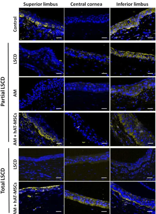
Figure 6. Immunofluorescence microscopy of CK15 expression, a limbal stem cell marker, in the ocular surface of the partial and total
The conjunctival epithelial cell marker CK7 [36] was expressed in the conjunctival epithelium and in some cells of the limbal epithelium in the healthy eyes (Supporting Infor-mation Fig. S4). There were no differences in CK7 expression among the different control and experimental groups of the partial and total LSCD models (Supporting Information Fig. S4; Table S4). Mucins in goblet cells stained by lectin were pre-sent in the conjunctiva while some were located in the limbus of healthy eyes. Additionally, there was a diffuse staining on
the surface of the conjunctival and limbal epithelia, indicating the presence of secreted mucins. Except as noted below, there were no differences in the staining of mucins by lectin among the different control or experimental groups of either the partial and total LSCD models. In the partial LSCD model, the central cornea remained free of lectin-stained goblet cells. In two of the six animals with untreated total LSCD, goblet cells were present in the central cornea. However, only one animal of the total LSCD group transplanted with AM1
hAT-Figure 7. Immunofluorescence microscopy of p63 expression, a limbal stem cell marker, in the ocular surface of the partial and total
limbal stem cell deficiency (LSCD) models at the end of the follow-up period. The expression of p63 (yellow) was lost in the damaged superior limbus of the untreated partial LSCD model and in the amniotic membrane (AM)-transplanted eyes. There was partial recovery in the AM1human adipose tissue-derived mesenchymal stem cell (hAT-MSC) transplanted group. In the total LSCD model, p63 expres-sion was lost in the limbal epithelium, but it was partially recovered in the AM1hAT-MSC transplanted group. Nuclei, blue. Representa-tive images from animals of each group. Scale bar5100 mm. Abbreviations: AM, amniotic membrane; hAT-MSCs, human adipose-derived mesenchymal stem cells; LSCD, limbal stem cell deficiency.
MSCs had goblet cells in the central cornea (Supporting Infor-mation Fig. S4).
D
ISCUSSIONTo study the tolerance and efficacy of the hAT-MSCs trans-planted onto the ocular surface, it was first necessary to develop an in vivo experimental model that closely resembles human LSCD disease. To that end, we used surgical limbec-tomy to develop two different rabbit models that mimicked the progressive LSCD grades of severity in humans. We chose rabbit to develop our models because the rabbit cornea is large enough to perform surgeries, the histological structures of the eye are similar to human eyes [37], and many of the in vivo LSCD models have been developed using rabbits [38–40].
By performing a n-heptanol-based denudation of the cor-neal surface followed by a surgical 1808 limbectomy in the superior and temporal ocular surface quadrants, we devel-oped a partial, mild LSCD model. Clinically and pathologically, the model resembled mild LSCD in humans [4, 5]. In our rab-bits, neovascularization and corneal opacification occurred during the first 3 weeks after the generation of the injury, but the epithelial defects arising from the heptanol denudation of the cornea partially recovered during the first 2 weeks, as also described by Chen and Tseng [41]. In our study, from the third week after induction of partial LSCD, even in the absence of further treatment, the clinical signs became stable and were maintained throughout the follow-up. Based on these findings and those of others [42], we selected the third week for the time of transplantation.
We generated a total LSCD model by performing n -hepta-nol-based denudation of the corneal surface followed by a 3608 limbectomy. This model clinically and pathologically resembled moderate-to-severe LSCD in humans [4, 5]. Neo-vascularization and corneal opacification were more severe than in the partial LSCD, and they developed over the first 3 weeks after the creation of the injury, as other authors have noted [23, 43–45]. Moreover, the epithelial defects underwent partial recovery during the first 2 weeks and were maintained during the follow-up period, as reported by others [30, 46]. In two of the six untreated rabbits, a thick conjunctival invasion occurred on the corneal surface, indicating that the total LSCD model was more severe than the partial LSCD model, as we expected. In addition, these results confirm that the lim-bus acts as a physical barrier between the conjunctiva and the cornea.
AMs without cells were transplanted to the ocular surface of three rabbits with partial LSCD to make sure that an adverse reaction would not occur after surgical suturing. The clinical signs did not change after the AM transplantation in comparison with the partial LSCD group without AM. Due to the lack of changes in the clinical signs after the AM trans-plantation, we decided that it was not necessary to include a group transplanted with cell-free AM in the total LSCD model. According to our results, previous in vivo studies did not find significant differences in the evolution of clinical signs in ani-mals with untreated LSCD or when treated with cell-free AM [40, 47]. Nevertheless, due to its re-epithelizing, anti-inflam-matory, antifibrotic, and anti-angiogenic properties, cell-free
AM transplantation is a procedure frequently performed in patients with mild and/or partial LSCD to improve their clini-cal situation [48, 49]. However, cell-free AM transplantation is insufficient for regeneration of the ocular surface in patients with severe and/or total LSCD [50].
The clinical signs did not improve in the partial LSCD model after hAT-MSC transplantation. The mildness of the model could partially explain this lack of improvement. How-ever, the fact that the clinical signs did not increase after the hAT-MSC transplantation indicates that hAT-MSCs were well tolerated in the rabbit ocular surface even without the use of immunosuppression. These results agree with other studies in which human MSCs were transplanted to the ocular surface of mice [21], rats [22, 51], and rabbits [20, 24] without any indications of rejection.
In eyes with total LSCD, the evolution of neovasculariza-tion and corneal opacificaneovasculariza-tion was restrained after hAT-MSC transplantation. Although a proangiogenic role has been described for MSCs [52], these cells seem to have anti-angiogenic properties in the ocular surface. Decreases in neo-vascularization and corneal opacity have been previously dem-onstrated after the hAT-MSC administration [23–25]. In addition, several authors have described the improvement of these clinical signs after MSC administration by different routes [25, 51, 53–56]. While the epithelial defects were larger in the non-transplanted group than in the transplanted group throughout the follow-up period, the differences were not statistically significant. However, other authors have noted that hAT-MSCs contribute to the recovery of corneal epithe-lium, even in humans [20–26]. Perhaps if the number of ani-mals had been higher, significant differences in epithelial defects would have been more apparent between the differ-ent experimdiffer-ental groups.
The presence of inflammatory cells was higher in the lim-bal stroma of the total LSCD model than in the partial LSCD model. In addition, the stroma was more disorganized in the total LSCD model, indicating again that the total model was more severe than the partial model, as shown by the clinical evaluation. These data suggest that the partial limbectomy produced a mild model of LSCD in rabbits. There were no his-tological differences among the study groups of the partial LSCD model. However, in the total LSCD model, there was a decrease of inflammatory cells and an improvement of the stromal organization in the AM1hAT-MSC transplanted group. These findings are consistent with the anti-inflammatory role of hAT-MSCs delivered by different routes for corneal chemical burn or in LSCD animal models [21–25]. Moreover, BM-MSCs or MSCs isolated from the limbal stroma can reduce the inflammation in the ocular surfaces [25, 55, 57, 58]. Some of the molecules that are implicated in this anti-inflammatory process are interleukin-10, transforming growth factor-b, and Tumor necrosis factor-stimulated gene 6 (TSG-6) [57, 59, 60].
numbers described for human chemical burns [61] and in rab-bits in the weeks after chemical injury [40, 62].
In both the partial and total LSCD models, hAT-MSCs migrated to areas of the limbal stroma that contained many inflammatory cells. Our results are consistent with the migration of topically applied hAT-MSCs to the burned cor-nea and limbus as described by other authors [21, 22]. Other groups have also demonstrated the migration of lim-bal stromal MSCs or BM-MSCs to the damaged cornea and limbus when they were applied by different routes [55, 58, 63]. On the other hand, some studies have reported that AT-MSCs or BM-MSCs administered by subconjunctival injec-tion did not migrate away from the injecinjec-tion site [21, 43]. Furthermore, some reports indicated that MSCs adminis-tered systemically or intraperitoneally in a LSCD model exert their therapeutic action without migration to the damaged area [59]. One of the well-known signaling pathways that regulate cell homing is mediated by C-X-C motif chemokine 12 or stromal cell-derived factor 1 (CXCL12/SDF-1) and its receptor C-X-C chemokine receptor type 4 (CXCR4), which is expressed by MSCs [64]. Under inflammatory conditions, tis-sues secrete CXCL12/SDF-1 that attracts MSCs [65]. In nor-mal conditions, CXCL12/SDF-1 is more abundant in the limbal than in the corneal epithelium, and CXCR4 is expressed in the limbal stroma rather than in the corneal stroma [66, 67]. Although there is a lack of data about the expression of these molecules in inflamed ocular surface tis-sues, these factors could be implicated in the migration of the AT-MSCs to the damaged cornea and limbus of our in vivo models.
In our LSCD models, we analyzed by immunofluores-cence microscopy the molecular changes in the expression profiles of corneal, limbal, and conjunctival cell markers. The expression of the corneal epithelial cell marker CK3 increased in the limbus of the partial LSCD model, indicating the possibility that limbal epithelial stem cells were differ-entiating and migrating from the limbus to the central cor-nea during the restoration of the corcor-neal epithelium. In contrast, the expression of CK3 decreased in the cornea of the untreated total LSCD model in agreement with the data published by other authors using different LSCD models [30, 46, 51, 68].
Although there were no clinical or histological differences between the partial LSCD group without treatment and the AM1hAT-MSC transplanted group, there were some molecu-lar changes. After AM1hAT-MSC transplantation, CK3 was expressed not only in the corneal epithelium, but also in the limbal epithelium. These data suggest that hAT-MSCs contrib-ute to recovery of the corneal epithelium by secreting factors that promote the proliferation and differentiation of the remaining limbal epithelial stem cells. This is consistent with the increased expression of CK3 and/or CK12 in the corneal epithelium after BM-MSC administration in corneal burn and LSCD models [40, 51].
The expression of E-cadherin, an intercellular junction pro-tein, decreased in the total LSCD model, indicating a possible loss of intercellular junctions of the corneal epithelium. Our results are consistent with the decrease in E-cadherin expres-sion similar to that which occurs in defective development of mouse corneal epithelium [69]. E-cadherin expression increased in both the corneal and limbal epithelia of the total
LSCD group transplanted with AM1hAT-MSCs. These data indicate that hAT-MSCs contribute to the recovery of the cor-neal epithelium. Similarly, the expression of connexin 43, a gap junction protein, occurs after BM-MSC transplantation in a different rabbit model of LSCD [40].
The expression of the limbal epithelial stem cell markers CK15 and p63 was lost from the superior limbus of the partial LSCD model and from the superior and inferior limbus of the total LSCD model, as we expected. The loss of limbal stem cell markers occurs in other LSCD models [51, 70]. However, the expression of CK15 and p63 recovered in the limbal epi-thelium of the partial and total LSCD groups after AM1 hAT-MSC transplantation, like that noted for p63 after BM-hAT-MSC transplantation in different LSCD models [51, 71]. However, Reinshagen et al. did not find any differences in corneal p63 expression after the transplantation of BM-MSCs in rabbits with LSCD [40].
Although some authors have proposed the transdifferen-tiation of MSCs into CK3-expressing corneal epithelial cells [21, 40, 51, 72], only two have demonstrated that MSCs transplanted in vivo expressed CK3 in the corneal epithelium [21, 72]. However, we and other authors have shown that MSCs express CK3 in normal culture conditions; thus the CK3 expression might not indicate a real transdifferentiation [18, 40]. Our results, together with those obtained by other authors, indicate that MSCs contribute to the recovery of the corneal epithelium by secreting factors that act at a paracrine level rather than by a transdifferentiation process. These fac-tors would reduce neovascularization and inflammation [43, 63] and provide an enabling environment to promote the pro-liferation and differentiation of the resident stem cells in the tissues [73, 74]. Although the angiogenic and the anti-inflammatory capacity of hAT-MSCs have been reported [22–24], this is the first time that the capability of these cells to recover the corneal and limbal phenotypes has been dem-onstrated in in vivo models.
C
ONCLUSIONOverall, our results indicate that hAT-MSCs transplanted to the ocular surface are well tolerated, migrate to inflamed tissues, reduce inflammation, restrain the evolution of neo-vascularization and corneal opacification, and partially restore limbal and corneal epithelial phenotypes. This sug-gests that hAT-MSC transplantation could be a novel ther-apy to treat patients suffering from ocular surface failure due to LSCD. hAT-MSCs represent an efficient source of stem cells with no availability limitations, and which have immunomodulatory and regenerative properties that may provide therapeutic benefits and reduce health care expenses.
A
CKNOWLEDGMENTSSciences of Xenofile Editing, www.xenofileediting.com) for his assistance in the final editing and preparation of this manuscript. This work was supported by Instituto de Salud Carlos III, CIBER-BBN, Spain (CB06/01/003 MINECO/FEDER, EU); Regional Center for Regenerative Medicine and Cell Therapy, Castilla y Leon, Spain; Ministry of Science and Inno-
vation, Spain (SAF2010–14900); Ministry of Economy and Competitiveness and European Regional Development Fund, Spain (SAF2015–63594-R MINECO/FEDER, EU); and scholar-ships by the Junta de Castilla y Leon and the European Social Fund (to S.G., M.L.-P., and M.P.-C.). This work was presented in part at the 2014 and 2015 annual meetings of the Associ-ation for Research in Vision and Ophthalmology (ARVO): Invest Ophthalmol Vis Sci 2014; 55:ARVO E-abstract 5162. Invest Ophthalmol Vis Sci 2015; 56:ARVO E-abstract 3474. Invest Ophthalmol Vis Sci 2015; 56:ARVO E-abstract 5638. E.R. is currently affiliated with the Hospital Universitario Santa Cristina, Instituto de Investigacion Sanitaria Princesa
(IIS-P), Madrid, Spain; A.d.l.M. is currently affiliated with the Department of Physiology and Membrane Biology, University of California, Davis, CA.
A
UTHORC
ONTRIBUTIONSS.G.: conception and design, collection and/or assembly of data, data analysis and interpretation, manuscript writing, approved the final manuscript; J.M.H.: conception and design, performance of surgical procedures, data interpretation, approved the final manuscript; M.L.-P., A.d.l.M., and M.P.-C.: collection and/or assembly of data, approved the final manu-script; E.R.: collection and/or assembly of data, technical sup-port, approved the final manuscript; M.C. and T.N.-M.: conception and design, data analysis and interpretation, man-uscript writing, financial support, approved the final manu-script. M.C. and T.N.-M. are senior co-authors.
D
ISCLOSURE OFP
OTENTIALC
ONFLICTS OFI
NTEREST The authors indicated no potential conflicts of interest.R
EFERENCES1 Cotsarelis G, Cheng SZ, Dong G et al. Existence of slow-cycling limbal epithelial basal cells that can be preferentially stimu-lated to proliferate: Implications on epithelial stem cells. Cell 1989;57:201–209.
2 Li W, Hayashida Y, Chen YT et al. Niche regulation of corneal epithelial stem cells at the limbus. Cell Res 2007;17:26–36.
3 Schlotzer-Schrehardt U, Kruse FE. Identi-fication and characterization of limbal stem cells. Exp Eye Res 2005;81:247–264.
4 Dua HS, Saini JS, Azuara-Blanco A et al. Limbal stem cell deficiency: Concept, aetiol-ogy, clinical presentation, diagnosis and man-agement. Indian J Ophthalmol 2000;48:83–92. 5 Sejpal K, Bakhtiari P, Deng SX. Presenta-tion, diagnosis and management of limbal stem cell deficiency. Middle East Afr J Oph-thalmol 2013;20:5–10.
6 Shortt AJ, Secker GA, Rajan MS et al. Ex vivo expansion and transplantation of limbal epithelial stem cells. Ophthalmology 2008; 115:1989–1997.
7 Rama P, Matuska S, Paganoni G et al. Limbal stem-cell therapy and long-term cor-neal regeneration. N Engl J Med 2010;363: 147–155.
8 Ramirez BE, Sanchez A, Herreras JM et al. Stem cell therapy for corneal epithelium regen-eration following good manufacturing and clini-cal procedures. Biomed Res Int 2015;2015: 408495.
9 Joe AW, Gregory-Evans K. Mesenchymal stem cells and potential applications in treating ocular disease. Curr Eye Res 2010;35:941–952. 10 Ren G, Chen X, Dong F et al. Concise review: Mesenchymal stem cells and transla-tional medicine: Emerging issues. STEM CELLS TRANSMED2012;1:51–58.
11 Yao L, Bai H. Review: Mesenchymal stem cells and corneal reconstruction. Mol Vis 2013;19:2237–2243.
12 Zhang L, Coulson-Thomas VJ, Ferreira TG et al. Mesenchymal stem cells for treating ocular surface diseases. BMC Ophthalmol 2015;15 Suppl 1:155.
13 Jackson L, Jones DR, Scotting P et al. Adult mesenchymal stem cells: Differentia-tion potential and therapeutic applicaDifferentia-tions. J Postgrad Med 2007;53:121–127.
14 Aggarwal S, Pittenger MF. Human mes-enchymal stem cells modulate allogeneic immune cell responses. Blood 2005;105: 1815–1822.
15 Holan V, Javorkova E. Mesenchymal stem cells, nanofiber scaffolds and ocular sur-face reconstruction. Stem Cell Rev 2013;9: 609–619.
16 Harkin DG, Foyn L, Bray LJ et al. Concise reviews: Can mesenchymal stromal cells dif-ferentiate into corneal cells? A systematic review of published data. STEMCELLS2015;33: 785–791.
17 Strioga M, Viswanathan S, Darinskas A et al. Same or not the same? Comparison of adipose tissue-derived versus bone marrow-derived mesenchymal stem and stromal cells. Stem Cells Dev 2012;21:2724–2752. 18 Nieto-Miguel T, Galindo S, Reinoso R et al. In vitro simulation of corneal epithe-lium microenvironment induces a corneal epithelial-like cell phenotype from human adipose tissue mesenchymal stem cells. Curr Eye Res 2013;38:933–944.
19 Martinez-Conesa EM, Espel E, Reina M et al. Characterization of ocular surface epi-thelial and progenitor cell markers in human adipose stromal cells derived from lipoaspi-rates. Invest Ophthalmol Vis Sci 2011;53: 513–520.
20 Lin HF, Lai YC, Tai CF et al. Effects of cul-tured human adipose-derived stem cells transplantation on rabbit cornea regenera-tion after alkaline chemical burn. Kaohsiung J Med Sci 2013;29:14–18.
21 Lin KJ, Loi MX, Lien GS et al. Topical administration of orbital fat-derived stem cells promotes corneal tissue regeneration. Stem Cell Res Ther 2013;4:72.
22 Zeppieri M, Salvetat ML, Beltrami AP et al. Human adipose-derived stem cells for the treatment of chemically burned rat cor-nea: Preliminary results. Curr Eye Res 2013; 38:451–463.
23 Almaliotis D, Koliakos G, Papakonstantinou E et al. Mesenchymal stem cells improve healing of the cornea after alkali injury. Graefes Arch Clin Exp Ophthalmol 2015; 253:1121–1135. 24 Espandar L, Caldwell D, Watson R et al. Application of adipose-derived stem cells on scleral contact lens carrier in an animal model of severe acute alkaline burn. Eye Contact Lens 2014;40:243–247.
25 Holan V, Trosan P, Cejka C et al. A com-parative study of the therapeutic potential of mesenchymal stem cells and limbal epithelial stem cells for ocular surface reconstruction. Stem Cells Trans Med 2015;4:1052–1063. 26 Agorogiannis GI, Alexaki VI, Castana O et al. Topical application of autologous adipose-derived mesenchymal stem cells (MSCs) for persistent sterile corneal epithelial defect. Graefes Arch Clin Exp Ophthalmol 2011;250:455–457.
27 Zuk PA, Zhu M, Mizuno H et al. Multili-neage cells from human adipose tissue: Implications for cell-based therapies. Tissue Eng 2001;7:211–228.
28 Dominici M, Le BK, Mueller I et al. Mini-mal criteria for defining multipotent mesen-chymal stromal cells. The International Society for Cellular Therapy position state-ment. Cytotherapy 2006;8:315–317. 29 Tseng SC. Amniotic membrane trans-plantation for ocular surface reconstruction. Biosci Rep 2001;21:481–489.
30 Wan P, Wang X, Ma P et al. Cell delivery with fixed amniotic membrane reconstructs corneal epithelium in rabbits with limbal stem cell deficiency. Invest Ophthalmol Vis Sci 2011;52:724–730.
31 Efron N. Grading scales for contact lens complications. Ophthalmic Physiol Opt 1998; 18:182–186.
32 Kasper M, Moll R, Stosiek P et al. Pat-terns of cytokeratin and vimentin expression in the human eye. Histochemistry 1988;89: 369–377.
34 Yoshida S, Shimmura S, Kawakita T et al. Cytokeratin 15 can be used to identify the limbal phenotype in normal and diseased ocular surfaces. Invest Ophthalmol Vis Sci 2006;47:4780–4786.
35 Pellegrini G, Dellambra E, Golisano O et al. P63 identifies keratinocyte stem cells. Proc Natl Acad Sci USA 2001;98:3156–3161. 36 Krenzer KL, Freddo TF. Cytokeratin expression in normal human bulbar conjunc-tiva obtained by impression cytology. Invest Ophthalmol Vis Sci 1997;38:142–152. 37 Hayashi S, Osawa T, Tohyama K. Compar-ative observations on corneas, with special reference to Bowman’s layer and Descemet’s membrane in mammals and amphibians. J Morphol 2002;254:247–258.
38 Higa K, Shimmura S, Kato N et al. Prolif-eration and differentiation of transplantable rabbit epithelial sheets engineered with or without an amniotic membrane carrier. Invest Ophthalmol Vis Sci 2007;48:597–604. 39 Omoto M, Miyashita H, Shimmura S et al. The use of human mesenchymal stem cell-derived feeder cells for the cultivation of transplantable epithelial sheets. Invest Oph-thalmol Vis Sci 2009;50:2109–2115.
40 Reinshagen H, Uw-Haedrich C, Sorg RV et al. Corneal surface reconstruction using adult mesenchymal stem cells in experimen-tal limbal stem cell deficiency in rabbits. Acta Ophthalmol 2009;89:741–748.
41 Chen JJ, Tseng SC. Corneal epithelial wound healing in partial limbal deficiency. Invest Ophthalmol Vis Sci 1990;31:1301– 1314.
42 Hayashida Y, Nishida K, Yamato M et al. Ocular surface reconstruction using autolo-gous rabbit oral mucosal epithelial sheets fabricated ex vivo on a temperature-responsive culture surface. Invest Ophthalmol Vis Sci 2005;46:1632–1639.
43 Yao L, Li ZR, Su WR et al. Role of mes-enchymal stem cells on cornea wound heal-ing induced by acute alkali burn. PLoS One 2012;7:e30842.
44 Xu B, Fan TJ, Zhao J et al. Transplanta-tion of tissue-engineered human corneal epi-thelium in limbal stem cell deficiency rabbit models. Int J Ophthalmol 2012;5:424–429. 45 Figueroa-Ortiz LC, Martin Rodriguez O, Garcia-Ben A et al. Neovascular growth in an experimental alkali corneal burn model. Arch Soc Esp Oftalmol 2014;89:303–307.
46 Zhang W, Yang W, Liu X et al. Rapidly constructed scaffold-free embryonic stem cell sheets for ocular surface reconstruction. Scanning 2014;36:286–292.
47 Ma Y, Xu Y, Xiao Z et al. Reconstruction of chemically burned rat corneal surface by bone marrow-derived human mesenchymal stem cells. STEMCELLS2006;24:315–321.
48 Anderson DF, Ellies P, Pires RT et al. Amniotic membrane transplantation for par-tial limbal stem cell deficiency. Br J Ophthal-mol 2001;85:567–575.
49 Dua HS, Gomes JA, King AJ et al. The amniotic membrane in ophthalmology. Surv Ophthalmol 2004;49:51–77.
50 Tseng SC, Prabhasawat P, Barton K et al. Amniotic membrane transplantation with or without limbal allografts for corneal surface reconstruction in patients with limbal stem cell deficiency. Arch Ophthalmol 1998;116:431–441. 51 Rohaina CM, Then KY, Ng AM et al. Reconstruction of limbal stem cell deficient corneal surface with induced human bone marrow mesenchymal stem cells on amniotic membrane. Transl Res 2014;163:200–210. 52 Rehman J, Traktuev D, Li J et al. Secre-tion of angiogenic and antiapoptotic factors by human adipose stromal cells. Circulation 2004;109:1292–1298.
53 Lee RH, Yu JM, Foskett AM et al. TSG-6 as a biomarker to predict efficacy of human mes-enchymal stem/progenitor cells (hMSCs) in modulating sterile inflammation in vivo. Proc Natl Acad Sci USA 2014;111:16766–16771. 54 Pinarli FA, Okten G, Beden U et al. Kera-tinocyte growth factor-2 and autologous serum potentiate the regenerative effect of mesenchymal stem cells in cornea damage in rats. Int J Ophthalmol 2014;7:211–219. 55 Acar U, Pinarli FA, Acar DE et al. Effect of allogeneic limbal mesenchymal stem cell therapy in corneal healing: Role of adminis-tration route. Ophthalmic Res 2015;53:82–89. 56 Ke Y, Wu Y, Cui X et al. Polysaccharide hydrogel combined with mesenchymal stem cells promotes the healing of corneal alkali burn in rats. PLoS One 2015;10:e0119725. 57 Li F, Zhao SZ. Mesenchymal stem cells: Potential role in corneal wound repair and transplantation. World J Stem Cells 2014;6: 296–304.
58 Ahmed SK, Soliman AA, Omar SM et al. Bone marrow mesenchymal stem cell trans-plantation in a rabbit corneal alkali burn model (a histological and immune histo-chemical study). Int J Stem Cells 2015;8:69–78. 59 Roddy GW, Oh JY, Lee RH et al. Action at a distance: Systemically administered adult stem/progenitor cells (MSCs) reduce inflam-matory damage to the cornea without engraftment and primarily by secretion of TNF-alpha stimulated gene/protein 6. STEM CELLS2011;29:1572–1579.
60 Oh JY, Roddy GW, Choi H et al. Anti-inflammatory protein TSG-6 reduces inflam-matory damage to the cornea following chemical and mechanical injury. Proc Natl Acad Sci USA 2010;107:16875–16880. 61 Fatima A, Iftekhar G, Sangwan VS et al. Ocular surface changes in limbal stem cell deficiency caused by chemical injury: A
histologic study of excised pannus from recipients of cultured corneal epithelium. Eye (Lond) 2008;22:1161–1167.
62 Wei ZG, Wu RL, Lavker RM et al. In vitro growth and differentiation of rabbit bulbar, fornix, and palpebral conjunctival epithelia. Implications on conjunctival epithelial trans-differentiation and stem cells. Invest Ophthal-mol Vis Sci 1993;34:1814–1828.
63 Oh JY, Kim MK, Shin MS et al. The anti-inflammatory and anti-angiogenic role of mesenchymal stem cells in corneal wound healing following chemical injury. STEM CELLS 2008;26:1047–1055.
64 Sohni A Verfaillie CM. Mesenchymal stem cells migration homing and tracking. Stem Cells Int 2013;2013:130763.
65 Dotan I, Werner L, Vigodman S et al. CXCL12 is a constitutive and inflammatory chemokine in the intestinal immune system. Inflamm Bowel Dis 2010;16:583–592. 66 Nieto-Miguel T, Calonge M, de la Mata A et al. A comparison of stem cell-related gene expression in the progenitor-rich limbal epithe-lium and the differentiating central corneal epithelium. Mol Vis 2011;17:2102–2117. 67 Xie HT, Chen SY, Li GG et al. Limbal epi-thelial stem/progenitor cells attract stromal niche cells by SDF-1/CXCR4 signaling to pre-vent differentiation. STEMCELLS2011;29:1874– 1885.
68 Kameishi S, Sugiyama H, Yamato M et al. Remodeling of epithelial cells and base-ment membranes in a corneal deficiency model with long-term follow-up. Lab Invest 2015;95:168–179.
69 Ng GY, Yeh LK, Zhang Y et al. Role of SH2-containing tyrosine phosphatase Shp2 in mouse corneal epithelial stratification. Invest Ophthalmol Vis Sci 2013;54:7933–7942. 70 Lin Z, He H, Zhou T et al. A mouse model of limbal stem cell deficiency induced by topical medication with the preservative benzalkonium chloride. Invest Ophthalmol Vis Sci 2013;54:6314–6325.
71 Lan Y, Kodati S, Lee HS et al. Kinetics and function of mesenchymal stem cells in corneal injury. Invest Ophthalmol Vis Sci 2012;53:3638–3644.
72 Gu S, Xing C, Han J et al. Differentiation of rabbit bone marrow mesenchymal stem cells into corneal epithelial cells in vivo and ex vivo. Mol Vis 2009;15:99–107.
73 Oh JY, Ko JH, Kim MK et al. Effects of mesenchymal stem/stromal cells on cultures of corneal epithelial progenitor cells with ethanol injury. Invest Ophthalmol Vis Sci 2014;55:7628–7635.
74 Hu N, Zhang YY, Gu HW et al. Effects of bone marrow mesenchymal stem cells on cell proliferation and growth factor expres-sion of limbal epithelial cells in vitro. Oph-thalmic Res 2012;48:82–88
See www.StemCells.com for supporting information available online.
1
Table S1. Treatment groups.Group (n) LSCD Treatment
0 (6) None None
1 (6) Partial None
2 (3) Partial AM
3 (6) Partial AM + hAT-MSCs
4 (6) Total None
5 (5) Total AM + hAT-MSCs
n, number of animals; LSCD, limbal stem cell deficiency; AM, amniotic membrane; hAT-MSCs, human
adipose tissue-derived mesenchymal stem cells. "Partial" refers to the LSCD model that covered 180o and
included the superior and temporal limbus. "Total" refers to the LSCD that included the entire limbus.
Table S2.Criteria for ocular surface evaluation.
Score Neovascularization Corneal opacity Epithelial defect Conjunctival invasion
0 None None None None
1 Area ≤ 1/4 Mild Area ≤ 1/4 Area ≤ 1/4
2 1/4 < area ≤ 1/2 Moderate 1/4 < area ≤ 1/2 1/4 < area ≤ 1/2
3 1/2 < area ≤ 3/4 Severe, pupil seen faintly 1/2 < area ≤ 3/4 1/2 < area ≤ 3/4
4 Area > 3/4 Severe, pupil not visible Area > 3/4 Area > 3/4
Table S3. Primary and secondary antibodies used in immunofluorescence experiments.
Antibody Specificity/Type Clone Source Dilution
Anti-BrdU-DyLight650 hAT-MSCs marked with BrdU Polyclonal Novus Biologicals 1:100
Anti-CK3 Corneal epithelial cells AE5 Mp Biomedical 1:50
Anti-CK7 Conjunctival cells OV-TL 12/30 Vector Laboratories 1:50
Anti-CK15 Limbal epithelial stem cells LHK15 Merck 1:50
Anti-E-cadherin Corneal epithelial cells 36 BD Biosciences 1:100
Anti-p63 Limbal epithelial stem cells 4A4 Santa Cruz 1:50
Anti-mouse (PECy5) Secondary antibody Polyclonal Santa Cruz 1:100
2
Table S4. CK3, E-cadherin, CK15, p63, and CK7 expression in the partial and total limbal stem cell deficiency (LSCD) models.Corneal epithelial cell markers Limbal stem cell markers Conjunctival cell marker
CK3 E-cadherin CK15 p63 CK7
SL CC IL SL CC IL SL CC IL SL CC IL CJ SL CC IL
Control + +++ + - +++ - +++ - +++ +++ - +++ +++ + - +
Partial LSCD ++ +++ ++ - +++ - - - +++ - - +++ +++ + - +
AM + + + - +++ - - - +++ - - +++ +++ + - +
AM + hAT-MSCs + +++ + + +++ - +++ - +++ ++ - +++ +++ + - +
Total LSCD - - - - + - - - - - - - +++ + - +
AM + hAT-MSCs ++ + ++ ++ +++ ++ +++ ++ +++ - - ++ +++ + - +
Subjective evaluation: – absence; + mild expression; ++ moderate expression; +++ high expression. AM: amniotic membrane, hAT-MSCs: human adipose tissue-derived mesenchymal stem cells, SL: superior limbus, CC: central cornea, IL: inferior limbus, CJ: conjunctiva.