Chiral capillary electrophoresis mass spectrometry of amino acids in foods / Coral Barbas [et al ]
11
0
0
Texto completo
(2) 1432 Carolina Simó1 Andreas Rizzi2 Coral Barbas3 Alejandro Cifuentes1 1. Department of Food Analysis, Institute of Industrial Fermentations (CSIC), Madrid, Spain 2 Institute of Analytical Chemistry, University of Vienna, Vienna, Austria 3 Dpto. Ciencias Básicas, Facultad de CC, Experimentales y Técnicas, Universidad San Pablo-CEU, Urbanización Montepríncipe, Madrid, Spain. Electrophoresis 2005, 26, 1432–1441. Chiral capillary electrophoresis-mass spectrometry of amino acids in foods In this work, the development of a new chiral capillary electrophoresis-mass spectrometry (CE-MS) method to separate D- and L-amino acids is shown. On-line coupling between CE and MS is established through an electrospray-coaxial sheath flow interface. Enantiomer separation is achieved by using a cheap, nonvolatile, chiral selector as b-cyclodextrin in the background electrolyte (BGE) together with a physically coated capillary that is aimed to prevent contamination of the electrospray. The capillary coating is simple and easy to obtain as it only requires flushing of the capillary with a polymer aqueous solution for 3 min. Optimization of CE parameters (pH of BGE, type and concentration of chiral selector, and capillary inner diameter) and electrospray-MS parameters (nature and flow rate of the sheath liquid, nebulizer pressure) is carried out. Two different derivatization protocols of amino acids using dansyl chloride (DNS) and fluorescein isothiocyanate (FITC) are compared in terms of MS sensitivity and chiral resolution. Under optimum CE-MS conditions it is observed that the MS sensitivity obtained for FITC- and DNS-amino acids is similar (with limit of detection (LOD) in the mM range, corresponding to amounts injected in the fmol range) while chiral resolution is better for FITC-amino acids. The optimized method is demonstrated to provide the simultaneous analysis of 15 selected amino acids (i.e., FITC-D/L-Asp, -Glu, -Ser, -Asn, -Ala, -Pro, -Arg, and FITC-g-aminobutyric acid (GABA) in a single chiral CE-MS run, corresponding to the main amino acids that can be found in orange. Moreover, as a result of the high resolution achieved, it is possible to detect down to 2% of D-Asp in the presence of 98% of L-Asp. The good possibilities of chiral CE-MS in food analysis are corroborated through the detection of the main amino acids in a commercial orange juice (i.e., FITC-L-Asp, -Glu, -Ser, -Asn, -Pro, -Arg, and the nonchiral FITCGABA) as well as the determination of the fraudulent addition of synthetic amino acids (containing D- and L-forms) to a fresh orange juice. Keywords: Amino acids / Chiral capillary electrophoresis-mass spectrometry / Cyclodextrin / Enantiomers / Fruits / Ion trap / Orange juice DOI 10.1002/elps.200406199. 1 Introduction Capillary electrophoresis-mass spectrometry (CE-MS) [1–3] has become a powerful analytical tool that allows combining the separation speed, high resolving power,. Correspondence: Dr. Alejandro Cifuentes, Department of Food Analysis, Institute of Industrial Fermentations (CSIC), Juan de la Cierva 3, E-28006 Madrid, Spain E-mail: [email protected] Fax: 134-91-5644853 Dr. Andreas Rizzi, Institute of Analytical Chemistry, University of Vienna, Währinger Straße 38, A-1090 Vienna, Austria E-mail: [email protected] Fax: 143-1-42779523 Abbreviations: DM-â-CD, heptakis(2,6-di-O-methyl)-b-cyclodextrin; DNS, dansyl chloride; EIE, extracted ion electropherogram; EPyM-DMA, ethylpyrrolidine methacrylate-N,N-dimethylacrylamide; GABA, g-aminobutyric acid; HP-â-CD, hydroxypropyl-b-cyclodextrin; TIE, total ion electropherogram. 2005 WILEY-VCH Verlag GmbH & Co. KGaA, Weinheim. and minimum sample consumption of CE with the selectivity and structural information provided by MS. In the last ten years, there have been many significant developments in CE-MS instrumentation and applications that have made CE-MS a competitive tool able to solve a wide number of analytical problems [4–10]. Among the multiple applications of CE-MS, analysis of chiral molecules are of special importance in different areas of knowledge as, e.g., pharmaceutical, clinical, environmental, or food science. Thus, it is already well-known that enantiomers may have different pharmacological or toxicological properties what have made CE-MS one of the techniques of choice for this type of assays. Moreover, as indicated by Armstrong et al. [11], enantioselective separations can be used in food and beverage studies for: (i) identifying adulterated foods and beverages, (ii) more exact control and monitoring of fermentation processes and products, (iii) evaluation and identification of age, (iv) treatment and storage effects, (v) more exact evaluation of some flavor.
(3) and fragrance components, (vi) fingerprinting complex mixtures, and (vii) analysis of chiral metabolites of many chiral and prochiral constituents of foods and beverages. From the above it can be concluded that nowadays development of efficient and sensitive chiral CE-MS methods is of major interest. However, although chiral CE-MS has extensively been applied to drugs and metabolites analysis [12], chiral CE-MS has scarcely been used to separate D- and L-amino acids. To our knowledge, only recently a sheathless interface coupling has been used in CE-MS for chiral separation of amino acids using (1)-(18-crown-6)-2,3,11,12-tetracarboxylic acid (18-C-6TCA) as running buffer [13, 14]. This is noteworthy taking into account that detection and identification of D- and L-amino acids are important issues in multiple areas of science [15] including clinical analysis [16–18], biological and biochemical studies [19, 20], pharmacochemical studies [21] or, as indicated above, food science [11, 22, 23]. Analysis of amino acids by CE can be done as underivatized compounds or, more frequently, after derivatization. Although it is clear that derivatization of amino acids introduces an additional analytical step, it has repeatedly been demonstrated that derivatization can bring about interesting advantages during the analysis of these molecules. Thus, apart of the evident sensitivity enhancement that can be achieved by using adequate probes together with fluorescence detection [22], derivatization frequently facilitates the chiral separation of amino acids, since inclusion in and interaction with the chiral selector becomes more discriminating. This is frequently not only due to the increase in size but also due to the new interactions involving the label [24]. Moreover, derivatization provides a better sensitivity for their analysis by ESI-MS, since the resulting larger molecules can be ionized with a higher yield and their molecular mass increases till a mass range (. 150 m/z) where the MS background noise is usually lower [25]. These well-known advantages took us in the present work to develop and compare two different derivatization procedures of amino acids (namely, using dansyl chloride (DNS) and FITC) in order to achieve their chiral determination by CE-MS. On the other hand, it has been repeatedly indicated that one of the main problems during any chiral analysis by CE-MS is the contamination of the ionization source induced by the chiral selector used in the BGE (typically nonvolatile cyclodextrin (CD) derivatives) [26]. As a result, different procedures have been developed in order to overcome this limitation, as reviewed by Shamsi [12]. Thus, sophisticated and expensive chiral selectors such as polymers and macrocycles have been demonstrated. 2005 WILEY-VCH Verlag GmbH & Co. KGaA, Weinheim. Chiral CE-MS of amino acids in foods. 1433. to be compatible with ESI-MS detection [13, 27]; also charged CDs and antibiotic macrocycles can be used due to their higher countercurrent electrophoretic mobility which prevents the entrance of these chiral selectors into the ionization source [28, 29]. Although some other complex procedures based on, e.g., column coupling with voltage switching [30], open-tubular or packed chiral CEC have also been reported [31, 32]; the most frequent chiral CE-MS procedure is based on the use of the so-called partial-filling technique (PFT) [33–35]. Although PFT makes the use of nonvolatile CDs compatible with CEMS, the use of PFT also has serious drawbacks that have to be considered [36]: (i) the total separation length is smaller as a result of the relatively short chiral zone [37]; (ii) the selectivity is altered since chiral and achiral CZE are combined; (iii) the efficiency is frequently decreased since an extra band-broadening mechanism occurs at the chiral selector zone buffer boundary; (iv) optimization of PFT-CE-MS analysis is tedious since it incorporates many variables, including the length and concentration of the zone that contains the chiral selector that affects the efficiency, selectivity, resolution, and sensitivity obtained [38, 39]. In general, these drawbacks result in a different selectivity, a lower resolution, and a lower peak capacity than in normal chiral CE. The goal of this work was, therefore, the development of a simple chiral CE-MS procedure using inexpensive CDs able to separate derivatized amino acids. A comparative study is carried out between FITC and DNS derivatization. A representative group of 14 D- and L-amino acids is used in this work (i.e., D/L-Asp, Glu, Ser, Asn, Ala, Pro, Arg) plus the nonchiral g-aminobutyric acid (GABA). Two reasons are behind the selection of this group of 15 amino acids: (i) they are a good representation of the different types of amino acids that can be found in nature (i.e., chiral and positively charged as Arg, neutral as Ala, Pro, Ser, Asn, negatively charged as Asp, Glu, and non-chiral as GABA); (ii) the mentioned group is responsible for more than 90% of the amino acidic content in orange juices [22, 40]. It is noteworthy that the fruit juice industry has become one of the most important agricultural businesses in the world with trades exceeding $10 billion per year, dominated by citrus juice [40]. Due to this economic impact, the adulteration of orange juice is an important issue that demands the development of new analytical procedures (as, e.g., chiral CE-MS) able to detect the everyday more sophisticated adulteration procedures tailored to defeat detection methods. The usefulness of the chiral CE-MS method developed in this work is demonstrated through the detection of the mentioned amino acids in orange juice and the recognition of an adulterated sample.. CE and CEC. Electrophoresis 2005, 26, 1432–1441.
(4) 1434. C. Simó et al.. 2 Materials and methods 2.1 Reagents and samples All chemicals were of analytical reagent grade and used as received. b-CD from Fluka (Buchs, Switzerland), hydroxypropyl-b-cyclodextrin (HP-b-CD), heptakis(2,6di-O-methyl)-b-cyclodextrin (DM-b-CD), and g-CD, all from Sigma (St. Louis, MO, USA) were used as chiral selectors. These selectors together with acetic acid from Acros Organics (Pittsburgh, PA, USA) were used for the CE running buffers at the pHs indicated. A 25% ammonia solution from Merck (Barcelona, Spain) was used to adjust the pH of the buffers. Methanol from Scharlau Chemie (Barcelona, Spain) was used for the sheath liquid preparation. All solutions were stored at 47C and warmed at room temperature before use. Distilled water was deionized by using a Milli-Q system (Millipore, Bedford, MA, USA). FITC from Fluka and DNS from Sigma, both dissolved in acetone HPLC-grade from Scharlau, were used for amino acid derivatization at the different concentrations indicated below. L- and D-amino acids and GABA, all from Sigma were directly prepared dissolving each amino acid in Milli-Q water to obtain a final concentration of 0.5 mM. A fresh juice obtained from Navelina oranges (Citrus sinensis, L. Osbeck) was used in this work. The orange juice was freshly squeezed and used after filtration (0.5 mm filter) to remove pulp. The orange juice was centrifuged for 10 min at 10 000 rpm, and the supernatant was used for derivatization.. 2.2 Derivatization procedures Two different derivatization procedures were used in this work. The FITC derivatization procedure consisted of mixing an aliquot of 100 mL of the supernatant from orange juice (adjusted to pH 10 with 5 M NaOH) or amino acid test sample solution with 100 mL of 0.5 M borate buffer at pH 10. This final solution were mixed with 200 mL of a 3.75 mM FITC solution in acetone, and stored in dark at room temperature overnight. A similar procedure was used for DNS derivatization. It consisted of mixing an aliquot of 100 mL of the supernatant from orange juice (adjusted to pH 9 with 5 M NaOH) or amino acid test sample solution with 100 mL of 0.2 M borate buffer at pH 9. This final solution was mixed with 200 mL of a 10 mM DNS solution in acetone and stored in dark at room temperature for 2 h.. 2.3 Polymer synthesis The synthesis of the copolymer ethylpyrrolidine methacrylate-N,N-dimethylacrylamide (EPyM-DMA) and the capillary coating protocol have been described in detail elsewhere [41]. In short, the monomers EPyM and DMA. 2005 WILEY-VCH Verlag GmbH & Co. KGaA, Weinheim. Electrophoresis 2005, 26, 1432–1441 were copolymerized by free radical polymerization 507C, using 2,2’-azo-bis(isobutyronitrile) (AIBN) ([I] 1.5 6 1022 mol L21) as a radical initiator, THF ([M] 1.0 mol?L21) as solvent, and a feed molar fraction 19% EPyM.. at = = of. 2.4 CE-UV conditions A Hewlett Packard CE instrument (Palo Alto, CA, USA) with UV detection was used to carry out the experiments for obtaining the optimum type of CD able to provide the best chiral separations in terms of resolution. Before first use, uncoated capillaries were conditioned in the following way: a 20 min rinse with 0.1 M NaOH was followed by a water rinse for another 20 min. Between injections, a very simple coating strategy was used consisting of flushing the capillary with a diluted polymer solution (0.1 mg/mL in water) for 3 min and next replacing this solution by flushing the capillary with the separation buffer for 3 min. The detection length to the UV detector was 50.1 cm and the total length 58.5 cm. Injections were made at the cathodic end using N2 pressure of 50 mbar for 8 s. The instrument was controlled by a PC running the 3D-CE ChemStation from Agilent Technologies (Waldbronn, Germany).. 2.5 CE-MS conditions The analyses were carried out in a P/ACE 5010 (Beckman Instruments, Fullerton, CA, USA) CE apparatus coupled with an orthogonal electrospray interface (ESI, model G1607A from Agilent Technologies, Palo Alto, CA, USA) to the MS detector (an Esquire 2000TM ion-trap mass spectrometer from Bruker Daltonik, Bremen, Germany). A bare fused-silica capillary with 50 mm ID and 87 cm length to MS detection was used. The coating procedure consisted of flushing the capillary with 0.1 mg/mL polymer solution for 3 min (with the nebulizing gas stopped) and next by flushing the capillary with the separation buffer for 3 min. Injections were made at the cathodic end using N2 pressure at 0.5 psi for 18 s (1 psi = 68.95 mbar). The CE instrument was controlled by a PC running the System GOLD software from Beckman. Electrical contact at the electrospray needle tip was established via a sheath liquid which consisted of methanol-water (50:50 v/v) containing 25% CE running buffer and delivered at a flow rate of 3.5 mL/min by a 74900-00-05 Cole Palmer syringe pump (Vernon Hills, IL, USA). The mass spectrometer was operated in the positive ion mode. The spectrometer was scanned at 150–700 m/z range at 13 000 u/s during separation and detection. MS operating conditions were optimized by adjusting the needle-counter electrode distance, liquid sheath flow rate, CE running buffer concentration in sheath liquid while a group of standard.
(5) Electrophoresis 2005, 26, 1432–1441 amino acids were analyzed by CE-ESI-MS. The nebulizer and drying gas conditions were 2 psi N2 and 3 L/min N2 at 3507C. The instrument was controlled by a PC running the Esquire NT software from Bruker Daltonics.. 3 Results and discussion 3.1 Preliminary study of the chiral separation of DNS- and FITC-amino acids First, in order to prevent the entrance of the nonvolatile CD into the mass analyzer, a physically coated capillary able to provide EOF values equal to zero at a separation pH at which the derivatized amino acids can bear electrical net-charge (and, therefore, move by electrophoretic mobility towards the MS) was used. In this sense, we have already demonstrated [41, 42] that the EOF of capillaries coated with EPyM-DMA can be conveniently tuned by selecting the adequate separation pH. Thus, the EPyMDMA-coated capillary shows an anodal EOF (i.e., negative values) at pHs lower than 6, a nearly zero EOF at pHs between 6 and 8 (the conditions at which we are interested most), and a low cathodal EOF at pHs higher than 8. This behavior has been explained [41, 42] considering that the global electrical charge onto the capillary wall is due to both the amine groups of the polymer (showing positive electrical charge) and the remaining silanol groups onto the silica wall (showing negative electrical charge). Thus, under acidic pHs the amine groups are the main charged groups on the capillary wall bringing about a global positive charge and, as a consequence, an anodal EOF. Under very basic pHs the number of positive charges on the polymer decreases and the negative silanol groups become predominant on the capillary wall, bringing about a cathodal EOF. It is noteworthy that this EOF is clearly reduced using the EPyM-DMA capillary compared with bare fused-silica capillary being a good indication of the shielding effect of the EPyM-DMA coating even at these basic pHs. It is also interesting to indicate that a reproducible coating is easily obtained just by flushing the capillary with a dilute solution containing the polymer. No peaks related to this coating were found to compromise the clarity of mass spectra and sensitivity of analysis. Once that the EPyM-DMA-coated capillary was prepared, a 100 mM ammonium acetate/acetic acid at pH 8 separation buffer was arbitrarily chosen to test the separation capability of different CDs in a CE-UV instrument applying 215 kV as separation voltage (i.e., the cathode is at the injection point). Under these conditions, the EOF of the coated capillary is zero while amino acids, showing a net negative electrical charge, move towards the detector.. 2005 WILEY-VCH Verlag GmbH & Co. KGaA, Weinheim. Chiral CE-MS of amino acids in foods. 1435. Four different CDs (namely, b-CD, HP-b-CD, DM-b-CD, and g-CD) were tested for the chiral separation of the derivatized DNS-amino acids. Thus, by using 2.5, 5, 7.5, 15, 22.5, or 30 mM HP-b-CD, baseline separation could only be observed for D- and L-Asp at 15 mM HP-b-CD, while D- and L-Ser could be partially resolved by using 7.5 mM HP-b-CD. The rest of D- and L-DNS-amino acids could not be separated by this procedure at any concentration. Under the same separation conditions as above using 2.5–22.5 mM g-CD, baseline separation could only be observed for D- and L-Glu at 7.5 mM, while D- and L-Asp, and D- and L-Pro could be partially resolved at the same concentration (i.e., 7.5 mM). The rest of D- and L-DNS-amino acids studied could not be separated by this procedure at any concentration. Using DM-b-CD concentrations between 2.5 and 22.5 mM added to the BGE, no chiral separation was observed for the D- and L-DNS-amino acids. Four different concentrations of b-CD (namely, 2.5, 5, 7.5, and 15 mM) were also tested under the same conditions as above. It could be observed that by using 5 mM of b-CD, except for D- and L-Ala for which only a partial separation was observed, baseline separation of the rest of amino acids could be achieved. Therefore, these conditions (100 mM ammonium acetate/acetic acid, 5 mM b-CD at pH 8) were selected for subsequent optimization for D- and L-DNS-amino acids separation (see below Section 3.2). In order to establish the aforementioned comparison between the two types of derivatized amino acids (i.e., DNS vs. FITC), b-CD was also selected as the optimum chiral selector for the FITC-amino acids. This was based on a previous work [22], in which we already demonstrated the better possibilities of b-CD to separate FITC-amino acids compared with other chiral selectors. However, in that study a MEKC-LIF protocol was used whose conditions were not compatible with CE-MS since it uses SDS, sodium tetraborate, and a high pH at which the EOF would pump this nonvolatile compounds (plus the b-CDs) used in the BGE to the ESI ruining the MS sensitivity. Therefore, a new separation BGE compatible with CE-MS had to be found. Moreover, as indicated above, the pH of the BGE employed for DNS-amino acids was arbitrarily chosen and, therefore, it had to be also optimized.. 3.2 Chiral CE-MS separation of DNS- and FITC-amino acids The effect of different separation parameters were studied in parallel for DNS- and FITC-amino acids in order to obtain good chiral CE-MS conditions for both types of.
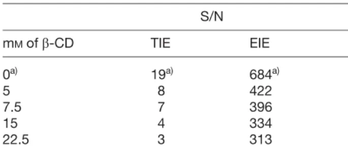
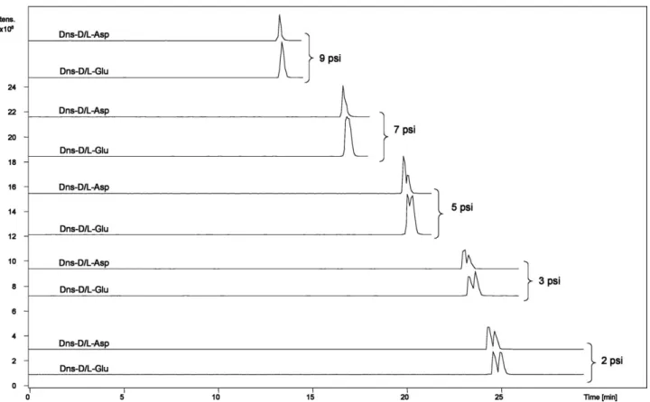
(6) 1436. C. Simó et al.. derivatized amino acids. Firstly, the effect of the pH of BGE on the chiral separation was studied. A BGE composed of 100 mM ammonium acetate/acetic acid was selected together with 5 mM b-CD. Three different pHs within the range at which the coated capillary provides an EOF equal to zero were tested, namely 6.0, 7.0, and 8.0. Although it is clear that the buffer capacity of this BGE is not very high at this range (mostly at pH 7) we decided to use it since this is a highly ESI-MS-compatible buffer with low electrical conductivity. These properties allow the use of relatively high BGE concentrations without deleterious effect on Joule heating generation or ESI-MS detection noise. It could be observed that the best results in terms of analysis speed and chiral resolution were obtained by using the buffer at pH 6.0 for both, DNS- and FITC-amino acids. Next, sheath liquid composition and flow rate were optimized in order to increase the MS sensitivity of the derivatized amino acids. Different sheath liquids compositions were tested. Table 1 shows, as an example, the signal/noise (S/N) ratios obtained for 0.25 mM L-Asp calculated from the total ion electropherogram (TIE) and the extracted ion electropherogram (EIE) depending on the percentage of BGE used in the sheath liquid. As can be seen, a sheath liquid composed of 25% of BGE (free of b-CD) in methanol/water (1:1 v/v) provided the best results for both TIE and EIE-MS signals in terms of S/N. In this experiment, an obstruction of the end of the capillary at the ESI-MS point was always observed when BGE percentages lower than 15% were added to the sheath liquid. At this moment we have not a clear explanation for this effect. The influence of the sheath liquid flow rate on the MS signal was next studied testing flow rates of 1.5, 2.5, 3.5, and 4.5 mL/min. It could be observed that the best results in terms of MS sensitivity were obtained using a flow rate of 3.5 mL/min. The nebulizer pressure was then optimized testing values of 2, 3, 5, 7, and 9 psi. Figure 1 shows, as an example, the results obtained for D/L-DNS-Asp and -Glu under these conditions. As can be seen, the higher the pressure the lower the resolution achieved for the enantiomers as a result of the sucking effect. Therefore, a nebulizer pressure of 2 psi (the minimum at which the ESI is stable) was chosen as optimum. In order to reduce even more the induced hydrodynamic flow and its deleterious effect on the efficiency and separation resolution [43], a capillary of smaller inner diameter was used, namely, a 25 mm ID capillary instead of 50 mm ID. No noticeable improvement could be observed by using the 25 mm capillary either in terms of resolution nor in terms of MS sensitivity. As it is well-known that narrower capillaries are prone to blockage, especially when real world samples are analyzed, the 50 mm capillary was selected.. 2005 WILEY-VCH Verlag GmbH & Co. KGaA, Weinheim. Electrophoresis 2005, 26, 1432–1441 Once suitable conditions were achieved (i.e., EPyMDMA-coated capillary with 87 cm ld and 50 mm ID; BGE composed of 100 mM ammonium acetate/acetic acid at pH 6 with 5 mM b-CD; sheath liquid composed of methanol-water (1:1 v/v) with 25% BGE without b-CD at a flow rate of 2.5 mL/min; nebulizer pressure of 2 psi), the possible effect (if any) of the b-CD concentration on the MS sensitivity under these conditions was studied. To do this, D- and L-DNS-Asp were separated under the described “optimum” conditions using a BGE with increasing quantities of b-CD. As expected, at 0 mM b-CD no chiral separation was observed, obtaining with 15 mM b-CD the maximum resolution (Rs = 1.4) between D- and L-DNSAsp. Table 2 shows the S/N ratios calculated from the TIE and EIE for this experiment. As can be deduced from the results of Table 2, b-CD addition to the BGE has some effect on the TIE and EIE signals. Thus, the S/N ratio of the TIE decreases from about 400 to about 300 (S/N of the EIE decreases from 8 to 3) corresponding to 5 mM and 22.5 mM b-CD, respectively. This can probably be due to the residual suction effect from the ESI interface discussed above. Table 1. DNS-L-Asp S/N values from the TIE and the EIE depending on the percentage of BGE added to the sheath liquid (methanol-water 1:1 v:v) S/N % BGE. TIE. EIE. 45 35 25 15 5 0. 6 7 9 3 –a) –. 449 355 514 322 – –. a) Obstruction of the capillary (see text). Table 2. DNS-L-Asp S/N values from the TIE and the EIE depending on the concentration of b-CD added to the running buffer S/N mM of b-CD. TIE. EIE. 0a) 5 7.5 15 22.5. 19a) 8 7 4 3. 684a) 422 396 334 313. a) At 0 mM b-CD no chiral separation is achieved and, therefore, the S/N ratios correspond to the sum of D- 1 L-amino acid..
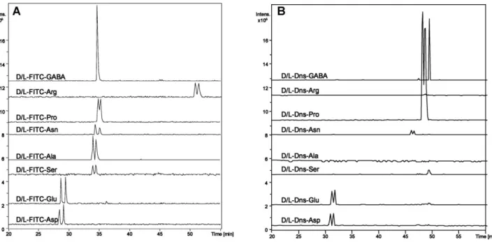
(7) Electrophoresis 2005, 26, 1432–1441. Chiral CE-MS of amino acids in foods. 1437. Figure 1. CE-MS EIE of D/L-DNS-Asp and -Glu at different ESI nebulizing pressures. CE conditions: polymer-coated capillary (87 cm ld, 50 mm ID); BGE, 100 mM ammonium acetate buffer at pH 6 with 5 mM b-CD; running voltage, 215 kV, injection at 0.5 psi during 18 s. MS conditions: positive ion mode; sheath liquid methanol-water (1:1 v/v) with 25% BGE without b-CD, at a flow rate of 3.5 mL/min, dry gas flow at 3 L/min; temperature, 3507C; mass scan, 150–700.. 3.3 Chiral CE-MS of DNS- and FITC-amino acids: a comparison A comparison of different figures of merit for the chiral CEMS analysis of FITC- and DNS-amino acids was carried out under “optimum” conditions. In order to establish an adequate comparison, the same concentration of amino acids was used in both experiments. Also, the mass target of the ion trap was set at 350 m/z for DNS-amino acids and at 530 m/z for FITC-amino acids for the same mass range (150–700 m/z), since optimum MS sensitivity was obtained under these conditions. Figure 2 shows the EIEs obtained from a single run of D/L-FITC- (Fig. 2A) and D/LDNS- (Fig. 2B) amino acids. In Table 3, the results obtained regarding experimental molecular mass (compared with theoretical), sensitivity and chiral resolution for each amino acid are also compared. The limits of detection (LODs, calculated as three times the S/N ratio) were between 1.6 and 29.4 mM for FITC-aa, and between 1.2 and 22.6 mM for DNS-aa, corresponding to minimum detected amounts of 19 fmol for FITC-aa, and 15 fmol for DNS-aa, what demonstrates the good possibilities of our CE-MS. 2005 WILEY-VCH Verlag GmbH & Co. KGaA, Weinheim. approach. These LODs are similar to the values usually obtained for chiral analysis of amino acids using CE-UV (in the mM range, see, e.g., [44]), and worse than the obtained using LIF as detector, with which LODs in the nM range can normally be obtained [22]. Also, the experimental mass values obtained are in all cases in good agreement with the theoretically expected, independently on the derivatization used. However, as can be seen in Fig. 2, although in general sensitive separations were obtained under these conditions for the D- and L-amino acids derivatized with FITC or DNS, Ala could not be detected when previously derivatized with DNS (see Fig. 2 and Table 3) under the derivatization conditions used in this work. DNS-Arg is netuncharged under these pH conditions and exhibits zero electrophoretic mobility; thus, it can not be detected. Besides, the chiral resolution values obtained for FITC amino acids were in all cases better than those obtained for DNS amino acids (see Table 3) and the migration was faster due to the higher anodic net-charge associated with this derivatization. Therefore, based on these results, FITC was preferred to DNS as derivatizing reagent to determine the selected amino acids in real samples..
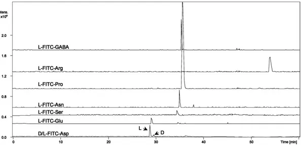
(8) 1438. C. Simó et al.. Electrophoresis 2005, 26, 1432–1441. Figure 2. CE-MS EIE of D/L-Asp, -Glu, -Ser, -Ala, -Asn, -Pro, -Arg, and GABA derivatized wih (A) FITC and (B) DNS, at a concentration of 125 mM of each amino acid. ESI nebulizer pressure, 2 psi. Other CE-MS separation conditions as in Fig. 1.. Table 3. Experimental molecular weight (MWe), theoretical molecular weight (MWt), limit of detection (LOD), and chiral resolution (Rs), obtained for amino acids derivatized with FITC and DNS under the optimum CE-MS separation conditions FITC. DNS LOD. Asp Glu Ser Ala Asn Pro Arg GABA a) b) c) d). MWe. MWt. 522.3 536.3 494.4 478.8 521.3 504.5 563.2 492.7. 522.5 536.5 494.5 478.5 521.5 504.5 563.6 492.5. (mM). a). 8.4 6.3 29.4 27.4 12.2 7.4 15.8 1.6. LOD b). (fmol). 103.1 77.4 360.2 335.7 149.3 91.0 194.3 19.4. Rs. MWe. MWt. 1.4 1.7 0.8 1.2 1.3 0.6 0.7 xd). 366.4 380.4 338.8 –c) 365.4 348.5 – 336.7. 366.4 380.4 338.4 322.4 365.4 348.4 407.5 336.4. (mM). a). 7.3 4.6 22.6 – 14.4 2.6 – 1.2. (fmol)b) 89.3 56.5 227.6 – 177.0 32.2 – 15.2. Rs 0.8 0.7 0.6 – 0.6 0.5 – x. LOD given as concentration of each amino acid LOD given as amount injected of each amino acid Not detected at the derivatization conditions used Nonchiral amino acid. 3.4 Some applications of the chiral CE-MS protocol The necessity to control the enantiomeric purity of foods, beverages, and nutritional supplements, has repeatedly been stressed [23, 44] since the different biological or physiological properties of amino acids could lead to nutritionally poorer and less safe products by generating. 2005 WILEY-VCH Verlag GmbH & Co. KGaA, Weinheim. nonmetabolizable and biologically nonutilizable forms of amino acids [23, 44]. This chiral CE-MS method also allows the adequate determination of the so-called enantiomeric excess where stringent requirements have to be met, i.e., baseline separation at overload conditions together with low detection limits [45, 46]. Our CE-MS method fulfils these requirements for a number of amino acids relevant in the context of food analysis and allows.
(9) Electrophoresis 2005, 26, 1432–1441. Chiral CE-MS of amino acids in foods. 1439. the determination of 2% of the D-FITC-Asp enantiomer in the presence of 98% of the optical antipode, as demonstrated in Fig. 3, what can be used as a good marker for controlling the quality and safety of different beverages and foods.. Figure 3. CE-MS EIE of D- and L-FITC-Asp with an enantiomeric ratio of 2:100. CE-MS separation conditions as in Fig. 1.. Figure 4 shows the EIE electropherograms obtained after FITC derivatization of an orange juice. As can be seen, the chiral CE-MS procedure allows the adequate determination of the main L-FITC-amino acids of this fruit, namely, L-FITC-Asp, -Glu, -Pro, -Asn, -Ser, -Arg, and the nonchiral FITC-GABA. It has often been mentioned that enantiomeric composition of amino acids in fruit juices may be. Figure 4. CE-MS EIE obtained from an orange juice sample derivatized with FITC. CE-MS separation conditions as in Fig. 1.. Figure 5. CE-MS EIE from a orange juice sample adulterated with 0.8% (with respect to the total amino acid content) of D/L-Asp. CE-MS separation conditions as in Fig. 1.. 2005 WILEY-VCH Verlag GmbH & Co. KGaA, Weinheim.
(10) 1440. C. Simó et al.. a very useful tool in the determination of several frauds [11, 22, 23]. Thus, the addition of inexpensive synthetic amino acids to mask water dilution can be detected through chiral analysis of amino acids due to the presence of D- and L-forms [11, 23, 47]. In this way, Fig. 5 shows an example of a fresh orange juice adulterated with a synthetic D/L-amino acid (i.e., D/L-Asp). Namely, the quantity of the synthetic D/L-Asp added was calculated to be 0.8% with respect to the total amino acid content detected by this procedure in the orange juice. As can be seen, our procedure allows the adequate detection of this fraudulent addition. To our knowledge, these applications are the first examples showing so far the good possibilities of chiral CE-MS analysis in food science.. 4 Concluding remarks A new, simple, and fast method for chiral CE-MS analysis of amino acids has been developed. The employment of a physically adsorbed coating onto the capillary wall permits the use of involatile chiral selectors as b-CD with no significant loss of ESI efficiency and MS sensitivity. A comparative study between DNS and FITC as derivatizing reagents of amino acids prior to their analysis by chiral CZE-MS has been carried out at a pH value of 6. It is concluded that under these conditions CZE resolution and MS sensitivity are better using FITC-amino acids. The usefulness of the chiral CZE-MS method is demonstrated through the analysis of the main amino acids found in a fresh orange juice as well as the detection of a fraudulent addition of synthetic amino acids used to mask water dilution of the fruit juice. The authors thank to Dr. Gerard Bruin and Novartis Pharma AG (Basel, Switzerland) for the gift of the P/ACE 5010 instrument used in this work. Financial support from a CICYT-AGL2002-04621-C02-02 project is acknowledged. CS thanks to Consejería de Educación y Cultura (Comunidad de Madrid) for a fellowship. Austrian Science Fundation, Project No. P15008, is acknowledged. Financial support from a bilateral project Spanish MCYT-University of Vienna (HU2002-0042) is acknowledged. Received May 4, 2004. 5 References [1] Olivares, J. A., Nguyen, N. T., Yonker, C. R., Smith, R. D., Anal. Chem. 1987, 59, 1230–1232. [2] Smith, R. D., Olivares, J. A., Nguyen, N. T., Udseth, H. R., Anal. Chem. 1988, 60, 436–441. [3] Smith, R. D., Barinaga, C. J., Udseth, H. R., Anal. Chem. 1988, 60, 1948–1952.. 2005 WILEY-VCH Verlag GmbH & Co. KGaA, Weinheim. Electrophoresis 2005, 26, 1432–1441 [4] Severs, J. C., Smith, R. D., in: Cole, R. B. (Ed.), Electrospray Ionization Mass Spectrometry, John Wiley & Sons, New York 1997. [5] Banks, J. F., Electrophoresis 1997, 18, 2255–2266. [6] Tomer, K. B., Deterding, L. J., Parker, C. E., in Khaledi, M. G. (Ed.), High-performance Capillary Electrophoresis. Theory, Techniques and Applications, John Wiley & Sons, New York 1998. [7] Brocke, A., Nicholson, G., Bayer, E., Electrophoresis 2001, 22, 1251–1266. [8] Tomer, K. B., Chem. Rev. 2001, 101, 297–328. [9] Moini, M., Anal. Bioanal. Chem. 2002, 373, 466–480. [10] Schmitt-Kopplin, P., Frommberger, M., Electrophoresis 2003, 24, 3837–3867. [11] Armstrong, D. W., Chang, C. D., Li, W. L., J. Agric. Food Chem. 1990, 38, 1674–1677. [12] Shamsi, S. A., Electrophoresis 2002, 23, 4036–4051. [13] Schultz, C. L., Moini, M., Anal. Chem. 2003, 75, 1508–1513. [14] Moini, M., Schultz, C. L., Mahmood, H., Anal. Chem. 2003, 75, 6282–6287. [15] Poinsot, V., Bayle, C., Couderc, F., Electrophoresis 2003, 24, 4047–4062. [16] D’aniello, A., Lee, J. M., Petrucelli, L., Di Fiere, M., Neurosci. Lett. 1998, 250, 131–134. [17] Schell, M. J., Cooper, O. B., Snyder, S. H., Proc. Natl. Acad. Sci. USA 1997, 94, 2013–2018. [18] Schell, M. J., Brady, R. O. Jr., Molliver, M. E., Zinder, S. H., J. Neurosci. 1997, 17, 1604–1615. [19] Kuhn, R., Stoecklin, F., Erni, F., Chromatographia 1992, 33, 32–36. [20] Verleysen, K., Sandra, P., Electrophoresis 1998, 19, 2798– 2833. [21] Prata, C., Bonnafous, P., Fraysse, N., Treilhou, M., Poinsot, V., Couderc, F., Electrophoresis 2001, 22, 4129–4138. [22] Simó, C., Barbas, C., Cifuentes, A., J. Agric. Food Chem. 2002, 50, 5288–5293. [23] Simó, C., Barbas, C., Cifuentes, A., Electrophoresis 2003, 24, 2431–2441. [24] Rizzi, A. M., Cladrowa-Runge, S., Jonsson, H., Osla, S., J. Chromatogr. A 1995, 710, 287–295. [25] Zöllner, P., Letnier, A., Berner, D., Kleinova, M., Jodlbauer, J., Mayer, B. X., Lindner, W., LC-GC Europe 2003, 16, 163–171. [26] Lu, W., Cole, R. B., J. Chromatogr. B 1998, 714, 69–75. [27] Samshi, S. A., Anal. Chem. 2001, 73, 5103–5108. [28] Fanali, S., Desiderio, C., Schulte, G., Heitmeier, S., Strickmann, D., Chankvetadze, B., Blaschke, G., J. Chromatogr. A 1998, 800, 69–76. [29] Schulte, S., Heitmeier, S., Chankvetadze, B., Blaschke, G., J. Chromatogr. A 1998, 800, 77–82. [30] Lamoore, M. H., Sprang, A. F. H., Tjaden, U. R., Van Der Greef, J., J. Chromatogr. A 1996, 742, 235–242. [31] Von Brocke, A., Wistuba, D., Gfrörer, P., Stahl, M., Schurig, V., Bayer, E., Electrophoresis 2002, 23, 2963–2972. [32] Schurig, V., Mayer, S., J. Biochem. Biophys. Methods 2001, 48, 117–141. [33] Jäverfalk, E. M., Aumini, A., Westerlund, D., Andrén, P. E., J. Mass Spectrom. 1998, 33, 183–186. [34] Tanaka, Y., Kishimoto, Y., Terabe, S., J. Chromatogr. A 1998, 802, 83–88. [35] Rudaz, S., Cherkaoui, S., Gauvrit, J.-Y., Veuthey, J.-L., Electrophoresis 2001, 22, 3316–3326. [36] Petersson, P., Jörntén-Karlsson, M., Stålebro, M., Electrophoresis 2003, 24, 999–1007..
(11) Electrophoresis 2005, 26, 1432–1441 [37] Muijselaar, P. G., Otsuba, K., Terabe, S., J. Chromatogr. A 1998, 802, 3–15. [38] Tanaka, Y., Otsuka, K., Terabe, S., J. Chromatogr. A 2000, 875, 323–330. [39] Grard, S., Morin, P., Dreux, M., Ribet, J. P., J. Chromatogr. A 2001, 926, 3–10. [40] Robards, K., Antolovich, M., Analyst 1995, 120, 1–28. [41] Simó, C., Elvira, C., González, N., San Román, J., Barbas, C., Cifuentes, A., Electrophoresis 2004, 25, 2056–2064. [42] González, N., Elvira, C., San Román, J., Cifuentes, A., J. Chromatogr. A 2003, 1012, 95–101.. 2005 WILEY-VCH Verlag GmbH & Co. KGaA, Weinheim. Chiral CE-MS of amino acids in foods. 1441. [43] Cifuentes, A., Rodríguez, M. A., García-Montelongo, F. J., J. Chromatogr. A 1996, 737, 243–253. [44] Boniglia, C., Carratù, B., Manzini, E., J. Food Sci. 2002, 67, 1352–1355. [45] D’Hulst, A., Verbeke, N., Electrophoresis 1994, 15, 854–861. [46] Simó, C., Gallardo, A., San Román, J., Barbas, C., Cifuentes, A., J. Chromatogr. B 2002, 762, 35–43. [47] Ooghe, W., Kasteleyn, H., Temmerman, I., Sandra, P., J. High Resolut. Chromatogr. 1984, 7, 284–285..
(12)
Figure
Documento similar
The characterization in amino acids, organic acids, sugars, trigonelline, volatiles compounds, fatty acids, total phenolic, carotenoids, vitamin C content, and antioxidant capacity
The cornerstone of the suspicion of a chiral phase transition is the singular behavior of the quark condensate < q ¯ q >, since this expectation value is an order parameter
This manuscript undertakes a review of the effects resulting from the intake of proteins and/or amino acids administered in carbohydrate drinks as well as whey protein and
Tumor cells (and other cells in the tumor) deplete nutrient levels (glucose, glutamine, amino acids, O 2 , etc.) in the TME, increase the levels of some metabolites, such as lactic
The stability and fragmentation dynamics of several positively charged molecules in the gas phase have been studied: thymidine, glycine, β-alanine, γ -aminobutyric acid,
Even though the 1920s offered new employment opportunities in industries previously closed to women, often the women who took these jobs found themselves exploited.. No matter
In the “big picture” perspective of the recent years that we have described in Brazil, Spain, Portugal and Puerto Rico there are some similarities and important differences,
ovalisporum UAM-MAO The influence of Arg and Gly in CYN production, considering that these amino acids could favor the synthesis of the toxin in two manners: first, by providing
