Effect of Chilean propolis on cariogenic bacteria Lactobacillus fermentum
10
0
0
Texto completo
(2) 118. ciencia e investigación agraria. 100% of the individuals from 35 to 44 year-old and from 65 to 74 year-old age groups had dental caries (MINSAL, 1997; Soto et al., 2007). Additionally, the World Health Organization (WHO) ranks our country among the highest in DMFT levels for individuals between 35 to 44 year-old (DMFT> 13.9) and moderate for 12 year-old-children (DMFT 2.7 to 4.4) (Pepersen, 2003), through DMFT, which describes the amount of dental caries of an individual, and is used to express prevalence. This is obtained by calculating the sum of decayed, missing and filled teeth. Among the microorganisms associated with the development of dental caries are mainly Streptococcus from the group mutans (species S. mutans, S. sobrinus), Lactobacillus spp. and Actinomyces, among others (Tanzer et al., 2001). The genus Lactobacillus is considered a powerful acidogenic that leads to demineralization of the dental surface (Byun et al., 2004). However, they only represent a small portion of dental plate microbiota, with a higher presence in more advanced teeth wounds (cavitation). Therefore, they are conferred a role in wound progression more than in the beginning of it (Van Houte, 1994). They colonize preferably in the back of the tongue and are carried in the saliva when the epithelium molts. Their cariogenicity depends on the consumption of a diet rich on carbohydrates by the host (Nishikawara et al., 2006). For some years, the pharmaceutical industry has centered its efforts on the discovery and achievement of new antimicrobial products, in order to solve the continuous problem of bacterial resistance to well-known antibiotics (Normark and Normark, 2002), and the collateral effects observed frequently after their use (Cuhna, 2001), where natural products used for these purposes since ancient times are the main target (Silver, 1990). Among these natural products, propolis has been considered a good candidate as adjuvant in the treatment and prevention for various infectious diseases. Propolis is relatively non-toxic (Cuesta et al., 2005) with a wide range of antimicrobial activity against a varied order of bac-. teria, fungi, parasites and viruses (Salomáo et al., 2005; Orsi et al., 2005; Freitas et al., 2006). More than 160 propolis components have been identified, commonly consisting on waxes, resins, water, inorganic compounds, phenolic compounds and essential oils (Mohammadzadeh and Shariatpanahi, 2007), where most of the biological activity is attributed to flavonoids (Santos and Bastos, 2002). These compounds are present in cells carrying out photosynthesis and that may be found in fruits, legumes, nuts, stems and flowers, as well as tea, wine, and obviously in apicultural products like honey and propolis (Cushnie and Lamb, 2005). In regard to the above, the present study was aimed to chemically characterize six Chilean commercial propolis products and evaluate their antimicrobial action on the cariogenic bacteria Lactobacillus fermentum, isolated from patients with dental caries. Materials and methods Patients A total of 40 individuals participated in this study with an age range varying between 6 and 78 years old. All presented diagnosis of dental pieces extraction due to deep dentine caries (D3). After signing an informed consent form, the piece destined to extraction was anaesthetized. Then, with a sterile excavator, the existing caries damage was scraped off. After the sample was obtained, it was impregnated in a sterile cotton swab and introduced in a Stuart medium. Microbiological culture In the laboratory, the sample was immediately sown on a plate with a Difco culture medium, Lactobacilli MRS agar (Winkler Ltda., Santiago, Chile), selective for Lactobacillus, streaking it after the medium swab was rubbed in the superior face of the plate. Then, it was incubated in an oven (Thermo HEPA CLASS100) at 37º C in 5% CO2 atmosphere for 24 hours..
(3) VOLUME 38 Nº1 JANUARY - APRIL 2011. Identification of Lactobacillus fermentum by PCR The colonies to be identified were diluted adding a colony of medium size in 500 µL of sterile distilled water; dilution from which the amplification technique was made directly with the technique of Polymerase Chain Reaction (PCR). A specific fragment of the subunit 16S of RNAr (334 bp) was amplified using and conditions described by Dickson et al. (2005). The amplification by PCR was made in a total volume of 50 mL, containing 2 µL of colonies dilution, and 48 µL of the reaction mixture including 1x Buffer [75mM Tris-HCl, 2.2 mM (NH4)2SO4, 0.01% Tween 20], 0.2 mM of dNTPs, 200 nM from each primer, 2.0 mM of MgCl2 and 1 unit of Taq DNA polymerase (Fermentas, Lithuania). The reaction mixture was prepared in a laminar flow chamber (ESCO, Singapur), that was previously decontaminated during 20 minutes with UV radiation. The amplification reaction was made in a Thermal Cycler MyCycler (BIORAD, EE.UU). It consisted of an initial denaturation at 98 oC for 3 minutes, followed by 35 cycles with denaturation at 94 oC for 1 minute, hybridization at 50 oC for 1 minute and extension at 72 oC for 1 minute, and ending with a 10-minute-final extension at 72 oC. For the visualization of amplification products obtained by PCR, an electrophoresis in agarose gel at 2% in buffer TBE 0.5x at 100V was made, during 35 minutes, using 100 bp-commercial Ladder as standard of molecular size. Subsequently, the gel was dyed with ethidium bromide (0.5 µg/ml), and visualized in a digital photodocumentation system EBox 1000 (Vilber Lourmat, France). Commercial propolis extracts. Six commercial Ethanolic Extracts of Propolis (EEP) were used, which were diluted in distilled water 1:4 and, subsequently, filtered in Wathman No 2 paper to discard the waxes. Afterwords, a new filtered process was applied with a 0.2 mm cellulose acetate filter in order to sterilize the solution.. 119. Determination of minimum inhibitory and bactericidal concentrations For the determination of the minimum inhibitory concentration-MIC (lowest antimicrobial concentration inhibiting the visible growth of a microorganism after the incubation period allowing its growth) and the minimum bactericidal concentration – MBC (lower antimicrobial concentration that hinders a microorganism growth, after a subcultivation in an antimicrobial substance-free-medium) (Andrews, 2001), the following took place: A 1 x 10⁵ UFC x mL inoculation was used for the MIC study, obtained by dilutions made from a tube with an inoculation equivalent to 0.5 McFarland (1.5 x 10⁸ UFC x mL), according to Andrews (2001). Trypticase Soy Agar contained in sterile microplates was used as a culture medium. A negative control (sterility control, culture medium and EEP), a positive control (culture medium and inoculation) and six dilutions for each EEP (1/8, 1/16, 1/32, 1/64, 1/128, 1/256) were considered. Subsequently, the colony developments were observed against the light. For the MBC determination, the wells that resulted negative to growth and, therefore, showing EEP antimicrobial activity were subject to subcultivations in EEP-free media, sowing again on agar plates, incubated at 37 oC in 5% CO2 atmosphere for 24 hours. All determinations were made in triplicate. Determination of total polyphenols For the determination of the total polyphenols present in the evaluated extracts, the method Folin-Ciocalteu was used (Singleton et al., 1999). Therefore, each extract 1:10 was diluted in ethanol 70% and then 1:10 in distilled water; subsequently 40 µL of this dilution was mixed with 560 µL of distilled water, 100 µL of the reactive Folin-Ciocalteu (Merck, Germany) and 300 µL of sodium carbonate at 7.5% (p/v). The absorbance was measured at 760 nm after 2 hours of incubation at room temperature. The concentra-.
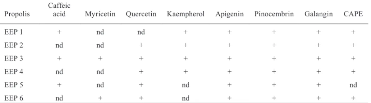
(4) 120. ciencia e investigación agraria. tions were calculated from a calibration curve and expressed in mg/mL equivalent to the mixture of the pinocembrin/galangin standards in a 2:1 proportion (Popova et al., 2007). Chromatographic analysis The analysis was performed in a High Pressure Liquid Chromatography (HPLC) Merck-Hitachi, equipped with an L-6200 model pump, a UV-visible detector, model L-4200 and a column heater Phenomenex Terma Sphere, model TS-130. The separation was made in a RP-18 column (12.5 x 0.4 cm, particle size 5 µm) (Merck, Germany), which was eluted at 25 oC using the mixture of 5% formic acid in water (A) and Methanol (B) as mobile phase. The compound separation was made through an isocratic run from 0 to 10 minutes, with the mixture A 70% and B 30%, followed by a gradient until 100% B at 70 minutes. The compounds were detected at a 290 nm wave length, with a sensitivity of 0.001; the injection volume was 10 µL. The identification of phenolic compounds was made by the use of the standards myricetin, kaempherol, quercetin, caffeic acid, galangin, pinocembrin, apigenin, caffeic acid phenethyl ester (CAPE) and resveratrol (Sigma, USA). Results Microbiological culture The 40 samples obtained from patients with diagnosis of deep dentine caries and indication of tooth extraction, were sown in appropriate culture media, obtaining bacterial development in all of them, at the end of the incubation period.. Lactobacillus fermentum isolation Nine Lactobacillus fermentum strains from the samples cultures of deep dentine caries were isolated from patients with indication of dental extraction, which were subject to a sensitivity test in triplicate. Determination of the minimum inhibitory concentration Only EEPs 2, 3, 4 and 5 showed antimicrobial activity, as they inhibited the visible growth of Lactobacillus fermentum (Figure 1). In this case, the minimum inhibiting concentrations from each propolis showing activity were: EEP2, 2.5% and EEP3, 2.0%. In the case of EEP4 and EEP5, we may only mention that the growth inhibition was observed in the initial dilution 1:4 for EEP4 and 1:16 for EEP5, as the initial concentration initial of these propolis extracts was not available. Similar results were observed for the 9 isolations analyzed. The wells content showing negativiness was transferred to an EEP-free medium. Those subcultures did not show development after they were observed when the incubation time had finished. This means that both the inhibiting and bactericidal concentrations matched. Determination of total polyphenols The propolis extracts evaluated showed large differences of total polyphenol concentrations, where the values found varied between 9 and 85 mg/mL. Additionally, there was no relation between the concentrations specified and the propolis percentages declared by the manufacturers. All the determinations were made in triplicate and the results obtained are shown in Table 1. Chromatographic analysis. Colonies identification by PCR Amplification by PCR was made to all the types of colonies developed, resulting 9 positive colonies (22.5%) for Lactobacillus fermentum at the end of the analysis.. The chromatographic analysis of the propolis studied, in the conditions mentioned before, indicated that they present caffeic acid, myricetin, quercetin, kaempherol, apigenin, pinocembrin, galangin and CAPE (Table 2). The dissenting re-.
(5) 121. VOLUME 38 Nº1 JANUARY - APRIL 2011. Figure 1. Determination of minimum inhibitory concentration (MIC) in microplates. EEP1 to EEP6, ethanolic extracts of propolis; D1-D6, dilutions of EEP; NC, negative control; PC, positive control; +/- indicates presence/absence of bacterial growth.. sults on antibacterial activity might be explained by the different content and/or presence of these ployphenols.. Discussion Dental caries is defined as a multifactorial pathology that begins after dental eruption, softening the hard tooth tissue and evolving to cavity formation (WHO, 1987). Additionally, it is categorized as a transmissible disease, induced by diet, where the main etiological factors responsible are cariogenic bacteria, fermentable carbohydrates and the host susceptibility (Harris et al., 2004). Among the microorganisms associated to dental caries are mainly Streptococcus from the mutans group (S. mutans, S. sobrinus), Lactobacillus spp. and Actinomyces, among others (Tanzer et. al., 2001; Cchour et al., 2005). Our study proposes the use of propolis as an alternative to the treatment of this pathology, due to a proven antimicrobial effect as previously mentioned by Hayacibara and Koo (2005), Sonmez et al. (2005), Olmez and Erdem (2004), Castaldo and Capasso (2002), Pietta et al. (2002) and Santos and Bastos (2002). In this study, Lactobacillus fermentum was detected by PCR in 9 out of 40 patients, that is, in 22.5% of the samples analyzed, which is coherent with the results by other authors, where 65 carious dentin samples taken from extracted dental pieces were analyzed, showing a prevalence of Lactobacillus fermentum (22%) (Byun and Madkarni, 2004). These results show the already proven capacity of molecular biology techniques for microorganism’s identification, which are less troublesome than identification by morphology and biochemical tests.. Table 1. Content of total polyphenols in extracts of commercial Chilean propolis determined by Folin-Ciocalteu method. Propolis extract. Propolis concentration1, %. Total polyphenols2, mg mL-1. EEP 1. 20. 85 ± 2.1. EEP 2. 10. 9 ± 0.3. EEP 3. 20. 41 ± 0.4. EEP 4. ND. 16 ± 0.2. EEP 5. ND. 19 ± 0.9. EEP 6. 20. 16 ± 0.4. Propolis concentration declared by manufacturers; 2Values expressed as mean ± standard deviation.ND: Not declared.. 1.
(6) 122. ciencia e investigación agraria. Table 2. Composition of flavonoids of six commercial Chilean propolis determined by HPLC. Caffeic acid. Myricetin. Quercetin. Kaempherol. Apigenin. Pinocembrin. Galangin. CAPE. EEP 1. +. nd. nd. +. +. +. +. +. EEP 2. nd. nd. +. +. +. +. +. +. EEP 3. +. +. +. +. +. +. +. +. EEP 4. nd. nd. +. +. +. +. +. +. EEP 5. +. nd. +. nd. +. +. +. nd. EEP 6. nd. +. +. nd. +. +. +. +. Propolis. CAPE: Caffeic acid phenethyl ester; + indicates presence; nd: no detected.. With the antimicrobial effect of the propolis selected for this study, differences in the action shown by each were observed. EEP 2, 3, 4 and 5 showed antimicrobial activity, obtaining minimum inhibitory concentration in the dilutions 1:8, 1:16, 1:8 and 1:32, respectively. These variations in the minimum inhibitory concentrations may be due to differences in the chemical composition of propolis, which depends on different factors like collection place, botanical origin and collection season (Sonmez et al., 2005). The inhibitory effect to the concentrations used in the study was not detected only in the cases of the EEP 1 and 6, which may be due to differences in the concentrations of the detected polyphenols. In a future study, it would be also interesting to characterize the selected propolis in sensitivity tests from a botanical point of view, and so, propolis with good antimicrobial quality may be associated with their original species. The determination of the minimum inhibitory and bactericidal concentrations was complicated by the lack of information on the propolis concentration contained in commercial products; therefore, this is necessary in order to know clearly which concentrations show antimicrobial efficiency, which may then be applied in future studies. The presence and identification of caffeic acid, myricetin, quercetin, kaempherol, apigenin, pinocembrin, galangin and caffeic acid phenethyl ester (CAPE) was determined through the chromatographic analysis, which is coherent to the results by other authors (Chaillou and Naza-. reno, 2009; Kalogeropoulos et al., 2009; Popova et al., 2005; Uzel et al., 2005), who analyzed propolis samples from other countries. Regardless the differences in the number of compounds detected among the samples analyzed, we may indicate that the chromatographic patterns presented wide similarities, and their differences would correspond mainly to differences in the concentration of each compound. Although the antimicrobial action of propolis is well known, the mechanisms of how this effect works are still unknown. Some components present in the propolis extracts have been described, like flavonoids (quercetin, galangin, pinocembrin) and caffeic, benzoic, and cinnamic acids. These probably act somewhere on the membrane or the bacterial wall, causing functional and structural damage (Scazzocchio et al., 2006; Kosalec et al., 2005). Other authors suggest that the ring B of the flavonoids structure may play a role in hydrogen integration or union of the bases, which might explain an action on DNA and RNA synthesis. It has also been proposed that the DNA gyrase and ATPase are inhibited from the components found in propolis. Likewise, bacterial membrane fluidity decrease, permeability increase and membrane potential dissipation have been also proven (Cushnie and Lamb, 2005). A recent study showed that EEP completely suppressed the virulence factor of the enzyme coagulase in Staphylococcus aureus and had a preventive effect on the formation of dose dependent biofilm (Scazzocchio et al., 2006). Therefore, we may indicate that, once the antimicrobial action has been proven, it is important.
(7) VOLUME 38 Nº1 JANUARY - APRIL 2011. to find out about the metabolic processes of the microorganism in order to detect which are altered by the EEP action, and then clarify the effect from this substance on the different microorganisms. Additionally, due to the wide range of biological activity exhibited by the propolis, the high variability and complexity of their chemical composition, and the variability existing among the concentration of total polyphenols present in commercial extracts, the need of regulation becomes more evident with both the determination of the botanical-geographical origin and the chemical characterization of the extracts. Therefore, it would be possible to standardize the particularities of this product and to know their. 123. composition clearly and thus, to develop biotechnological products for caries control and other infectious diseases and clinical syndromes. In summary, we may indicate that Chilean propolis has the capacity of inhibiting the development of cariogenic bacteria L. fermentum. However, this activity is variable and it depends on the chemical composition of the propolis used. Acknowledgments This work was financed by the FONDEF project of CONICYT Nº D05I-10021.. Resumen N. Saavedra, L. Barrientos, C.L. Herrera, M. Alvear, G. Montenegro y L.A. Salazar. 2011. Efecto de propóleos chilenos sobre la bacteria cariogénica Lactobacillus fermentum. Cien. Inv. Agr. 38(1): 117-125. La caries dental es una de las enfermedades infecciosas más prevalentes en el mundo. Entre las bacterias involucradas en esta patología se encuentran Streptococcus mutans, Streptococcus sobrinus, Actinomyces spp. y Lactobacillus spp. La industria farmacéutica ha volcado sus esfuerzos al descubrimiento de nuevos productos antibacterianos ante el aumento de resistencia a los ya conocidos. El propóleos se ha utilizado como tal, desde tiempos antiguos, por lo que se ha investigado su efecto contra variados microorganismos. En este estudio se evaluó el efecto antimicrobiano de seis extractos etanólicos comerciales de propóleos, sobre la bacteria Lactobacillus fermentum. Ésta fue aislada luego de su identificación mediante PCR con el uso de primers especie específicos, posterior al cultivo microbiológico de muestras de caries de pacientes con indicación de extracción de pieza dental, y se detectó en 9 de 40 pacientes, correspondiendo a un 22%. El estudio de susceptibilidad se realizó mediante dilución en microplacas y se comprobó la actividad antimicrobiana en cuatro de los seis extractos etanólicos de propóleos utilizados, difiriendo en la concentración efectiva contra el microorganismo, lo que puede ser atribuido a factores como el origen botánico, el lugar geográfico y la estación de recolección. Los propóleos mostraron concentraciones de polifenoles que variaron entre 9 ± 0,3 y 85 ± 2,1 mg/mL. El análisis cromatográfico permitió detectar la presencia de ácido cafeico, miricetina, quercetina, kaempferol, apigenina, pinocembrina, galangina y ácido cafeico fenil éster (CAPE). Nuestro estudio demuestra la acción antimicrobiana del propóleos sobre L. fermentum, patógeno relacionado al desarrollo de caries. Palabras clave: Lactobacillus fermentum, caries dental, propóleos, actividad antibacteriana.. References Andrews, J. 2001. Determination of minimum inhibitory concentrations. Journal of Antimicrobial Chemotherapy 48: 5-16. Becker, MR, B. Paster, E. Leys, M. Moeschberger, S. Kenyon, J.L. Galvin, S. Boches, F. Dewhirst, and. A. Griffen. 2002. Molecular analysis of bacterial species associated with childhood caries. Journal of Clinical Microbiology 40(2): 1001-1009. Byun, R., M. Madkarni, K.L. Chhour, E. Martin, N. Jaques, and N. Hunter. 2004. Quantitative analysis of diverse Lactobacillus species present in advanced dental caries. Journal of Clinical Microbiology 42(7): 3128-3136..
(8) 124. ciencia e investigación agraria. Castaldo, S., and F. Capasso. 2002. Propolis, an old remedy used in modern medicine. Fitoterapia 73(1): S1-S6. Cchour, K.L., M. Nadkarni, R. Byun, E. Martin, N. Jacques, and N. Hunter. 2005. Molecular analysis of microbial diversity in advanced caries. Journal of Clinical Microbiology 43(2): 843-849. Chaillou, L., and M. Nazareno. 2009. Chemical variability in propolis from Santiago del Estero, Argentina, related to the arboreal environment as the sources of resins. Journal of the Science of Food and Agricultura 88(6): 978-983. Cuesta, A., A. Rodríguez, M. Esteban, and J. Meseguer. 2005. In vivo effects of propolis, a honeybee product, on gilthead seabream innate immune responses. Fish and Shellfish Immunology 18: 71-80. Cuhna, B. 2001. Antibiotic side effects. Medical Clinics of North America 85: 149-185. Cushnie, T., and A.J. Lamb. 2005. Antimicrobial activity of flavonoids. International Journal of Antimicrobial Agents 26: 343-356. Dickson, E.M., M.P. Riggio, and L. Mcpherson. 2005. A novel species-specific PCR assay for identifying Lactobacillus fermentum. Journal of Medical Microbiology 54: 299-303. Freitas, S., L. Shinohara, J. Sforcin, and S., Guimarães. 2006. In vitro effects of propolis on Giardia duodenalis trophozoites. Phytomedicine 13, 170–175. Harris, R., A.D. Nicoll, P.M. Adair, and C.M. Pine. 2004. Risk factor for dental caries in young children: a systematic review of the literature. Community Dental Health 21:71-85. Hayacibara, M.F., and H. Koo. 2005. In vitro and in vivo effects of isolated fractions of Brazilian propolis on caries development. Journal of Ethnopharmacology 101(1-3): 110-115. Kalogeropoulos, N., S. Konteles, E. Troullidou, I. Mourtzinos, and V. Karathanos. 2009. Chemical composition, antioxidant activity and antimicrobial properties of propolis extracts from Greece and Cyprus. Food Chemistry 116. 452–461. Kosalec, I., S. Pepeljnjak, M. Bakmaz, and S. Vladimir-Knezevic. 2005. Flavonoid analysis and antimicrobial activity of commercially available propolis products. Acta Pharmaceutica 55(4): 423–430. MINSAL - Ministerio de Salud. 1997. Situación de Salud Bucal en Chile. Ministerio de Salud. Departamento de Salud Bucal. Available online at: http://www.minsal.cl/ici/salud_bucal/. saludbucal.html (Website accessed July 02, 2007). Mohammadzadeh, S., and M. Shariatpanahi. 2007. Chemical composition, oral toxicity and antimicrobial activity of Iranian propolis. Food Chemistry 103(4): 1097-1103. Nishikawara, F., S. Katsumura, A. Ando, Y. Tamaki, Y. Nakamura, K. Sato, Y. Nomura, and N. Hanada. 2006. Correlation of cariogenic bacteria and dental caries in adults. Journal of Oral Science 48(4): 245-251. Normark, B., and S. Normark. 2002. Evolution and spread of antibiotic resistance. Journal of Internal Medicine 252:91-106. Olmez, S., and G.B. Erdem. 2004. Inhibitory effect of bursa propolis on dental caries formation in rats inoculated with Streptococcus sobrinus. Turkish Journal of Zoology 28: 29-36. Orsi, R., J. Sforcin, S. Funari, and V. Bankova. 2005. Effects of Brazilian and Bulgarian propolis on bactericidal activity of macrophages against Salmonella typhimurium. International Immunopharmacology 5: 359–368. Petersen, P.E. 2003. The World Oral Health Report 2003: continuous improvement of oral health in the 21st century--the approach of the WHO Global Oral Health Programme. Community Dentistry and Oral Epidemiology 31 (Suppl 1): 3-23. Pietta, P.G., C. Gardana, and A.M. Pietta. 2002. Analytical methods for quality control of propolis. Fitoterapia 73(1): S7-S20. Popova, M., S. Silici, O. Kaftanoglu, and V. Bankova. 2005. Antibacterial activity of Turkish propolis and its qualitative and quantitative chemical composition. Phytomedicine12 (3): 221-228. Popova, M.P., V. Bankova, S. Bogdanov, I. Tsvetkova, C. Naydenski, G.L. Marcazzan, and A.G. Sabatini. 2007. Chemical characteristics of poplar type propolis of different geographic origin. Apidologie 38: 306-311. Salomão, K., P. Pereira, L. Campos, C. Borba, P. Cabello, M.C. Marcucci, and S. de Castro. 2005. Brazilian Propolis: Correlation between Chemical Composition and Antimicrobial Activity. Evidence-based Complementary and Alternative Medicine. 5: 317-324. Santos, F.A., and E.M. Bastos. 2002. Antibacterial activity of Brazilian propolis and fractions against oral anaerobic bacteria. Journal of Ethnopharmacology 80(1): 1-7. Scazzocchio, F., F.D. D’Auria, D. Alessandrini, and F. Pantanella. 2006. Multifactorial aspects of an-.
(9) VOLUME 38 Nº1 JANUARY - APRIL 2011. timicrobial activity of propolis. Microbiological Research 161: 327-333. Silver, L., and K. Bostian. 1990. Screening of natural products for antimicrobial agents. European Journal of Clinical Microbiology & Infectious Diseases 9(7): 455-461. Singleton, V. L., R. Orthofer, and R.M. LamuelaRavento´s. 1999. Analysis of total phenols and other oxidation substrates and antioxidants by means of Folin-Ciocalteu reagent. Methods Enzymol 299: 152-178. Sonmez, S., L. Kirilmaz, M. Yucesoy, B. Yucel, and B. Yilmaz. 2005. The effect of bee propolis on oral pathogens and human gingival fibroblasts. Journal of Ethnopharmacology 102(3): 371376. Soto L., R. Tapia R, G. Jara, G. Rodríguez, and T. Urbina. 2007. Diagnóstico Nacional de Salud Bucal del Adolescente de 12 años y Evaluación. 125. del Grado de Cumplimiento de los Objetivos Sanitarios de Salud Bucal 2000-2010. Facultad de Odontología. Ediciones Universidad Mayor, Serie Documentos Técnicos. 312 pp. Tanzer, J.M., J. Livingston, and A.M. Thompson. 2001. The microbiology of primary dental caries in humans. Journal of Dental Education 65(10): 1028-1037. Uzel, A., K. Sorkun, O. Oncag, D. Cogulu, O. Gencay, and B. Salih. 2005. Chemical compositions and antimicrobial activities of four different Anatolian propolis samples. Microbiol Res 160:18995. Van Houte, J. 1994. Role of microorganisms in caries etiology. Journal of Dental Research 73(3): 672-681. World Health Organization (WHO). 1987. Oral health surveys. Basic Methods. 3rd. Geneve, Suiza, WHO..
(10)
(11)
Figure

Documento similar
The present study had the objective to study the influence of the presence of bacteria on the tribological response. For that pin-on-plate reciprocating wear tests were
Abstract: Transepidermal water-loss (TEWL), stratum-corneum hydration (SCH), erythema, elas- ticity, pH and melanin, are parameters of the epidermal barrier function and
To evaluate the effect of the flocculant on the suspended particle size, the initial sample (Sample A) and a sample of the biogenic sulfur present clarified effluent after the
Government policy varies between nations and this guidance sets out the need for balanced decision-making about ways of working, and the ongoing safety considerations
Therefore, this study aimed to evaluate the effect of the mother kangaroo method on mothers of preterm infants and its influence on maternal stress and anxiety in the first days
The aim of the present study was to compare behavior, performance, and meat quality in pigs subjected to heat conditions (above 30 ◦ C six hours a day) in relation to lower
The objective of the present study was to evaluate the effect of two exogenous GA enzymes, alone or in combination, with a neutral metalloprotease on apparent total tract
The Dwellers in the Garden of Allah 109... The Dwellers in the Garden of Allah
