Modulación del procesamiento de la proteína precursora del amiloide por la tirosina quinasa C ABL y su implicancia en la enfermedad de Niemann Pick tipo C /
109
0
0
Texto completo
(2) Pontificia Universidad Católica de Chile Facultad de Ciencias Biológicas Programa Doctorado en Ciencias Biológicas Mención Biología Celular y Molecular. TESIS DOCTORAL “MODULACIÓN DEL PROCESAMIENTO DE LA PROTEÍNA PRECURSORA DEL AMILOIDE POR LA TIROSINA QUINASA C-ABL Y SU IMPLICANCIA EN LA ENFERMEDAD DE NIEMANN-PICK TIPO C”. Tesis entregada a la Pontificia Universidad Católica de Chile en cumplimiento parcial de los requisitos para optar al grado de Doctor en Ciencias Biológicas Mención en Biología Celular y Molecular Por MARÍA JOSÉ YÁÑEZ HENRÍQUEZ Directoras de tesis: Comisión de tesis:. Dra. Alejandra Álvarez R. Dra. Silvana Zanlungo M. Dr. Enrique Brandan S. Dra. Francisca Bronfman C. Dra. Patricia Burgos H. Dr. Alfonso González de la R. ENERO 2016. . II .
(3) INDEX. INDEX ............................................................................................... III FIGURE INDEX .................................................................................... VI RESUMEN............................................................................................. X ABSTRACT ........................................................................................ XII. 1. INTRODUCTION ............................................................................... 1 1.1 Niemann-Pick type C disease ...................................................... 1 1.2 An overview of APP processing and Aβ production ..................... 2 1.3 NPC disease presents an increase in the Aβ levels ...................... 6 1.4 APP and its trafficking route within the cell ................................ 8 1.5 Searching for a possible regulator of APP processing: c-Abl tyrosine kinase ............................................................................... 12 1.6 Hypothesis and Objectives ........................................................ 14. 2. CHAPTER I ..................................................................................... 16 Abbreviated title ............................................................................. 17 Abstract .......................................................................................... 17 Significance Statement ................................................................... 19 Introduction ................................................................................... 19 Materials & Methods ....................................................................... 22 Antibodies and Reagents .................................................................. 22 Plasmid .......................................................................................... 22 . III .
(4) Primary cortical cell cultures ............................................................. 24 Sample preparation and immunoblotting analyses ............................... 24 Coimmunoprecipitation assay............................................................ 25 Site-directed mutagenesis ................................................................ 25 Confocal microscopy and Fluorescence Lifetime Imaging Microscopy (FLIM) .................................................................................................... 26 Statistical analyses .......................................................................... 27 Results ........................................................................................... 27 Alteration in APP processing in CHO NPC models.................................. 27 c-Abl inhibition decreases Aβ and βCTF levels and increases sAPPα levels in a cellular NPC model ........................................................................ 29 Imatinib treatment impairs APP-c-Abl interaction ................................. 33 c-Abl inhibition affects APP binding to BACE1 in a cellular NPC model ..... 38 Discussion ...................................................................................... 42 Acknowledgments .......................................................................... 48 References ...................................................................................... 48. 3. CHAPTER II ................................................................................... 55 Materials & Methods ....................................................................... 56 Antibodies and Reagents .................................................................. 56 Plasmid .......................................................................................... 56 Cell lines and transfection................................................................. 56 Sample preparation and immunoblotting analyses ............................... 57 Immunofluorescence ....................................................................... 58. . IV .
(5) Results ........................................................................................... 58 c-Abl inhibition decreases Aβ levels in a cellular NPC model ................... 58 c-Abl inhibition does not change the APP localization in a cellular NPC model ............................................................................................ 59. 4. DISCUSSION .................................................................................. 65 4.1 APP amyloidogenic processing is increased in NPC models ....... 65 4.2 c-Abl inhibition decreases APP amyloidogenic processing and increases sAPPα in NPC models ...................................................... 67 4.3 Imatinib or APPY682A mutation leads to reduced APP-c-Abl and APP-BACE1 interactions in a cellular NPC model ............................. 68 4.4 c-Abl inhibition does not alter APP localization in NPC cells ...... 71 4.5 Graphical conclusion ................................................................. 72. 5. CONCLUSIONS ............................................................................... 73. 6. REFERENCES .................................................................................. 76. . V .
(6) FIGURE INDEX. Figure 1. Introduction. Sequential cleavage of the amyloid precursor protein (APP) occurs by two alternative pathways .................................................. 4. Figure 1. Chapter I. CHO NPC models show increased APP processing through the β-secretase pathway ............................................................ 28 Figure 2. Chapter I. c-Abl inhibition decreases β-secretase cleaved APP and increases sAPPα levels in a cellular NPC model .......................................... 30 Figure 3. Chapter I. Imatinib treatment decreases β-secretase cleavage products in an NPC animal model ............................................................ 32 Figure 4. Chapter I. The APP Y682A mutation impairs the APP-cAbl interaction in a cellular NPC model .......................................................... 36 Figure 5. Chapter I. c-Abl inhibition impairs APP-BACE1 interaction in a cellular NPC model ................................................................................ 39 Figure 6. Chapter I. c-Abl does not interact with BACE1 in a cellular NPC model ................................................................................................. 41. Figure 1. Chapter II. c-Abl inhibitors decreases β-secretase cleaved APP in a cellular NPC model ................................................................................ 61. . VI .
(7) Figure 2. Chapter II. c-Abl inhibition decreases βCTF levels in a cellular NPC model ................................................................................................. 62 Figure 3. Chapter II. c-Abl inhibition does not alter APP localization in a cellular NPC model ................................................................................ 63 Figure 4. Chapter II. c-Abl inhibition does not affect APP levels in the cell surface of NPC cells ............................................................................... 64. Figure 1. Discussion. Proposal model of c-Abl role in APP processing in NPC cells .................................................................................................... 73. . VII .
(8) ABBREVIATIONS. NPC: Niemann-Pick type C. NPC1: Niemann-Pick type C 1 protein. NPC2: Niemann-Pick type C 2 protein. AD: Alzheimer disease. CHO: Chinese hamster ovary. Aβ: Amyloid beta. APP: Amyloid precursor protein. αCTF: Alpha carboxyl terminal fragment. βCTF: Beta carboxyl terminal fragment. sAPPα: Soluble amyloid precursor protein alpha. sAPPβ: Soluble amyloid precursor protein beta. AICD: APP intracellular domain. PTB: Phosphotyrosine binding. SH2: Src homology 2. IF: Immunofluorescence. FLIM: Fluorescence Lifetime Imaging Microscopy. CoIP: Co-immunoprecipitation. c-Abl: Non-receptor tyrosine kinase. TL: Total lysate. WB: Western blot. WT: Wild type. Y or Tyr: Tyrosine.. . VIII .
(9) A: Alanine. APPY682A: Mutated amyloid precursor protein, tyrosine residue 682 in carboxiterminal domain was mutated to alanine residue. APP3Y/A: Mutated amyloid precursor protein, the three tyrosine residues in carboxiterminal domain were mutated to alanine residues.. . IX .
(10) RESUMEN. La enfermedad de Niemann-Pick tipo C (NPC) es un desorden hereditario autosómico recesivo causado por mutaciones en dos genes que codifican para las proteínas NPC1 y NPC2. Estas proteínas participan en el tráfico intracelular de lípidos y su deficiencia causa la acumulación de colesterol en los lisosomas. Sorpresivamente en distintas regiones del SNC de pacientes NPC, se han detectado aumentos del péptido β-amiloide (Aβ), el elemento patogénico causante de la pérdida sináptica y la muerte neuronal en la Enfermedad de Alzheimer (EA). Estudios in vitro e in vivo indican de que la pérdida de NPC1 conduce a un aumento significativo en los niveles de βCTF y sAPPβ. Resultados de nuestro laboratorio indican que la quinasa c-Abl se encuentra activada en la enfermedad de NPC. Además, c-Abl interactúa y fosforila la proteína precursora amiloide (APP), sin embargo, la relevancia de esta interacción no se ha definido aun. En este trabajo, se observó que la inhibición de c-Abl, mediante el uso de Imatinib un inhibidor específico de c-Abl o expresando un ARN interferente (shRNA) específico para c-Abl, reduce los niveles de Aβ y βCTF y aumenta los niveles de sAPPα en células deficientes de NPC1 que sobreexpresan APP. Consistentemente el tratamiento con Imatinib resultó en una disminución en el procesamiento amiloidogénico de APP en ratones nulos para NPC1. Por otra parte, también encontramos disminución de los niveles de βCTF en cultivos de neuronas corticales derivadas de ratones c-Ablfloxo/floxo Nestin Cre (neuronas nulas para c-Abl). . X .
(11) Además, encontramos que c-Abl interactúa con APP y que el motivo -YENP- en la cola citoplásmica de APP es esencial para su interacción con c-Abl. Mediante el uso de imágenes de fluorescencia de vida media (FLIM), se observó que Imatinib redujo significativamente la interacción de APP con c-Abl. Sin embargo, más relevante fue que se observó que la inhibición de c-Abl reduce la interacción de APP con BACE1, lo que es consistente con que c-Abl potencia la interacción APP-BACE1 y promueve el procesamiento amiloidogénico de APP y la secreción de Aβ en modelos de NPC. En este trabajo, nosotros reportamos que específicamente la mutación Y682A afecta a la formación del complejo de APP con BACE1. Estos. resultados. dan. nuevos. antecedentes. para. comprender. el. papel. desempeñado por c-Abl en su interacción con APP y en la progresión de la degeneración neuronal. Además, muestran el papel crucial que desempeña el residuo Tyr682 en el control del procesamiento de APP en las células.. . XI .
(12) ABSTRACT. Niemann-Pick type C (NPC) disease is an autosomal recessive disorder caused by mutations in either of the two genes encoding for the lysosome-associated lipid trafficking proteins, NPC1 and NPC2. NPC1 and NPC2 participate in intracellular lipid transport and their deficiency cause intracellular lysosomal accumulation. of. many. lipids,. particularly. unesterified. cholesterol. and. sphingolipids in multiple organs. Interestingly NPC patient’s brains also show increased levels of amyloid-β peptide (Aβ), the key molecule in Alzheimer's disease (AD) pathogenesis. Previous in vitro and in vivo findings indicate that the loss of NPC1 leads to a significant increase in βCTF levels and sAPPβ release. We previously reported that the tyrosine kinase c-Abl is activated in NPC. Additionally, c-Abl interacts and phosphorylates the amyloid precursor protein (APP), however the relevance of this interaction has not been defined yet. In this work, we found that c-Abl inhibition, by using Imatinib a c-Abl specific inhibitor or by expressing a small interfering RNA (shRNA) specific for c-Abl, reduces Aβ and βCTF levels and increases sAPPα levels in NPC1-deficient cells that overexpress APP. Consistently, Imatinib treatment resulted in a decrease in APP amyloidogenic processing in NPC1 null mice. Moreover, we also found decreased levels of βCTF in cortical neuronal cultures derived from c-Ablfloxo/floxo Nestin Cre (c-Abl null neurons) mice. Also, we found that c-Abl interacts with APP and that the -YENP- motif in the APP cytoplasmic tail is essential for its interaction with c-Abl. By using fluorescence lifetime imaging microscopy (FLIM), we observed that Imatinib . XII .
(13) significantly reduced the APP-c-Abl interaction. However the most relevant finding was that c-Abl inhibition reduced APP-BACE1 association, indicating that c-Abl links and enhances the APP-BACE1 interaction promoting the amyloidogenic APP processing and Aβ-secretion in NPC models. Furthermore, c-Abl inhibition affects the localization of APP. Here, we report that the Y682A mutation affects the formation of the APP complex with BACE1. These results give new antecedents for comprehending the role of c-Abl in its interaction with APP and in the progression of neuronal degeneration. In addition, they further highlight the crucial role of the Tyr682 residue in controlling APP processing in cells.. . XIII .
(14) 1. INTRODUCTION. 1.1 Niemann-Pick type C disease.. Niemann-Pick type C (NPC) disease is an autosomal recessive disorder caused by mutations in either of the two genes encoding for the lysosome-associated lipid trafficking proteins NPC1 and NPC2. NPC has an estimated incidence of 1:120.000 (Vanier and Millat 2003). Most cases are caused by loss-of-function mutations in NPC1, a gene enconding a multipass transmembrane protein that contains a sterol sensing domain with homology to the regulators of cholesterol metabolism and the Hedgehog signaling receptor Patched (Ioannou 2001). NPC1 is localized primarily to late endosomes and lysosomes, where it is involved in lipid sorting and vesicular trafficking, and is thought to act as an efflux pump for cholesterol from these compartments (Neufeld, Wastney et al. 1999, Garver, Heidenreich et al. 2000). This pathway is essential for the delivery of extracellular LDL-derived cholesterol to the endoplasmic reticulum for esterification and redistribution to other intracellular sites, including the plasma membrane and Golgi apparatus (Sokol, Blanchette-Mackie et al. 1988, Liscum, Ruggiero et al. 1989). NPC2 is a small lysosomal soluble protein, which functions in concert with NPC1 (Peake and Vance 2010) allowing cholesterol exit from the lysosome. Dysfunction of these proteins leads to impaired intracellular trafficking and accumulation of unesterified cholesterol, glycosphingolipids, sphingosine, LBPA and sphingomyelin in multiple organs (Mukherjee and Maxfield 2004, Vance . 1 .
(15) 2006). The lipid storage pattern differs markedly in visceral and neuronal tissues (Vanier 1983, Goldin, Roff et al. 1992, Vanier 1999). Liver and spleen mainly accumulate cholesterol and other lipid compounds, while in brain tissue accumulation of glycosphingolipids prevails (Walkley and Suzuki 2004). Cholesterol is an important structural component of cellular membranes and myelin and a precursor of oxysterols, steroid hormones, and bile acids. Cholesterol is a major constituent of the human brain (Dietschy and Turley 2001) and plays an essential role in regulation of membrane fluidity (Jurevics and Morell 1995, Bjorkhem 2006). Visceral cholesterol storage leads to early clinical. symptoms. splenomegaly cholesterol. including. with. or. storage. prolonged. without. results. in. neonatal. hepatomegaly meganeurite. jaundice. (Vanier. and. 2010).. formation. and. isolated Neuronal ectopic. dendritogenesis, neuroaxonal dystrophy with demyelination and neuronal loss (Walkley and Suzuki 2004, Walterfang, Fietz et al. 2006, Vanier 2010). Neurodegeneration seems to follow a distinctive distribution pattern, and loss of neurons is primarily evident in the prefrontal cortex, thalamus and cerebellum but not in the hippocampus (Li, Repa et al. 2005, Kodam, Maulik et al. 2010). A number of recent studies have shown that NPC disease exhibits some. intriguing. parallels. with. Alzheimer’s. disease. (AD),. including. neurofibrillary tangles (Auer, Schmidt et al. 1995) and trafficking abnormalities in endosomes and lysosomes (Nixon, Yang et al. 2008). Interestingly, previous studies have shown that Npc1-/- models accumulate Aβ peptide.. . 2 .
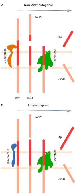
(16) 1.2 An overview of APP processing and Aβ production.. The 37-43 amino acid amyloid β-peptide (Aβ) is generated by proteolytic processing from its precursor, the β-amyloid precursor protein (APP) in a physiologically normal pathway (Haass, Koo et al. 1992). APP is a type 1 transmembrane protein whose physiological role is yet to be fully defined. The predominant transcripts are APP695, APP751 and APP770. All of these transcripts encode multidomain proteins with a single membrane-spanning region. APP695 is the predominant form in neuronal tissue. There are two proteolytic processing pathways of APP and its metabolic derivatives are depicted in Figure 1. Cleavage of APP by either α or β-secretases produces large soluble N-terminal fragments sAPPα and sAPPβ and αCTF and βCTF membrane-bound C-terminal fragments, respectively. γ-secretase cleavage of αCTF and βCTF will result in the generation of non-pathogenic p3 peptide and 4kDa Aβ, respectively, as well as the amino-terminal APP intracellular domain (AICD). BACE1 is the β-secretase, and the components of the γ-secretase complex are presenilin (PS), nicastrin (NCT), presenilin enhance 2 (Pen-2) and anterior pharynxdefective 1 (Aph-1). The activity of α-secretase is associated with several members of the ADAM (a distintergrin and metalloproteinase) family, ADAM9, ADAM10 and tumour necrosis factor-α convertase (also named ADAM17), although other proteases may also contribute. Both nascent APP and β-secretase molecules mature through the constitutive secretory pathway from the endoplasmic reticulum (ER) to the plasma. . 3 .
(17) Figure 1. Sequential cleavage of the amyloid precursor protein (APP) occurs by two alternative pathways. (A) Non-amyloidogenic processing of APP involving α-secretase followed by γ-secretase cleavage is shown. (B) Amyloidogenic processing of APP involving β-secretase followed by γ-secretase clevage is shown. Both processes generate soluble ectodomains (sAPPα and sAPPβ) and identical intracellular C-terminal fragments (AICD).. . 4 .
(18) membrane (PM) (Capell, Steiner et al. 2000) The majority of APP localizes to the Golgi complex (Caporaso, Takei et al. 1994). Only a small proportion of APP is detected at the cell surface and over 50% is internalized within 10 minutes (Koo, Squazzo et al. 1996, Perez, Soriano et al. 1999) and sorted into early endosomes (Koo and Squazzo 1994), where one fraction of APP is recycled back to the PM and another fraction is targeted to the lysosome for degradation (Yamazaki, Koo et al. 1996). α-secretase is particularly enriched at the cell surface and competes with βsecretase for APP processing (Parvathy, Hussain et al. 1999). α-secretase also competes with β-secretase in the trans-Golgi network (TGN), whereas protein kinase C stimulates α-secretase activity to relatively decrease β-cleavage (Skovronsky, Moore et al. 2000). However, β-secretase is predominantly localized in the TGN and endosomes (Vassar, Bennett et al. 1999). These acidic endosomal compartments provide a low pH environment, which is more favorable for β-secretase activity (Hook, Toneff et al. 2002). Moreover, βsecretase is rapidly internalized from the cell surface (Pastorino, Ikin et al. 2002) and is degraded by the ubiquitin-proteasome pathway (Qing, Zhou et al. 2004). Accordingly, accelerating β-secretase degradation by ubiquitin carboxylterminal hydrolase L1 (UCHL1) reduces βCTF and Aβ production (Zhang, Deng et al. 2012). Therefore, the majority of cell surface APP is processed through the. non-amyloidogenic. pathway,. whereas. intracellular. APP. processing. predominantly involves the amyloidogenic pathway (Koo and Squazzo 1994). Only a small fraction of the γ-secretase complex components are located on the. . 5 .
(19) cell surface, the rest are mainly localized at the ER, Golgi/TGN and endosome (Chyung, Raper et al. 2005). The oligomerization and aggregation of the Aβ peptide has been implicated in the pathogenesis of AD.. 1.3 NPC disease presents an increase in the Aβ levels.. Several antecedents suggest that the metabolism of Aβ – previously mainly studied in Alzheimer`s disease – is altered in NPC disease. First, lipid metabolism changes the APP processing and Aβ secretion (Grimm, Grimm et al. 2007). The α-secretase pathway, which releases the N-terminal ectodomain. sAPPα. and. prevents. Aβ. formation,. occurs. mainly. outside. cholesterol rich lipid raft membrane domains. In contrast, β and γ-secretases, which release Aβ peptides, are more active within lipid rafts. Consequently, cholesterol depletion inhibits Aβ formation (Simons, Keller et al. 1998), while cholesterol enrichment may reduce sAPPα secretion (Bodovitz and Klein 1996) and increase Aβ deposition (Refolo, Malester et al. 2000). Second,. several. studies. have. examined. Aβ. metabolism. using. a. NPC. pharmacological model by treating cells with the U18666A drug, which induces lysosomal cholesterol accumulation in cells finding different effects. Davis found that U18666A regulate APP trafficking, increasing the level of the holoprotein at the cell surface, reducing its internalization, the β-secretase processing and reducing Aβ secretion (Davis 2008). In contrast, Yamazaki et al. found no effect of U18666A treatment on secretion of Aβ from Chinese. . 6 .
(20) hamster ovary (CHO) cells, although the treatment lead to intracellular accumulation of Aβ (especially Aβ42) in late endosomes (Yamazaki, Chang et al. 2001). Jin et al. found that U18666A led to accumulation of Aβ and CTFs in APP695. transfected. primary. mouse. cortical. neurons,. but. not. in. βCTF. transfected cells, suggesting that the treatment increased the activity of other secretases than γ-secretase (Jin, Shie et al. 2004). Few studies have explored systems with endogenous APP expression, but Koh et al. found that U18666A treatment reduced the release of Aβ while the Aβ intracellular levels increased in mouse cortical neurons only expressing endogenous APP (Koh, Whiteman et al. 2006). U18666A has several effects, in addition of inducing cholesterol accumulation,. including. transcriptional. upregulation. of. the. γ-secretase. components presenilin-1 and presenilin-2 (Crestini, Napolitano et al. 2006) and altered glycosylation of β-secretase (Sidera, Parsons et al. 2005). Therefore, it is difficult to determine if the effects on APP metabolism due to U18666A treatment are directly related to the accumulation of cholesterol on lysosomes. Furthermore, in vitro and in vivo findings indicate that loss of NPC1 leads to a significant increase in βCTF levels and sAPPβ release (Yamazaki, Chang et al. 2001, Jin, Shie et al. 2004). Moreover, cells lacking the NPC1-protein have reduced APP surface levels (Kosicek, Malnar et al. 2010, Malnar, Kosicek et al. 2010) and increased brain activity of Aβ generating enzymes (Kodam, Maulik et al. 2010). Besides Mattsson et al. found that in CHO NPC1-nulls cells had reduced media levels of sAPPα and Aβ, and increased levels of sAPPβ (Mattsson, Olsson et al. 2012). Third, it have been shown that Npc1-/- mice accumulate βCTF and Aβ in their. . 7 .
(21) brains (Yamazaki, Chang et al. 2001, Burns, Gaynor et al. 2003).. However,. Npc1-/- mice brain have unchanged levels of APP, sAPP, PS1 and β-secretase proteins and only slightly increased γ-secretase activity (Burns, Gaynor et al. 2003). In another hand, Kodam et al found increased β-secretase activity along with increased levels of APP, β-secretase and all four components of the γ-secretase complex in NPC mice cerebellum and hippocampus compared to controls (Kodam, Maulik et al. 2010). Also, NPC patients receiving treatment with miglustat and β-cyclodextrin, that reduce the glicosphingolipids and cholesterol accumulation repectively, showed a decrease in Aβ and sAPPβ levels over time of treatment (Mattsson, Olsson et al. 2012). Additionally, NPC1depletion. in. the. AD-transgenic. mouse. model. (PS1xAPP). enhances. the. progression of AD by increasing Aβ accumulation (Borbon and Erickson 2011). More recently, altered expression of NPC1 mRNA/protein was reported in AD brains (Ginsberg, Alldred et al. 2010, Kagedal, Kim et al. 2010), indicating a bidirectional link between NPC1 dysfunction and Alzheimer's disease. NPC disease may thus be an innovative model to study the molecular mechanisms of cholesterol-mediated APP-CTF/Aβ accumulation in which a defect in a single gene involved in cholesterol trafficking causes AD-like phenotype.. 1.4 APP and its trafficking route within the cell.. APP is a transmembrane protein that is actively sorted among numerous compartments in the cell and is known that its subcellular localization affects its processing. APP, like other proteins, is biosynthesized in the ER and. . 8 .
(22) transported via the constitutive secretary pathway from the ER, through the Golgi apparatus/TGN, to the cell membrane. As APP traffics through the secretory pathway, it undergoes post-translational modifications, including N and O-glycosylation, ubiquitination, phosphorylation, and tyrosine sulfation (De Strooper and Annaert 2000). Mature APP is then internalized by clathrinmediated endocytosis, incorporated into the endosomal-lysosomal system and returns to the cell surface. Relevant is that the trafficking and localization of APP affects the balance between the two APP processing pathways — mediated by α and β-secretases — so directly impact Aβ production. Since 1992, evidence had demonstrated that Aβ is mainly produced in the endosome/lysosome system. Impairing APP trafficking to the cell surface or enhancing APP internalization increases βsecretase-mediated processing of it (Haass, Koo et al. 1992, Lee, Zhang et al. 2005), while enhancing APP routing to, or reducing its internalization from, the cell surface facilitates α-secretase-mediated processing (Cataldo, Barnett et al. 1997). The endocytosis motif located at the carboxyl terminus of APP (682YENPTY687, referring to APP 695 numbering) is responsible for the efficient internalization of APP, in clathrin-coated vesicles, to early endosomes (Lai, Sisodia et al. 1995). Deletion or mutation of this motif led to APP endocytosis-deficiency and significantly reduced Aβ production (Selkoe, Yamazaki et al. 1996). The Swedish. double. mutant. (KM/NL). APP. produced. significantly. more. Aβ. (approximately threefold) than wild-type APP (Mullan, Crawford et al. 1992); however, abolishing the endocytic process of Swedish APP by removing its. . 9 .
(23) endocytic motif still resulted in substantially more Aβ than with normal APP. This result indicates that β-cleavage on Swedish APP does not require an intact cytoplasmic domain and Aβ can also be produced in the Golgi during its biosynthetic transport (Haass, Lemere et al. 1995). Furthermore, inhibition of protein transport from the ER to Golgi and redistributing Golgi proteins into the ER by brefeldin A treatment, or retention of APP in the ER with an ER-retrieval signal, significantly reduced but did not abolish intracellular Aβ production (Chyung, Greenberg et al. 1997). All this evidence indicates that intracellular trafficking of APP clearly plays a central role in APP processing; amyloidogenic cleavage of APP and Aβ production occurred in multiple subcellular organelles, including the ER/ER-Golgi intermediate compartment, the Golgi during its biosynthetic transport, and the endosome/lysome after endocytosis from the plasmatic membrane. YENPTY is a typically motif present in many Tyr-kinase (TK) receptors and nonreceptor TKs; it is generally phosphorylated and represents the docking site for multiple interacting proteins involved in cell signaling and gene transcription. As APP — and all the CTFs and AICD fragments — contain the 682YENPTY687 sequence, which can be bound by different adaptor proteins. Mutations within the YENPTY motif selectively inhibit APP internalization and decrease Aβ generation (Perez, Soriano et al. 1999). One position of particular functional significance in the amino acid sequence YENPTY is Tyr682, which levels of phosphorylation are increased in AD patients (Russo, Salis et al. 2001). Moreover, it has been shown that the brain of mice expressing APP with the Tyr682Gly mutation have a large redistribution of APP towards the non-. . 10 .
(24) amyloidogenic pathway, with increased sAPPα and αCTF levels and decreased sAPPβ and Aβ levels (Barbagallo, Weldon et al. 2010). Interestingly in the brain of. both. AD. and. age-matched. normal. subjects,. most. βCTFs. are. Tyr-. phosphorylated while αCTFs are not phosphorylated (Russo, Salis et al. 2001, Russo, Dolcini et al. 2002). In another hand, Takahashi et al. reported that in HEK293 cells the phosphorylation of APP at Tyr687, is important for its processing by α and γ-secretases, increasing αCTF and AICD generation (Takahashi, Niidome et al. 2008). As the processing of APP occurs at several subcellular localizations and αCTF fragments are produced at the cell surface level, it is suggested that the alternative phosphorylation of amino acid residues can deliver APP to different subcellular compartments. This event can be relevant in preparing the APP substrate for different enzymatic processing steps that may or may not lead to the formation of amyloidogenic fragments. These findings indicate that alternative phosphorylation of Tyr682 or Tyr687, directing APP to different subcellular sites, could be relevant for sorting α or βsecretase cleavage. The resulting CTFs could either play a functional role as a whole or be further processed by γ-secretase to yield Aβ or P3 fragments and AICD. Therefore, the phosphorylation of APP may be very tightly regulated, as the. kinases. involved. can. affect. not. only. cell. signaling. but. also. the. amyloidogenic pathway. The C-terminal region of APP, CTFs and AICD is also the docking site for interacting proteins such as Fe65 (Fiore, Zambrano et al. 1995, Borg, Ooi et al. 1996), X11 (Borg, Ooi et al. 1996), mDab (Howell, Gertler et al. 1997), Numb (Roncarati, Sestan et al. 2002), JIP-1 (Scheinfeld, Roncarati et al. 2002) and. . 11 .
(25) c-Abl (Zambrano, Bruni et al. 2001). The aforementioned adaptor proteins are able to recognize, via their PTB domain, the NPXpY motif, which is generated by phosphorylation. They can also bind the C-terminus of APP and related compounds. regardless. Interestingly,. Zambrano. of. the et. al.. phosphorylation demonstrated. of. the. that. YENPTY. APP. is. motif.. tyrosine-. phosphorylated in cells expressing a constitutively active form of c-Abl (Zambrano, Bruni et al. 2001).. 1.5 Searching for a possible regulator of APP processing: the c-Abl tyrosine kinase.. We want to address the question of the possible involvement of c-Abl in the protein-protein interaction network centered at the cytosolic domain of APP. We previously reported that in NPC models the tyrosine kinase c-Abl and the apoptotic system c-Abl/p73 is actived and strikingly, the inhibition of c-Abl kinase with Imatinib (STI571, Gleevec) reduces weight loss, neurological symptoms and cerebellar apoptosis, increasing the number of Purkinje cells and survival of NPC mice (Alvarez, Klein et al. 2008). Interestingly, Imatinib treatment. also. reduced. the. number. and. size. of. Aβ. deposits. in. the. APPsw/PSEN1∆E9 mice (Netzer, Dou et al. 2003, Cancino, Toledo et al. 2008). Moreover, the ~50 amino acids long APP intracellular region contains seven residues that can be phosphorylated and several of these amino acids are hyperphosphorylated in human AD brain. Upon phosphorylation, they become docking sites for intracellular signaling proteins containing specific SH2 (Src. . 12 .
(26) homology 2), SH3, PH (pleckstrin homology) and PTB (phosphotyrosine binding) domains (Cattaneo and Pelicci 1998). As mentioned before, one of these sites of particular functional significance is Tyr682 of the amino acid sequence YENPTY. In particular, Tyr682 modulates the interaction with adaptor proteins through its phosphorylation and dephosphorylation, suggesting that this residue functions as a switch that activates certain APP signaling pathways. Relevantly, Tyr682 residue is phosphorylated by c-Abl being this phosphorylation recognized by the Src-Homology 2 (SH2) domain of c-Abl itself (Zambrano, Bruni et al. 2001). c-Abl can also regulate AICD formation and. the. modulation. of. AICD-dependent. cellular. responses,. such. as. transcriptional induction and apoptotic cell death (Vazquez, Vargas et al. 2009). Site-direct mutagenesis of Tyr682 to phenylalanine, but not of Tyr653 or 687, abrogates APP phosphorylation by c-Abl (Zambrano, Bruni et al. 2001). Moreover, the APP Y682G mutation in mice brain results in: i) a redistribution of APP towards the non-amyloidogenic pathway (Barbagallo, Weldon et al. 2010) and ii) alternative APP trafficking toward late endosomes and lysosomes, ensuing functional alterations of the lysosomal system (La Rosa, Perrone et al. 2015). Previously we showed that c-Abl is activated in NPC models and interestingly Imatinib treatment reduced the number and size of Aβ deposits in the APPsw/PSEN1∆E9 mice (Netzer, Dou et al. 2003, Cancino, Toledo et al. 2008). Although the Imatinib effects on Aβ burden were been linked to the GSAP inhibition and c-Abl participation was hastily discarded, the mechanism involved is controversial (He, Luo et al. 2010, Hussain, Fabregue et al. 2013).. . 13 .
(27) In this work we evaluated i) APP processing following Aβ, βCTF and sAPPα levels in: a) CHO WT and NPC cells treated with Imatinib or transfected with short hairpin RNA (shRNA) construct against c-Abl and b) NPC mice injected with Imatinib for 4 weeks, ii) the interaction between APP, c-Abl and βsecretase (BACE1) in CHO NPC cells treated with vehicle or Imatinib. Moreover we used a mutant APPY682A to investigate whether the binding of c-Abl to APP is dependent on the YENPTY motif and iii) APP localization in CHO NPC cells that overexpress the pathogenic AD mutation APPswe.. 1.6 Hypothesis and Objectives.. Hypothesis. c-Abl promotes APP amyloidogenic processing in Niemann-Pick type C disease.. Objectives. Aim: To evaluate the role of c-Abl in the processing of APP and its implication in Niemann-Pick type C disease models.. . 14 .
(28) Specific Objectives.. 1.. To characterize the effect of Imatinib in the processing of APP and Aβ accumulation in in vivo and in vitro NPC models.. 2.. To demonstrate that c-Abl is mediating the effect of Imatinib on the processing of APP in CHO NPC1-null cells.. 3.. To evaluate the mechanism by which c-Abl promotes the cutting of the βsecretase in CHO NPC1-null cells.. . 15 .
(29) 2. CHAPTER I. The next section presents the results that support the specific objectives 1, 2 and 3. This work has been submitted to The Journal of Neuroscience on August 27th, 2015 (Manuscript ID: JN-RM-3223-15). This work is entitled: “c-Abl links APP-BACE1 interaction promoting APP amyloidogenic processing in Niemann-Pick type C Disease” from the authors: María José Yáñez, Olivia Belbin, Lisbell Estrada, Nancy Leal, Pablo Contreras, Alberto Lleó, Patricia Burgos, Silvana Zanlungo and Alejandra Alvarez. In this work we establish that: c-Abl inhibition reduces the levels of the amyloidogenic pathway fragments; Aβ and βCTF and increases the levels of the non-amyloidogenic sAPPα fragments in NPC cells. Moreover, we found decreased levels of βCTF in cortical neuronal cultures derived from c-Ablfloxo/floxo Nestin Cre (c-Abl null) mice. With this evidence, we analyzed BACE1-APP interaction. We observed that c-Abl interacts with APP through the Y682ENPT motif and we found that c-Abl inhibition results in decreased BACE1-APP interaction, supporting the idea that c-Abl inhibition reduces the availability of APP to interact with BACE1. This manuscript addresses the participation of the c-Abl kinase in the processing of APP in NPC models.. . 16 .
(30) c-Abl links APP-BACE1 interaction promoting APP amyloidogenic processing in Niemann-Pick type C Disease Yáñez MJ. 1,2,3. , Belbin O5, Estrada LD1,3, Leal N1,3, Contreras PS1,2,3, Lleó. A5, Burgos PV6, Zanlungo S2,4, Alvarez AR1,3. 1. Department of Cell & Molecular Biology, 2Department of Gastroenterology,. 3. CARE-Chile-UC, 4FONDAP Center for Genome Regulation (CGR), Pontificia Universidad Catolica de Chile, Santiago 8331010, Chile, 5Neurology. Department, IIB-Santpau, Hospital de la Santa Creu i Sant Pau, Barcelona 08026, Spain, 6Instituto de Fisiología, Facultad de Medicina, and Centro de Investigación Sur-Austral en Enfermedades del Sistema Nervioso, Universidad Austral de Chile, Valdivia 5110566, Chile.. Abbreviated title: c-Abl regulates APP amyloidogenic processing in NPC.. Key words: NPC, Niemann-Pick type C; NPC1, Niemann-Pick type C 1 protein; NPC2, Niemann-Pick type C 2 protein; AD, Alzheimer disease; Aβ, amyloid beta; APP, amyloid precursor protein; αCTF, alpha carboxyl terminal fragment;. βCTF, beta carboxyl terminal fragment; sAPPα, soluble amyloid precursor protein alpha; sAPPβ, soluble amyloid precursor protein beta.. Abstract. Niemann-Pick type C (NPC) disease is characterized by lysosomal accumulation of cholesterol. Interestingly, NPC patients’ brains also show increased levels of. . 17 .
(31) amyloid-β. (Aβ). peptide,. a. key. protein. in. Alzheimer's. disease. (AD). pathogenesis. We previously reported that the c-Abl tyrosine kinase is active in NPC neurons and in AD animal models and that Imatinib, a specific c-Abl inhibitor, decreased the amyloid burden in brains of the AD mouse model. The Aβ peptide is produced by sequential cleavage of the amyloid precursor protein (APP) first by β-secretase (BACE1), resulting in a carboxy-terminal fragment (βCTF), and subsequently by γ-secretase, releasing Aβ. While active c-Abl has been shown to interact with and phosphorylate APP, the relevance of this interaction has yet to be defined. Here, we show that c-Abl inhibition, using Imatinib or shRNA-mediated c-Abl knockdown, reduces Aβ oligomer and βCTF levels and increases the soluble sAPPα levels in CHO NPC cells that overexpress the pathogenic AD mutation APPswe. Moreover, Imatinib decreased APP amyloidogenic processing in a NPC mouse model. In addition, we found decreased levels of βCTF in neuronal cultures derived from c-Abl null mice. Moreover, we found that c-Abl interacts with both APP and BACE1 and that Tyr682 of the amino acid sequence YENPTY in the cytoplasmic APP tail is essential for its interaction with c-Abl. Using fluorescence lifetime imaging microscopy, we observed that Imatinib significantly inhibits both APP-c-Abl and APP-BACE1 interactions. We conclude that c-Abl is a linker that facilitates APPBACE1 interaction, thereby promoting amyloidogenic processing of APP in NPC models.. . 18 .
(32) Significance Statement. Our findings show that c-Abl-mediated phosphorylation at the APP Tyr682 residue is a key molecular mechanism regulating its interaction with BACE1, promoting BACE1-APP interaction, amyloidogenic APP cleavage and favoring Aβ accumulation in NPC models. These results strongly suggest that the pharmacological inhibition of c-Abl underlies the decrease in amyloid burden by Imatinib observed in AD mouse models. Therefore, inhibition of c-Abl could be a pharmacological target for preventing the deleterious effects in the NPC and AD amyloid pathology. Finally, these results implicate c-Abl in amyloid pathology and provide insights for future research in APP processing in AD.. Introduction. Niemann-Pick type C (NPC) disease is an autosomal recessive disease produced by mutations in the Npc1 and Npc2 genes that cause intracellular lysosomal accumulation of many lipids, particularly unesterified cholesterol and sphingolipids, in multiple organs (Vance 2006). The Central Nervous System is particularly affected in this disease and loss of neurons is evident primarily in the prefrontal cortex, thalamus and cerebellum (Li, Repa et al. 2005, Kodam, Maulik et al. 2010). A number of recent studies have shown that NPC disease exhibits some intriguing parallels with Alzheimer’s disease (AD), including the presence of neurofibrillary tangles (Auer, Schmidt et al. 1995), impaired cholesterol homeostasis, trafficking abnormalities in endosomes and lysosomes. . 19 .
(33) (Nixon, Yang et al. 2008), and altered amyloid precursor protein (APP) metabolism (Malnar, Kosicek et al. 2010, Mattsson, Olsson et al. 2012). Notably, Aβ peptide levels are increased in the vulnerable regions of the NPC brain (Yamazaki, Chang et al. 2001, Jin, Shie et al. 2004). The Aβ peptide, believed to be the initiating factor in AD pathogenesis (Hardy and Selkoe 2002), is generated by the proteolytic processing of the APP protein by two proteases termed β-secretase (BACE1) and γ-secretase (Kang, Lemaire et al. 1987). The cleavage of APP by β-secretase generates the soluble sAPPβ fragment and the membrane-bound carboxy-terminal fragment βCTF, which is the substrate of γ-secretase and generates the Aβ peptide. Previous in vitro and in vivo findings indicate that loss of NPC1 leads to a significant increase in βCTF levels and sAPPβ release (Yamazaki, Chang et al. 2001, Jin, Shie et al. 2004). Moreover, levels of APP at the cell surface are reduced in NPC1-deficient Chinese hamster ovary (CHO) cells (Malnar, Kosicek et al. 2010); and expression of APP, β and γ-secretases are elevated in the brain of an NPC mouse model (Kodam, Maulik et al. 2010). We previously reported that the tyrosine kinase c-Abl is active in NPC neurons and mouse models (Alvarez, Klein 2008). Strikingly, inhibition of c-Abl with Imatinib reduces weight loss, neurological symptoms and cerebellar apoptosis, increasing the number of Purkinje cells and survival of NPC mice (Alvarez, Klein et al. 2008). Interestingly, Imatinib treatment also reduces the number and size of Aβ deposits in the APPswe/PSEN1∆E9 mouse model (Netzer, Dou et al. 2003, Cancino, Toledo et al. 2008). Other studies have reported that Imatinib prevents Aβ generation but inhibiting the γ-secretase activating. . 20 .
(34) protein (GSAP) and preventing γ-secretase cleavage of the βCTF (Netzer, Dou et al. 2003, He, Luo et al. 2010, Hussain, Fabregue et al. 2013). However, we believe that the effects of Imatinib on APP metabolism and the role of c-Abl should be re-evaluated. The APP tail contains 3 tyrosine residues (653, 682 and 687 of APP. 695). that are. potential targets for tyrosine kinases such as c-Abl (Zambrano, Bruni et al. 2001). Additionally, it has been shown that the brain of mice expressing a Tyr682Gly mutation show i) a large redistribution of APP towards the nonamyloidogenic pathway with increased sAPPα and αCTF levels and decreased sAPPβ and Aβ levels (Barbagallo, Weldon et al. 2010); and ii) alternative APP trafficking toward late endosomes and lysosomes associated with functional alterations of the lysosomal system (La Rosa, Perrone et al. 2015). Therefore, we propose that both, the phosphorylation state of the APP-Tyr682 residue and c-Abl, could be involved in the APP-centered molecular machinery that modulates APP processing. Here, we show that NPC1-deficient CHO cells (NPC) and the brains of NPC mice have increased levels of Aβ. We found that c-Abl inhibition, using Imatinib or shRNA-mediated c-Abl knockdown, reduces Aβ and βCTF levels and increases sAPPα levels in NPC cells that overexpress APP. Consistently, Imatinib treatment resulted in a decrease in APP amyloidogenic processing in the brains of NPC mice. Moreover, we also found decreased levels of βCTF in cortical neuronal cultures derived from c-Ablfloxo/floxo Nestin Cre (c-Abl null) mice. Our results confirm previous reports showing that c-Abl interacts with APP, through the Y682ENPT motif, and they also sustain that this interaction has a crucial. . 21 .
(35) functional. effect. promoting. APP-BACE1. interaction,. amyloidogenic. APP. cleavage and Aβ accumulation.. Materials & Methods. Antibodies and Reagents Mouse anti-c-Abl (sc-23) was purchased from Santa Cruz biotechnology (Dallas, United States of America). Mouse anti-BACE1 (61-3E7), mouse antiphosphotyrosine (p-Tyr) 4G10 (05-321), mouse anti-APP (22C11) and mouse anti-Aβ (WO2) were purchased from Millipore (Billerica, United States of America). Mouse anti-Aβ (4G8) was purchased from Covance. Mouse anti-HA (HA.C5) was purchased from Abcam (Cambridge, United Kingdom). Imatinib mesylate (13139) was purchased from Cayman Chemical Company (Ann Arbor, United States of America). The inhibitors for α and β-secretase (CC1000 and 565749, respectively) were obtained from Millipore. Plasmid APP-GFP, APP3Y/A-GFP, APP wild-type and APP tyrosine mutations (653, 682 and 687 of APP695) plasmids were kindly donated by Patricia Burgos, PhD (Instituto de Fisiología, Facultad de Medicina, Universidad Austral de Chile). BACE1-GFP, APP-RFP and GFP-RFP plasmid were kindly donated by Alberto Lleó, PhD (Neurology Department, IIB-Santpau, Hospital de la Santa Creu i Sant Pau). c-Abl-DsRed was purchased from Genscript (New Jersey, United States of America). The shRNA-c-Abl (SHDNA-NM_009594) and scrambled shRNA plasmids were purchased from Sigma (St. Louis, Missouri).. . 22 .
(36) Animals, cell lines and transfection BALB/c mice carrying a heterozygous mutation in the Npc1 gene (NPC mice), c-Ablfloxo/floxo Nestin Cre and c-Ablfloxo/floxo were obtained from Jackson Laboratory (Maine, United States of America). Genotyping was performed using a PCRbased screening as previously described (Amigo, Mendoza et al. 2002). All protocols were approved and followed local guidance documents generated by the ad hoc committee of Chile (CONICYT) and were approved by the Bioethics Committee of the School of Medicine from Pontificia Universidad Católica de Chile (CEBA Protocol # 10-017). The recommendations of the Guide for Care and Use of Laboratory Animals the Institute for Laboratory Animal Research in agreement to US Public Health Service Policy on Humane Care and Use of Laboratory Animals were strictly followed. Chinese hamster ovary (CHO) wild-type (WT) cells and NPC1-deficient CHO cells (NPC) (kindly provided by Laura Liscum, PhD., Tufts University, Boston) were maintained in DMEM:F12 medium (1:1) containing 0.5 mM Na-pyruvate supplemented with 10% FBS, 2 mM L-glutamine, 100 IU/ml penicillin and 100 µg/ml streptomycin. For experiments with α and β-secretases inhibitors (inhibitor II and GM6001 MMP, respectively), cells were treated with 25 mM and 10 mM respectively. CHO WT and NPC cell lines stably expressing the APP swedish construct were established by transfection using Lipofectamine LTX (Invitrogen, California, United States of America) and selected using media supplemented with the appropriate antibiotic according to the supplier´s instructions.. . 23 .
(37) Transient transfection of 2µg of plasmid was performed using Lipofectamine LTX (Invitrogen) according to the supplier’s instructions. 24 h after transfection medium was removed, fresh medium was added and cells were further incubated for 24 h. Primary cortical cell cultures Cortical tissues were dissected from c-Ablfloxo/floxo Nestin Cre (c-Abl null) and cAblfloxo/floxo (wild-type) mice at embryonic day 18. Cortical cells were seeded onto poly-L-lysine-coated wells. Cultures were maintained at 37°C in 5% CO2 with. neurobasal. growth. medium. (Invitrogen). supplemented. with. B27. (Invitrogen) plus antibiotics (2mM L-glutamine, 100 U/ml penicillin and 100 µg/ml streptomycin) for 5 days. On day 2, cultured neurons were treated with 2 µM AraC for 24 h to prevent glial cell proliferation. Sample preparation and immunoblotting analyses Cell medium was collected and centrifuged at 16,000g for 10 min at 4°C. For detection of secreted APP (sAPPα), aliquots of conditioned medium normalized according to protein concentration in the cell lysate were directly analyzed by SDS-PAGE. For lysate preparation, cells were washed with PBS and lysed in radioimmunoprecipitation assay (RIPA) buffer (0.5% Nadeoxycholate, 0.1% SDS, 1% NP40, 5 mM EDTA, 150 mM NaCl, 50 mM Tris-HCl ph 8.0) containing a protease inhibitor cocktail (Roche Applied Science) on ice for 10 min and centrifuged at 4°C for 10 min at 16,000g. Supernatants were mixed with 6x sample buffer (60% glycerol, 12% SDS, 3% DTT, 1/8 v/v Tris pH 6.8, bromophenol blue), heated at 95°C for 10 min and subjected to SDS-PAGE, transferred to Nitrocellulose membrane (Thermo Scientific, Massachusetts,. . 24 .
(38) United States of America), then blocked for 1 hr at room temperature in 5% nonfat dry milk in PBS 1X, and finally incubated overnight with primary antibodies against APP, Aβ, c-Abl, pTyr or BACE1. Membranes were washed, incubated. with. appropriate. horseradish. peroxidase. labeled. secondary. antibodies (Thermo Scientific), and developed using the ECL technique (Thermo Scientific). The protein levels were quantified using the ImageJ software. Coimmunoprecipitation assay Protein extracts were obtained from CHO NPC cells lysed in RIPA buffer supplemented with a protease inhibitors cocktail (Roche Applied Science). Immunoprecipitations from protein extracts (500 µg) were performed using 2 µg of anti-APP and anti-c-Abl antibody. The immunocomplexes were then precipitated with protein G Sepharose. Cell lysates were separated by SDSPAGE, transferred to PVDF membranes (Thermo Scientific), and immunoblotted with pTyr, anti-APP and anti-c-Abl antibodies. Site-directed mutagenesis APP mutants were generated by PCR using the proofreading Pfu polymerase (Stratagene, Santa Cruz, United States of America), followed by DpnI (New England Biolabs, Massachusetts, United States of America) digestion of the methylated. parental. plasmid.. Oligonucleotides. used. were. as. follows:. Y682Asense 5`ACCTGTCCAAGATGCAGCAGAACGGCGCCGAAAATCCAACCTACAAGTTCTTTGA G3`;. . Y682Aantisense. 25 .
(39) 5`CTCAAAGAACTTGTAGGTTGGATTTTCGGCGCCGTTCTGCTGCATCTTGGACAGGT 3`. Each clone was verified by automated sequencing. Confocal microscopy and Fluorescence Lifetime Imaging Microscopy (FLIM) Confocal microscopy was performed using a Leica inverted fluorescent confocal microscope (Leica TCD SP2-AOBS, Wetzlar, Germany). This microscope is equipped with a 405 diode pulsed laser, a PMC-100 detector (Leica, Wetzlar, Germany) and a time-correlated single photon counting module (SPC730) to perform FRET/FLIM. The hardware/software package (SymphoTime) allows the measurement of fluorescence lifetimes on a pixel-by-pixel basis. Values were fitted to two-exponential decay curves to represent a “non-FRETing” population with a longer lifetime (t2) and a “FRETing” population with a shorter lifetime (t1). FLIM has been described as a novel technique for the analysis of protein proximity. The technique is based on the observation that fluorescence lifetimes of a donor fluorophore shorten in the presence of a FRET acceptor in close proximity (<10 nm). The fluorescence lifetime of the donor is directly proportional to the distance between the donor and the acceptor. CHO NPC cells were grown for 24 h on 10 cm plates to 80% confluency and then transfected using X-tremeGENE (Roche) with APP-GFP, APP3Y/A-GFP, cAbl-DsRed. or. BACE1-GFP.. For. APP-c-Abl. and. APP-BACE1. FRET/FLIM. experiments, cells were fixed and double-immunostained for APP, c-Abl and BACE1 as described previously (Lleo, Berezovska et al. 2004). All samples were compared with a negative control in which the donor fluorophore fluorescence lifetime was measured in the absence of the acceptor (no FRET. . 26 .
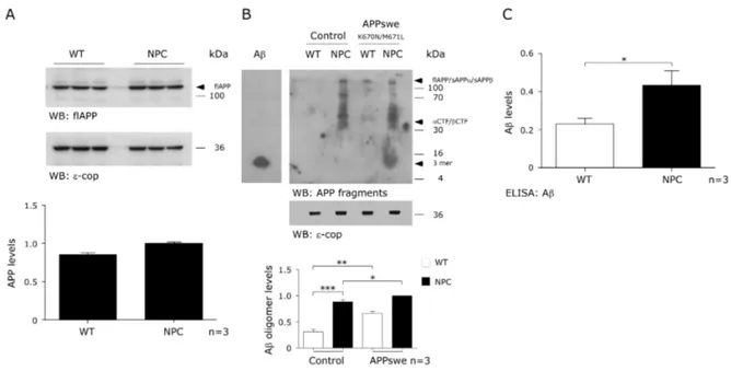
(40) ~3.5 ns). The degree of GFP donor lifetime shortening due to the presence of FRET was used as an indicator of the proximity between the GFP donor and RFP acceptor fluorophores in APP-RFP co-transfected cells (~0.5 ns). Statistical analyses Mean and SEM values and the number of experiments are indicated in each figure. Statistical analysis was performed by one-way ANOVA, followed by Bonferroni posttest using the Prisma Software.. Results. Alteration in APP processing in CHO NPC models Since it was previously reported that the amyloidogenic cleavage of APP is stimulated in NPC cells (Yamazaki, Chang et al. 2001, Burns, Gaynor et al. 2003, Jin, Shie et al. 2004), we first sought to determine the effect of NPC1deficient CHO cells (NPC) on APP expression. In accordance with previous reports, we did not observe any changes in full length APP levels (flAPP) in NPC cells compared to CHO wild-type (WT) cells (Figure 1A), suggesting that the increased amlyoidogenic processing is not due to an increase in APP expression. To determine the effects on APP processing, we used the 4G8 antibody, which detects full length APP, Aβ sequence-containing cleavage products and the Aβ peptide (soluble and oligomeric). In NPC cells, but not in WT cells, we detected high molecular weight species (>100kDa), which could include flAPP, sAPPβ and sAPPα (fragment released in non- amyloidogenic pathway) (Figure 1B). In agreement with Malnar et al 2010, we also observed. . 27 .
(41) Figure 1. CHO NPC models show increased APP processing through the β-secretase pathway. APP (A) and APP-derived proteolytic fragments (B) were analyzed between CHO WT, NPC cells and APPswe-transfected CHO WT and NPC cells. (C) ELISA analysis of Aβ levels from brain homogenates of CHO WT and NPC mice. Mean and SEM of three independent experiments are shown. Statistical analysis was performed using Student`s t-test: *p<0.05, **p<0.01, ***p<0.001.. . 28 .
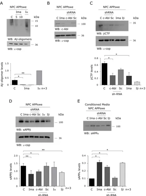
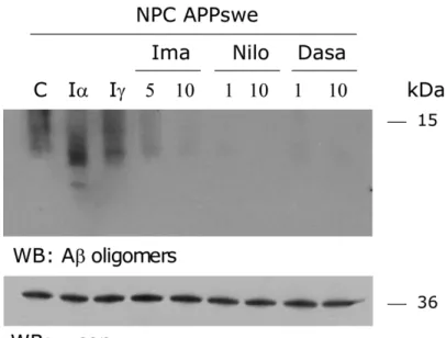
(42) a substantial increase in α and βCTFs (between 30–70 kDa) in NPC cells. Moreover, in NPC cells we observed the same increase in all APP species compared to WT cells when both lines were stably transfected with the APPswe construct (Figure 1B), as previously described (Yamazaki, Chang et al. 2001, Burns, Gaynor et al. 2003, Jin, Shie et al. 2004). A band corresponding to 6– 20 KDa (which could be 3-mer Aβ oligomers) was detected in the CHO NPC APPswe-transfected cells (NPC APPswe) only. As shown in Figure 1C, these results in cell lines are supported by our finding that increased levels of soluble Aβ are found in NPC compared to wild-type mouse brains, as measured by ELISA, in agreement with previous studies (Burns, Gaynor et al. 2003, Jin, Shie et al. 2004). Overall, these data demonstrate that due to their enhanced amyloidogenic processing, NPC APPswe-transfected cells and NPC mice are good models for studying the regulation of APP processing.. c-Abl inhibition decreases Aβ and βCTF levels and increases sAPPα levels in a cellular NPC model In order to evaluate whether c-Abl kinase modulates APP processing in NPC APPswe cells, we analyzed the effect of the c-Abl kinase inhibitor, Imatinib, on Aβ production in the NPC APPswe cells. Interestingly, an alternative nonamyloidogenic processing of APP by α and γ-secretases can also occur. We found that Imatinib treatment reduces Aβ oligomer levels (WO2 positive bands between 6–20 KDa) in a dose dependent manner (Figure 2A). In contrast, the accumulation of lower molecular weight bands (<10kDa, corresponding to p3) following treatment with the α-secretase inhibitor. . 29 .
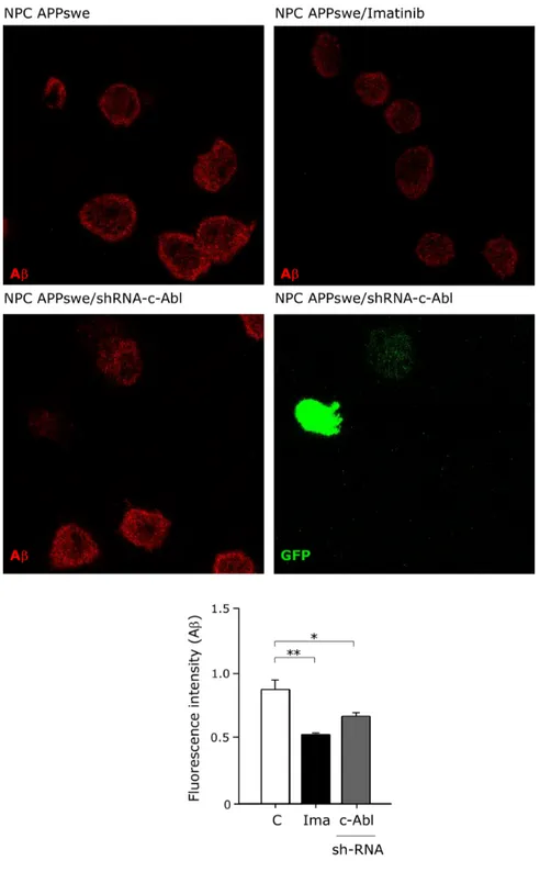
(43) Figure 2. c-Abl inhibition decreases β-secretase cleaved APP and increases sAPPα levels in a cellular NPC model. (A) Western blot of APPderived proteolytic fragments of CHO NPC APPswe cells treated either with vehicle (C), Imatinib (Ima; 5 and 10 µM) or α-secretase inhibitor (Iα; 25 µM) for 16 hrs. (B) Western blot of c-Abl of CHO NPC APPswe treated either with vehicle (C), Imatinib (Ima; 10 µM) or transfected with a plasmid expressing a shRNA against c-Abl (c-Abl) or scrambled shRNA (Sc) for 16 hrs (C-E) Western blot analysis of βCTF (C), sAPPβ (D) and sAPPα (E) levels of CHO NPC APPswe cells. CHO NPC APPswe cells transfected with a plasmid expressing a shRNA against c-Abl (c-Abl) or scrambled shRNA (Sc) or treated with vehicle (C), Imatinib (Ima; 10 µM), β-secretase inhibitor (Iβ; 10 µM) or α-secretase inhibitor (Iα; 25 µM) for 16 hrs. Mean and SEM of three independent experiments are shown. Statistical analysis was performed using Student`s ttest: *p<0.05, **p<0.01. . 30 .
(44) GM6001, confirms that Imatinib treatment affects the amyloidogenic rather than the non-amyloidogenic pathway. Concomitantly, Imatinib-treated NPC APPswe cells presented significantly reduced βCTF (Figure 2C) and sAPPβ (Figure 2D) levels and increased sAPPα levels (Figure 2E). We used the β and α-secretases inhibitors, inhibitor II and GM6001 MMP, respectively, to confirm the identity of the βCTF and sAPPα APP fragments in our samples. To confirm that the inhibitory effect of Imatinib on β-secretase processing of APP was mediated by c-Abl inhibition, we transfected NPC APPswe cells with a short hairpin RNA (shRNA) construct against c-Abl or a scrambled shRNA (Sc) (Figure 2B). We found that the shRNA-c-Abl transfection, similar to the Imatinib treatment, significantly reduced βCTF (Figure 2C) and sAPPβ (Figure 2D) levels and increased sAPPα levels (Figure 2E). Consistent with the idea that c-Abl promotes β-secretase mediated APP processing we observed (using the 22C11 APP antibody) that c-Abl inhibition induced a decrease of the high molecular weight APP immunoreactive band. The fact that the β-secretase inhibitor also induced a reduction of this band implies that it is likely to correspond to sAPPβ. However, we cannot discard a contribution of APP-FL to this APP signal. Together, these results show that c-Abl inhibition either by treatment with Imatinib or by shRNA-knockdown of c-Abl expression, induce a decrease in Aβ oligomers, βCTF, and sAPPβ, and an increase in sAPPα levels, supporting the notion that c-Abl promotes the β-secretase processing of APP. Additionally, using NPC mice that show enhanced amyloidogenic APP processing, we investigated the effect of c-Abl inhibition using Imatinib. We treated 4-week-. . 31 .
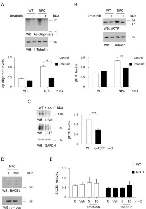
(45) Figure 3. Imatinib treatment decreases β-secretase cleavage products in an NPC animal model. Western blot analysis of Aβ oligomers (A) or βCTF (B) levels from brain homogenates of CHO WT and NPC mice injected with NaCl 0,9% or Imatinib (12,5 mg/kg) for 4 weeks. (C) Western blot analysis of c-Abl, and βCTF levels in cortical neuronal cultures derived from Ablfloxo/floxo Nestin Cre mice. (D) Immunoblot analyses of of BACE1 protein levels in CHO NPC cells treated with vehicle or Imatinib (10 µM). (E) Extracts from CHO NPC cells were subjected to an in vitro BACE1 activity assay. The control samples contained BACE1 and the fluorogenic substrate in assay buffer. β-secretase inhibitor III (inhibitor III) served as a positive control. Mean and SEM of three independent experiments are shown. Statistical analysis was performed using Student`s t-test: *p<0.05, **p<0.01, ***p<0.001. . 32 .
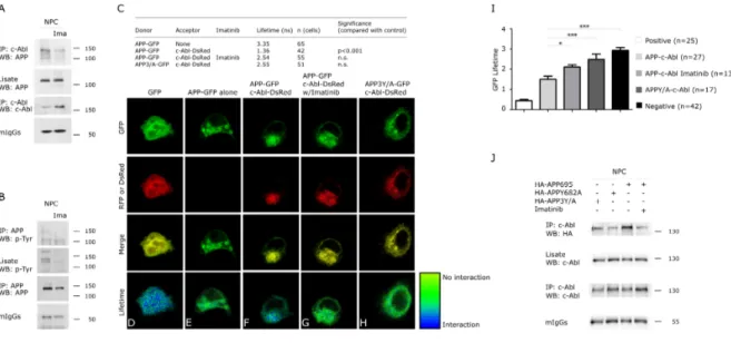
(46) old NPC mice daily with vehicle or 12.5 mg/kg Imatinib through intraperitoneal injection for 4 weeks. Aβ sequence-containing APP cleavage products were quantified in the brain using the WO2 antibody. Interestingly, Imatinib significantly reduced Aβ oligomers (Figure 3A) and βCTF levels (Figure 3B), consistent with a role of c-Abl in promoting β-secretase APP processing in NPC mice. Next, we prepared primary cultures of cortical neurons from c-Ablfloxo/floxo Nestin Cre (c-Abl null) and c-Ablfloxo/floxo (wild-type) mice embryos. Interestingly, we observed that the c-Abl null cultures showed lower levels of βCTF than the wild-type cells (Figure 3C). We also measured β-secretase activity using an in vitro assay. We observed that Imatinib did not change BACE1 expression levels (Figure 3D) or β-secretase activity (Figure 3E) in WT and NPC cells, thus confirming that the reduction in βCTF levels is not due to increased β-secretase activity. These results show that Imatinib reduces APP amyloidogenic processing in cell culture and mouse models of NPC, suggesting that c-Abl is required for APP processing through the β-secretase pathway.. Imatinib treatment impairs APP-c-Abl interaction Next, we evaluated whether the interaction of c-Abl with APP could mediate the observed increase in APP β-secretase cleavage. Previous results in AD cell models have shown that c-Abl interacts with and phosphorylates APP (Zambrano, Bruni et al. 2001). Here, we show that c-Abl immunoprecipitates. . 33 .
(47) with APP and that treatment with the c-Abl inhibitor Imatinib resulted in a reduction of the c-Abl-APP complex in NPC cells (Figure 4A). Moreover, and in agreement with c-Abl activation in NPC cells, the APP immunoprecipitated. from. NPC. cells. was. phosphorylated. and. this. phosphorylation decreased following c-Abl inhibition (Figure 4B). These results suggest that APP-c-Abl interaction and specifically APP phosphorylation, could underlie the increased β-secretase APP processing and Aβ generation observed in the NPC models. In. order. to. further. characterize. the. APP-c-Abl. interaction,. we. used. Fluorescence Lifetime Imaging Microscopy (FLIM). Using this technique, the lifetime of an excited fluorophore (donor) depends on the proximity of a FRET acceptor. fluorophore. and. can. be. visualized. as. a. pseudo-color. image. (Berezovska, Bacskai et al. 2003, Lleo, Berezovska et al. 2004). We measured changes in the lifetime of the donor fluorophore (GFP) attached to the C-terminal of wild-type APP (APP-GFP) in NPC cells. As shown in Figure 4C, in the absence of an acceptor fluorophore, the lifetime of GFP attached to wild-type APP (APP-GFP) alone was 3.4 nanoseconds, indicative of a long lifetime as demonstrated by the pseudocolored image at the green end of the spectrum (Figure 4E). In comparison, the lifetime of the GFP-RFP fusion FRET positive control was 0.6 nanoseconds, as demonstrated by the blue color (Figure 4D). When APP-GFP was co-expressed with c-Abl C-terminal labeled with Ds-Red, the FLIM image was closer to the blue end of the spectrum (Figure 4F) due to the short lifetime (1.4 nanoseconds) (Figure 4C), indicating FRET between the two fluorophores, and that APP and c-Abl interact closely.. . 34 .
(48) Although the APP–c-Abl interaction has previously been described, this is the first time that this interaction has been evaluated in situ. Moreover, the FLIM data indicates a direct c-Abl-APP interaction. Interestingly, in the cells coexpressing. APP-GFP. and. c-Abl-DsRed,. Imatinib. treatment. significantly. increased the APP-GFP lifetime to 2.5 nanoseconds (Figure 4C), indicating a decrease in the APP-c-Abl interaction (Figure 4G), and that c-Abl activity is required for this interaction. The APP intracellular region (~50 amino acids long) contains seven residues that can be phosphorylated and several of these amino acids are known to be hyperphosphorylated. in. the. human. AD. brain.. Of. particular. functional. significance is the Tyr682 residue included in the amino acid sequence YENPTY (amino acids 682-687, using the numbering of the 695 long brain APP isoform), which is a docking site for numerous cytosolic adaptor proteins, such as Grb2 (Zhou, Noviello et al. 2004), Shc (Russo, Dolcini et al. 2002, Tarr, Roncarati et al. 2002), Grb7 and Crk (Tamayev, Zhou et al. 2009). Moreover, the Src-Homology 2 (SH2) domain of c-Abl phosphorylates the Tyr682 residue (Zambrano, Bruni et al. 2001). Moreover, phosphorylation of these tyrosines regulates APP processing (Rebelo, Vieira et al. 2007, Barbagallo, Weldon et al. 2010). To investigate whether the binding of c-Abl is dependent on the tyrosine residues in the APP tail, we performed FLIM using a mutant construct of APP (APP3Y/A-GFP), in which tyrosine residues were mutated to alanine residues (653, 682 and 687 of APP695). Interestingly, the lifetime of APP3Y/AGFP when co-expressed with c-Abl-DsRed was 2.6 nanoseconds. This is very similar to the lifetime of APP-GFP in the absence of an acceptor, and. . 35 .
(49) Figure 4. The APP Y682A mutation impairs the APP-cAbl interaction in a cellular NPC model. Extracts from CHO NPC cells were treated with vehicle or Imatinib (10 µM) and immunoprecipitated with anti-c-Abl (A) or anti-APP (B) antibodies and analyzed by western blot with anti-APP (A) or anti-pTyr (B) antibodies. (C-H) APP-c-Abl interactions in CHO NPC cells were monitored by FLIM. CHO NPC cells were transfected with APP-GFP and c-Abl-DsRed and then treated with Imatinib (10 µM). Also, CHO NPC cells were transfected with APP3Y/A-GFP (Y653A, Y682A and Y687A) and c-Abl-DsRed. (I) GFPs lifetimes are shown for APP-c-Abl interaction in CHO WT cells. (J) CHO NPC cells were treated with Imatinib (10 µM) or transfected with the plasmid encoding HA-APP or the mutant versions HA-APPY682A or HA-APP3Y/A. The proteins containing the HA epitope were immunoprecipitated, and western blot analysis was performed using HA antibodies. Mean and SEM of three independent experiments are shown.. . 36 .
(50) significantly longer than the lifetime of wild-type APP-GFP plus c-Abl-DsRed, indicating that APP3Y/A does not interact efficiently with c-Abl (Figure 4H). These results were confirmed in WT cells (Figure 4I). Based on these data, we propose that the tyrosine residues in the APP tail are required for APP-c-Abl interaction. To evaluate whether tyrosine 682 is relevant for APP interaction with c-Abl we generated. an. Y682A. mutant. of. APP. (APPY682A).. As. expected,. immunoprecipitation of the APP-c-Abl complex was reduced in NPC cells transfected with mutant APP constructs (APP3Y/A and APPY682A) compared to wild-type APP. These data suggest that this residue is the main tyrosine required for APP–c-Abl interaction (Figure 4J). Our results clearly demonstrate the direct interaction between APP and c-Abl in WT and NPC cells and, interestingly, that the binding of APP and c-Abl involves the GYENPTY motif in the cytoplasmatic tail of APP. Similarly, the mutation of the tyrosine at position 682 of APP to alanine (Y682A) severely inhibited the binding with c-Abl in NPC cells. These results are consistent with Zambrano et al., 2001, who described that c-Abl interaction with APP requires the phosphorylation of Tyr682. More interestingly, it was described that the Y682G APP mutation shifts the processing of APP towards a non-amyloidogenic pathway in vivo, sustaining the participation of Tyr682 in β-secretase APP processing (Barbagallo et al. 2010).. . 37 .
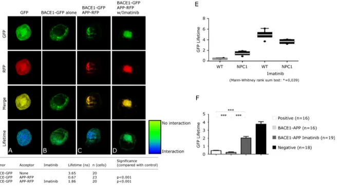
(51) c-Abl inhibition affects APP binding to BACE1 in a cellular NPC model In order to investigate the molecular mechanisms that are responsible for the reduced β-secretase APP processing when c-Abl is inhibited, we evaluated BACE1-APP interaction following inhibition of c-Abl.. We used FRET-FLIM to. verify BACE1-APP interaction in NPC cells. The capture of FRET was confirmed using NPC cells transfected with a GFP(donor)-RFP(acceptor) construct (Figure 5A; pseudocolor = blue). The lifetime of GFP attached to the C terminus of BACE1. (BACE1-GFP) in. the. absence. of an. acceptor (RFP), was. 3.65. nanoseconds (Figure 5B; pseudocolor = green). In NPC cells transfected with both BACE1-GFP (donor) and APP-RFP (acceptor), the lifetime was shortened to 0.67 nanoseconds, confirming the APP-BACE1 interaction (Figure 5C; pseudocolour = green/blue). Treatment with Imatinib reversed this lifetime shortening (Figure 5D; lifetime = 1.86 nanoseconds; pseudocolour = green), similar to the condition in which no acceptor was present. These data indicate that c-Abl inhibition decreases APP-BACE1 interaction. The pseudocolored FLIM image in green, particularly in the perinuclear region, where the interaction occurs, supports a loss of APP-BACE1 interaction (Figure 5D). In both cell models, Imatinib significantly impaired APP-BACE1 association, increasing the lifetime of the BACE1 donor (Figure 5E). Interestingly, although no significant differences were observed in APP-BACE1 interaction in NPC cells compared to WT cells, a trend towards a greater interaction was observed (Figure 5E). These results suggest that c-Abl activation in NPC cells favors APP-BACE1 interaction and that c-Abl inhibition decreases the proximity between APP and BACE1, reducing β-secretase-dependent amiloidogenic processing of APP.. . 38 .
(52) Figure 5. c-Abl inhibition impairs APP-BACE1 interaction in a cellular NPC model. (A-D) CHO NPC cells were transfected with BACE1-GFP and APPRFP and then treated with vehicle or Imatinib (10 µM). (E) Comparison of APPBACE1 interaction in CHO WT and NPC cells, with and without Imatinib treatment. (F) GFPs lifetimes are shown for BACE1-APP interaction in CHO WT cells. Mean and SEM of three independent experiments are shown.. . 39 .
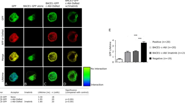
(53) These results sustain that c-Abl activity, and probably phosphorylation of APP Tyr682, are required for the intimate interaction of APP-BACE1 allowing APP cleavage by β-secretase. Also we evaluated APP-BACE1 association in WT cells by FLIM obtaining similar results (Figure 5F). We next sought to determine whether BACE1 interacts with c-Abl in NPC cells. First, the capture of FRET was confirmed by transfecting the cells with a GFP(donor)-RFP(acceptor) construct (Figure 6A; pseudocolor = blue/green). The lifetime of BACE1-GFP in the absence of acceptor (RFP) was 3.3 nanoseconds (Figure 6B; pseudocolour = green), whereas in the presence of the acceptor (c-Abl-DsRed), the lifetime was 1.95 nanoseconds (Figure 6C; pseudocolor = green), indicating that there is no interaction between c-Abl and BACE1.. Similarly,. the. lifetime. following. Imatinib. treatment. was. 1.8. nanoseconds (Figure 6D; pseudocolour = green). Similarly, we found no interaction between APP and c-Abl in WT cells (Figure 6E). Taken together, our findings show that the interaction of c-Abl with APP requires phosphorylation of APP on Tyr682. We propose a scenario whereby cAbl-APP interaction leads to APP phosphorylation on Tyr682, and that phosphorylated APP has an increased ability to interact with BACE1.. . 40 .
(54) Figure 6. c-Abl does not interact with BACE1 in a cellular NPC model. (A-D) BACE1-c-Abl interactions in CHO NPC cells were evaluated by FRETFLIM. Pseudo-coloured images and fluorescence lifetimes are shown for CHO NPC cells transfected with (A) a GFP-RFP fusion construct, (B) BACE1-GFP, (CD) BACE1-GFP and c-Abl-DsRed with (C) and without (D) Imatinib (10 µM) treatment. (E) GFPs lifetimes are shown for BACE1-c-Abl interaction in CHO WT cells. Mean and SEM of three independent experiments are shown.. . 41 .
(55) Discussion. Here, we report evidence of a direct interaction between APP and c-Abl. Using biochemical and FRET-FLIM analysis of cellular and mouse models of NPC, we show that this interaction involves the GY682ENPTY motif in the cytoplasmatic tail of APP and results in its phosphorylation. As a result, APP is directed towards the amyloidogenic pathway. Importantly, we demonstrate that this interaction can be reversed by treatment with the c-Abl inhibitor Imatinib. We propose. that. c-Abl. activity. promotes. the. BACE1-APP. interaction. via. phosphorylation of APP, thereby favoring Aβ accumulation and contributing to the pathogenesis of NPC disease. The data presented here could explain our previous findings showing decreased brain Aβ burden in a transgenic mouse model of Alzheimer’s disease when treated with Imatinib (Cancino, Toledo et al. 2008). It could also explain why NPC mouse and cellular models, which have active c-Abl (Alvarez, Klein et al. 2008), present increased Aβ production (Yamazaki, Chang et al. 2001, Jin, Shie et al. 2004). While some studies have linked c-Abl to APP cytosolic domain signaling (Zambrano, Bruni et al. 2001, Perkinton, Standen et al. 2004, Vazquez, Vargas et al. 2009), this is the first study that demonstrates that c-Abl can direct APP towards amyloidogenic processing. In this study, we used CHO NPC1-null cells and an NPC mouse model that have active c-Abl (Alvarez, Sandoval et al. 2004, Klein, Maldonado et al. 2011), prominent amyloidogenic processing and increased Aβ production. Therefore, they are suitable models to study APP processing and to investigate if c-Abl. . 42 .
Figure
+7
Documento similar
An increase in the ad-valorem commodity tax will increase output per firm, decrease the number of firms and decrease total output of oligopolistic firms if the inverse
In conclusion, the present study shows that high levels of anxiety and depression are associated with higher clinical variables (BMI and fat mass, %) and lower levels of anxiety
In 2H -NbS 2 , the strong decrease of the zero-temperature upper critical field below 8.7 GPa, associated with a slight increase in critical temperature, can be explained by a
These results correlated with our previous kinetics of arginase I expression in the hearts of both C57BL/6 and BALB/c mice (19) and indicated that during in vivo infection, arginase
Tumor cells (and other cells in the tumor) deplete nutrient levels (glucose, glutamine, amino acids, O 2 , etc.) in the TME, increase the levels of some metabolites, such as lactic
In diabetic mice treated with SOCS1 peptidomimetic the number of Iba-1 positive cells was significantly reduced in comparison with diabetic mice treated with vehicle (Figure
Stun- ningly, downregulation of NMDAR1 in HK-2 induced changes in the epithelial phenotype, evident as a decrease of E-cadherin (Figure 2, C and D) and an increase of
Similar to the results obtained in the genetic model, we observed a time- dependent decrease of the CD133+ cell fraction in ATR siRNA-treated cells along with a
