D
OCTORAL
T
HESIS
Tissue-scale, patient-specific modeling
and simulation of prostate cancer growth
GUILLERMO
LORENZO
GÓMEZ
SUPERVISOR
DR. HÉCTOR
GÓMEZ
DÍAZ
PROGRAMA DEDOUTORAMENTO ENENXEÑARÍACIVIL
This has been an intense and challenging journey, but also a unique opportunity. I had the pleasure to share the PhD experience with many people that have contributed to my research either directly or indirectly. These lines are intended to show my profound appreciation to them.
First of all, I would like to express my most sincere gratitude to my thesis advisor, Dr. Hector Gomez. Thank you for your continuous support and guidance, for your patience, for your encouragement and motivation, and for your wise advice. You have not only guided my first steps into research but also helped me to grow personally. It has been a privilege to have you as my academic father and I appreciate the good relationship that we have built over the last few years.
I am grateful to Dr. Michael Scott for accepting me for a research stay at Brigham Young University. I want to thank him and his team for their valuable collaboration to the work presented herein. Special thanks to Dr. Kevin Tew for his close assistance with the hierarchical adaptivity code. My deepest appreciation also goes to Dr. Alessandro Reali, who has welcomed me in his group at Univeristà degli Studi di Pavia and has actively supported and collaborated with me in the last part of my PhD. I would also like to extend my deepest gratitude to Dr. Thomas J.R. Hughes for his insightful comments and advice all along my PhD.
I would like to acknowledge Kent Beasley, MD, and the Austin Radiological Association (Austin, TX, USA) for providing anonymized medical images and reports of prostate cancer patients. I also acknowledge the Fulton Supercomputing Lab at Brigham Young University (Provo, UT, USA) and the Centro de Supercomputación de Galicia (Santiago de Compostela, Spain) for providing HPC resources that contributed to the results presented in this thesis.
thank you all for your invaluable help during my research, for creating an extraordinary environment in the lab, as well as for the pleasant time we spent out together. Special thanks to Alba López for assisting with the administrative work, for her continuous encouragement, and for our enjoyable conversations. Besides, I would like to extend this acknowledgment to those who temporarily joined our group: Pablo Orosa, Nati Velay, Miguel Burés, Sandra Fernández, Manuel Tasende, Esteban Sañudo, and Adrián López. I am also thankful to those other colleagues with whom I have shared time during my PhD both inside and outside the office. In particular, I would like to specially thank Iris, Iván, Miguel, Raquel, Luis, Sindi, Miriam, Gemma and Jorge. Furthermore, my appreciation also goes to my colleagues at PAIPUDC, with whom I collaborated to make the life of other PhD candidates at our university a bit better. Many thanks to Zaida, Adrián, and Marta for their active support and the good times together. Furthermore, I would like to thank the colleagues at Dr. Michael Scott’s and Dr. Alessandro Reali’s groups for welcoming me and sharing a nice time both inside and outside the lab.
Regardless of the remote corner of the world where they might be living, I also want to show my gratitude to my friends from my urban family and college. Thank you for making my life better through all this years. I am deeply thankful to Ana, Sarai, Cornide, Mallo, Inés, Ana del Valle, Anita, Gael, Ricardo, and Quico, whose constant support, encouragement, and advice have also contributed to this thesis. I feel priviliged to have you as friends.
Last but not least, I want to express my dearest gratitude to my parents, Moncho and Marisa, and my siblings, Ramón e Irene. Thank you for your unconditional support, love, and advice. Finally, thank you, Naya, for being an essential part of my life, for your understanding, and for your unlimited encouragement in every decision.
Prostate cancer is a major health problem among aging men worldwide. This pathology is easier to cure in its early stages, when it is still organ-confined. However, it hardly ever produces any symptom until it becomes excessively large or has invaded other tissues. Hence, the current approach to combat prostate cancer is a combination of prevention and regular screening for early detection. Indeed, most cases of prostate cancer are diagnosed and treated when it is localized within the organ. Despite the wealth of accumulated knowledge on the biological basis and clinical management of the disease, we lack a comprehensive theoretical model into which we can organize and understand the abundance of data on prostate cancer. Additionally, the standard clinical practice in oncology is largely based on statistical patterns, which is not sufficiently accurate to individualize the diagnosis, prediction of prognosis, treatment, and follow-up.
scale, patient-specific modeling and simulation of organ-confined PCa growth within the context of mathematical oncology. We present a model for localized prostate cancer growth that reproduces the growth patterns of the disease observed in experimental and clinical studies. To capture the coupled dynamics of healthy and tumoral tissue, we use the phase-field method together with reaction-diffusion equations for nutrient consumption and prostate specific antigen production. We leverage this model to run the first tissue-scale, patient-specific simulations of prostate cancer growth over the organ anatomy extracted from medical images. Our results show similar tumor progression as observed in clinical practice.
We leverage isogeometric analysis to handle the nonlinearity of our set of equations, as well as the complex anatomy of the prostate and the intricate tumoral morphologies. We further advocate dynamical mesh adaptivity to speed up calculations, rationalize computational resources, and facilitate simulation in a clinically relevant time. We present a set of efficient algorithms to accommodate localh-refinement andh-coarsening of hierarchical splines in isogeometric analysis. Our methods are based on Bézier projection, which we extend to hierarchical spline spaces. We also introduce a balance parameter to control the overlapping of basis functions across the levels of the hierarchy, leading to improved numerical conditioning. Our simulations of cancer growth show remarkable accuracy with very few degrees of freedom in comparison to the uniform mesh that the same simulation would require.
El cáncer de próstata es un gran problema de salud en hombres de edad avanzada en todo el mundo. Esta patología es más fácil de curar en sus estadios iniciales, cuando aún es órgano-confinada. Sin embargo, casi nunca produce ningún síntoma hasta que es demasiado grande o ha invadido otros tejidos. Por tanto, el enfoque actual para combatir el cáncer de próstata es una combinación de prevención y exámenes rutinarios para una detección precoz. De hecho, la mayoría de casos de cáncer de próstata son diagnosticados y tratados cuando aún está localizado dentro del órgano. A pesar de la riqueza del conocimiento acumulado sobre las bases biológicas y la gestión clínica de la enfermedad, carecemos de un modelo teórico completo en el que podamos organizar y comprender la enorme cantidad de datos existentes sobre el cáncer de próstata. Además, la práctica clínica estándar en oncología está basada en gran medida en patrones estadísticos, lo cual no es suficientemente preciso para individualizar el diagnóstico, la predicción de la prognosis, el tratamiento y el seguimiento.
modelos matemáticos. Finalmente, las tecnologías de oncología matemática pueden guiar las investigaciones futuras sobre cáncer de próstata, por ejemplo, proponiendo nuevas estrategias de tratamiento o descubriendo mecanismos involucrados en el crecimiento tumoral.
Por tanto, el objeto de esta tesis es proporcionar un marco computacional para la mo-delización y simulación del crecimiento del cáncer de próstata órgano-confinado de forma personalizada y a escala tisular dentro del contexto de la oncología matemática. Presentamos un modelo de crecimiento de cáncer de próstata localizado que reproduce los patrones de crecimiento de la enfermedad observados en estudios experimentales y clínicos. Para capturar las dinámicas acopladas de los tejidos sano y tumoral, usamos el método de campo de fase junto con ecuaciones de reacción-difusión para el consumo de nutriente y la producción de antígeno prostático específico. Empleamos este modelo para realizar las primeras simulaciones personalizadas a escala tisular del crecimiento de cáncer de próstata sobre la anatomía del órgano extraída de imágenes médicas. Nuestros resultados muestran una progresión tumoral similar a la observada en la práctica clínica.
Utilizamos el análisis isogeométrico para resolver la no-linealidad de nuestro sistema de ecuaciones, así como la compleja anatomía de la próstata y las intricadas morfologías tumorales. Adicionalmente, proponemos el uso de adaptatividad dinámica de malla para acelerar los cálculos, racionalizar los recursos computacionales y facilitar la simulación en un tiempo clínicamente relevante. Presentamos un conjunto de algoritmos eficientes para introducir el refinamiento y el engrosado locales tipohen análisis isogeométrico. Nuestros métodos están basados en la proyección de Bézier, que extendemos a los espacios de splines jerárquicas. También introducimos un parámetro de balance para controlar la superposición de funciones de base a través de los niveles de la jerarquía, lo cual conduce a un condicionamiento numérico mejorado. Nuestras simulaciones de crecimiento de cáncer muestran una notable precisión con muy pocos grados de libertad en comparación con la malla uniforme que la misma simulación requeriría.
O cancro de próstata é un gran problema de saúde en homes de idade avanzada en todo o mundo. Esta patoloxía é máis fácil de curar nos seus estadios iniciais, cando aínda é órgano-confinada. Porén, case nunca produce ningún síntoma ata que é demasiado grande ou ten invadido outros tecidos. Polo tanto, o enfoque actual para combater o cancro de próstata é unha combinación de prevención e exames rutinarios para unha detección precoz. De feito, a maioría de casos de cancro de próstata son diagnosticados e tratados cando aínda está localizado dentro do órgano. Malia a riqueza do coñecemento acumulado sobre as bases biolóxicas e a xestión clínica da doenza, carecemos dun modelo teórico completo no que podamos organizar e comprender a enorme cantidade de datos existentes sobre o cancro de próstata. Ademais, a práctica clínica estándar en oncoloxía está baseada en gran medida en patróns estatísticos, o cal non é suficientemente preciso para individualizar a diagnose, a predición da prognose, o tratamento e o seguimento.
sobre cancro de próstata, por exemplo, propoñendo novas estratexias de tratamento ou descubrindo mecanismos involucrados no crecemento tumoral.
Polo tanto, o obxecto desta tese é proporcionar un marco computacional para a mode-lización e simulación do crecemento do cancro de próstata órgano-confinado de forma personalizada e a escala tisular dentro do contexto da oncoloxía matemática. Presentamos un modelo de crecemento de cancro de próstata localizado que reproduce os patróns de crecemento da enfermidade observados en estudos experimentais e clínicos. Para capturar as dinámicas acopladas dos tecidos san e tumoral, usamos o método de campo de fase xunto con ecuacións de reacción-difusión para o consumo de nutriente e a produción de antíxeno prostático específico. Empregamos este modelo para realizar as primeiras simulacións personalizadas a escala tisular do crecemento de cancro de próstata sobre a anatomía do órgano extraída de imaxes médicas. Os nosos resultados amosan unha progresión tumoral similar á observada na práctica clínica.
Utilizamos a análise isoxeométrica para resolver a non-linealidade do noso sistema de ecuacións, así como a complexa anatomía da próstata e as intricadas morfoloxías tumorais. Adicionalmente, propoñemos o uso de adaptatividade dinámica de malla para acelerar os cálculos, racionalizar os recursos computacionais e facilitar a simulación nun tempo clinicamente relevante. Presentamos un conxunto de algoritmos eficientes para introducir o refinamento e o engrosado locais tipohen análise isoxeométrica. Os nosos métodos están baseados na proxección de Bézier, que estendemos aos espazos de splines xerárquicas. Tamén introducimos un parámetro de balance para controlar a superposición de funcións de base a través dos niveis da xerarquía, o cal conduce a un condicionamento numérico mellorado. As nosas simulacións de crecemento de cancro amosan unha notable precisión con moi poucos graos de liberdade en comparación coa malla uniforme que a mesma simulación requiriría.
Acknowledgments. . . i
Abstract . . . iii
Resumen. . . v
Resumo . . . vii
Contents . . . ix
List of Figures. . . xv
List of Tables . . . xix
List of Symbols . . . xxi
1 Introduction 1 1.1 Motivation. . . 1
1.2 Research objectives . . . 4
1.3 Thesis overview . . . 6
2 Background 9 2.1 The fundamentals of cancer. . . 9
2.2.2 Physiology of the prostate . . . 14
2.2.3 Prostate pathologies . . . 15
2.3 Benign prostatic hyperplasia . . . 16
2.3.1 Description . . . 16
2.3.2 Origin . . . 16
2.3.3 Development . . . 16
2.3.4 Clinical management . . . 18
2.3.4.1 Detection and diagnosis . . . 18
2.3.4.2 Treatment . . . 19
2.4 Prostate cancer . . . 19
2.4.1 Classification . . . 19
2.4.2 Origin . . . 20
2.4.3 Development . . . 20
2.4.4 Risk factors . . . 21
2.4.5 Clinical management . . . 23
2.4.5.1 Prevention . . . 23
2.4.5.2 Screening and detection . . . 24
2.4.5.3 Diagnosis and staging . . . 26
2.4.5.4 Treatment . . . 31
2.5 Mathematical modeling of cancer growth . . . 37
2.5.1 General concepts . . . 38
2.5.2 Classification of tumor growth models according to mathematical description . . . 39
2.5.3 The tumor mass effect . . . 40
2.6 The phase-field method . . . 41
2.7 Isogeometric analysis . . . 44
2.8 Bernstein bases . . . 46
2.8.1 Univariate Bernstein basis . . . 46
2.8.4 The Gramian of the Bernstein basis and its inverse . . . 48
2.9 B-splines and NURBS . . . 49
2.9.1 B-splines . . . 49
2.9.2 Non-uniform rational B-splines (NURBS) . . . 51
2.10 Hierarchical splines . . . 52
2.10.1 Hierarchical spline spaces . . . 52
2.10.2 The geometric map for hierarchical splines . . . 53
2.10.3 Bézier extraction . . . 53
2.10.3.1 Bézier elements . . . 54
2.10.3.2 Hierarchical extraction operator . . . 55
3 A computational approach for the tissue-scale, patient-specific prediction of PCa growth 57 3.1 Introduction . . . 57
3.2 A mathematical oncology framework for PCa . . . 58
3.3 Mathematical model . . . 58
3.4 Patient data . . . 62
3.5 Numerical methods . . . 63
3.6 Simulation of PCa growth. . . 64
3.6.1 Growth patterns of localized PCa . . . 64
3.6.2 Patient-specific simulation . . . 69
3.6.3 Tissue and serum PSA . . . 72
3.7 Conclusions . . . 74
4 Hierarchical adaptivity strategies for the resolution of phase-field models of PCa growth 75 4.1 Introduction . . . 75
4.2 Hierarchical reconstruction operator . . . 76
4.3 Hierarchical refinement . . . 77
4.6 Application to a phase-field model of PCa growth . . . 80
4.6.1 Spatial discretization . . . 82
4.6.2 Time integration . . . 83
4.6.3 Automatic adaptivity . . . 83
4.6.3.1 Indicator . . . 83
4.6.3.2 Time step offsets. . . 84
4.6.3.3 Adaptive solver . . . 84
4.6.4 Initial conditions . . . 85
4.6.5 PCa growth in a square domain . . . 85
4.6.6 PCa growth in a cubic domain . . . 95
4.6.7 Tissue-scale, patient-specific simulation of PCa growth . . . 97
4.7 Conclusions . . . 99
5 A mechanically-coupled model for PCa growth and interaction with BPH 103 5.1 Introduction . . . 103
5.2 Patient data . . . 104
5.3 Mathematical model . . . 104
5.4 Numerical methods . . . 109
5.5 Deformation of the prostate due to BPH . . . 110
5.6 Deformation of the prostate due to PCa . . . 110
5.7 BPH hampers PCa growth . . . 114
5.8 Conclusions . . . 118
6 Summary, conclusions, and future work 121 6.1 Summary . . . 121
6.2 General conclusions. . . 123
6.3 Future work . . . 126
A.1 Articles in peer-reviewed international journals . . . 131
A.2 Contributions in national and international conferences . . . 132
B Collaboration in other works 133 B.1 Prostate Meshes for Isogeometric Analysis. . . 133
B.2 The equations of cancer. . . 135
B.3 Ongoing research on computational methods for prostate cancer . . . . 135
C Extended summary in Spanish 139 C.1 Introducción. . . 139
C.2 Objetivos . . . 143
C.3 Contribuciones de la tesis . . . 145
C.4 Conclusiones . . . 146
C.5 Futuras líneas de trabajo . . . 151
D Extended summary in Galician 155 D.1 Introdución . . . 155
D.2 Obxectivos . . . 159
D.3 Contribucións da tese . . . 160
D.4 Conclusións . . . 162
D.5 Futuras liñas de traballo. . . 166
2.1 The hallmarks of cancer. . . 11
2.2 The spatial and time scales of cancer . . . 12
2.3 Anatomic location of the prostate. . . 13
2.4 Local anatomy of the prostate. . . 14
2.5 Benign prostatic hyperplasia . . . 17
2.6 Current standard for the clinical management of PCa . . . 22
2.7 The Gleason scale. . . 27
2.8 The TNM system . . . 30
2.9 Sharp interface versus diffuse interface models . . . 42
2.10 Bernstein basis functions . . . 46
2.11 B-spline basis functions. . . 50
2.12 Basis functions for a univariate quadratic hierarchical spline space . . . 54
2.13 The Bézier mesh for a two-dimensional hierarchical spline space and the distribution of the elements inHEacross the hierarchical levels. . . 56
3.3 Estimation of tissue PSA parameters . . . 63
3.4 Extraction of the geometry of the prostate and the initial tumor from CT data . . . 65
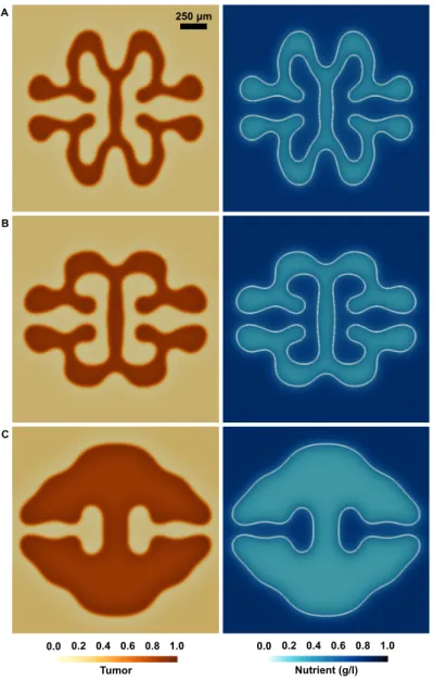
3.5 Different growth morphologies adopted by initially spheroidal prostatic tumors . . . 66
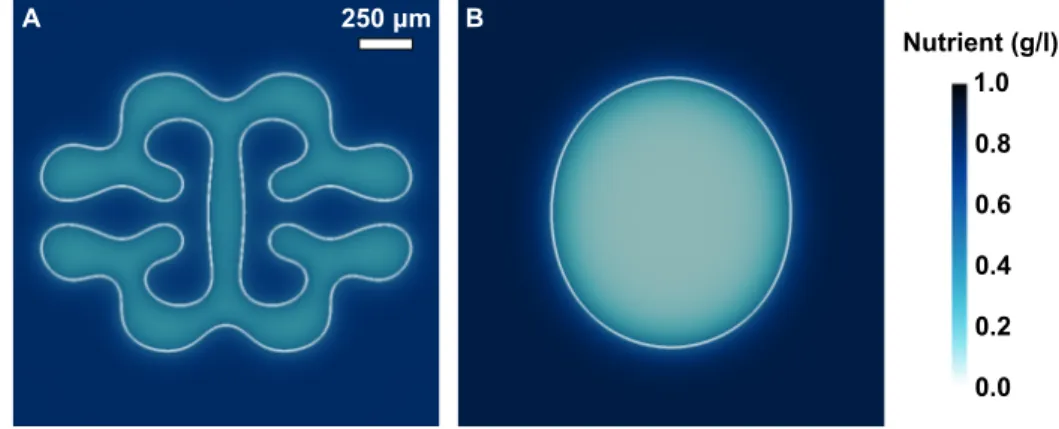
3.6 Nutrient distribution in PCa morphologies . . . 67
3.7 Details on the onset and evolution of the shape instability . . . 68
3.8 Fingered morphology of PCa growth in the presence of decreased and increased nutrient supply . . . 70
3.9 Tissue-scale, patient-specific simulation of PCa growth . . . 71
3.10 Tissue PSA distribution across the prostate gland att=0.5 years, tumor volume evolution and serum PSA history. . . 73
4.1 Bézier extraction operatorCeand reconstruction operatorRefor a bilin-ear hierarchical spline . . . 77
4.2 Ring balancing of a bilinear hierarchical mesh over an elementefor
nrb=1 andnrb=2 . . . 81
4.3 Function support balancing of a bilinear hierarchical mesh over an ele-mentefornf sb=1 andnf sb=2 . . . 81
4.4 Tumor phase field and adaptive mesh for a simulation of 2D PCa growth using a quadratic hierarchical B-spline space with 5 levels and balance parameternf sb=1 . . . 87
4.5 Tumor phase field and adaptive mesh for a simulation of 2D PCa growth using a quadratic hierarchical B-spline space with 5 levels and balance parameternf sb=2. . . 88
4.6 The distribution of elements across the hierarchical levels and the corre-sponding Bézier meshes fornf sb=1 andnf sb=2 att=0.4 years . . . 89
4.7 Analysis of the number of hierarchical basis functions (HBF) and ele-ments along the simulations in Fig. 4.4 (nf sb=1) and Fig. 4.5 (nf sb=2)
. . . 90
4.7 (Continued) Analysis of the number of hierarchical basis functions (HBF) and elements along the simulations in Fig. 4.4 and Fig. 4.5 . . . 91
4.10 Our adaptive algorithms are compatible with globalk-refinement.. . . . 94
4.11 Tumor phase field and adaptive mesh for a simulation of 3D PCa growth using a quadratic hierarchical B-spline space with 5 levels and balance parameternf sb=1. . . 96
4.11 (Continued) Tumor phase field and adaptive mesh for a simulation of 3D PCa growth using a quadratic hierarchical B-spline space with 5 levels and balance parameternf sb=1 . . . 97
4.12 Patient-specific, tissue scale simulation of PCa growth (posterior view). 100
4.13 Patient-specific, tissue scale simulation of PCa growth (posterior view). 101
5.1 Patient-specific local anatomy of the prostate . . . 105
5.2 Deformation of the prostate caused by BPH over 1 year . . . 110
5.3 Detail of the deformation of the prostate caused by BPH over 1 year . . 111
5.4 Analysis of prostate deformation caused by artificial tumors in different locations. . . 112
5.5 Detail of prostate deformation caused by artificial tumors in different locations. . . 113
5.6 BPH hampers PCa growth . . . 115
5.7 Prostate deformation caused by the patient’s tumor with and without considering BPH . . . 116
4.1 Parameters in the PCa growth model: square and cubic domain simulations 86
4.2 Parameters in the PCa growth model: patient-specific, tissue-scale simu-lation . . . 98
Main abbreviations
ASG Analysis suitable geometries BPH Benign prostatic hyperplasia CAD Computer aided design CG Central gland of the prostate
CT Computed tomography
DRE Digital rectal examination FEA Finite element analysis
GS Gleason score
IGA Isogeometric analysis
mpMR Multiparametric magnetic resonance
MR Magnetic resonance
NURBS Non-uniform rational B-splines PCa Prostate cancer
PIA Proliferative inflammatory atrophy PIN Prostatic intraepithelial neoplasia PSA Prostate specific antigen
PZ Peripheral zone of the prostate TRUS Transrectal ultrasound
TURP Transurethral resection of the prostate
φ Tumor phase field
η Nutrient
ρ Tissue PSA
P Serum PSA
t Time
x Position vector
u Displacement vector
σσσ Stress tensor
σv Von Mises stress
σh Hydrostatic stress
Main parameters of the mathematical models
Dφ Diffusivity of the phase field
τ Time scale for the phase field χ Nutrient-induced tumor growth rate
A Apoptosis rate
Dη Nutrient diffusivity
S Nutrient supply
δ Nutrient consumption rate
γη Nutrient natural decay rate Dρ Tissue PSA diffusivity
αh Tissue PSA production rate in healthy tissue
αc Tissue PSA production rate in cancerous tissue
γρ Tissue PSA natural decay rate
M Mechanotransductive coupling function
β1,β2 Mechanotransductive empirical constants
λ,µ Lamé parameters
κ Compressive pressure exerted by the tumor
K Bulk modulus
g Estimated linear volumetric growth of the prostate due to BPH VMRI Volume of the prostate on T2-weighted MR images
ζ Adjusting coefficient to estimate CG growth rate kw Winkler constant
Main notation in numerical methods
p Polynomial degree
B Vector of Bernstein basis functions
A Transformation matrix between Bernstein spaces over different intervals
G Gramian matrix
∂Ω Boundary of the physical space
N B-spline basis function
N Functional space spanned by a B-spline basis
N Vector of B-spline basis functions
P Vector of spline control points
wA NURBS weight
w(s) NURBS weighting function
R NURBS basis function
R Vector of NURBS basis functions nh Number of hierarchical levels
α Level of the hierarchy
H Set of hierarchical spline basis functions
nf Number of functions in the hierarchical spline basisH
H Functional space spanned by a hierarchical spline basis ng Number of geometric blending functions
e Bézier element
Eα Set of Bézier elements corresponding to the spline basis in levelα
HE Set of Bézier elements corresponding to a hierarchical spline basis ne Number of Bézier elements inHE
ˆ
Ωe Parametric domain of elemente Ωe Physical domain of elemente
He Vector of hierarchical basis functions with support over elemente Ce Bézier extraction operator of elemente
Re Reconstruction operator of elemente
Le Transmission refinement operator of elemente Pe Vector of spline control points of elemente Qe Vector of Bézier control points of elemente
M Refinement operator
nrb Ring balance parameter
nf sb Function support balance parameter
CR Coefficient to control the intensity of refinement
CC Coefficient to control the intensity of coarsening
∆T SR Time step offset to perform refinement
1
Introduction
1.1 Motivation
According to the World Health Organization (WHO), prostate cancer (PCa) is the second most common cancer and the fifth leading cause of death from cancer among men worldwide [89]. The data are revealing: in 2012, there was an estimate of 1,095,000 new cases and 308,000 deaths worldwide associated with this cancer and it will be responsible for about 161,360 new cases and 26,730 deaths only in the U.S.A. in 2017 [5]. Additionally, national epidemiologic studies in Spain revealed an estimate of 33,370 new PCa cases in 2015 and 5,855 deaths from this disease in 2014 [96,224].
The development of a prostate adenocarcinoma requires a gradual accumulation of mutations in a number of different genes, which varies from patient to patient, but is usually at least seven [259,265]. Thus, PCa is more frequent among older men. As a result of these alterations of the genome over years, an initial moderate disorder of cell behavior evolves gradually towards an advanced cancer. As the tumor develops, it becomes more malignant and cell differentiation decreases. This evolutive phenomenon is called tumor progression and, in general, comprises four steps: (1) local overproliferation of cells, (2) extensive overproliferation of cells, (3) invasion of the surrounding tissues and, finally, (4) metastasis, which is the process whereby some malignant cells escape from the original tumor, enter the bloodstream and migrate to other tissue, which they invade and colonize [3,259,265].
Medical practice for PCa has been developed upon the aforementioned genetic and biological bases as well as on the accumulated experience of physicians treating this disease [259,265]. The current medical protocols include all these sources of knowledge on PCa in the form of statistics for the probability of cancer stage and treatment success. In brief, PCa is easier to cure in its early stages, before it becomes excessively aggressive and spreads out of the prostate but, unfortunately, this disease hardly ever produces any symptom until the tumor is either very large or has invaded other tissues. Therefore, the best way to combat PCa is a combination of prevention and regular screening for early detection. PCa is usually diagnosed and treated when it is still localized within the prostate. Localized PCa is usually managed with a curative approach such as surgery or radiotherapy, while advanced PCa is normally addressed with palliative strategies based on hormonal therapy or chemotherapy. Patients with low-risk PCa may delay their treatment until the tumor reaches an optimal stage (active surveillance). Patients that are not eligible for local curative treatment, or those with a short life expectancy, may benefit from a deferred palliative treatment, aimed at tackling specific symptoms of the disease, while they continue on regular screening (watchful waiting).
Apart from PCa, the prostate is also the site of other major pathology affecting older men: benign prostatic hyperplasia (BPH) [259,265]. This disease consists of the pathological enlargement of the prostate with age, with a prevalence increasing from 50% in men in their fifties to about 70% in men in their seventies [26]. BPH obstructs urinary and ejaculatory flow, hence causing bothersome lower-urinary tract symptoms. PCa and BPH may coexist in the same patient and share numerous similarities [4,191,265], which have suggested several connections or some interplay between them. However, solid evidence confirming their existence is lacking. Recent studies on extensive series of surgical prostate specimens have shown that tumors originating in larger prostates present favorable pathological features [36,55,92,126,145,184]. These studies provide solid evidence to suggest that large prostates may exert a protective effect against PCa.
how tumors originate, grow, and spread within the body, also analyzing new options for PCa treatment. Despite the number of experimental and clinical investigations, we lack a comprehensive theoretical model into which all this abundance of data can be understood and organized, also guiding future research efforts efficiently so as to enrich such model. The emphasis of these investigations, as well as the standard clinical practice in oncology, are based on statistical patterns, which is not sufficiently accurate for individualized diagnosis, estimation of prognosis, treatment, and follow-up.
Alternatively,predictive medicine[231,232] is an interdisciplinary approach whose aims are the determination of patient-specific disease progression and optimal treatment. Methods of predictive medicine are based on mathematical modeling and computer simulation of both the disease and treatments. This new approach complements the statistical and experiential basis of diagnosis and treatment planning, which constitute current medical practice. In the particular case of cancer research,mathematical oncology [7,53,97,188,274] is a new and promising field devoted to the development of models and computational tools to simulate tumor growth and cancer treatment. Although significant progress has been made in recent decades, accurately modeling the growth of cancerous tumors remains an outstanding challenge [7,9,42,52,53,97,160,167,170,
174,188,206,211,212,236,251,274].
The nonlinearity that usually characterizes the set of equations comprising tumor growth models, the complex geometry of patient-specific anatomical models, and the intricate morpohologies that growing tumors may exhibit demand very efficient methods of res-olution in terms of memory and time of computation. Isogeometric analysis (IGA) is a rapidly growing and cutting-edge technology that can handle all these computational challenges [66,130]. IGA can be seen as a generalization of the classic Finite Ele-ment Analysis (FEA) [132]. Instead of using standard piecewise polynomials, IGA leverages richer functions coming from computer graphics and computational geometry, such as B-splines [66,84,130,195,204], Non-Uniform Rational B-splines (NURBS) [66,84,130,195,204], and T-splines [18,21,22,154]. Isogeometric methods based on these functional spaces are geometrically exact, provide enhanced accuracy per degree of freedom, and enable higher global continuity (Cp−1for spline spaces with polynomial degree p) [1,66]. Additionally, IGA is compatible with dynamic mesh-adaptivity tech-niques based on the concept of hierarchical refinement and coarsening [215,219,257], which enables the local adaptation of the computational mesh dynamically throughout the simulation. Hence, the mesh features finer elements with richer approximation functions close to the tumor, and coarser elements elsewhere. This leads to dramatic savings of memory and compute time. Thus, dynamic mesh-adaptivity techniques hold the potential to speed up the calculations and create isogeometric methods that facilitate obtaining solutions in a clinically relevant time.
tissue-scale, patient-specific modeling of cancer growth for several reasons:
• PCa is a major health problem, and a paradigmatic example in which a predictive model could make a difference in clinical practice because there are currently many patients being overtreated and undertreated [164,260].
• Medical imaging is emerging as a diagnostic tool in PCa. Medical imaging is not widely used in PCa diagnosis due to insufficient confidence in current imaging modalities. However, multiparametric magnetic resonance (mpMR) imaging is commonly used to monitor the evolution of the disesase and is emerging as a promising technology that might be also used for PCa diagnosis [51,75,87,111,
116,123,153,227,238].
• The prostate is a small organ, typically between 20 and 80 cc [24,26,31,259,265,
270], so it is feasible to create a patient-specific anatomical model of the prostate from medical images to perform predictive simulations.
• The growth of the tumor in a given patient may be estimated using the concentration of a PCa biomarker known as prostate specific antigen (PSA), which is measured with a blood test and plays a crucial role during the diagnosis, treatment selection, and follow-up of the patients [253,254,259,265].
• Some PCa patients do not receive any treatment but have their PSA monitored and are periodically imaged, for instance, during the time between diagnosis and treatment or in active surveillance and watchful waiting protocols. This potentially may open the door toin vivomodel validation [261,274].
• IGA provides a rich computational framework that guarantees the required accuracy and efficiency to perform predictive tissue-scale, patient-specific simulations of PCa growth.
• Mathematical oncology is also a promising approach to investigate the relationship between PCa and BPH because it can provide solid mechanisms of interaction to explain the current evidence and to guide further clinical research.
1.2 Research objectives
u
Definition of a computational framework for the clinical management of PCa.Our first objective will be to devise a framework for the clinical management of PCa in which the technologies of mathematical oncology complement the standard medical protocols for this condition. For this purpose, we reviewed the current knowledge on the biological basis of prostatic tumors, the standard procedures in the clinical management of PCa, and the recent developments in mathematical modeling and simulation of tumor growth. We also reviewed the current and developing technologies of medical imaging for PCa because they provide a wealth of patient-specific data to inform a computational tool for the prediction of tumor growth.
u
Design of a tissue-scale, patient-specific mathematical model of PCa growth.We aim at defining a tissue-scale, patient-specific mathematical model to describe localized PCa growth, for this is the stage at which it is normally diagnosed and treated. The definition of the model will be based on the current biological and medical knowledge of this condition. We will identify and mathematically and physically describe the fundamental mechanisms that enable the construction of a model for prostatic tumor growth. We will require the model to capture the coupled dynamics of healthy and tumoral tissue and to describe the patient’s PSA dynamics. Additionally, we will study the incorporation of the dynamics of other key substances in PCa growth to the model.
u
Development and implementation of algorithms to solve the equations in themodel and construct patient-specific prostate meshes. To perform numerical
simulations, we will develop algorithms based on the concept of IGA. This is an ideal technology to handle the nonlinearity that characterizes tumor growth problems, the complex anatomy of the prostate, and the intricate morphologies of PCa in an efficient and accurate manner. We will also explore dynamic mesh adaptivity techniques based on hierarchical spline spaces to rationalize the required computational resources, speed up the calculations, and create a clinically relevant technology. We will utilize our own libraries or available packages (e.g.: Trilinos [120], PETSc [12], PetIGA [70]) together with message passing interface (MPI) to obtain a powerful and flexible parallel implementation of our algorithms. A computational approach for the tissue-scale, patient-specific prediction of PCa growth requires an accurate mesh of the patient’s prostate. Hence, we will also develop an algorithm to extract a NURBS mesh from the patient’s medical images, such as computed tomography (CT) and magnetic resonance (MR) images. This mesh can be globally and/or locally refined afterwards to perform simulations with a good level of accuracy.
u
Extension of the model to include the mechanical deformation of the prostatediseases that may coexist in the same patient and deform the prostate over time. The idea is to couple our mathematical model for PCa growth with the equations of mechanical equilibrium in order to compute the displacement and stress fields created by the growing tumor and BPH. We will also introduce a term in the equation describing tumor dynamics to account for the inhibitory effect of the stress fields generated by PCa and BPH. Leveraging this mechanically-coupled model, we will study the deformation of the prostate induced by PCa and BPH both independently and simultaneously. We will also utilize this model to analyze how the deformational state of the prostate affects tumor growth and investigate why larger prostates are less prone to tumors showing an aggressive demeanor and adverse pathological features. Additionally, we will discuss several applications of a mechanically-coupled model within a computational framework for the clinical management of PCa.
u
Calibration of the model and numerical methods to reproduce PCa growthas observed in clinical practice efficiently and accurately.We will analyze the
value range of the parameters in the equations of our mathematical models and numerical methods in order to reproduce the growth morphologies and clinical features that characterize PCa growth in medical practice in an accurate and efficient manner.
1.3 Thesis overview
Following this introduction, Chapter2presents a brief review of the current knowledge that provided the basis for the research work presented herein, from the biological mechanisms and clinical management of PCa to the latest advances in mathematical modeling of cancer and IGA.
In Chapter 3, we present a continuous model that reproduces the growth patterns of localized PCa, as this is the stage at which most prostatic tumors are diagnosed and treated. This model reproduces the common clinical morphologies of PCa and the transition between them based on nutrient availability and tumor metabolism. We also used our model to perform the first tissue-scale, patient-specific simulation of PCa growth based on a patient’s prostatic anatomy extracted from CT images.
simple 2D and 3D domains as well as a more compelling tissue-scale, patient-specific simulation.
In Chapter5, we extend our model of PCa growth to include the equations of mechanical equilibrium and define the coupling terms between them and tumor dynamics. We use this mechanically-coupled model to explore the prostate deformation and the inhibition of prostatic tumor growth due to mechanical stress fields caused by both the tumor and BPH. This study provided a feasible mechanism to explain an unresolved clinical debate: why tumors developing in larger prostates tend to be less aggressive.
2
Background
2.1 The fundamentals of cancer
With the generic name of cancer we refer to a vast host of diseases [3]. The different types of cancer are classified and named depending on the type of cell within each tissue of our body that brings forth the condition. The most common group of cancers in human beings arecarcinomas, which originate in epithelial tissues, i.e., those tissues that internally line the organs, cavities, and ducts in our body. Other common types of cancer are: melanomas, which originate in skin cells; lymphomas and leukimias, which are derived from blood cells or their precursors; sarcomas, which originate in the cells that constitute connective tissues and muscles; and cancers derived from cells of the nervous system, such as glioma and glioblastoma multiforme.
apoptosis, to avoid the propagation of genetic errors if necessary. However, some genetic alterations might escape this control process and some of them may even confer the cancerous cell the ability to avoid this DNA quality control mechanisms.
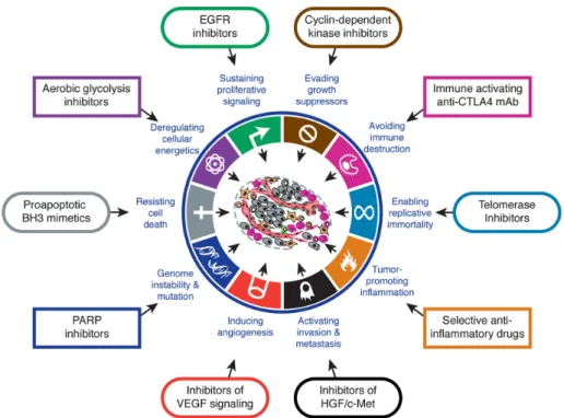
Cancerous cells present an asocial and competitive behavior within the organized cellular architecture of the tissues in our body. This demeanor is characterized by a high rate of net proliferation and invasiveness (see Fig.2.1). Most of the tissues of our body undergo continuous cell turnover under a rigorous balance between cell division and cell death. The increase in the net proliferation cell in tumors can be caused by genetic alterations that produce a direct increment in cell proliferation, a reduction in cell death rate, or a combination of both. A deficient DNA quality control combined with a high net proliferation rate promotes the gradual accumulation of more mutations and epigenetic changes. Hence, this genetic drift contributes to increase the aggressiveness of cancer and grants cancerous cells other abilities that characterize them (see Fig.2.1), such as conveniently adjusting their metabolism, resisting the attacks from the body immune system, enabling replicative immortality, promoting favorable tumor environment (e.g.: inflammation), and promote the formation of new blood vessels to nourish the tumor, a process called angiogenesis [112,113].
Angiogenesis begins in a situation of hypoxia, which is characterized by a low con-centration of oxygen and nutrients that limits tumor growth. In such situation, tumor cells produce a proangiogenic factor. This substance activates the tip endothelial cells present in neighboring blood vessels, which change their quiescent phenotype to become migratory. These cells begin their migration guided by chemotactic mechanisms towards hypoxic regions and creating new capillaries as they advance. The incipient blood vessels grow by cell proliferation and connect among themselves in order to bring forth a vascular network that supplies oxygen and nutrients to the hypoxic cancerous cells, hence favoring tumor growth [45].
Some malignant cells may acquire the ability to leave the tumor, enter the blood or lymphatic vessels, migrate to other place in the body and create a secondary tumor or metastasis. The ability to colonize a different tissue and thrive in it requires the alteration of certain genes too. Additionally, angiogenesis creates capillaries with different diameters, multiple branches, and high permeability, which might constitute an entrance to the bloodstream for these independent cells. Thus, tumor growth does not only jeopardize the structure and function of the organ where it originates, but might also endanger other tissues in our body.
Figure 2.1. The hallmarks of cancer. This figure illustrates the characteristic capabilities of growing cancers and examples of therapeutic drugs that have been developed to combat them. Reproduced from [112].
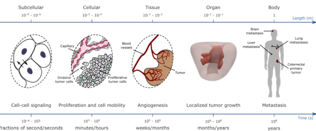
Therefore, tumor growth takes place at different spatial scales, from the initial proliferative mass, measured in microns, to metastatic stage, featuring several tumor masses of various millimeters or centimeters. As shown in Fig.2.2, the processes involved in tumor growth also occur at very different spatial scales: subcellular, such as mutations and metabolism; cellular, such as cell division and intercellular adhesion; tissue, such as angiogenesis and invasion; and corporal, such as metastasis. Additionally, the time scales of these processes span several orders of magnitude (see Fig.2.2). While cell metabolism takes place in fractions of a second, cell division may take hours, and the invasion of tissues prolongs over months or even years. The variability of the spatial and time scales of the processes involved in tumor growth is one of the central challenges to study cancer.
10⁻ - 10² 105 - 106
Length (m)
Time (s)
10⁻9 - 10⁻6
Subcellular Cellular Tissue Organ
fractions of second/seconds minutes/hours weeks/months months/years
Capillary
Cell-cell signaling Proliferation and cell mobility Angiogenesis Localized tumor growth Metastasis
Figure 2.2. The spatial and time scales of cancer.The biological and physiological processes involved in tumor growth span a wide range of spatial and time scales. This is one of the central challenges to investigate these pathologies. Adapted from [167].
different tumor cellular types can also collaborate via cell specialization. In any case, the structure, global behavior, and evolution of a tumor depend on the genetic alterations that originated it and supported its development. For a thorough review on the biological bases of cancer, the interested reader is referred to references [3,45,112,113].
2.2 The prostate
2.2.1 Anatomy of the prostate
The prostate is a tiny gland, whose volume is approximately 20 cc in healthy men [26,259,265]. It lies in the intersection of men’s urinary and reproductive tracts, deeply confined in the pelvic area (see Fig.2.3). Consequently, the prostate is surrounded by vulnerable structures, such as the bladder, the rectum, the sphincters, several arteries and veins, and a host of delicate nerves. The prostate is often described as an inverted pyramidal body that surrounds the urethra from its base at the bladder wall to its apex in contact with the membranous urethra. Though this organ has a capsule that protects it, the prostate is still poorly insulated [259,265].
Figure 2.3. Anatomic location of the prostate. The prostate is a tiny organ placed deeply within the pelvic area at the intersection of the male urinary and reproductive tracts. It is surrounded by several delicate organs and tissues, as shown in this figure. Provided by the National Cancer Institute of the USA at
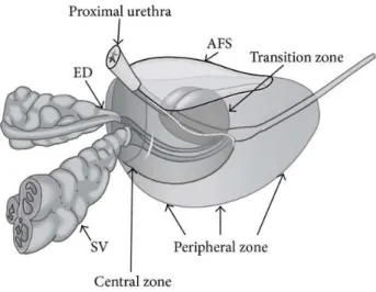
Figure 2.4. Local anatomy of the prostate.ED: ejaculatory ducts; SV: seminal vesicles; AFS: anterior fibromuscular stroma. The periurethral glands surround the proximal part of the prostatic urethra. Reproduced from [28].
From an histological perspective, the prostate is divided into five anatomical regions: peripheral zone (PZ), transition zone, central zone, anterior fibromuscular stroma, and periurethral glands [62,252,259,265]. These zones are depicted in Fig.2.4. While the transition zone and the periurethral glands surround the proximal part of the prostatic urethra, the central zone surrounds them and the ejaculatory ducts posterosuperiorly. In turn, the PZ encloses the central zone and the distal prostatic urethra except anteriorly, where there is a region of fibromuscular stroma. The glandular tissue of the prostate is distributed among the PZ (70%), the central zone (25%), the transition zone (5%), and the periurethral glands (<1%). The PZ and the transition zone are very similar histologically.
2.2.2 Physiology of the prostate
We call semen to the ejaculate which is made of sperm, made in the testes, and fluid, which is composed by a number of substances made in the prostate and mostly in the seminal vesicles [259,265]. This fluid is the most abundant component of semen in terms of volume.
admixture, semen is immediately pumped into the penis toward the exterior thanks to muscular action through the final section of the male urinary tract.
These prostatic secretions sustain sperm to survive their trip and remain active as long as possible. After ejaculation, the sperm immediately coagulates inside the vagina, lingering therein to increase the odds of reproduction. After a certain lapse of time, the process of breaking down again is due to the PSA, a prostatic enzyme with a major role in the the clinical management of PCa. Infections in the reproductive tract can produce scar tissue in the ducts that drain the testes, a situation that might cause infertility. The substances produced in the prostate also protect the urinary tract and the reproductive system from harmful bacteria or hazardous substances that may enter the urethra.
The prostate depends on hormones for its development, growth, and function. The hor-monal activation of the prostate takes place during orgasm. First of all, the hypothalamus, within the brain, produces the luteinizing hormone-releasing hormone. This hormone travels to the nearby pituitary gland, where the luteinizing hormone is effectively pro-duced. The luteinizing hormone controls the testes and once it reaches them, it boosts the production of testosterone. This hormone is also produced in the adrenal glands, placed on top of the kidneys. Testosterone is considered the chief male hormone because it is the cause of fertility and secondary sex characteristics, such as body hair and deep voice. Testosterone circulates in the bloodstream and, eventually, seeps into a prostate cell by diffusion. Then, the combination of testosterone and an enzyme called 5-α reductase gives raise to dihydrotestosterone. Finally, the combination of dihydrotestosterone and a certain protein in prostatic cells’ nucleus activates the required genes that are behind the production of the prostatic secretions.
2.2.3 Prostate pathologies
The prostate is the source of three major health problems in men: PCa, BPH, and prostatitis [259,265]. Indeed, the same man may endure more than one of these conditions during his lifetime, even at the same time and more than once. However, a wide array of treatments are available for these three diseases and continuous research is being carried out to improve them and derive new alternatives, which aim at reducing the side effects and optimizing both sensitivity and specificity.
prostatitis present similar symptoms to chronic bacterial prostatitis, but they are not originated by a bacterial infection. Indeed, the origin of these variants of prostatitis is not clear yet. Asymptomatic prostatitis produces no symptoms and is found by chance during a biopsy or the examination of the removed tissue samples obtained in a surgical procedure. This type of prostatitis is thought to be linked to the early stages of PCa.
2.3 Benign prostatic hyperplasia
2.3.1 Description
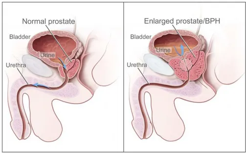
BPH consists of the pathological enlargement of the prostate with age, with a prevalence increasing from 50% in men in their fifties to about 70% in men in their seventies [26,259,265]. BPH may arise in the transition zone or the periurethral glands of the prostate. This condition is caused by an imbalance between proliferation and programmed cell death, which leads to an increase in cell number. The growing tissue constricts the urethra and may obstruct urinary flow, hence causing bothersome lower-urinary tract symptoms. The difference between a healthy prostate and and BPH is depicted in Fig.2.5.
2.3.2 Origin
There is not a clear origin for the BPH [259,265]. This condition has been associated with the imbalance of androgens and estrogens that comes with aging, due to the empowering effects of estrogens on testosterone and a decrease only in testosterone in older men. Alternatively, growth factors have been also thought to be the cause of this disorder, possibly those produced by the stromal cells in the prostate. These growth factors may alter the normal life span of the prostatic cells. This results in a decrease in the cell death rate with no variation in cell births, hence breaking the natural homeostatic balance between both rates and provoking the abnormal growth.
Additionally, around 7% of men are prone to BPH due to certain inherited genes. Plus, a series of genes seems to be altered both in BPH and PCa, so maybe there is a connection between both diseases which worths investigating in order to shed light on the origin and development of both disorders [4,191,259,265].
2.3.3 Development
Figure 2.5. Benign prostatic hyperplasia. This pathology provokes the enlargement of the prostate, which presses on the bladder and the urethra hence obstructing the flow of urine. Provided by the National Cancer Institute of the USA athttps://visualsonline.cancer.gov/.
urethral obstruction. As the disease develops, it may cause initial irritating but still tolerable symptoms. However, when it progresses beyond a certain point of nuisance, when the bladder is never empty or when the bladder and the kidneys become damaged, it needs to be treated.
During BPH, the affected tissue grows developing bulbous nodules that form character-istic clusters or lobes. The muscular tissue grows organizing its spatial configuration according to the lobe growth. These lobes arrange themselves in three main configura-tions: lateral lobe enlargement, which takes place at both sides of the urethra and causes mild obstruction; middle lobe enlargement, which occurs around the bladder neck and causes a great difficulty in urination; and trilobar enlargement, which is a combination of both, i.e., obstruction takes place both around the urethra and the bladder neck.
becomes unstable and overly reactive, what usually leads to incontinence and nocturia.
2.3.4 Clinical management
2.3.4.1 Detection and diagnosis
The detection of BPH is based on digital rectal examination (DRE) and a PSA test [259,
265]. Because these two tests are the baseline of PCa screening, we will discuss them in Section2.4. The DRE relies on the experience of the physician performing it. False negatives may be caused by the inner growth of the lobes, when they can’t be felt by the doctor. The prostate size can also be measured more accurately with medical imaging, such as ultrasounds (US) or MR. However, the degree of symptoms does not only depend on the size of the prostate, but also on the particular type of BPH growth (see Section2.3.3).
As BPH does not develop in the PZ, the glandular prostate is segmented into two zones in radiological studies: the PZ and the central gland (CG), which contains the central, transition, and periurethral zones [62,252,265]. The PZ appears as an hyperintense region in T2-weighted MR imaging due to the abundance of glandular components with sparsely interwoven smooth muscle. As BPH develops, the central zone is increasingly compressed until it becomes an almost imperceptible dark border that separates the PZ and the CG. The CG has a larger and denser stromal component that becomes more compact during BPH, what results in an overall lower signal intensity in T2-weighted MR images. However, the signal intensity of the CG is usually rather heterogeneous due to the varying proportions of stromal and glandular hyperplasia [62,75,186,252,265].
Additionally, the International Prostate Symptom Score (IPPS) aims at describing to what extent BPH bothers each patient and helps in the choice of the best treatment. The IPPS is comprised of eight questions that are graded by the patient. In particular, seven questions describe the symptoms and one question about the quality of life due to urinary symptoms. The patient’s medical history is also of major importance in the diagnosis of BPH because many symptoms of this disease might be caused by other disorders, for instance: an urethral stricture due to previous surgical procedures, urinary infections, bladder cancer or even some neurological disorders.
2.3.4.2 Treatment
There are three options for treating BPH [259,265]:
• Watchful waiting:This option is only recommended for patients showing mild
symptoms of the disease. It consists of closely monitoring the evolution of BPH and postpone treatment until the symptoms aggravate.
• Drug-based treatments: Patients with small prostates and moderate symptoms
are recommended alpha-blockers. These drugs act by relaxing the muscular tissue in the prostate to relieve urinary obstruction. Patients with larger prostates are normally treated with 5-αreductase inhibitors, which prevent testosterone from converting into dihydrotestosterone. These drugs shrink the prostate and decrease the obstructive symptoms, halting the progression of BPH. 5-αreductase inhibitors do not alter the testosterone levels, so the patient’s libido is unaffected. However, their effect is slow and gradual, they are mostly effective in large prostates, and they may lower the PSA level artificially, hence causing a potential delay in the detection of PCa. There exist combined approaches using both alpha blockers and 5-α reductase inhibitors, but they have been reported to alter PSA too and to have
controversial efficiency and accuracy.
• Surgery:Patients showing severe symptoms or no response to drug-based therapies
are recommended a transurethral resection of the prostate (TURP). This procedure is performed under anesthesia and consists of the removal of excess tissue in the prostate. The resected pieces of tissue are then taken to laboratory to look for PCa evidence.
2.4 Prostate cancer
2.4.1 Classification
Approximately 95% of PCa cases are adenocarcinomas, which originate in the glandular cells that configure the prostate. Thus, we will only consider this kind of tumors in this thesis. The remaining 5% of cases correspond to more rare variants of PCa, namely: small-cell carcinoma, transitional-cell carcinoma, and sarcoma of the prostate [259,265].
2.4.2 Origin
PCa is most likely caused by oxidative damage, i.e., the incremental damage to cell’s DNA caused by a combination of inflammation and certain carcinogens and cancer-promoting substances [259,265]. Some of these substances are present directly in our diet or lifestyle, while others may originate within our organism, for instance, free radicals resulting from metabolism. Free radicals are highly reactive, unstable and electrically charged molecules that can destroy tissue, melt membranes, and kill cells in an instant. Their role is to eliminate bacteria and other foreign invaders of our organism but their levels are kept low thanks to neutralization by scavenger enzymes and antioxidant nutrients within cells. However, an overload of free radicals may overtake the scavenger enzymes abilities and hence cause oxidative damage.
Inflammation can cause many types of cancer because it damages both cells and their DNA. Inflammatory cells act in two ways: they produce harmful forms of oxygen and nitrogen, which can mutate DNA directly, and they can injure healthy cells. This urges the body to replace them quickly, thus contributing to initiate an event of rapid cell proliferation during which replication mistakes may lead to the rising and propagation of mutations and epigenetic changes. Hence, these events may lead to precancerous stages. Scavenger enzymes also protect cells from the oxidative damage produced by inflammation.
One of the most important beneficial enzymes against oxidative damage is called glutathion-S-transferase-π (GST-π), whose job is to render free radicals into
harm-less, water-soluble products and provide toxic clean-up on a cellular level. This enzyme is present in normal prostatic cells but not in precancerous or cancerous cells. Thus, these cells are prone to experimenting further oxidative damage, which promotes cancer progression.
Before PCa is effectively originated, the action of oxidative damage over time may lead to precancerous stages that share a common feature: an abnormal proliferation of cells. The specific precursor lesion of PCa is proliferative inflammatory atrophy (PIA), which consists of hot spots of inflammation surrounded by areas of atrophy, that is, there are cells that appear to be dying but they are actually proliferating rapidly. PIA lesions pave the way for next precancerous stage: prostatic intraepithelial neoplasia (PIN), in which the prostatic glands show a disorderly appearance but are not cancerous yet. Indeed, PIN is the previous stage to PCa.
2.4.3 Development
healthy tissue in the prostate, normally within the anatomical zone where it originated. Should the tumor grow close to the urethra it might cause lower-urinary tract symptoms similar to those created by BPH. During tumor growth, new mutations and epigenetic changes are accumulated in the cancerous tissue. This genetic drift increases the malig-nancy of tumor cells, promoting their ability to conquer other tissues and conferring them competing advantages over other cancerous species and healthy tissue. Aggressive PCa can distort the internal tissue architecture of the prostate and invade other anatomical zones and structures. As the prostate is poorly insulated, advanced PCa cells may escape the organ and invade the capsule and the prostate bed, including the seminal vesicles, the bladder, and local lymph nodes. The last stage of PCa involves the generation of metastases typically in bones, lungs, lymph nodes, the liver, and the brain.
2.4.4 Risk factors
The main risk factors of PCa can be classified in five categories:
• Age:Oxidative damage is a gradual process. Additionally, mutations and epige-netic changes must originate and propagate for a cancer to develop. It has been estimated that 7 or 8 mutations are required for the onset and development of PCa, an then, 10 to 12 years are needed from initiation to clinical presentation. Thus, PCa is usually more frequent among older men.
• Family history:Approximately 25% men with PCa present family history of the
disease, which may be associated to environmental causes or the inheritance of some genetic alterations. However, only 3-4% of them actually have hereditary PCa, which involves a set of mutated genes that can be passed on by either parent. Consequently, these men may develop PCa earlier, but it is as curable as regular PCa.
• Diet:In general, diet can help preventing PCa by incrementing body’s defenses.
• Race:Asian men present lower rates of PCa mostly due to their diet, which is rich in vegetables, fruits, and natural antioxidants (soy, green tea). They also consume less animal fats and do more exercise than western men. Additionally, Asian men have been reported to be genetically protected against PCa. Conversely, black men tend to develop more severe forms of PCa, experiment cancer recurrence, and die from PCa. Several reasons have been pointed for this. We may highlight their genetic susceptibility to PCa, the increased sensitivity of their prostate to hormones, and their lower levels of vitamin D.
• Hormones: Hormonal supplements used to compensate the natural drop in a
man’s hormonal levels associated with age may boost PCa. Healthy men willing to use them are recommended a previous complete examination for PCa and close monitoring afterwards. These measures aim at preventing the early progression of an undetected tumor and to closely follow these men, whose PSA levels may be altered by some of these hormonal supplements.
2.4.5 Clinical management
A large body of research work is aimed at studying the biological mechanisms of PCa growth and developing new strategies for detecting and eliminating prostatic tumors in a more accurate and efficient manner [259,265]. Due to the variety of factors that affect the onset and development of PCa, statistical methods are leveraged to quantitatively determine which of them have a significant role. Our current knowledge on PCa is based on the experimental results and the statistical patterns that result from these investigations as well as on the accumulated experience of the physicians treating the disease. Clinical protocols translate this knowledge on the pathology to medical practice [63,177,181,259,
260,265]. These protocols include recommendations about PCa prevention, screening strategies, a set of diagnostic variables, and recommendations for treatment and follow-up. Patients are classified in risk groups according to certain clinical variables and each group is assigned more specific protocols for treatment and follow-up. Fig.2.6summarizes the current approach for the clinical management of PCa.
2.4.5.1 Prevention
Prevention of PCa is still a developing clinical strategy, with numerous ongoing research projects. There are two basic approaches: chemoprevention and dietary prevention [259,265]. Chemoprevention consists of adding a drug or a dietary agent to our usual regime in order to hinder cancer initiation or progression. Some examples are 5-α
substances that mitigate oxidative damage and strengthen the body’s defenses to obstruct the development of cancer. In general, both balance and moderation are the key to dietary prevention: excessive concentrations of some nutrients may be hazardous for other organs in our body and a balanced diet avoids the lack of beneficial nurients, promotes a synergistic effect from several cancer-fighting substances, and lowers the intake of cancer-promoting nutrients. Reducing animal fat intake and increasing the consumption of fruits and vegetables are widely accepted guidelines to prevent cancer. Soy, green tea, red wine, carotenoids (e.g.: lycopene, vitamin A), cruciferous vegetables (e.g.: kale, cauliflower, cabbage), and pomegranate juice have also been shown to prevent the onset and progression of PCa. Nonetheless, research in dietary prevention is full of difficulties because it does not only require determining the singular nutrient of interest in a certain food, but also the appropriate dose and the frequency for an individual to benefit from it.
Additionally, regular exercise and reducing or avoiding the consumption of alcohol, tobacco, and certain drugs contributes to minimize the risk of developing PCa. This way, the presence of toxic substances in the organism is reduced and the defenses can focus on other potential threats that may lead to developing cancer.
2.4.5.2 Screening and detection
PCa is easier to cure in its lower stages, before it gets excessively aggressive and spreads out of the prostate. However, this disease hardly ever produces any symptom until the tumor reaches such advance stage. The symptoms of PCa include urinary problems as in BPH, less rigid erection, blood in urine or the ejaculate, impotence, decrease in the volume of ejaculate, and pain in the back, pelvis, rectum, hips or thighs. However, some of these symptoms can also be attributed to other pathologies in aging men, such as BPH, prostatitis, arthritis, fybromialgia, or aging itself.
Nowadays, the best way to combat PCa is a combination of prevention and regular screening for early detection [259,260,265]. This strategy facilitates the diagnosis and treatment of PCa when it is localized in the prostate. Indeed, a wide variety of treatments have been developed to effectively target and eliminate PCa in this stage. Regular screening for PCa usually consists of a PSA test and a DRE, which are usually performed periodically in men over age fifty.
Digital rectal examination
A DRE informs about the location and extension of PCa. This test can also tell if cancer has spread out of the prostate into the pelvic side wall or the seminal vesicles. However, the DRE misses the tumors out of the doctor’s finger reach, e.g., in the CG or anterior PZ, tumors growing in naturally firmer prostates, and smaller tumors that may be aggressive. Additionally, multifocal tumors are typical in PCa and they are also difficult to detect with a DRE because the prostate feels uniformly firm, which may be confused with its natural compliance.
PSA test
The PSA test is a blood test for measuring the serum level of this prostate activity biomarker. The rationale for this test is that PSA usually rises during PCa growth because the tumor blocks some parts of the prostatic ductal system and leaves glandular areas unconnected with the urethra, so that prostatic secretions are leaked to the bloodstream. A PSA test normally covers the array of tumor types that cannot be detected with a DRE. The worldwide accepted threshold for the PSA level to begin further investigation for potential PCa is 4.0 ng/mL. Some studies and clinical protocols recommend specific thresholds according to patient age or race in order to focus on the diagnosis and effective treatment of relevant PCa cases more efficiently [175,189,201].
However, PSA testing is a controversial issue in the urologic community [259,265]. First, PSA is prostate-specific, not cancer-specific: a higher level of PCa can be attributed to other causes, such as BPH, a vigorous rectal exam, trauma to the prostate, a cytoscopy, recent surgery for BPH, sexual activity, prostate biopsy, prostatitis, prostate infarction, or even a mistake in the laboratory. Additionally, fake low PSA levels may come from the treatment with 5-α reductase inhibitors, obesity or TURP, because of the removal of
prostatic glandular tissue during the surgery.
During the last three decades, alternative serum tests and PSA-based measurements have been proposed with the aim to reduce overdetection and overtreatment of nonaggressive PCa [63,177,181,259,265]. The most common ones are:
• Free and bound PSA: If PSA circulated in the blood in its native form, it could
• PSA velocity: It is the rate of change from year to year in PSA. It normally requires at least three PSA measurements obtained during a two-year period in order to avoid the natural fluctuations of PSA. The rationale for this measurement is that PCa cells double and produce PSA at a much faster rate than in BPH, so that the yearly rate of increase in PSA is likely to be larger in PCa than in BPH. A consistent increase greater than 0.75 ng/mL/y in patients with PSA>4 ng/mL or greater than 0.2-0.4 ng/mL/y in patients with PSA level 1-4 ng/mL might be attributed to PCa. Similarly, the PSA doubling time provides the exponential increase in PSA over time. While PSA velocity and doubling time may have a prognostic role in PCa treatment, their diagnostic use is limited because of the background noise (prostate volume and BPH), different intervals between the PSA tests, and acceleration and deceleration effects over time [117,187,247].
• PSA density: It is the PSA score divided by the volume of the prostate, measured
on medical images (e.g.: US, MR). This measurement helps to differentiate between BPH and PCa because in BPH the PSA level takes values between 10% and 15% of the prostate volume, while PSA levels in PCa are much higher. Therefore, a greater PSA density is likely to be associated with clinically relevant PCa [114].
2.4.5.3 Diagnosis and staging
Biopsy
If either the DRE or the PSA test is positive, the patient will be asked to undergo a biopsy, an invasive procedure performed with a hollow needle guided by TRUS to obtain an average of 8 to 12 tissue cores. This procedure entails a very reduced risk of complications, which include mild to moderate pain and presence of blood in urine or semen. In rare cases, a biopsy may provoke an infection, rectal bleeding, or temporary impotence.
Figure 2.7. The Gleason scale.The histopathologic patterns corresponding to Gleason grades 1 to 5 to determine the aggressiveness of prostatic adenocarcinoma. Reproduced from [240].
during and after the procedure. Repeated biopsies usually involve more samples taken and a broadened search including the transition zone and the prostate base.
Gleason score
If cancerous cells are found in the biopsy, the structure and organization of the aberrant cells will be analyzed by a pathologist to determine the aggressiveness of the tumor, which is measured by an heuristic histopathological indicator known as the Gleason score (GS). The Gleason scale consists of five possible grades numbered 1 to 5 with decreasing cell differentation, as shown in Fig.2.7. The GS is computed as the sum of the number of the most common grade and the number of the second most common one.

![Figure 3.3. Estimation of tissue PSA parameters. Correlation of the serum PSA time history between our model and the average values of prostate volumes and PSA scores of healthy men presented in [31] and [201], respectively](https://thumb-us.123doks.com/thumbv2/123dok_es/4003114.676611/89.722.187.524.130.401/figure-estimation-parameters-correlation-history-prostate-presented-respectively.webp)