Article
Structural, Dermal and Ungual Characteristics of the
Foot in Patients with Type II Diabetes
Cristina Gonzalez-Martin1,* , Sonia Pertega-Diaz1, Teresa Seoane-Pillado1, Vanesa Balboa-Barreiro1, Alfonso Soto-Gonzalez2and Raquel Veiga-Seijo1
1 Research Group of Clinical Epidemiology and Biostatistics, Biomedical Research Institute of A
Coruña (INIBIC), University Hospital Complex of A Coruña (CHUAC), SERGAS, University of A Coruña, 15006 A Coruña, Spain; [email protected] (S.P.-D.); [email protected] (T.S.-P.); [email protected] (V.B.-B.); [email protected] (R.V.-S.)
2 Endocrinology Service of the University Hospital Complex of A Coruña (CHUAC), SERGAS, University of A Coruña, 15006 A Coruña, Spain; [email protected]
* Correspondence: [email protected]; Tel.:+34-981-337400 (ext. 3535); Fax:+34-981-337420
Received: 24 July 2019; Accepted: 20 September 2019; Published: 25 September 2019
Abstract:Background and Objectives: Diabetes is a chronic and metabolic disease, considered as an important public health problem. The objective of this study was to determine the prevalence of podiatric pathology in type II diabetic patients.Materials and Methods: An observational descriptive study of prevalence in the endocrinology service of Complexo Hospitalario Universitario A Coruña (CHUAC) (A Coruña-Spain) was carried out (n=153). Type II diabetic patients included, of legal age who signed the informed consent. Sociodemographic variables were studied (age, sex, body mass index (BMI), smoking habit, alcohol consumption, family history), disease variables (time of evolution of diabetes, treatments, low-density lipoprotein (LDL), high-density lipoprotein (HDL), glucose), podiatric variables: measurement of the footprint, metatarsal and digital formula, nail, skin, hindfoot and forefoot alterations. The data collection was done in 2018 and the data analysis was carried out in 2019.Results: The patients with type II diabetes had greater age, obesity and arterial hypertension it compared to the general population. Diabetic patients had a higher prevalence of flat feet than the general population (71.2% vs. 20.7%,p<0.001), with a predominance of normal foot according to the podoscope. The predominant podological pathology was the presence of claw toes (94.8%), followed by dermal (78.4%) and nail (71.9%) alterations, and the Hallux Valgus (66.0%). The Clarke angle and the Chippaux index showed a Kappa concordance index of 0.26 with the type of footprint measured with the podoscope. The Staheli index showed a Kappa index of 0.27 associated with an observed agreement of 54%.Conclusions:This study shows that foot problems continue to be prevalent in subjects with type II diabetes mellitus and for this reason, podiatry is essential in its treatment.
Keywords: foot; diabetes; podiatry
Highlighting
Updated information on podiatric pathology data in patients with type II diabetes mellitus. These data can help to establish prevention criteria in clinical practice.
1. Introduction
Type II diabetes is increasing worldwide due to the current sedentary lifestyle, high obesity and longer life expectancy [1].
Diabetes is a chronic disease that presents a high morbimortality due to the complications that develop during the evolution of the disease [2].
In Spain, the prevalence of diabetes mellitus (DM) is estimated at 9.4% (10.6% men, 8.2% women) [3]. The most important risk factors for DM are age, obesity and family history of DM. The prevalence of the different chronic complications varies depending on the type of DM, the time of evolution of the disease and the degree of metabolic control, with an estimate of 25% of neuropathy, 32% of retinopathy and 23% of nephropathy [4].
Diabetes can cause serious complications at the level of the foot, among these complications is diabetic neuropathy (loss of normal nervous function) that affects 40% of this population [5] and peripheral vascular disease (loss of normal circulation) [6].
Diabetic neuropathy and vascular diseases are usually present in many diabetic patients. Neuropathy is associated with metabolic abnormalities of diabetes and will cause insensibility and deformities in the foot, which usually occur with a gait pattern with alterations. By joining the diabetic neuropathy, peripheral vascular disease will cause any external pressure or friction of footwear on the foot and can lead to an injury that may end in ulceration. The most frequent areas of ulceration will be the fingers, the heel, and the bones of the ankle [6,7].
84% of ulcers that do not heal lead to amputations of the lower extremities of diabetic patients. The frequency of mortality of diabetic patients after a major amputation varies from 11 to 41% in the first year [6–8].
DM is one of the diseases with the greatest sociosanitary repercussions, not only because of the frequency of this disease, but also because of the impact of the complications with which these patients attend, as it happens with the feet. In this way, the complications in the feet affect to quality of life, social participation and livelihood [1,8,9].
The fact of knowing the main foot problems can help to prevent and treat diabetic foot and avoid serious complications such as amputations [7]. For all the above and the absence of relevant studies on pathology at the level of the foot, the objective of this work was to determine the prevalence of podiatric pathology in type II diabetic patients.
2. Materials and Methods
2.1. Design and Field of Study
An observational descriptive study of prevalence was carried out. It was carried out in the Service of endocrinology and in the Clinical Epidemiology and Biostatistics Unit of the University Hospital Complex of A Coruña (CHUAC) (A Coruña, Spain). This study is part of a broader multidisciplinary study with endocrine, vascular, digestive, ophthalmologists and nursing staff(n=505). From the sample of patients included in the study (n=505), a subsample was taken (n=153) where the podiatry scan was performed. The general and chiropody characteristics of these patients were compared with a randomized population sample of the same geographical area (n=1844). The foot examinations of the patients included in the study were performed by podiatrists.
The data collection was done in 2018 and the data analysis was carried out in 2019.
2.2. Inclusion and Exclusion Criteria of the Sample Studied
2.3. Variables Studied and Procedure
The following variables were collected from each person included in the study: sociodemographic variables (age, sex, Body Mass Index (BMI), smoking habit, alcohol consumption, and family history), disease variables (time of evolution of diabetes, pharmacological treatments, low-density lipoprotein (LDL), high-density lipoprotein (HDL), glucose), podiatric variables: measurements of the footprint through podoscopy and pedigraphy. To study the footprint by pedigraph, three footprint measurements were used: Clarke’s angle, Chippaux–Smirak and Staheli index [10]. These parameters are usually used to categorize the footprint as cavus, flat or normal foot. The footprints were obtained by placing a reticulated piece of rubber sheeting, tensed and impregnated with ink, between the subject’s foot and a piece of stretched paper. In order to get an accurate footprint, it was performed while the participants were sitting. They were also studied metatarsal and digital formula, nail changes, dermal, of the hindfoot and forefoot, the absence of hair, skin color and temperature and the presence of edema were also determined. The participants were on a stretcher to collect this data.
2.4. Ethical and Legal Aspects
The study is approved by the ethics committee of Galicia (CAEIG 2016/72; approval date: 25 May 2016). Furthermore, ethical and legal aspects were considered in this study. All participants were informed of the objective of the study and its procedure. Informed consent was necessary to participate in the study.
2.5. Statistical Analysis
A descriptive analysis of the variables included in the study was carried out, the quantitative variables were expressed as mean±standard deviation (SD) and the qualitative variables as frequency (n) and percentage.
We compared the random sample with the general population, in turn, we compared the podological characteristics of the sample studied according to sex. For the comparison of means, the Student’st-test or Mann–Whitney U test was used depending on the nature of the variables. The association between qualitative variables was analyzed using the Chi-square test or Fisher’s test. The concordance between the different image diagnoses and the diagnosis by means of a podoscope were analyzed through the Kappa concordance index.
3. Results
3.1. Sociodemographic Characteristics of the Sample Studied
We analyzed 505 patients diagnosed with type II diabetes, whose general characteristics and comorbidity (Table 1) were compared with the characteristics presented in the healthy general population sample belonging to a previous study [11].
Table 1.General characteristics and comorbidity of diabetic patients, and comparison with those of healthy general population (*).
General Population (n=1844)
Diabetic (n=505)
Mean (SD) Mean (SD) p
Age (years) 61.8(12.4) 62.9(7.9) 0.016
BMI (Kg/m2) 29.2(4.7) 31.7(5.4) <0.001
Abdominal perimeter (cm) 95.4(12.7) 105.6(13.8) <0.001
SAT 131.4(17.2) 82.2(10.1) <0.001
DAT 75.2(10.3) 141.1(17.7) <0.001
LDL 132.0(31.4) 101.3(30.2) <0.001
Glucose 99.2(26.6) 136.6(44.04) <0.001
Diabetes evolution time (years) 12.9(8.6)
n (%) n (%)
Sex 0.995
Men 840(45.7) 231(45.7)
Woman 997(54.3) 274(54.3)
BMI <0.001
Normal weight 327(17.9) 41(8.2)
Overweight 784(42.9) 172(34.6)
Obesity 717(39.2) 283(56.9)
Smoking habit <0.001
No 1008(55.0) 488(96.6)
Former smoker 505(27.6) 4(0.8)
Yes 320(17.5) 13(2.6)
Consumption of alcohol
No 497(98.4)
Casual drinker 6(1.2)
Chronic drinker 2(0.4)
Hypertension 0.018
No 1155(63.5) 291(57.6)
Yes 663(36.5) 214(42.4)
Treatments
Insulin 35(1.9) 111(22.0) <0.001
Oral antidiabetics 174(9.6) 406(80.4) <0.001
Diet 297(63.6)
Exercise 287(61.2)
BMI: body mass index, SAT: systolic blood pressure, DAT: diastolic blood pressure, LDL: light density lipoproteins. Numbers written in bold indicate statistically significant differences.
3.2. Podiatric Alterations
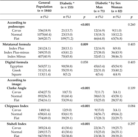
Table 2.Description and concordance of the footprint type according to podoscope and image.
Index Plus 241(24.1) 20(13.1) 12(16.9) 8(9.8)
Index Plus-minus 349(35.0) 63(41.2) 27(38.0) 36(43.9)
Index Minus 407(40.9) 70(45.8) 32(45.1) 38(46.3)
Digital formula 0.058 0.403
* Subsample of diabetic patients in which podiatric examination was performed. Numbers written in bold indicate statistically significant differences.
More than 60% of the 153 diabetic patients presented normal foot according to the Clarke angle and the Chippaux index (60.4% and 61.9% respectively), whereas the Staheli index determined a higher prevalence of flat foot (38.8%) followed by a 30.6% normal foot. These classifications were similar in terms of the sex of the patients.
3.2.1. Podiatric Alterations Depending on Sex
Table 3.Podiatry characteristics of diabetic patients.
Hallux Extensus 38(24.8) 20(28.2) 18(22.0) 0.375 Hallux Valgus 101(66.0) 44(62.0) 57(69.5) 0.326
Grades 0.662
Grade 1 47(46.5) 23(52.3) 24(42.1) Grade 2 30(29.7) 12(27.3) 18(31.6)
Grade 3 13(12.9) 4(9.1) 9(15.8)
Grade 4 11(10.9) 5(11.4) 6(10.5)
Claw fingers 145(94.8) 68(95.8) 77(93.9) 0.725
2nd 132(86.3) 62(87.3) 70(85.4) 0.726
* Subsample of diabetic patients in which podiatric examination was performed. Numbers written in bold indicate statistically significant differences.
3.2.2. General Characteristics According to the Type of Footprint and the Podiatric Alterations
Table 4. Characteristics of diabetic patients according to the type of footprint and the podiatric alterations: 2nd claw toe and Hallux Valgus.
Type of Footprint
Mean (SD) Mean (SD) Mean (SD) p
Normal Cavus Flat
Age(years) 61.4(9.0) 64.7(6.6) 62.8±8.3 0.422
BMI (Kg/m2) 28.4(3.4) 26.7(3.1) 32.2(5.3) <0.001
n (%) n (%) n (%)
Sex 0.260
Men 13(56.5) 12(57.1) 46(42.2)
Woman 10(43.5) 9(42.9) 63(57.8)
Time of evolution (years) 11.7(6.9) 19.6(10.3) 12.6(8.5) 0.013 Abdominal perimeter (cm) 99.7(10.5) 97.5(10.1) 108.7(14.5) 0.001 Hyperkeratosis 0.006
No 14(60.9) 10(47.6) 31(28.4)
Yes 9(39.1) 11(52.4) 78(71.6)
2nd claw toe
No Yes p
Age(years) 61.8(10.7) 63.0(7.8) 0.985
BMI (Kg/m2) 30.7(5.79) 30.9(5.2) 0.674
n (%) n (%)
Sex 0.726
Men 9(42.9) 62(47.0)
Woman 12(57.1) 70(53.0)
Time of evolution (years) 16.2(7.2) 13.0(9.1) 0.054
Abdominal perimeter (cm) 104.0(14.0) 106.0(14.2) 0.676
Hyperkeratosis 0.825 No 8(38.1) 47(35.6)
Yes 13(61.9) 85(64.4)
Hallux Valgus
No Yes p
Age (years) 62.1(9.8) 63.2(7.3) 0.603
BMI (Kg/m2) 31.3(5.5) 30.6(5.1) 0.461
n (%) n (%)
Sex 0.326
Men 27(51.9) 44(43.6)
Woman 25(48.1) 57(56.4)
Time of evolution (years) 14.6(8.3) 12.9(9.1) 0.109
Abdominal perimeter (cm) 106.3(14.2) 105.5(14.1) 0.679
Hyperkeratosis 0.239 No 22(42.3) 33(32.7)
Yes 30(57.7) 68(67.3)
Onychocryptosis 0.882 No 19(45.2) 43(43.9)
Yes 23(54.8) 55(56.1)
3.2.3. Concordance Between Footprint According to the Podoscope
The concordance between the type of footprint according to the podoscope and the measurements using the Clarke angle and the Chippaux and Staheli indexes was analyzed. A greater agreement percentage was observed between the footprint type according to the podoscope and the Staheli index (54%), followed by the Chippaux index (50%) and, to a lesser extent, the Clarke angle (49%). A Kappa index between 0.26 (Chippaux index and Clarke angle) and 0.27 (Staheli index) was observed.
4. Discussion
This work constitutes a research that tries to cover the gaps of knowledge found in the literature during the course of this work in Spain with a podiatric perspective, in which there has been a lack of studies that tried to know the repercussion that type II diabetes mellitus triggers at the level of the foot.
Although we find in the literature different studies that tried to know the prevalence of diabetic foot, and different alterations in the foot in people with this condition [12–14], we wanted to study the impact in a broad way, knowing the repercussions at all levels of the foot: structural, dermal and nail aspects.
Taking into account that among the risk factors for diabetes are age [15] and obesity [16,17], our sample has a high age (62.9±7.9 years) with a minimum age of around 40 years compared with other studies found in the literature [18,19], likewise, we are struck by the high obesity present in our study, a fact that is higher than the literature consulted [19].
In the present study we found a high presence of podiatric pathology, finding 94.8% of claw fingers, data that are higher than that found in the general population as well as in the diabetic population (9%) [17]. In relation to the deformity of Hallux valgus, we found in our study a prevalence of 66%; we have not found any study with data on this pathology.
Alavi et al. and Vural et al. [19,20] studied nail changes as in our study, finding similar data on onychodystrophy (13% in our study vs. 11% in the study by Vural et al. [19]). On the other hand, we found a higher prevalence of onychogyphosis in our study (12.9% vs. 4% in the study by Vural et al. 19). The results obtained in relation to onycholysis (2.9%) were lower than those found in the literature [19,20].
Regarding the skin alterations, we found studies that dealt with xerosis and hyperkeratosis. Regarding xerosis, we found a lower prevalence in this work compared to the literature reviewed [18,19]. On the other hand, the prevalence of hyperkeratosis was higher in this study, compared to the data consulted [19,21–23].
In relation to the measurements in footprints, Plumarom et al. [24] discovered that the Staheli index could be considered as the screening or diagnostic method for flatfoot. In our study, the major of the footprints corresponded with flatfoot. Furthermore, we analyzed the concordance between the methods with the podoscope, and we got the best agreement between the podoscope and the Staheli Index.
Based on the findings of this study, it is possible to observe the importance that type II diabetes mellitus triggers at the level of the lower limb, revealing the need for adequate podiatric prevention [25] in patients with this pathology in collaboration with a multidisciplinary team.
Limitations
The results of the present study should be interpreted taking into account their possible limitations. A potential problem with this study is that no data were collected from patients who refused to participate, so the sample may not be representative of the population with diabetes. However, the percentage of patients who declined to participate in the study was less than 5%, so it can be considered that the representativeness of the sample has not been affected.
observer, the podiatric study has been carried out by two podiatrists who have agreed on the findings. On the other hand, the measurement of subjective concepts that can be perceived differently by different people, such as pain, functionality or health related to the foot, can be sources of bias. The use of validated questionnaires such as those used in this study limits this problem, and also makes it possible to compare the findings obtained with those of other similarly themed publications.
5. Conclusions
This study concluded with findings including a high prevalence of podiatric pathology in diabetic patients, mainly flat feet, hallux valgus and claw toes. We found a predominance of skin alterations in female patients, whereas abnormal color in the feet was more frequent in the male patients.
In order to substantiate the concordance between the type of footprint according to the podoscope and the measurements studied in the footprint, the Staheli index showed a greater percentage according to the type of footprint according to the podoscope than the Chippaux index and the Clarke angle, showing a weak concordance in all three cases.
Author Contributions:Data collection: C.G.-M., R.V.-S.; conception and design of the study: C.G.-M., A.S.-G., S.P.-D.; analysis of data: T.S.-P., V.B.-B.; the writing of the article or the critical review of a substantial part of its content: C.G.-M., S.P.-D., T.S.-P., V.B.-B., A.S.-G., R.V.-S.
Funding:This research received no external funding.
Conflicts of Interest:The authors declare no conflicts of interest.
References
1. International Diabetes Federation.IDF Diabetes Atlas, 6th ed.; International Diabetes Federation: Brussels, Belgium, 2013; Available online:http://www.idf.org/diabetesatlas(accessed on 10 March 2018).
2. Alonso-Morán, E.; Orueta, J.F.; Fraile Esteban, J.I.; Arteagoitia Axpe, J.M.; Marqués González, M.L.; Toro Polanco, N.; Ezkurra Loiola, P.; Gaztambide, S.; Nuño-Solinis, R. The prevalence of diabetes-related complications and multimorbidity in the population with type 2 diabetes mellitus in the Basque Country. BMC Public Health2014. [CrossRef] [PubMed]
3. WHO.Informe Mundial Sobre la Diabetes; World Health Organization: Geneva, Switzerland, 2016; Available online:https://apps.who.int/iris/bitstream/handle/10665/204877/WHO_NMH_NVI_16.3_spa.pdf?sequence= 1(accessed on 10 March 2018).
4. Goday, A. Epidemiology of diabetes and its non-coronary complications.Revista Española de Cardiología2002, 55, 657–670. [CrossRef]
5. American Diabetes Association. Microvascular complications and foot care.Diabetes Care2016,39(Suppl. 1). 6. Chandra-Mishra, S.; C-Chhatbar, K.; Kashikar, A.; Mehndiratta, A. Diabetic foot.BMJ2017, 359. [CrossRef] 7. Armstrong, D.G.; Boulton, A.J.; Bus, S.A. Diabetic foot ulcers and their recurrence.N. Engl. J. Med.2017,376,
2367–2375. [CrossRef] [PubMed]
8. Zhang, P.; Lu, J.; Jing, Y.; Tang, S.; Zhu, D.; Bi, Y. Global epidemiology of diabetic foot ulceration: A systematic review and meta-analysis.Ann. Med.2017,49, 106–116. [CrossRef] [PubMed]
9. Guisado-Vasco, P.; Cano-Megías, M.; Carrasco-de la Fuente, M.; Corres-González, J.; Matei, A.M.; González-Albarrán, O. Clinical features, mortality, hospital admission, and length of stay of a cohort of adult patients with diabetic ketoacidosisattending the emergency room of a tertiary hospital in Spain. Endocrinol. Nutr.2015,62, 277–284. [CrossRef] [PubMed]
10. Moreno de la Fuente, J.L.; Catena Toledano, M.; González, M.Podología General y Biomecánica; Masson: Barcelona, Spain, 2003.
11. Pita-Fernandez, S.; González-Martín, C.; Seoane-Pillado, T.; Pertega-Diaz, S.; Perez-Garcia, S.; López-Calviño, B. Podiatric medical abnormalities in a random population sample 40 years or older in Spain.J. Am. Podiatr. Med. Assoc.2014,104, 574–582. [CrossRef]
13. Nather, A.; Cheng, J.C.C.; Devi, G.P.; Chionh, S.B.; Erasmus, A.Foot Screening for Diabetics. Diabetic Foot Problems; World Scientific: Singapore, Singapore, 2008; pp. 169–177.
14. Mehra, B.R.; Thawait, A.P.; Karandikar, S.S.; Gupta, D.O.; Narang, R.R. Evaluation of foot problems among diabetics in rural population.Indian J. Surg.2008,70, 175. [CrossRef]
15. Davis, T.M.; Stratton, I.M.; Fox, C.J.; Holman, R.R.; Turner, R.C. UK Prospective Diabetes Study 22. Effect of age at diagnosis on diabetic tissue damage during the first 6 years of NIDDM.Diabetes Care1997,20, 1435–1441. [CrossRef]
16. Franch Nadal, J.; Alvarez Torices, J.C.; Alvarez Guisasola, F.; Diego Domínguez, F.; Hernández Mejía, R.; Cueto Espinar, A. Epidemiology of diabetes mellitus in the province of Leon.Med. Clin.1992,98, 607–611. 17. Tamayo-Marco, B.; Faure-Nogueras, E.; Roche-Asensio, M.J.; Rubio-Calvo, E.; Sánchez-Oriz, E.; Salvador-Oliván, J.A. Prevalence of diabetes and impaired glucose tolerance in Aragón, Spain.Diabetes Care 1997,20, 534–536. [CrossRef] [PubMed]
18. Vázquez, J.A.; Gaztambide, S.; Soto-Pedre, E. 10-year prospective study on the incidence and risk factors for type 2 diabetes mellitus.Med. Clin.2000,115, 534–539. [CrossRef]
19. Vural, S.; Bostanci, S.; Koçyigit, P.; Çaliskan, D.; Baskal, N.; Aydin, N. Risk Factors and Frequency of Ingrown Nails in Adult Diabetic Patients.J. Foot Ankle Surg.2018,57, 289–295. [CrossRef] [PubMed]
20. Alavi, A.; Sanjari, M.; Haghdoost, A.; Sibbald, R.G. Common foot examination features of 247 Iranian patients with diabetes.Int. Wound J.2009,6, 117–122. [CrossRef] [PubMed]
21. Litzelman, D.K.; Marriott, D.J.; Vinicor, F. The role of footwear in the prevention of foot lesions in patients with NIDDM. Conventional wisdom or evidence-based practice?Diabetes Care1997,20, 156–162. [CrossRef] [PubMed]
22. Pavicic, T.; Korting, H.C. Xerosis and callus formation as a key to the diabetic foot syndrome: Dermatologic view of the problem and its management.J. Dtsch. Dermatol. Ges.2006,4, 935–941. [CrossRef] [PubMed] 23. Chuback, J.; Embil, J.M.; Sellers, E.; Trepman, E.; Cheang, M.; Dean, H. Foot abnormalities in Canadian
Aboriginal adolescents with Type 2 diabetes.Diabet. Med.2007,24, 747–752. [CrossRef] [PubMed] 24. Plumarom, Y.; Imjaijitt, W.; Chaiphrom, N. Comparison between Staheli index on Harris mat footprint and
Talar-first metatarsal angle for the diagnosis of flatfeet.J. Med. Assoc.2014,97(Suppl. 2), S131–S135. 25. Chiwanga, F.S.; Njelekela, M.A. Diabetic foot: Prevalence, knowledge, and foot self-care practices among
diabetic patients in Dar es Salaam, Tanzania—A cross-sectional study.J. Foot Ankle Res.2015,8, 20. [CrossRef] [PubMed]