The Rockefeller University Press $30.00 J. Gen. Physiol. 2016 Vol. 148 No. 4 277–291 Introduction
That the charged nature of K+ ions impairs their free
movement across the plasma membrane derives from elementary physics. The calculation of the Born self-en-ergy for K+ within the low dielectric constant of the
membrane shows the nonspontaneity of this process (Parsegian, 1969). Nevertheless, in K+ channels, nature
found a low-energy mechanism to move K+ ions across
the plasma membrane by developing proteins able to mimic its water coordination (Zhou et al., 2001). Thanks to these membrane proteins, K+ is the most
per-meable ion in resting cells, and because K+ is also the
most abundant intracellular ion, the resting membrane potential in most living cells is close to the Nernst po-tential for K+ (Hodgkin and Huxley, 1952). K+ channels
are probably an ancient protein family and are present in every living being (Armstrong, 2015). These mem-brane proteins belong to one of the biggest gene fami-lies, with ∼90 representatives in the mammalian genome (Yu et al., 2005). Their physiological role is widespread: they guard the resting membrane potential, stabilize os-motic imbalance, set the excitability threshold in excit-able membranes, and shape the neuronal action potential (Hille, 2001; Armstrong, 2015).
K+ channels are endowed with an unsurpassed
archi-tectural mechanism that allows K+ ions to permeate
se-lectively across the cell membrane. However, they show a wide variability in unitary conductance (or ion trans-port rate), which spans approximately two orders of magnitude when measured under similar experimental conditions. In this viewpoint, we propose that the struc-tural determinants for selectivity and conductance are
segregated to two structures within the pore of K+
chan-nels: the selectivity filter and the internal vestibule, re-spectively. We raise the idea that the structure of the selectivity filter seems to be so dedicated to selective and efficient K+ transport that it is unlikely to be the
structural determinant of conductance diversity. On the contrary, the physical dimensions of the hydrophobic inner vestibule seem to be the factors that limit K+
trans-port, accounting for the difference in unitary conduc-tance among K+ channels.
The structure of the K+ permeation pathway
K+ channels allow selective passage of K+ ions,
thermo-dynamically lured to flow against their own electro-chemical gradient, to the exclusion of all other physiological cations. K+ channels select for K+ over Na+
by almost 1,000-fold, a surprising task considering a dif-ference of <0.5 Å between the ionic radii of these two cations (Hille, 1973). Nevertheless, because the differ-ence in their hydration energies is ∼16 kcal/mol, re-moval of the hydration water should be ∼1010 harder
for Na+ (Robinson and Stokes, 2002). Thus, the
sim-plest explanation for the high K+ selectivity should
in-volve, at least in part, the need for partial dehydration of the ion, excluding Na+ because replacing its
hydra-tion waters is energetically costlier (Bezanilla and Arm-strong, 1972). It has also been argued that the binding sites within the selectivity pore must be precisely shaped around a partially dehydrated K+ so that it fits snugly
(Mullins, 1959). The selectivity sequence of K+
chan-nels for alkali metal cations (K+≈ Rb+ > Cs+ > Na+ > Li+) Ion channels are membrane proteins that mediate efficient ion transport across the hydrophobic core of cell membranes, an unlikely process in their absence. K+ channels discriminate K+ over cations with similar radii with
extraordinary selectivity and display a wide diversity of ion transport rates, covering differences of two orders of
magnitude in unitary conductance. The pore domains of large- and small-conductance K+ channels share a
gen-eral architectural design comprising a conserved narrow selectivity filter, which forms intimate interactions with permeant ions, flanked by two wider vestibules toward the internal and external openings. In large-conductance K+ channels, the inner vestibule is wide, whereas in small-conductance channels it is narrow. Here we raise the
idea that the physical dimensions of the hydrophobic internal vestibule limit ion transport in K+ channels,
ac-counting for their diversity in unitary conductance.
Pore size matters for potassium channel conductance
David Naranjo,1 Hans Moldenhauer,1 Matías Pincuntureo,1,4 and Ignacio Díaz‑Franulic1,2,3 1Centro Interdisciplinario de Neurociencia de Valparaíso, Universidad de Valparaíso, Playa Ancha, Valparaíso 2360103, Chile 2Center for Bioinformatics and Integrative Biology, Universidad Andrés Bello, Santiago 8370146, Chile
3Fraunhofer Chile Research, Las Condes 7550296, Chile
4Programa de Doctorado en Ciencias, mención Biofísica y Biología Computacional, Universidad de Valparaíso, Valparaíso 2360103, Chile
© 2016 Naranjo et al. This article is distributed under the terms of an Attribution– Noncommercial–Share Alike–No Mirror Sites license for the first six months after the publication date (see http ://www .rupress .org /terms). After six months it is available under a Creative Commons License (Attribution–Noncommercial–Share Alike 3.0 Unported license, as described at http ://creativecommons .org /licenses /by -nc -sa /3 .0 /).
Correspondence to David Naranjo: david.naranjo@uv.cl; or Ignacio Díaz-Franulic: ignacio.diaz@cinv.cl
The Journal of General Physiology
on April 28, 2017
indicates that K+ channel permeation is biased against
larger hydration energy and larger size, as expected for a relatively “low-field-strength” site (Eisenman, 1962). Thus, Bezanilla and Armstrong (1972) postulated “Na+
ions do not enter the narrower part of the pore be-cause they are too small to fit well in the coordination cages provided by the pore as replacements for the water molecules surrounding an ion.” Because the ob-served selectivity sequence is virtually identical among K+ channels, it anticipates a highly conserved ion
selec-tivity structure (Latorre and Miller, 1983; Heginbotham and MacKinnon, 1993).
The crystallographic structure of the KcsA bacterial K+ channel resolved at 3.2 Å by Doyle et al. (1998)
re-vealed for the first time how the pore of a K+ channel
looks (Fig. 1, A and B). The protein has a tetrameric organization around the pore placed in its axis of sym-metry. Although the structure corresponded to that of a closed channel, it showed several, previously antici-pated, functional features: (a) The pore hosts several K+ ions in single-file order as Hodgkin and Keynes
pre-dicted 60 years ago (Hodgkin and Keynes, 1955). (b) The pore has a narrow selectivity filter located toward the external entrance and is flanked internally by a wider internal vestibule as anticipated by Armstrong
and Bezanilla (Armstrong, 1971; Bezanilla and Arm-strong, 1972; Miller, 1982; Latorre and Miller, 1983). (c) K+ ions are partially hydrated in the narrow section
of the pore, as Mullins, Bezanilla, and Armstrong pro-posed half a century ago (Mullins, 1959; Armstrong, 1971; Bezanilla and Armstrong, 1972; Hille, 1973). Although the structural analysis could not resolve in-teratomic bond orientation, it was hypothesized that carbonyl oxygens from the signature sequence in the peptide backbone, TVG YG, shape the anticipated low-field-strength K+ binding sites by forming surrogate
hydration cages in the filter (Eisenman, 1962; Hegin-botham et al., 1994). The presence of these expected features in a single crystallographic structure gave this study immediate acceptance.
Later on, crystallization of KcsA channels with im-proved resolution (2.2 Å) provided a detailed picture of K+ ions and their carbonyl cages in the selectivity filter
(Fig. 1, C and D). At the internal and external en-trances, K+ ions are fully or partially hydrated, whereas
those located inside the filter fit perfectly into the four carbonyl-lined binding sites of the selectivity filter (Zhou et al., 2001). Constrained by the K+/water 1:1
stoichiometry flux ratio, determined from streaming potentials by Alcayaga et al. (1989), it was proposed that
Figure 1. Structural features of the KcsA channel and K+ coordination structure in the pore. (A and B)
Membrane‑omitted side and top views of the KcsA K+ channel (PDB ID 1K4C). Each monomer is a two–
transmembrane segment peptide position around the pore at the axis of fourfold symmetry forming the K+ selective pore (green spheres). (C) High‑res‑
olution electronic density map showing the two di‑ agonal subunits and the orientation of the carbonyl oxygen atoms to coordinate K+ ions. The numbers
correspond to the four binding sites determined by the sequence TVG YG. (D) Antiprism and cubic cages forming the selectivity filter binding sites, the dis‑ tances d1–d5 and heights h1–h4 correspond to the
inter‑oxygen separations described in Table 1 for several K+ channel structures. A and B were inspired
by Doyle et al. (1998), C was modified from Zhou et al. (2001) with permission from Macmillan Publishers Ltd., and D was inspired by Chen et al. (2014).
on April 28, 2017
the four ion-binding sites at the selectivity filter are en-ergetically equivalent for K+ in alternate occupancy of
sites 1–3 and 2–4, with intervening waters at the vacant sites (Bernèche and Roux, 2000; Morais-Cabral et al., 2001). This arrangement makes near to zero the energy cost to put two K+ ions inside the selectivity filter. In
con-trast, a solitary K+ would not be able to permeate
mea-surably because it would be too tightly bound (Neyton and Miller, 1988; Liu and Lockless, 2013). In contrast, double occupancy in sites separated by ∼7 Å (either sites 1 and 3 or 2 and 4) in the selectivity filter affords enough electrostatic repulsion to allow efficient ion translocation along the pore (Åqvist and Luzhkov, 2000; Morais-Cabral et al., 2001).
The geometry of cation coordination in the selectivity filter
The K+ ions along the selectivity filter are coordinated
in a square prism fashion (Fig. 1, C and D), with eight carbonyls groups each contributing a binding site in the selectivity filter, four on top and four below the cation. For sites 1, 2, and 3, the top four carbonyls are rotated
∼45°, forming a squared antiprism with vertices sepa-rated by 3–4 Å, whereas a cube encases site 4. Notably, the K+ located internally to the selectivity filter appears
to coordinate eight water molecules also in antiprism fashion, and the most external K+ is coordinated on top
by four waters as if these two cations were caught “in flagrante” getting stripped from their waters before en-tering the selectivity filter (Miller, 2001; Zhou et al., 2001). Neutron scattering, spectroscopy, statistical mechani-cal, and molecular dynamic simulation converge on a mean center to center distance between the K+ ion and
the oxygen atoms of the hydration shell of ∼2.6–2.8 Å (Enderby, 1995; Glezakou et al., 2006; Mancinelli et al., 2007; Bankura et al., 2013). Such a distance matches the center to center separation between K+ and the
car-bonyl’s oxygen atoms in the selectivity filter.
The mean K+ coordination geometry in solution is
unknown; however, using the above K+-O separation of
2.6–2.8 Å, it is possible to calculate a vertex to vertex distance of 3.0–3.2 Å in a squared hydration cage. These distances fit well for most of the selectivity filter cages in Table 1. Moreover, a cube, or a squared antiprism, formed by a cage composed of eight water molecules separated by 3.0–3.2 Å would fill a volume of 195–220 Å3, which is the volume of a 3.6–3.8-Å-radius sphere,
consistent with the estimated hydrodynamic radius of K+ (Díaz-Franulic et al., 2015; Moldenhauer et al.,
2016). Thus, from the geometrical point of view, the ox-ygen cages can be regarded as a surrogate water cages. Moreover, this argument favors a homotetrameric struc-ture in K+ channels as a requisite for highly selective K+
binding sites, as was proposed by Zhou et al. (2001).
The rigidity of the K+ selective filter
Electrophysiological, structural, and calorimetry stud-ies support the equilibrium-binding hypothesis for ionic selectivity in K+ channels: selectivity is attained by
making the energy wells along the reaction coordinate deeper for K+ than for Na+ (Eisenman, 1962; Neyton
and Miller, 1988; Zhou et al., 2001; Piasta et al., 2011; Liu et al., 2012). In light of the conserved structure of the selectivity filter among different channels (Fig. 1, C and D; and Table 1), we are tempted to consider this structure as static and immutable. Nevertheless, func-tional evidence and intuitive thinking indicate that this temptation is dangerous. Proteins are flexible, and the carbonyl atoms of the selectivity filter fluctuate around 0.4 Å root mean square (Allen et al., 2004), nearly the difference among Na+ and K+ ionic radii, suggesting
that the snug-fit mechanism requires some amend-ments (Allen et al., 2004; Noskov et al., 2004). Function-ally speaking, selectivity filter flexibility is evidenced in, for instance, C-type inactivation of Kv channels. C-type inactivation is a Na+-permeable pore conformation in
K+ channel usually triggered by the removal of
exter-nal K+ or by long depolarizations (see, for example,
Starkus et al. [1997]). This phenomenon has also been seen in the KcsA channel, being associated with the Table 1. O–O distances at the edges of the water surrogating cages in the selectivity filter, Å
Selectivity filter edge Structure (resolution)
KcsA 1K4C (2.0 Å) Kv1.2/2.1 2R9R (2.4 Å) MthK 4HYO (1.65 Å) KvAP 1ORQ (3.2 Å)
d1 3.6 3.5 3.5 4.1
d2 3.3 3.3 3.2 3.9
d3 3.3 3.3 3.3 3.9
d4 3.2 3.3 3.1 3.1
d5 3.7 3.9 3.6 3.4
h1 3.1 3.1 3.1 2.9
h2 3.0 3.0 3.0 2.7
h3 3.1 3.1 3.2 3.3
h4 3.0 2.9 2.9 2.9
Center to center inter-oxygen distances at the selectivity filter edges shown in Fig. 1 D. Distance calculations are directly from the Biological Assembly PDB file
coordinates. Distances di result from the construction of the Biological Assembly. We chose the above structures because they showed the best resolution available
at the time.
on April 28, 2017
loss of the second and third selectivity filter K+ binding
sites (Cuello et al., 2010). Selectivity filter stability has been proposed to be important for K+ discrimination
in KcsA, as mutations to surrounding residues (E71; Fig. 1 C) increase Na+ permeability (Cheng et al., 2011).
Thus, a complex network of interactions must support the functional integrity of the selectivity filter. In fact, despite their identical signature sequence and ion dis-tribution along the KcsA and MthK channels selectivity filter, the pore of KcsA, but not MthK, collapses upon K+ removal (Morais-Cabral et al., 2001; Ye et al., 2010).
Thus, the selectivity filter emerges as a dynamic struc-ture, able to adopt a collection of stable conformations, of which the fully conducting ones seem to be also the most often crystallized.
The bacterial nonselective NaK channel, which al-lows permeation of both K+ and Na+, has contributed
significantly to our understanding of ionic selectivity in K+ channels. The NaK channel selectivity filter possesses
the sequence TVG DG instead of the canonical TVG YG, failing to show S1 and S2 ion-binding sites (Alam and Jiang, 2009). Variants having the number of binding sites restored to four are K+ selective, indicating that the
ability to make transitions between 1,3 and 2,4 configu-rations is essential for K+-selective permeation, revealing
a strong marriage between selectivity and conductance (Derebe et al., 2011; Liu and Lockless, 2013; Sauer et al., 2013). Excellent reviews covering the mechanisms of ion selectivity in rich detail are available (Noskov and Roux, 2006; Nimigean and Allen, 2011; Lockless, 2015).
We excluded the inward rectifier family from consid-eration in this review. Although these channels share with BK and Kvs the selectivity filter and the internal vestibule structures, unlike the latter, their
tetrameriza-tion domain contributes significantly to ion conduc-tion, adding several K+-binding sites in series with the
pore. Their conduction mechanism looks more com-plex, and their structural determinants for unitary con-ductance may be different from Kv and BK channels (Nishida and MacKinnon, 2002; Lu, 2004).
Pore architecture of K+ channels: Implications
for ion permeation
Selectivity filters among different K+ channels are so
similar that we hypothesized that, regardless of their specific unitary conductance, they should be equally competent to allow permeation of K+ at high rates.
However, different K+ channels display large differences
in their unitary conductance measured under similar experimental conditions. Permeation should occur at very high rates because the energy cost of putting two K+ ions in the selectivity filter is to zero (Åqvist and
Lu-zhkov, 2000; Morais-Cabral et al., 2001; Zhou et al., 2001; Bernèche and Roux, 2003; Allen et al., 2004; Jen-sen et al., 2013). Accordingly, in standard recording solutions, the selectivity filter accounts for, at most, 3 GΩ (333 pS) of the total pore resistance in both small- and large-conductance K+ channels (Díaz-Franulic et
al., 2015). Direct estimation of electrical resistance, or electrostatic field calculation along the pore, showed that 3 GΩ accounts for 50–90% of the total resistance of BK or MthK channels (Morais-Cabral et al., 2001; Con-treras et al., 2010; Díaz-Franulic et al., 2015). In con-trast, in Shaker Kv channels, such resistance accounts for only ∼10% of the total (Díaz-Franulic et al., 2015). Thus, in small-conductance Kv channels, ion transport must be limited in structures that are separate from the selectivity filter (Moscoso et al., 2012). Table 2 com-Table 2. Single-channel conductance in selected K+ channels
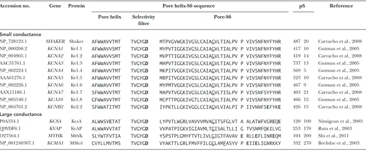
Accession no. Gene Protein Pore helix-S6 sequence pS Reference
Pore helix Selectivity filter
Pore-S6
Small conductance
NP_728123.1 SHA KER Shaker AFW WAV VTMT TVG YGD MTP VGV WGK IVG SLC AIAGVLT IAL PV P VIV SNF NYF YHR 487 20 Carvacho et al., 2008 NP_000208.2 KCNA1 Kv1.1 AFW WAV VSMT TVG YGD MYP VTI GGK IVG SLC AIAGVLT IAL PV P VIV SNF NYF YHR 417 10 Gutman et al., 2005 NP_004965.1 KCNA2 Kv1.2 AFW WAV VSMT TVG YGD MVP TTI GGK IVG SLC AIAGVLT IAL PV P VIV SNF NYF YHR 419 14 Carvacho et al., 2008 AAC31761.1 KCNA3 Kv1.3 AFW WAV VTMT TVG YGD MHP VTI GGK IVG SLC AIAGVLT IAL PV P VIV SNF NYF YHR 737 13 Gutman et al., 2005 NP_002224.1 KCNA4 Kv1.4 AFW WAV VTMT TVG YGD MKP ITV GGK IVG SLC AIAGVLT IAL PV P VIV SNF NYF YHR 569 5 Gutman et al., 2005 AAA61276.1 KCNA5 Kv1.5 AFW WAV VTMT TVG YGD MRP ITV GGK IVG SLC AIAGVLT IAL PV P VIV SNF NYF YHR 523 10 Carvacho et al., 2008 NP_002226.1 KCNA6 Kv1.6 AFW WAV VTMT TVG YGD MYP MTV GGK IVG SLC AIAGVLT IAL PV P VIV SNF NYF YHR 467 9 Gutman et al., 2005 AAX11186.1 KCNA7 Kv1.7 SFW WAV VTMT TVG YGD MAP VTV GGK IVG SLC AIAGVLT ISL PV P VIV SNF SYF YHR 403 21 Carvacho et al., 2008 NP_005540.1 KCA10 Kv1.8 GFW WAV VTMT TVG YGD MCP TTP GGK IVG TLC AIAGVLT IAL PV P VIV SNF NYF YHR 466 12 Gutman et al., 2005 NP_004761.2 KCNB2 Kv2.2 SFW WAT ITMT TVG YGD IYP KTL LGK IVG GLC CIAGVLV IAL PI P IIV NNFSEFYKE 426 15 Carvacho et al., 2008
Large conductance
P0A334.1 KCSA KcsA ALW WSVETAT TVG YGD LYP VTL WGR LVA VVV MVAGITS FGL VT A ALA TWF VGREQE 120 100 Nimigean et al., 2003 Q9YDF8.1 KVAP KvAP ALW WAV VTAT TVG YGD VVP ATP IGK VIG IAV MLTGISA LTL LI G TVS NMF QKI LVG 253 170 Ruta et al., 2003 O27564.1 MTHK MthK SLY WTF VTIA TVG YGD YSP STP LGM YFT VTL IVLGIGT FAV AV E RLLEFLI NREQM 104 200 Shi et al., 2011 NP_001240307.1 KCMA1 MSlo1 CVY LLM VTMS TVG YGD VYA KTT LGR LFM VFF ILGGLAMFASYV P EIIELIG NRK KY 332 270 Brelidze et al., 2003 Multiple alignment of the primary structure of K+ channel pores. The signature sequence TVG YGD on the selectivity filter, used as reference for the alignments, is separated for
clarity. Shaker’s Gly466 and aligning residues are underlined. Shaker’s 475 and aligning residues in Kv channels are individualized for clarity. Aligning with Shaker 475 are KcsA’s Ala108, KvAP’s Gly241, MthK’s Glu92 and MSlo1’s Pro320. Aspartates and glutamates are in bold (table modified from Moscoso et al. [2012] with permission from Elsevier).
on April 28, 2017
pares the amino acidic sequences of several K+-selective
channels between the pore helix and the C terminus of S6, where the structural determinants for single-chan-nel conductance reside. It is apparent that, although K+
channels share identical selectivity filter sequences, their unitary conductance ranges from 5 to 270 pS and, accordingly, they fit into two categories: small and large conductance (top and bottom groups in Table 2). Se-quence diversity in both groups leads to the idea that the determinants of unitary conductance are located at or near the internal cavity.
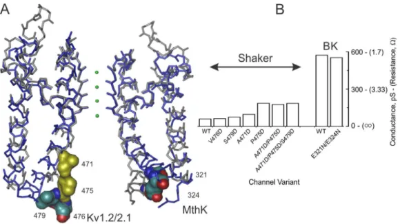
Architecture of BK and MthK channels. BK channels (also known as Slo1 or Maxi-K) display the largest uni-tary conductance among K+ channels, ranging from ∼250 pS under standard recording conditions (100– 150 mM K+) to 600 pS under saturating K+
concentra-tions (Eisenman et al., 1986; Brelidze et al., 2003). Sequence analysis of BK channels reveals two conserved glutamate rings (Glu rings) at the internal entrance, comprising Glu321 and Glu324 in each subunit of MSlo (Fig. 2 A). In 100 mM K+, these eight negatively charged
residues double unitary conductance (Brelidze et al., 2003; Zhang et al., 2006). However, such increments vanish at saturating K+ concentration, indicating that
the charged rings contribute to channel conductance by attracting cations and are not an essential part of the efficient K+ transport mechanism (Fig. 2 B). Therefore,
maximum conductance, measured at saturating K+
con-centrations, is required to separate permeation from
binding (Díaz-Franulic et al., 2015; Sack and Tilley, 2015). In contrast, Phe380, located at the inner cavity of HSlo (F315 in MSlo), was shown to be critical for ion permeation when replacement with isoleucine or tyro-sine decreased unitary conductance by ∼70% or ∼50%, respectively (Carrasquel-Ursulaez et al., 2015). How-ever, the impact of these mutations on the maximum conductance is unknown.
The internal entrance of BK channels seems wider than in Kv channels. The association rate of internally applied quaternary ammonium to the pore is higher than for Kv channels (Li and Aldrich, 2004; Wilkens and Aldrich, 2006). Also, overlapping dual cysteine modifi-cation in BK channels revealed an inner vestibule wider than that of Kv channels (Zhou et al., 2011). Thus, a wide, and possibly short, vestibule could afford a low-re-sistance pathway for ion permeation in BK channels. This suggests that the dimension of the internal pore entrance limits ionic conduction, such that the presence of large side chain amino acids at the inner entrance reduce BK’s unitary conductance, whereas smaller side chain substitutions have little effect (Geng et al., 2011).
In the absence of crystallographic data of BK in the open conformation, the bacterial calcium activated MthK K+ channel has been validated as a bona fide
structural model for the pore domain of BK channels (Geng et al., 2011; Shi et al., 2011; Posson et al., 2013; Moldenhauer et al., 2016). MthK is a large-conductance two–transmembrane segment (TM) channel coupled to a calcium gating ring similar to that of BK (Jiang et Figure 2. Occupancy and dimensions of the internal cavity and the maximum conductance of Kv and BK channel. (A) Align‑ ment of the Kv1.2/2.1 chimera (gray backbone) with MthK (blue) pore domain structures. Shown are the diagonal subunits, with front and back subunits omitted for clarity. K+/water in the selectivity filter are green spheres. The side chains shown on CPK color
correspond to the glutamate ring equivalent positions (right for MthK and left for Kv1.2/2.1). The residues in yellow are the internal cavity residues able to tolerate aspartate substitution. Kv2.1/1.2 chimera: PDB ID 2R9R; MthK: PDB ID 4HYO. (B) Role of charged residues in the inner cavity on maximum conductance of Shaker and BK. Maximal conductance is defined as the unitary conductance at saturating K+ concentration.
on April 28, 2017
al., 2002; Yuan et al., 2011). Consistent with BK’s guessed internal pore dimensions, the MthK structure displays a wide ∼15-Å internal entrance (Fig. 2 A), lined with hy-drophobic residues (Table 2).
A direct functional assessment of BK’s pore architec-ture came from Magleby’s laboratory with measurements of the “radius of capture” (Brelidze and Magleby, 2005). If the pore is assumed to be a hemispheric sink into which approaching ions vanish, it is possible to use diffu-sional collisions theory to infer the dimension of the pore entrance in a condition where the rate-limiting step for ion transport is the diffusion of K+ ions into the
en-trance of the pore (Läuger, 1976; Andersen, 1983). As-suming K+ is a rigid sphere approaching the mouth of
the channel, the radius of capture corresponds to the difference between the radius of the ion and the radius of the pore entrance (Ferry, 1936; Läuger, 1976) as
r P = r C + r i , (1)
where rP, rC, and ri are the pore, the capture, and the
ion radii, respectively. If the ion is a point charge (ri =
0), the radius of capture is the same as the radius of the pore. Experimentally, increasing the viscosity of the solution through the addition of high concentrations of sugar, the amplitude of K+ current becomes
asymptoti-cally voltage independent, revealing the limiting K+
dif-fusional access to the pore (Läuger, 1976; Brelidze and Magleby, 2005; Díaz-Franulic et al., 2015). In BK chan-nels, the internal rC is 2.2 Å, a surprising, and debated,
number suggesting that the pore is barely wide enough to fit a hydrated K+ ion (Brelidze and Magleby, 2005).
Furthermore, we must consider the hydrodynamic di-mensions of K+ to calculate the pore radius, but the size
of hydrated K+ is not well defined because hydrating
water molecules bind with dissimilar energies and life-times, forming a fuzzy arrangement. Then, one has to decide how many hydration shells should be added to the radius of the ion. The simplest assumption is to add just one water layer, but this seems arbitrary (Brelidze and Magleby, 2005).
Let’s consider that rP, the pore radius (a functional
estimate arising from the radius of capture), is equiva-lent to the “effective pore radius” (rE; a structural
esti-mate defined as the radius of the largest sphere that is able to enter the pore cavity; Ferry, 1936). rE is very easy
to estimate from the wealth of K+ channel structures
available, and for MthK it is 5.7–5.9 Å (Moldenhauer et al., 2016). Thus, by replacing rE for rP in Eq. 1, we
ob-tain ri = 3.5–3.7 Å for K+. These figures are consistent
with K+ ions carrying only one hydration layer
(En-derby, 1995; Glezakou et al., 2006; Mancinelli et al., 2007; Bankura et al., 2013). Bearing in mind that the radius of capture is measured at room temperature, in liquid phase, and with operative friction forces, the harmonious outcome with structural studies, made at
very low temperature, in solid phase, and in equilib-rium, is satisfying.
Architecture of Shaker and Kv channels. We have seen that side chain volume and charge of residues at the internal entrance of large-conductance channels re-strict K+ conductance (Brelidze et al., 2003; Nimigean et
al., 2003; Geng et al., 2011). Thus, K+ channels having
narrower pores should display smaller unitary conduc-tance. Among the small-conductance K+ channels the
most extensively studied is the Drosophila melanogas-ter Shaker K+ channel. As expected, Shaker ionic
selec-tivity is lost upon mutation of some of the selecselec-tivity filter residues (Heginbotham et al., 1994). Shaker’s uni-tary conductance ranges from ∼20 pS in ∼100 mM K+ to
60 pS in saturating 3,000 mM K+ (Heginbotham and
MacKinnon, 1993; Díaz-Franulic et al., 2015). Intra- and intersubunit metal coordination at the internal en-trance suggests that this region is narrower than that in BK or MthK and/or that the opening conformational change implies a small physical displacement (Webster et al., 2004; del Camino et al., 2005). Consistent with a narrow pore, the crystal structure of Kv1.2, a mamma-lian homologue of Shaker, shows an internal entrance of ∼10 Å, which, as occurs in MthK, is lined with hydro-phobic side chains (Long et al., 2005a, 2007). Thus, the narrow Kv internal cavity is just large enough to let ions go across, and “…may help to explain why Shaker has an approximately tenfold lower conductance than its bacterial relatives” (Webster et al., 2004).
As with BK, measurements of diffusion limited out-ward currents allowed us to estimate that Shaker’s cyto-solic radius of capture was ∼0.8 Å, ∼1.4 Å narrower than BK’s (Díaz-Franulic et al., 2015). As seen with MthK/BK in section Architecture of BK and MthK channels, this estimate of rC is consistent with the effective opening of
the open Kv1.2/2.1 chimera if we assume that the hy-drodynamic radius for K+ is ∼3.5–4.4 Å (Díaz-Franulic et
al., 2015; Moldenhauer et al., 2016). As hypothesized, such a narrow cavity contributes decisively to the total electrical resistance of the pore. Estimations of the sec-tional resistance along the pore of Shaker showed that, in contrast to BK, the inner cavity entails an electrical resistance ∼55–fold larger (or 1/55 lower conductance) than that of BK channels (Díaz-Franulic et al., 2015). Thus, these results reinforce the idea that the size of the internal vestibule severely limits single-channel conduc-tance of small-conducconduc-tance K+ channels.
The internal pore dimensions and K+ channels cytosolic
structure. For both Kv and MthK structures, the effec-tive entrance is consistent with the estimates of the ra-dius of capture of Shaker and BK channels, respectively, when the K+ hydrodynamic radius is taken as ∼4 Å
(Díaz-Franulic et al., 2015; Moldenhauer et al., 2016). However, the canonical Kv channel activation bundle
on April 28, 2017
crossing gate, located at the lower end of S6, appears to be missing in BK channels. Instead, these K channels may gate the ion pathway near the selectivity filter (Wilkens and Aldrich [2006], Garg et al. [2013], and Posson et al. [2013]; but see Hite et al. [2015]). Thus, the estimates of radius of capture could represent the pore dimensions at different depths in the permeation pathway. Because the estimates of the hydrodynamic size of K+ produced by both comparisons (MthK with
BK and Kv1.2 with Shaker) are so similar, the possible discrepancy in deepness should not account for more than 1 Å in radius. In addition, the resulting size of K+
agrees well with estimations for the hydrated K+ in
aque-ous solutions obtained using unrelated techniques (En-derby, 1995; Glezakou et al., 2006; Mancinelli et al., 2007; Bankura et al., 2013).
There is one more problem with the estimation of the internal pore dimension based on the radius of capture for BK and Kv channels. These measurements disregard the contribution of important intracellular domains in both proteins, leading to an oversimplified structural image (Long et al., 2005a; Hite et al., 2015). Both BK and Kv channels have tetramerization domains near the internal entrance (Kobertz and Miller, 1999; Krishna-moorthy et al., 2005; Hite et al., 2015). In Kv channels, the four tetramerization domains (T1) form a structure known as “the hanging gondola” because it hangs from the pore domain through four linkers, leaving four side-facing openings for ion transport (Fig. 3; Kobertz and Miller, 1999; Long et al., 2007). Meanwhile, the structure of Slo2.2, a BK channel, shows the calcium-de-pendent gating ring forming a funnel structure that could guide ions into the pore cavity (Hite et al., 2015). Both structures project negative electrostatic potential into the permeation pathway, raising local K+
concen-tration, but contribute, at most, to 30% to the unitary
current (Kobertz and Miller, 1999; Budelli et al., 2013; Hite et al., 2015).
Why does a narrow pore cause larger resistance? How is it possible that the internal cavity of Kv chan-nels, with a radius only 1.4 Å smaller than that of BK, causes a 55-fold increase in resistance? The seminal paper by Parsegian (1969) predicted that the energy required to put an ion inside an aqueous pore embed-ded in the low dielectric membrane could decrease by up to 28 kcal/mol for every Å increase in sectional ra-dius. Thus, a mere 1-Å narrower pore would represent a much larger energy barrier for the ion to overcome. Energy calculations show that K+ transport in the
non-polar nanotube membrane model is highly sensitive to the dimensions of the pore. In narrow pores (<5-Å opening diameter) ion transport cannot occur, but wider pores (∼10-Å opening diameter) are highly per-meable, with ionic mobilities comparable with those seen in bulk solution (Peter and Hummer, 2005). In contrast, narrow pores would strip off solvation waters, paying a large energetic penalty; in contrast, wider pores would keep the first hydration shell intact. The free energy calculated for K+ ions inside the cavity of K+
channels is indeed a few kcal lower in the wider parts of the pore (Chung et al., 2002; Jogini and Roux, 2005; Treptow and Tarek, 2006). Thus, higher resistances may result from the reduced probability of finding an ion inside narrower pores, and, conversely, we expect that decreasing the energy required to put an ion in-side the cavity will result in an increased conductance (see section Turning Shaker into a large-conductance K+ channel below). We have to keep in mind that these
considerations involve using equilibrium energies to describe the nonequilibrium phenomenon that is ion transport. Nevertheless, because diffusion and water Figure 3. K+ channel structural
to-pology. Surface representation of Kv (Kv1.2/2.1 chimera; left) and BK (Slo2.2; right) structures. Green and yellow colors represent the voltage‑sensing domain (VSD) and the pore domain (PD), respectively. The green arrows show the putative conduction paths for ions that in Kv channels K+ access/
exit through the lateral windows of the “hanging” T1 domain (the gondola in cyan), whereas in BK the ions cross the entire gating ring formed by the RCK domains (in cyan). The pink spheres are K+ ions, and the horizontal discontinued
lines indicate the approximate inner and outer boundaries of the mem‑ brane. External side is up. The Kv figure is a 6‑Å slab prepared with VMD, with
−6 < x < 0 (Humphrey et al., 1996). BK front and rear subunits are removed for clarity (inspired by Hite et al. [2015]).
on April 28, 2017
turnover are orders of magnitude faster than perme-ation, an equilibrium approximation should be at least partially satisfactory (Hille, 2001; Grossfield, 2005; Mancinelli et al., 2007).
Because ion conduction occurs away from equilib-rium, we should also consider the action of frictional forces on unitary conductance. We are required to ac-knowledge that ions must interact physically with their surroundings (Eisenberg, 2013). Also, molecular dy-namics simulations have shown drastic reductions of ion and water diffusion coefficients inside wide pores (Wilson et al., 2011; Zhu and Hummer, 2012b). As ex-pected for friction, such reduction seems to be caused by the pore shape and wall tortuosities. Because of the lack of room inside the inner vestibule of Kv channels, fully or partially hydrated ions would be bumping into the walls of the pore. In contrast, in BK channels, with a 1.4-Å wider cavity, there is enough room for a loosely attached extra layer of water molecules interfacing with the walls of the inner pore. In principle, these interfa-cial waters should bear low mobility (because of their low entropy); nevertheless, recent molecular dynamic simulations show highly mobile and chaotic interfacial waters in narrow nanotubes organic-aqueous contact (Garate et al., 2014). Such loosely attached water mol-ecules may lubricate the passage of the K+ cage, working
as ball bearing sliders.
Turning Shaker into a large-conductance K+ channel In agreement with the equilibrium energy hypothesis, lowering the K+ energy inside the cavity is expected to
increase unitary conductance, by increasing pore occu-pancy. Indeed, introducing negatively charged residues at Shaker’s internal entrance (position Pro475) increases unitary conductance by eightfold (Sukhareva et al., 2003; Moscoso et al., 2012). Such an increment was cor-related with the appearance of a cytosolically accessible K+ site that could enhance Mg2+ blockade by preventing
its exit toward the cytosol. Molecular dynamics simula-tions showed a largely increased K+ occupancy and Mg2+
binding at the inner cavity. Two distal K+-binding sites
flanked Mg2+ exit (Moscoso et al., 2012). Because of the
deep impact of mutations of Pro475 on channel gating (Hackos et al., 2002), it was quite possible that P475D had enlarged pore size, accounting for such an increased conductance. However, the radius of capture was found to be identical to that of Shaker-WT (Díaz-Franulic et al., 2015). This is in satisfying agreement with the Parsegian hypothesis because it shows that it is possible to raise conductance by several-fold just by increasing pore oc-cupancy, i.e., by lowering the work needed to put an ion in the cavity. Several other charges added to the internal vestibule (471D, 476D, and 479D) also increased unitary conductance; nevertheless, no additional change could increase maximum conductance beyond 200 pS, which is one third of BK’s conductance (Díaz-Franulic et al.,
2015). Part of the other two thirds of the difference in unitary conductance between Kv and BK channels might have different causes: fluctuations of the selectivity filter or friction. In the former case, differences in the selec-tivity sequence should be expected (Starkus et al., 1997; Allen et al., 2004; Cheng et al., 2011).
Electromechanical coupling and permeation determinants map to the same hotspot
Voltage-activated K+ channels are impressive examples
of evolutionary functional tuning. On one hand, the ad-dition of several sensing modules to the seemingly per-meation-optimized voltage- and calcium-activated BK pore domain gives them the ability to integrate complex stimuli into transmembrane K+ fluxes. On the other
hand, voltage sensitivity of Kv channels seems to be near the maximal possible, limited only by the capacity of the voltage-sensing domain to host more charged residues without disrupting the trans-protein electric field (Ahern and Horn, 2004; Tombola et al., 2007; González-Pérez et al., 2010). The voltage-sensing domain is ener-getically linked to the cytosolic end of S6 in the pore domain, mostly through the S4-S5 linker. Open proba-bility increases 20-fold the for every 6-mV depolarization (Aggarwal and MacKinnon, 1996; Seoh et al., 1996; Islas and Sigworth, 1999; Lu et al., 2002; Long et al., 2005b; Batulan et al., 2010; González-Pérez et al., 2010).
In the Shaker Kv channel, residues located toward the internal end of S6 are important for both perme-ation and electromechanical coupling (Ding and Horn, 2002; Hackos et al., 2002; Sukhareva et al., 2003; Bat-ulan et al., 2010). Because the electromechanical cou-pling is tight, mutual interference between permeant cations and channel gating is expected. In fact, we’ve known for a long time that Rb+ and K+ slow channel
closure in the squid axon and in oocytes, resembling a “foot in the door” mechanism, suggesting the existence of a cation-selective site near the inner end of the pore (Swenson and Armstrong, 1981; Matteson and Swen-son, 1986). Such a site was supported by the observation that K+ ions applied internally lock in quaternary
am-monia or Mg2+ (Thompson and Begenisich, 2001;
Mos-coso et al., 2012). Thus, tampering with pore occupancy to increase conductance also affects voltage sensitivity (Sukhareva et al., 2003; Moscoso et al., 2012).
Is it possible that the reduced unitary conductance of Kv channels is a consequence of the tight functional cou-pling between the voltage sensor and the pore domain? Faure et al. (2012) proposed an ∼4-Å radial displace-ment during the opening transition based on lumines-cence resonance energy transfer (LRET) measurements of the bacterial Kv channel, KvAP. This displacement is about the same as the effective radial difference between closed and open pore structures of KcsA and Kv1.2/2.1, respectively (Moldenhauer et al., 2016). Molecular dy-namics simulations of Kv1.2-2.1 under an external
elec-on April 28, 2017
tric field show channel openings as small conformational changes at the activation gate. Ensuing pore hydration drives the channel toward its conductive state (Jensen et al., 2012). This process, coined “hydrophobic gating,” has been reported for several other ion channels. Small, not physically occluded, pores reside in a de-wetted state because water molecules refuse to interact with their hy-drophobic linings (Beckstein et al., 2003; Anishkin and Sukharev, 2004; Jensen et al., 2010, 2012; Zhu and Hum-mer, 2012a; Neale et al., 2015). The critical radius for pore hydration is ∼4–5 Å, very close to the radius of the internal entrance in the Kv1.2/2.1 structure (Beckstein et al., 2003; Webster et al., 2004; Long et al., 2005b; Wang et al., 2008; Díaz-Franulic et al., 2015). Thus, according to these considerations, pore opening in Kv channels might be barely wide enough to allow permeation.
The closed–open transitions seem to be ruled by eco-nomical conformational changes in other ion channels too. In fact, several ion channels (K+-CNG and TRP)
switch between open and closed states with physically small conformational changes in the inner cavity and/ or at the selectivity filter (Flynn and Zagotta, 2001; Bruening-Wright et al., 2002; Proks et al., 2003; Salazar et al., 2009; Rapedius et al., 2012; Cao et al., 2013; Garg et al., 2013; Liao et al., 2013; Posson et al., 2013).
Although small movements during channel gating are not mandatory by any biophysical principle, they may possibly result from an evolutionary selective pres-sure to reduce the energy required to control the pore open probability. Assuming 25–33 cal/mol per Å2 of
hy-drocarbon exposed to water (Reynolds et al., 1974), a cylindrical greasy cavity of 15-Å depth and 5-Å radius requires 11–15 kcal/mol to open. This figure may be considered a lower limit for the work the electrome-chanic gear has to perform to open the pore of a Kv channel because it prefers to be closed (Yifrach and MacKinnon, 2002; Jensen et al., 2010, 2012).
On the opposite sidewalk, a large-conductance chan-nel such as BK, endowed with a wider pore, would re-quire larger, energetically expensive gate movements (Beckstein and Sansom, 2004). To open the gate of a BK-like channel, a 7-Å radius pore would require 16–21 kcal/mol, that is 5–6 kcal/mol more than those re-quired to open a Kv channel (Reynolds et al., 1974). If, as recent crystal structures suggest, the Slo2.2 BK chan-nel primary gate is located at the internal bundle cross-ing, as in Kv channels (Hite et al., 2015), the extra energy required to open BK’s pore can be translated into a shift along the voltage axis. The voltage shift, ΔV, is ΔV = ΔΔG/zF, where ΔΔG is the difference between the energies required to open both pores, z is the effec-tive valence of the voltage dependence, and F the Fara-day constant. Interestingly, if we take BK’s voltage dependency as z = 1.5–2.0, the additional energy re-quired to gate the wider pore would correspond to a positive 100–170-mV shift in the conductance-voltage
relationship with respect to Kv channels. Aware that this calculation oversimplifies gating energetics, and as-suming similar voltage-responsive gears, it is remark-able that such a voltage shift agrees well with the actual difference in half-activation voltage between BK and Kv channels (Díaz et al., 1998; Horrigan et al., 1999). In addition, other larger-conductance ion channels such as TRPV1 also exhibit similarly shifted conductance-volt-age relationships (Matta and Ahern, 2007). Several larger-conductance and multimodal ion channels have evolved additional, physiologically relevant gates, dis-tinct from those at the cytoplasmic end of the pore, which gate ion access with 1–2-Å movements (Flynn and Zagotta, 2001; Bruening-Wright et al., 2002; Proks et al., 2003; Salazar et al., 2009; Rapedius et al., 2012; Garg et al., 2013).
This rationale of small movement gates is per se specu-lative and simplistic because there aren’t obvious bio-physical or physiological constraints to limit pore gating to small conformational adjustments. Worse, x-ray struc-tures have to be taken with certain skepticism because they are obtained under nonphysiological conditions. Nevertheless, we should bear in mind that, on the one hand, energy economy is not uncommon in the opera-tion of K+ channels (for example, in cation coordination
in the selectivity filter), and, on the other hand, smaller energy requirements to open the pore would permit a sensitive tuning of the channel open probability.
The physiological role of unitary conductance is mostly unknown
The large functional diversity and regulatory mecha-nisms of K+ channels surely underlie physiological
fine-tuning within each cell type. These channels are present in almost every tissue, and most cells express dif-ferent K+ channels. Possibly, because of this functional
redundancy, K+ channel pathologies are rarely fatal,
al-though they often seriously challenge an individual’s happiness. Finding the physiological significance for the spectrum of unitary conductances in K+ channels is also
challenging because there is not a clear connection with cell type. In this regard, congenital channelopathy anal-ysis could inform us about the physiological relevance of unitary conductance if we focus on missense mutations occurring at the internal end of S6, the functional hotspot for conductance and electromechanical cou-pling (Ding and Horn, 2002; Lu et al., 2002; Sukhareva et al., 2003; Long et al., 2005b; Posson et al., 2013). A myriad of mutations in S6, with pathological conse-quences, may potentially be affecting unitary conduc-tance (Table 3), but in most cases, research has focused largely on the macroscopic phenotype, such as current density and kinetics, failing to explore microscopic elec-trophysiological behavior. For example, episodic ataxia type 1 (EA1; characterized by spells of incoordination and imbalance) is caused by mutations in the KCNA1
on April 28, 2017
gene (Kv1.1). Likewise, cerebellar ataxia (CA), charac-terized by coordination imbalance, and epileptic en-cephalopathy early infantile 32 (EIEE32), characterized by refractory seizures and neurodevelopmental impair-ment, are both caused by mutation of the KCNA2 gene (Kv1.2; Imbrici et al., 2006; Xie et al., 2010; Syrbe et al., 2015). Mutations could diminish ionic currents by de-creasing protein maturation, trafficking, or activity level, or interaction with accessory subunits, and/or by domi-nant-negative effects. Nevertheless, we will miss the whole pathological picture as long as we ignore their impact on unitary conductance.
We must not think that the unitary conductance is a fixed character of the channel. There are beautiful ex-amples of how accessory subunits change conductance. For example, the unitary conductances of Kv4.2 and Kv7.1 (KCNQ1) increase several fold when they are coex-pressed with accessory subunits: dipeptidyl-peptidase– like protein-6 (DPP6) and KCNE1 (MinK), respectively (Melman et al., 2004; Kaulin et al., 2009). The mecha-nism behind this DPP6 gain of function is similar to that of the Glu ring in BK (Kaulin et al., 2009), whereas in MinK it seems to involve intimate interaction with the S6 domain of Kv7 (Melman et al., 2004). Are these changes Table 3. S6 channelopathies of voltage-dependent K+ channels
Channel Gene Unitary
cond.
Tissue expression S6 limits Mutation Disease Reference
pS
Kv1.1 KCNA1 8.7–20 Central nervous
system, kidney, and heart
387–415 V404I, V408A Episodic ataxia EA1 Browne et al., 1994;
Scheffer et al., 1998
Kv1.2 KCNA2 14–18 Neocortex,
hippocampus, main olfactory bulb, and cerebellum
389–417 P405L Early infantile epileptic
encephalopathy, 32
EIEE32 Syrbe et al., 2015
Kv2.1 KCNB1 14 Hippocampal neurons
and cortical neurons
392–420 G401R Early infantile epileptic
encephalopathy, 26
EIEE26 Saitsu et al., 2015
Kv3.3 KCNC3 32–38 Cerebellum, basal
ganglia, and spinal cord
518–539 V535M Spinocerebellar ataxia
13
SCA13 Duarri et al., 2015
Kv4.3 KCND3 4 Substantia nigra pars
compact, retrosplenial
382–402 S390N; V392I Spinocerebellar ataxia
19; Brugada syndrome
Kv7.1 KCNQ1 0.7–4 Heart, uterus,
stomach, small and
LQT1 Jongbloed et al.,
1999; Tester et al., 2005; Kapplinger et al., 2009
Kv7.2 KCNQ2 6.5 Hippocampal and
cortical neurons
292–312 A306T Benign familial neonatal
seizures
BFNS1 Singh et al., 2003
Kv7.4 KCNQ4 2.1 Brain, cochlea, heart,
and skeletal muscle; neuron derived from embryonic stem cells
297–317 G321S Deafness autosomal
dominant 2A
DFNA2A Coucke et al., 1999
Kv10.1 EAG KCNH1 8.5 Brain, kidney, lung,
and pancreas; in brain: in cortex, hippocampus, caudate, putamen, amígdala, and substantia nigra
478–498 L489F, I494V Temple-Baraitser
syndrome and epilepsy
TMB TS Simons et al., 2015
Kv11.1 ERG KCNH2 10–13 Brian: reticular
thalamic nucleus,
Long QT syndrome 2 LGT2 Napolitano et al.,
2005; Tester et al., 2005; Kapplinger et al., 2009
KCa3.1 KCNN4 30–80 Nonexcitable tissues 265–285 V282E/M Dehydrated hereditary
stomatocytosis
DHS2 Glogowska et al.,
2015
TASK 3 KCNK9 16–32 Cerebellum and
external plexiform layer of the olfactory bulb; hippocampus
219–239 G236R Birk-Barel mental
retardation dysmorphism
BIB AS Barel et al., 2008
Non-exhaustive listing of mutations potentially affecting unitary conductance in voltage-gated K+ channel. Mutational data as well as topological composition of S6
transmembrane segments were obtained from UniProt (http ://www .uniprot .org /uniprot /).
on April 28, 2017
in unitary conductance relevant or just collateral conse-quences of the protein–protein interaction controlling gating kinetics and inactivation? By dissociating kinetics from conductance phenotypes, these cases present an opportunity to understand how relevant the changes in the unitary conductance are to physiology.
Concluding remarks
K+ channels are finely tuned to allow the selective
pas-sage of K+ across the membrane at high rates. The
chan-nel selectivity filter is in charge of this task, dropping to near zero the energy for ion transfer from the bulk solution (Morais-Cabral et al., 2001). K+ ions pass across
the selectivity filter so efficiently that, even in the largest conductance channels, the physical dimensions of the internal vestibule limit channel conductance (Geng et al., 2011). Thus, we propose that the main difference between large- and small-conductance channels arises from the size of the entrance to the internal pore; large-conductance channels have wider vestibules than do smaller conductance ones. The effect of vestibule size on unitary conductance is clearly nonsteric because it is not proportional to the sectional area available for permeation. Inside narrow aqueous pores, embedded in low dielectric lipid membranes, ions require larger en-ergies to become stabilized, limiting K+ current
(Parse-gian, 1969). These equilibrium energy considerations would reduce the conductance gap from approximately two orders of magnitude to one third of the maximal transport rate, corresponding to ∼1 kT in activation energy terms. The rest of the difference remains to be accounted for (Díaz-Franulic et al., 2015). Although Kv channels open just wide enough to let hydrated ions enter the permeation pathway, larger-conductance channels would require larger energies to open because of the hydrophobic nature of their inner walls. Thus, larger-conductance channels may host other activation gates as functional and structural data suggest (Zhou et al., 2011; Hite et al., 2015). Because structural de-terminants for unitary conductance and for electrome-chanical coupling colocalize toward the cytosolic end of S6, a mutual interference between pore occupancy and gating is expected. Therefore, S6 mutant Kv chan-nelopathies require unitary conductance studies to fully understand their pathophysiology.
A C K N O W L E D G M E N T S
We thank John Ewer (Centro Interdisciplinario de Neurociencia de Valparaíso [CINV]) for critical reading of the manuscript.
This work is supported by Fondo Nacional de Desarrollo Científico y Tecnológico (Fondecyt) grant #1120819 and by Ini-ciativa Cientifica Milenio grant PO9-022. I. Díaz-Franulic and H. Moldenhauer were funded by Fraunhofer Chile Research and Fondecyt postdoctoral grant #3160321, respectively. The CINV is a Millennium Institute supported by the Millennium Scientific Initiative of the Ministerio de Economía, Fomento y Turismo.
The authors declare no competing financial interests. Lesley C. Anson served as editor.
Submitted: 19 May 2016 Accepted: 10 August 2016
R E f E R E N C E S
Aggarwal, S.K., and R. MacKinnon. 1996. Contribution of the S4 segment to gating charge in the Shaker K+ channel. Neuron. 16:1169–
1177. http ://dx .doi .org /10 .1016 /S0896 -6273(00)80143 -9
Ahern, C.A., and R. Horn. 2004. Specificity of charge-carrying residues in the voltage sensor of potassium channels. J. Gen. Physiol. 123:205–216. http ://dx .doi .org /10 .1085 /jgp .200308993 Alam, A., and Y. Jiang. 2009. High-resolution structure of the open
NaK channel. Nat. Struct. Mol. Biol. 16:30–34. http ://dx .doi .org /10 .1038 /nsmb .1531
Alcayaga, C., X. Cecchi, O. Alvarez, and R. Latorre. 1989. Streaming potential measurements in Ca2+-activated K+ channels from skeletal and smooth muscle. Coupling of ion and water fluxes. Biophys. J. 55:367–371. http ://dx .doi .org /10 .1016 /S0006 -3495(89)82814 -0 Allen, T.W., O.S. Andersen, and B. Roux. 2004. On the importance
of atomic fluctuations, protein flexibility, and solvent in ion permeation. J. Gen. Physiol. 124:679–690. http ://dx .doi .org /10 .1085 /jgp .200409111
Andersen, O.S. 1983. Ion movement through gramicidin A channels. Studies on the diffusion-controlled association step. Biophys. J. 41:147–165. http ://dx .doi .org /10 .1016 /S0006 -3495(83)84416 -6 Anishkin, A., and S. Sukharev. 2004. Water dynamics and dewetting
transitions in the small mechanosensitive channel MscS. Biophys. J. 86:2883–2895. http ://dx .doi .org /10 .1016 /S0006 -3495(04)74340 -4
Åqvist, J., and V. Luzhkov. 2000. Ion permeation mechanism of the potassium channel. Nature. 404:881–884. http ://dx .doi .org /10 .1038 /35009114
Armstrong, C.M. 1971. Interaction of tetraethylammonium ion derivatives with the potassium channels of giant axons. J. Gen. Physiol. 58:413–437. http ://dx .doi .org /10 .1085 /jgp .58 .4 .413 Armstrong, C.M. 2015. Packaging life: the origin of ion-selective
channels. Biophys. J. 109:173–177. http ://dx .doi .org /10 .1016 /j .bpj .2015 .06 .012
Bankura, A., V. Carnevale, and M.L. Klein. 2013. Hydration structure of salt solutions from ab initio molecular dynamics. J. Chem. Phys. 138:014501. http ://dx .doi .org /10 .1063 /1 .4772761 Barel, O., S.A. Shalev, R. Ofir, A. Cohen, J. Zlotogora, Z. Shorer, G.
Mazor, G. Finer, S. Khateeb, N. Zilberberg, and O.S. Birk. 2008. Maternally inherited Birk Barel mental retardation dysmorphism syndrome caused by a mutation in the genomically imprinted potassium channel KCNK9. Am. J. Hum. Genet. 83:193–199. http ://dx .doi .org /10 .1016 /j .ajhg .2008 .07 .010
Batulan, Z., G.A. Haddad, and R. Blunck. 2010. An intersubunit interaction between S4-S5 linker and S6 is responsible for the slow off-gating component in Shaker K+ channels. J. Biol. Chem.
285:14005–14019. http ://dx .doi .org /10 .1074 /jbc .M109 .097717 Beckstein, O., and M.S. Sansom. 2004. The influence of geometry,
surface character, and flexibility on the permeation of ions and water through biological pores. Phys. Biol. 1:42–52. http ://dx .doi .org /10 .1088 /1478 -3967 /1 /1 /005
Beckstein, O., P.C. Biggin, P. Bond, J.N. Bright, C. Domene, A. Grottesi, J. Holyoake, and M.S. Sansom. 2003. Ion channel gating: insights via molecular simulations. FEBS Lett. 555:85–90. http ://dx .doi .org /10 .1016 /S0014 -5793(03)01151 -7
Bernèche, S., and B. Roux. 2000. Molecular dynamics of the KcsA K+ channel in a bilayer membrane. Biophys. J. 78:2900–2917. http
://dx .doi .org /10 .1016 /S0006 -3495(00)76831 -7
on April 28, 2017
Bernèche, S., and B. Roux. 2003. A microscopic view of ion conduction through the K+ channel. Proc. Natl. Acad. Sci. USA. 100:8644–8648. http ://dx .doi .org /10 .1073 /pnas .1431750100 Bezanilla, F., and C.M. Armstrong. 1972. Negative conductance
caused by entry of sodium and cesium ions into the potassium channels of squid axons. J. Gen. Physiol. 60:588–608. http ://dx .doi .org /10 .1085 /jgp .60 .5 .588
Brelidze, T.I., and K.L. Magleby. 2005. Probing the geometry of the inner vestibule of BK channels with sugars. J. Gen. Physiol. 126:105–121. http ://dx .doi .org /10 .1085 /jgp .200509286 Brelidze, T.I., X. Niu, and K.L. Magleby. 2003. A ring of eight
conserved negatively charged amino acids doubles the conductance of BK channels and prevents inward rectification. Proc. Natl. Acad. Sci. USA. 100:9017–9022. http ://dx .doi .org /10 .1073 /pnas .1532257100
Browne, D.L., S.T. Gancher, J.G. Nutt, E.R. Brunt, E.A. Smith, P. Kramer, and M. Litt. 1994. Episodic ataxia/myokymia syndrome is associated with point mutations in the human potassium channel gene, KCNA1. Nat. Genet. 8:136–140. http ://dx .doi .org /10 .1038 /ng1094 -136
Bruening-Wright, A., M.A. Schumacher, J.P. Adelman, and J. Maylie. 2002. Localization of the activation gate for small conductance Ca2+-activated K+ channels. J. Neurosci. 22:6499–6506.
Budelli, G., Y. Geng, A. Butler, K.L. Magleby, and L. Salkoff. 2013. Properties of Slo1 K+ channels with and without the gating ring.
Proc. Natl. Acad. Sci. USA. 110:16657–16662. http ://dx .doi .org /10 .1073 /pnas .1313433110
Cao, E., M. Liao, Y. Cheng, and D. Julius. 2013. TRPV1 structures in distinct conformations reveal activation mechanisms. Nature. 504:113–118. http ://dx .doi .org /10 .1038 /nature12823
Carrasquel-Ursulaez, W., G.F. Contreras, R.V. Sepúlveda, D. Aguayo, F. González-Nilo, C. González, and R. Latorre. 2015. Hydrophobic interaction between contiguous residues in the S6 transmembrane segment acts as a stimuli integration node in the BK channel. J. Gen. Physiol. 145:61–74. http ://dx .doi .org /10 .1085 /jgp .201411194
Carvacho, I., W. Gonzalez, Y.P. Torres, S. Brauchi, O. Alvarez, F.D. Gonzalez-Nilo, and R. Latorre. 2008. Intrinsic electrostatic potential in the BK channel pore: role in determining single channel conductance and block. J. Gen. Physiol. 131:147–161. http ://dx .doi .org /10 .1085 /jgp .200709862
Chen, H., F.C. Chatelain, and F. Lesage. 2014. Altered and dynamic ion selectivity of K+ channels in cell development and excitability.
Trends Pharmacol. Sci. 35:461–469. http ://dx .doi .org /10 .1016 /j .tips .2014 .06 .002
Cheng, W.W., J.G. McCoy, A.N. Thompson, C.G. Nichols, and C.M. Nimigean. 2011. Mechanism for selectivity-inactivation coupling in KcsA potassium channels. Proc. Natl. Acad. Sci. USA. 108:5272– 5277. http ://dx .doi .org /10 .1073 /pnas .1014186108
Chung, S.H., T.W. Allen, and S. Kuyucak. 2002. Conducting-state properties of the KcsA potassium channel from molecular and Brownian dynamics simulations. Biophys. J. 82:628–645. http :// dx .doi .org /10 .1016 /S0006 -3495(02)75427 -1
Contreras, J.E., J. Chen, A.Y. Lau, V. Jogini, B. Roux, and M. Holmgren. 2010. Voltage profile along the permeation pathway of an open channel. Biophys. J. 99:2863–2869. http ://dx .doi .org /10 .1016 /j .bpj .2010 .08 .053
Coucke, P.J., P. Van Hauwe, P.M. Kelley, H. Kunst, I. Schatteman, D. Van Velzen, J. Meyers, R.J. Ensink, M. Verstreken, F. Declau, et al. 1999. Mutations in the KCNQ4 gene are responsible for autosomal dominant deafness in four DFNA2 families. Hum. Mol. Genet. 8:1321–1328. http ://dx .doi .org /10 .1093 /hmg /8 .7 .1321
Cuello, L.G., V. Jogini, D.M. Cortes, and E. Perozo. 2010. Structural mechanism of C-type inactivation in K+ channels. Nature. 466:203–
208. http ://dx .doi .org /10 .1038 /nature09153
del Camino, D., M. Kanevsky, and G. Yellen. 2005. Status of the intracellular gate in the activated-not-open state of Shaker K+
channels. J. Gen. Physiol. 126:419–428. http ://dx .doi .org /10 .1085 /jgp .200509385
Derebe, M.G., D.B. Sauer, W. Zeng, A. Alam, N. Shi, and Y. Jiang. 2011. Tuning the ion selectivity of tetrameric cation channels by changing the number of ion binding sites. Proc. Natl. Acad. Sci. USA. 108:598–602. http ://dx .doi .org /10 .1073 /pnas .1013636108 Díaz, L., P. Meera, J. Amigo, E. Stefani, O. Alvarez, L. Toro, and
R. Latorre. 1998. Role of the S4 segment in a voltage-dependent calcium-sensitive potassium (hSlo) channel. J. Biol. Chem. 273:32430–32436. http ://dx .doi .org /10 .1074 /jbc .273 .49 .32430 Díaz-Franulic, I., R.V. Sepúlveda, N. Navarro-Quezada, F.
González-Nilo, and D. Naranjo. 2015. Pore dimensions and the role of occupancy in unitary conductance of Shaker K channels. J. Gen. Physiol. 146:133–146. http ://dx .doi .org /10 .1085 /jgp .201411353 Ding, S., and R. Horn. 2002. Tail end of the S6 segment: role in
permeation in Shaker potassium channels. J. Gen. Physiol. 120:87– 97. http ://dx .doi .org /10 .1085 /jgp .20028611
Doyle, D.A., J. Morais Cabral, R.A. Pfuetzner, A. Kuo, J.M. Gulbis, S.L. Cohen, B.T. Chait, and R. MacKinnon. 1998. The structure of the potassium channel: molecular basis of K+ conduction and
selectivity. Science. 280:69–77. http ://dx .doi .org /10 .1126 /science .280 .5360 .69
Duarri, A., J. Jezierska, M. Fokkens, M. Meijer, H.J. Schelhaas, W.F. den Dunnen, F. van Dijk, C. Verschuuren-Bemelmans, G. Hageman, P. van de Vlies, et al. 2012. Mutations in potassium channel kcnd3 cause spinocerebellar ataxia type 19. Ann. Neurol. 72:870–880. http ://dx .doi .org /10 .1002 /ana .23700
Duarri, A., E.A. Nibbeling, M.R. Fokkens, M. Meijer, M. Boerrigter, C.C. Verschuuren-Bemelmans, B.P. Kremer, B.P. van de Warrenburg, D. Dooijes, E. Boddeke, et al. 2015. Functional analysis helps to define KCNC3 mutational spectrum in Dutch ataxia cases. PLoS One. 10:e0116599. http ://dx .doi .org /10 .1371 /journal .pone .0116599
Eisenberg, B. 2013. Interacting ions in biophysics: real is not ideal. Biophys. J. 104:1849–1866. http ://dx .doi .org /10 .1016 /j .bpj .2013 .03 .049
Eisenman, G. 1962. Cation selective glass electrodes and their mode of operation. Biophys. J. 2:259–323. http ://dx .doi .org /10 .1016 / S0006 -3495(62)86959 -8
Eisenman, G., R. Latorre, and C. Miller. 1986. Multi-ion conduction and selectivity in the high-conductance Ca++-activated K+ channel from skeletal muscle. Biophys. J. 50:1025–1034. http :// dx .doi .org /10 .1016 /S0006 -3495(86)83546 -9
Enderby, J.E. 1995. Ion solvation via neutron scattering. Chem. Soc. Rev. 24:159–168. http ://dx .doi .org /10 .1039 /cs9952400159 Faure, É., G. Starek, H. McGuire, S. Bernèche, and R. Blunck. 2012.
A limited 4 Å radial displacement of the S4-S5 linker is sufficient for internal gate closing in Kv channels. J. Biol. Chem. 287:40091– 40098. http ://dx .doi .org /10 .1074 /jbc .M112 .415497
Ferry, J.D. 1936. Statistical evaluation of sieve constants in ultrafiltration. J. Gen. Physiol. 20:95–104. http ://dx .doi .org /10 .1085 /jgp .20 .1 .95
Flynn, G.E., and W.N. Zagotta. 2001. Conformational changes in S6 coupled to the opening of cyclic nucleotide-gated channels. Neuron. 30:689–698. http ://dx .doi .org /10 .1016 /S0896 -6273(01)00324 -5 Garate, J.A., T. Perez-Acle, and C. Oostenbrink. 2014. On the
thermodynamics of carbon nanotube single-file water loading: free energy, energy and entropy calculations. Phys. Chem. Chem. Phys. 16:5119–5128. http ://dx .doi .org /10 .1039 /c3cp54554g
on April 28, 2017