Author’s Accepted Manuscript
Incidence, associated factors and clinical impact of
severe infections in a large multicentric cohort of
patients with Systemic Lupus Erythematosus
Íñigo Rúa-Figueroa, Francisco Javier
López-Longo, María Galindo-Izquierdo, Jaime
Calvo-Alén, Víctor Del Campo, Alejandro
Olivé-Marqués,
Sabina
Pérez
Vicente,
Antonio
Fernández-Nebro,
Mariano
Andrés,
Celia
Erausquin, Eva Tomero, Loreto Horcada, Esther
Uriarte, Mercedes Freire, Carlos Montilla, Ana
Sánchez-Atrio, GregoCE: Plesae chcek this author
name.rio f, Alina L Boteanu, Elvira Díez-Álvarez,
Javier Narváez, Víctor Martínez-Taboada, Lucía
Silva-Fernández, Esther Ruiz-Lucea, José Luis
Andreu, José Ángel Hernández-Beriain, Marian
Gantes, Blanca Hernández-Cruz, José J.
Pérez-Venegas,
Ángela
Pecondón-Español,
Carlos
Marras, Mónica Ibáñez-Barceló, Gema Bonilla,
Vicente Torrente, Iván Castellví, Juan José Alegre,
Joan Calvet, Jose Luis Marenco, Enrique Raya,
Tomás Vázquez, Victor Quevedo, Santiago
Muñoz-Fernández,
Manuel
Rodríguez-Gómez,
Jesús Ibáñez, José M. Pego-Reigosa
PII:
S0049-0172(17)30075-6
DOI:
http://dx.doi.org/10.1016/j.semarthrit.2017.01.010
Reference:
YSARH51145
To appear in: Seminars in Arthritis and Rheumatism
Cite this article as: Íñigo Rúa-Figueroa, Francisco Javier López-Longo, María
Galindo-Izquierdo, Jaime Calvo-Alén, Víctor Del Campo, Alejandro
Olivé-Marqués, Sabina Pérez Vicente, Antonio Fernández-Nebro, Mariano Andrés,
Celia Erausquin, Eva Tomero, Loreto Horcada, Esther Uriarte, Mercedes Freire,
Carlos Montilla, Ana Sánchez-Atrio, GregoCE: Plesae chcek this author name.rio
f, Alina L Boteanu, Elvira Díez-Álvarez, Javier Narváez, Víctor
Martínez-Taboada, Lucía Silva-Fernández, Esther Ruiz-Lucea, José Luis Andreu, José
Ángel Hernández-Beriain, Marian Gantes, Blanca Hernández-Cruz, José J.
Pérez-Venegas, Ángela Pecondón-Español, Carlos Marras, Mónica
Ibáñez-Barceló, Gema Bonilla, Vicente Torrente, Iván Castellví, Juan José Alegre, Joan
Calvet, Jose Luis Marenco, Enrique Raya, Tomás Vázquez, Victor Quevedo,
Santiago Muñoz-Fernández, Manuel Rodríguez-Gómez, Jesús Ibáñez and José
M. Pego-Reigosa, Incidence, associated factors and clinical impact of severe
infections in a large multicentric cohort of patients with Systemic Lupus
E r y t h e m a t o s u s ,
Seminars
in
Arthritis
and
Rheumatism,
http://dx.doi.org/10.1016/j.semarthrit.2017.01.010
This is a PDF file of an unedited manuscript that has been accepted for
publication. As a service to our customers we are providing this early version of
the manuscript. The manuscript will undergo copyediting, typesetting, and
review of the resulting galley proof before it is published in its final citable form.
Please note that during the production process errors may be discovered which
could affect the content, and all legal disclaimers that apply to the journal pertain.
Title: Incidence, associated factors and clinical impact of severe infections in a large
multicentric cohort of patients with Systemic Lupus Erythematosus.
Authors
Íñigo Rúa-Figueroa, MD Rheumatology Department
Doctor Negrín University Hospital Las Palmas de Gran Canaria (Spain)
Francisco Javier López-Longo, MD Rheumatology Department
Gregorio Marañón University Hospital Madrid (Spain)
María Galindo-Izquierdo, MD Rheumatology Department
Doce de Octubre University Hospital Madrid (Spain)
Jaime Calvo-Alén, MD Rheumatology Department Sierrallana Hospital
Víctor Del Campo, MD Preventive Medicine Service
University Hospital Complex of Vigo. Biomedical Research Institute of Vigo (IBIV) Vigo (Spain)
Alejandro Olivé-Marqués, MD Rheumatology Department German Trials i Pujol Hospital Badalona (Spain)
Sabina Pérez Vicente, MD Statistical Department
Research Unit. Spanish Society of Rheumatology (SER) Madrid (Spain)
Antonio Fernández-Nebro, MD Rheumatology Department Carlos Haya University Hospital Málaga, (Spain)
Mariano Andrés, MD Rheumatology Department Hospital of Alicante Alicante (Spain)
Celia Erausquin, MD Rheumatology Department
Doctor Negrín University Hospital Las Palmas de Gran Canaria (Spain)
Eva Tomero, MD
Rheumatology Department La Princesa University Hospital Madrid (Spain) Loreto Horcada, MD Rheumatology Department Hospital of Navarra Pamplona (Spain) Esther Uriarte, MD Rheumatology Department Donosti Hospital Guipuzcoa (Spain) Mercedes Freire, MD Rheumatology Department
University Hospital Complex of A Coruña A Coruña (Spain)
Carlos Montilla, MD Rheumatology Department Salamanca University Hospital Salamanca (Spain)
Ana Sánchez-Atrio, MD
System Diseases and Oncology Service Príncipe de Asturias University Hospital Alcalá de Henares, Madrid (Spain)
Gregorio f, MD
Rheumatology Department Marina Baixa Hospital Alicante (Spain)
Alina L Boteanu, MD Rheumatology Department
Ramon y Cajal University Hospital Madrid, (Spain)
Elvira Díez-Álvarez, MD
Rheumatology Department Hospital of León
Javier Narváez, MD Rheumatology Department Bellvitge Hospital Barcelona (Spain) Víctor Martínez-Taboada, MD Rheumatology Department
Marqués de Valdecilla University Hospital Santander (Spain)
Lucía Silva-Fernández, MD Rheumatology Department
University Hospital Complex of Ferrol Ferrol (Spain)
Esther Ruiz-Lucea, MD Rheumatology Department Basurto Hospital
Bilbao (Spain)
José Luis Andreu, MD Rheumatology Department
Puerta de Hierro-Majadahonda Hospital Madrid (Spain)
José Ángel Hernández-Beriain, MD Rheumatology Department
Hospital Insular of Gran Canaria Las Palmas de Gran Canaria (Spain)
Marian Gantes, MD
Rheumatology Department University Hospital of Canarias Tenerife (Spain)
Blanca Hernández-Cruz, MD Rheumatology Department Virgen Macarena Hospital Sevilla (Spain)
José J. Pérez-Venegas, MD Rheumatology Department
Jerez de la Frontera University Hospital Cádiz (Spain)
Ángela Pecondón-Español, MD Rheumatology Department
Miguel Servet University Hospital Zaragoza (Spain)
Carlos Marras, MD
Rheumatology Department
Virgen de la Arrixaca University Hospital Murcia (Spain)
Mónica Ibáñez-Barceló, MD Rheumatology Department Son Llatzer Hospital Mallorca (Spain)
Gema Bonilla, MD
Rheumatology Department La Paz University Hospital Madrid (Spain)
Vicente Torrente, MD Rheumatology Department
Hospital of Hospitalet-Moisés Broggi CSI Barcelona (Spain)
Iván Castellví Rheumatology Unit
Santa Creu i Sant Pau Hospital Barcelona (Spain)
Juan José Alegre Rheumatology Department Dr. Peset Hospital Valencia (Spain) Joan Calvet Rheumatology Department Parc Taulí Hospital
Barcelona (Spain)
Jose Luis Marenco
Rheumatology Department
Virgen de Valme University Hospital, Sevilla (Spain)
Enrique Raya
Rheumatology Department San Cecilio Hospital Granada (Spain)
Tomás Vázquez
Rheumatology Department Lucus Augusti Hospital Lugo (Spain)
Victor Quevedo Rheumatology Unit Monforte Hospital Lugo (Spain) Santiago Muñoz-Fernández Rheumatology Department Infanta Sofía University Hospital Madrid (Spain)
Manuel Rodríguez-Gómez Rheumatology Department Hospital Complex of Ourense Orense (Spain)
Jesús Ibáñez Rheumatology Unit POVISA Medical Center Vigo (Spain)
José M. Pego-Reigosa Rheumatology Department
University Hospital Complex of Vigo. Biomedical Research Institute of Vigo (IBIV) Vigo (Spain)
Financial supporters: Spanish Foundation of Rheumatology. FIS/ISCIII (grant number
PI11/02857). Dr. Pego-Reigosa is supported by Grant 316265 (BIOCAPS) from the European Union 7th FrameworkProgramme (FP7/REGPOT-2012–2013.1).
Corresponding author:
Iñigo Rúa-Figueroa Hospital Doctor Negrín Bco. de la Ballena s/n 35020 Las Palmas de GC [email protected] Phone number: 34+928450604 FAX: +34 928450094 ABSTRACT
Objectives To estimate the incidence of severe infection and investigate the associated
factors and clinical impact in a large Systemic Lupus Erythematosus (SLE) retrospective cohort.
Methods All patients in the Spanish Rheumatology Society Lupus Registry
(RELESSER) who meet ≥ 4 ACR-97 SLE criteria were retrospectively investigated for severe infections. Patients with and without infections were compared in terms of SLE severity, damage, comorbidities and demographic characteristics. A multivariable Cox regression model was built to calculate hazard ratios (HRs) for the first infection.
Results A total of 3,658 SLE patients were included: 90% female, median age 32.9
years (DQ 9.7) and mean follow-up (months) 120.2 (±87.6). A total of 705 (19.3%) patients suffered ≥ 1 severe infection. Total severe infections recorded in these patients numbered 1,227. The incidence rate was 29.2 (95% CI:27.6 - 30.9) infections per 1,000 patient years. Time from first infection to second infection was significantly shorter than time from diagnosis to first infection (p<0.000). Although respiratory infections were the most common (35.5%), bloodstream infections were the most frequent cause of mortality by infection (42.0%). In the Cox regression analysis, the following were all associated with infection: age at diagnosis (HR 1.016; 95% CI:1.009-1.023), Latin-American (Amerindian-Mestizo) ethnicity (HR 2.151; 95% CI:1.539-3.005),
corticosteroids (≥10 mg/day) (HR 1.271; 95% CI: 1.034-1.561), immunosuppressors (HR 1.348; 95% CI:1.079-1.684), hospitalization by SLE (HR 2.567; 95% CI:1.905-3.459) , Katz severity index (HR 1.160; 95% CI:1.105-1.217), SLICC/ACR damage index (HR 1.069; 95% CI:1.031-1.108) and smoking (HR1.332; 95% CI:1.121-1.583). Duration of antimalarial use (months) proved protective (HR 0.998; 95% CI: 0.997-0.999).
Conclusions Severe infection constitutes a predictor of poor prognosis in SLE patients,
is more common in Latin Americans and is associated with age, previous infection and smoking. Antimalarials exerted a protective effect.
Keywords
1. Introduction
Infection remains an important cause of mortality and morbidity in patients with Systemic Lupus Erythematosus (SLE). Severe infection occurred in 11-45% of SLE patients depending on case definition, study population, observation period, etc (1-7). Although several centres reported their incidence of infection, specific studies on severe infection from large multicenter SLE cohorts are lacking. Roughly 30% of deaths in SLE patients are related to infections and recentdata from a multicenter French registry suggests that overall mortality for infectious diseases in SLE is higher than thought (8). Additionally, severe infections account for 11-23% of all hospitalization in SLE patients (3, 9,10), with hospitalization rates for serious infections 12 times higher than that in patients without SLE (11), thus forming a significant part of the direct health-related costs associated with SLE (12).
Several factors are associated with a predisposition to infection in SLE patients, including disease-related factors, immunosuppressant and corticosteroid use and intrinsic immune deregulations (13-15). However, the relative contribution of each is not well known and comprehensive infection risk factor analysis is lacking. To
determine the density of incidence of severe infection, delineate any associated factors and explore its clinical impact, we analyzed cumulative infection data from the
RELESSER-T registry (Spanish Society of Rheumatology Lupus Registry -
retrospective phase), a very large, non-selected multicenter, well-characterized SLE patient cohort containing abundant data on comorbidities (16).
2. Methods
Patients from the RELESSER-T registry who met at least 4 ACR-97 SLE criteria were included. The variables, definitions, processes and methodological characteristics of the registry have been previously described in detail (17, 18). In short, RELESSER- T is a multicenter, hospital-based registry, with retrospective cross-sectional collection of data from a large representative sample of adult non-selected patients with SLE attending Spanish rheumatology services from the public national health system. The patients were consecutively included until deadline of the study. Forty-five centers were involved and all of the participating research carried a specific training on the study procedures and on the use of SLE assessment tools (i.e., activity, severity and damage indexes). A total of 359 variables per patient were collected, with highly standardized definitions, encompassing sociodemographic data, cumulative clinical and laboratory characteristics as well as comorbidities and Charlson index, all of which were
retrospectively recorded until the last available visit per RELESSER, including deceased. SLICC/ACR/Damage index (SDI), Severity Katz index (SKI) (19) and activity (SELENA-SLEDAI) (S-SLEDAI) at the last visit (when enrollment occurred) were calculated. Regarding treatments, the use of glucocorticoids (GC) and
immunosuppresants (IS) was recorded in three ways: “use at last visit”, “any use” and “use at the time of the infection”. Only in the case of antimalarials was time of exposure (months) also recorded.
The first patient was enrolled in October 2011 and data collection was completed in August 2012.
Severe infection was defined as either the need for hospitalization with parenteral anti-biotherapy for a potentially fatal infection or death caused by the infection. Isolation of the causative agent was not required in every case, with final classification as an
infection being made using standard clinical criteria. Nevertheless, just in case of no-isolation, a strict clinical diagnosis of infection, with a response to antibiotics, was also classified as an infectious event. Only infections recorded during the follow-up period were included. We considered death due to infection only when it had a decisive influence on the death, based on RELESSER investigator criteria.
2.1 Statistical analysis
Numerical variables are expressed as mean and standard deviation for those having a normal distribution, and as median and interquartile range for non-normal distributions (Kolmogorov test). The categorical variables are described by absolute frequency and percentage.
The rate (density) of incidence of infection during periods of patient monitoring was calculated, comparing incidence densities based on exposure (assessed retrospectively) to various factors including comorbidities etc., and calculating the relative risks for each.
Survival analysis (Kaplan-Meier) was conducted to assess when and how frequently the infections occurred as a function of the length of the follow-up period. Subsequent comparisons between survival curves were made using a log-rank test.
Comparisons of numerical variables were performed using a Student t test or a Mann-Whitney U test, according to normality adjustments, and categorical variables using a Chi square (or Fisher’s exact test as necessary).
To assess the temporal evolution, annual rates were smoothed using moving averages (five-year periods) to depict time trends, thereby avoiding short-term variations.
Variables reaching statistical significance in the bivariate analysis and those considered clinically meaningful (e.g., alcohol abuse etc.) were entered into a multivariate model (Cox proportional hazards regression) using the forward LR entering method. Given
that the dependent variable (severe infection) corresponded to a model of "repeated events", a variation of the Cox model (Andersen-Gill), which takes into account the given circumstances, was also carried out (20).
Variables ultimately included in the multivariable models were sex, ethnicity, age at diagnosis, previous severe infection, tobacco use, Charlson index, corticoid (any time), immunosuppressants (any time), time on antimalarials (months), hospitalization by active SLE (excluding by infection), lupus nephritis, last S-SLEDAI, SDI and SKI index, chronic hepatopathy, HCV infection, AISD, COPD, diabetes, malignancy, splenectomy and alcohol abuse.
The IBM-SPSS for Windows statistical software package (v. 19.0) was used for all statistical analyses. Significance was defined as p < 0.05.
2.2 Ethical issues
Study procedures complied with the Helsinki Declaration (2008 Seoul update).
Authorizations were obtained from the corresponding research ethics committee at each facility
3. Results
A total of 3,658 SLE patients were included, 90% female, median age 32.9 years, interquartile range (IQR):19.4. Regarding ethnic distribution, 93% were Caucasian, 5% Hispanic (Latin-American: Amerindian or Mestizo) (N=185) and less than 1% other ethnic groups. Mean follow-up (months) was 120.2 (SD: ±87.6; range: 508).
The main clinical characteristic of the cohort has been extensively described elsewhere (15). The ACR97 SLE criteria distribution is showed in table 1. The median S-SLEDAI score at time of last evaluation was 2.00 (IQR: 4), median SDI was 1 (IQR: 2) and median SKI score was 2 (IQR: 2). SLE-related treatments at the time of the
RELESSER-T assessment (corresponding to the last visit before enrolment), at the time of the infection, and cumulative figures are shown in Table 2. GC doses at the infection time were: <10 mg/day 53.1%, between 10-30 mg/day, 30.2% and >30mg/day, 16.7%. Mean time (months) on antimalarial treatment during the disease course was 78.4 (±78.4).
A total of 705 (19.3%) patients suffered ≥ 1 severe infection. The total number of severe infections recorded was 1,227. The mean infection rate per patient was: 1.7 (SD±1.25) and the median was 1; 426 patients (60.4%) had only one infection, 266 (37.7%) 2 to 5 infections and 13 (1.9%) ≥ 6. The incidence rate was 29.2 (95% CI: 27.6 - 30.9)
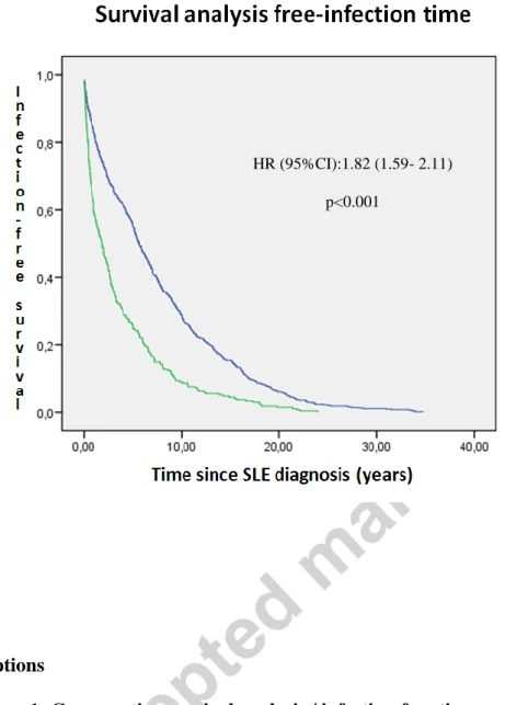
infections / 1000 patient years. The incidence rate for a second infection was 71.1 (95% CI: 63.1 -79.8) infections/1000 patient years. The time elapsed from first infection to second infection was lower than the time from baseline to first infection (median time 1.86 vs. 5.61 years, log rank p<0.000). (Figure 1),
The SKI was higher in patients with ≥ 1 severe infection compared to the whole cohort, even when excluding patients with GC (>10mg) or IS (whichever) at the time of the severe infection (N=244; SKI: 3.41±1.65 vs. 2.65±1.67, p <0.0001). When comparing patients with only one infection versus those with ≥ 2, those with > 1 infection had a higher SKI score (4.35 ±2.05 vs. 3.42 ±1.86, p=0.000) and a higher SDI score (3.33 ±2.75 vs. 1.84 ±1.98, p=0.000] at the last assessment.
Regarding etiologic agents, there was a clear predominance of bacterial causes (51.9%), with 30.4% corresponding to unknown cause (Table 3). The rate of incidence of
mycobacterial infections was 1/1000 patient-years.Analysing the soft rates (mobile-means every 5 years), a steady incidence rate was observed throughout the follow-up period. However, when mycobacterial infection rates were analysed, the incidence rate showed an annual decrement (p=0.02).
Concerning localization, the respiratory tract was the most frequently involved (35.5%), followed by the urinary tract (15.0%) (Table 3). A total of 208 (5.7%) patients died during follow-up, 24.5% by infection. The predominant localization for the fatal infection was the circulatory stream (bacteraemia, regardless of their source) (42.0%), followed by respiratory-related ones (34.0%).
3.1 Bivariate analysis
Several variables were associated with severe infection in the bivariate analysis, with density of incidence serving as a dependent variable (table 4), highlighting the
hospitalization by SLE (Relative Risk: 5.67) and immunosuppressant use (RR: 10.20). Several co-morbidities/situations were also associated with severe infection, such as occurred with tobacco smoking (any history) (RR: 1.35) (Table 4). Furthermore, in the bivariate analysis any history of smoking was also associated with respiratory infections (RR: 1.36).
Regarding antimalarials, current and past use of antimalarials were protective factors [RR 0.49 (95% CI: 0.43-0.57) and 0.76 (95% CI: 0.65-0.88), respectively p=0.0000]. Furthermore, when quintiles of time on antimalarials (months) were analyzed, we observed an inverse relationship with the incidence of severe infection; namely, a proportional decrease in the incidence ratio with each quintile, from RR: 1 for Q1 (9.4 months on antimalarials) to RR: 0,66 (95% CI: 0,53-0,84) for Q5 (68.3 months). Specifically, antimalarial users suffered viral infections less frequently than non-users (2.8% in users vs. 5.8% in non-users, p<0.001).
GC use at the time of the infectious event was associated with increased viral (14.6% vs. 11.1%) and fungal infections (3.8% vs. 0.8%), with a higher percentage of agent isolations (75.8% vs. 62.8% in non-GC users) (p<0.0001).
We also observed an association between GC at time of infection and mycobacterium as an etiologic agent: 3.7% in GC users vs. 0.2% in non-GC users (any dose), p <0.00001. As shown in Table 4, there were differences in cumulative IS use between patients with and without severe infection. Interestingly, 45.1% of patients that died by infection were on IS at that time vs. 9.6% of the surviving patients with severe infection (p<0.001). Furthermore, 45.1% of the patients that died by infection were on GC (> 10 mg/day) vs. 9.5% of survivors (p<0.001).
In a bivariate sub-analysis of mycobacterial infections, the following factors were all associated with this group of microorganism: Hospitalization by SLE (RR = 2.88; 95%CI:1.33-6.23; p= 0.007), renal disease (RR = 1.98; 95%CI:1.08-3.66; p = 0.04), severity [SKI>4: RR = 2.10 (1.10-4.03), p=0.04] and comorbidity [Charlson>4: RR=2.47 (95%CI:1.29-4.76), p=0.009].
3.2 Multivariable analysis
In the Cox proportional-hazards regression model, which used time until first infection as the dependent variable, the following were associated with severe infection: age at diagnosis, Latin-American ethnicity, any history of glucocorticoid (≥10 mg/day), immunosuppressor or tobacco use, time on antimalarials (months), hospitalization due to SLE, renal involvement, SKI, and SDI (Table 5). When the IS were considered separately, Rituximab (HR=1.61, p<0.001), Abatacept (HR=1.58, p=0.01) and
Mycophenolate mofetil or Mycophenolic acid (HR=1.41, p=0.01) showed all of them statistical significative association with severe infection.
When using a time repeated-events Cox regression (Andersen-Gill variant) model, with time of follow-up to infection being the dependent variable, results proved similar (Table 5), although immunosuppressant use, time on antimalarials and SDI lost
statistical significance. Worth noting, however, is that previous severe infection reached significance only in the last model.
4. Discussion
The incidence of severe infection in SLE patients in the RELESSER registry (19.3%) was not very different than previous reports involving other large cohorts (4, 9, 21, 22), although certainly lower than in previous studies from monocentric cohorts in Spain, ranging from 29 to 38% (23, 24), or than was recently reported by Feldman et al in a study involving an extensive administrative SLE database from the USA (25) . Differences in patient selection and/or time of follow-up could explain these
discrepancies or, as in the case of the North American cohort, the explanation might lie rather in ethnic or heath system differences. In fact, these patients were treated under Medicaid, a public health insurance program for low-income individuals, a
socioeconomic status that could influence the infection risk (25).
Consistent with most other studies, the RELESSER data confirm that respiratory infections are the most common severe infection in SLE (3, 21-28).
Regarding aetiology, bacteria were the most common agent involved followed by virus and fungus, which is also consistent with previous reports (25, 29). The percentage of infections of unknown origin was relatively high (a third of the total), but that was not unexpected, given the high percentage of respiratory infections in this cohort. In fact, in SLE, as in the general population the aetiology of pneumonias remains unknown in ~50% of all cases (15, 30-32).
There are scarce, reliable data concerning mycobacterium infections in the largest cohorts of SLE. The reported prevalence of M. tuberculosis infection in SLE patients ranges widely, from 5% to 30% (33, 34). A total of 42 mycobacterium infections (3.5%) were registered in RELESSER, equivalent to a prevalence of 1.14% in the global
RELESSER cohort. This prevalence is higher than the mycobacterium tuberculosis infection rate expected in the general Spanish population (34). Furthermore, consistent with other studies (33, 35, 36), we found a higher proportion of extrapulmonary mycobacterium infections than expected, with an even higher percentage of
extrapulmonary (vs. pulmonary) in our patients (57.1%). Additionally, we observed that this infection tended to appear during the first years of the SLE course and was
associated with GC use at the time of infection, suggesting a relationship exists between higher disease activity and higher levels of immunosuppressive therapy.
We found an association, previously unreported, between tobacco use and recurrent severe infection in the RELESSER database. This is not surprising, since active
smoking increases the risk of developing community-acquired pneumonia in the general population (37, 38). Intriguingly, despite the fact that pneumonia is the most common severe infection in SLE, studies specifically addressing pneumonia in SLE have failed to demonstrate tobacco use as a specific risk factor (15, 31). This apparent absence of association could be explained by its limited statistical power or, alternately, tobacco use could be a risk factor for any type of infection, not just respiratory ones. In fact, tobacco use increases an individual’s susceptibility to bacterial infections in general, compromising the anti-bacterial function of leukocytes, including neutrophils, monocytes, T and B cells (39). In any case, we have demonstrated an association between tobacco use and respiratory infections in the RELESSER data.
According to our findings, a previous infectious event appears to increase the risk of a new infection in SLE patients, a fact only sporadically explored (40). While this risk factor could be a mere consequence of persistent predisposing factors that increase the susceptibility to recurrent infections, our data provides information about the global risk of infection, shedding light on unknown predisposing factors.
The association of severe infection with IS or GC use is well known (1-5,13). Any use of GC ≥ 10mg increases the risk of infection in our study, and 84% of the patients were been treated with GC at the infection time. These findings reinforce the role of GC as severe infection risk factor in SLE. Perhaps more interesting from our analysis is that the use of these treatments at the time of infection increases the risk of mortality from severe infection. On the other hand, a negative association between antimalarials exposure time and severe infection was observed in our study, reinforcing the possible role these drugs play in protecting against infection, a finding previously reported (25, 27, 41, 42). Our results, however, do not agree with the other available multicenter study, which analyzed the exposure time to antimalarials in the GLADEL cohort. In that multi-nation inception cohort, no association was found between infection
prevalence or death by infection and antimalarial exposure time. However, the follow-up period in this study was relatively short, with low numbers of infection and lesser mean durations of antimalarial drug exposure (48.5 months vs. 78.4 in RELESSER) (43). An alternative, and somewhat provocative, explanation could lie in ethnic factors, given the Latin-American origins of the entire GLADEL cohort. While those
differences may reflect the low severity of lupus in the European Caucasian population, one could also speculate that antimalarials might be less effective in Hispanics. This would also explain why we found a higher incidence of severe infection in this ethnic minority in our own study. Interestingly, in our analysis, the association of exposure
duration with antimalarials was independent of severity, as measured by a validated quantitative severity index. This suggests the absence of a cofounding variable since it points to a less severe cases bias. The alkalinization of phagolysosomes and the inhibition of DNA replication has been posited as one mechanism explaining the
protective ability of antimalarials, particularly against intracellular microorganisms such a salmonella, viral agents, etc. (44-48). Consistent with this, protection against viral infections was observed in our analysis.
Finally, the protective effect of the antimalarials on infection risk could be reduced by the tobacco use, even though both factors showed an independent effect on infection risk in our statistical models.
We identified age at diagnosis as an independent factor associated with infection in this study, even when adjusted for co-morbidity as measured by the Charlson comorbidity index. This finding suggests that lupus exerts perhaps more impact over the immune system with increasing age than previously realized, an explanation that makes
biological sense. In fact, in the general population the incidence of pneumonia increases with aging (49).
A higher risk of infection was identified in the Latin-American (Amerindian-Mestizo) minority. This ethnic difference has been previously noted in Afro-American SLE patients (25, 50) but not, to the best of our knowledge, in Latin Americans. Indeed, Feldman et al observed a slightly reduced risk of serious infection among Hispanics (HR 0.90 [95% CI 0.82–0.99]) in their previously named analysis. If confirmed, our results are particularly interesting, given that the difference was adjusted by severity and that the Latin American patients from the RELESSER cohort lived in Spain during the observation period and had complete access to the same public and universal health system as native Caucasian patients. This suggests that ethnicity, and not simply
severity or socioeconomic factors, influences the incidence of infection.
The primary objective of our study was to explore the differences between SLE patients with or without previous severe infection. According to our data, SLE patients with serious infection suffer more severe disease. Not only are they more frequently hospitalized for SLE activity, but they also seem to have lower survival rates than patients without such infections. An independent relationship between SLE severity and pneumonia has been previously reported (15). This is not an unexpected finding given the fact that SLE itself entails several immunologic disorders predisposing individuals to infection (1, 14). However, weighing the impact of severity and immunosuppressive treatment on severe infection incidence remains problematic due to the presence of collinearity, as the most severely affected patients tends to receive more
immunosuppressive therapy.
Some limitations of this study must be acknowledged. Several variables, such as measures of activity and damage, were not documented at the time of infection due to the multi-purpose nature of the RELESSER-T registry, which was never specifically designed to study infection in SLE. Although its retrospective design has an inherent bias, we believe that a bias towards unreported infections was unlikely given the definition of severe infection used in our study, which encompassed hospitalization. This definition of severe infection could exclude some patients suffering from
pneumonia or others severe infections not subject to hospitalization. However, given the characteristics of our Sanitary Health System, where the vast majority of patients with severe infections are managed inpatient, we believe our cohort sufficiently
Concerning SLE characteristics and covariates, the large number of patients and the special standardization effort carried out (17) minimize the impact of the retrospective design in missing data and quality.
The relative rates of mortality by infection in SLE patients are consistent in most cohorts, accounting for at least a quarter of all deaths (2, 3, 51-53). Interestingly, blood stream infections were the first cause of fatal infection in our cohort, supplanting respiratory-related causes at the top of the list. These results reinforce previous findings that lupus patients with bacteraemia episodes have poor outcomes (54-56). Strategies to reduce bacteraemia-related mortality - such as immediate instauration of antibiotic therapy if sepsis is suspected and others (e.g., the so-called “surviving sepsis campaign” strategy) should be implemented with particular care in SLE febrile patients,
particularly patients on IS (57). In fact, delayed or inadequate antibiotic therapy was the most significant independent factor for mortality in SLE-infected patients from one intensive care unit (58).
5. Conclusions
Our results need to be confirmed in prospective studies; specifically, with a more exhaustive inclusion of variables related to infection. Nonetheless, several remarkable conclusions can be drawn from our data:
1. Severe infection is associated with severity and damage in SLE patients. There are ethnic differences in the prevalence of infection in SLE, with rates appearing to be higher in Latin Americans.
2. Respiratory bacterial infections are the most commonly found severe infection in SLE, although bacteraemia are more often fatal. Indeed, there is an urgent need to implement improvements in both the early detection and aggressive management of this dangerous complication of SLE.
3. A previous infectious event increases the risk of a subsequent infection in SLE patients. Tobacco use seems to be a risk factor for severe infection in SLE patients. In addition, antimalarials exert a time-dependent protective effect.
Acknowledgments
We are grateful to all of the employees of theSpanish Rheumatology Society Research Unit for their commitmentand professionalism.
Bibliography
1. Iliopoulos AG, Tsokos GC. Immunopathogenesis and spectrum of infections in systemic lupus erythematosus. Semin Arthritis Rheum 1996; 25:318–36.
2. Noël V, Lortholary O, Casassus P Cohen P, Généreau T, André MH et al. Risk factors and prognostic influence of infection in a single cohort of 87 adults with systemic lupus erythematosus. Ann Rheum Dis 2001; 60:1141-44.
3. Goldblatt F, Chambers S, Rahman A, Isenberg DA. Serious infections in British patients with systemic lupus erythematosus: hospitalisations and mortality.
Lupus 2009; 18:682-89.
4. Cervera R, Khamashta MA, Font J, Sebastiani GD, Gil A, Lavilla P et al. Morbidity and mortality in systemic lupus erythematosus. A multicenter prospective study of 1000 patients. Medicine (Baltimore) 1999; 78:167–75.
5. Jacobsen S, Petersen J, Ullman S, Junker P, Voss A, Rasmussen JM et al. A
multicentre study of 513 Danish patients with systemic lupus erythematosus. II. Disease mortality and clinical factors of prognostic value. Clin Rheumatol 1998; 17:478-84.
6. Halberg P, Alsbjørn B, Balslev JT, Lorenzen I, Gerstoft J, Ullman S et al. Systemic lupus erythematosus. Follow-up study of 148 patients. II: Predictive factors of
importance for course and outcome. Clin Rheumatol 1987; 6:22-26.
7. Hernandez-Cruz B, Tapia N, Villa-Romero AR, Reyes E, Cardiel MH. Risk factors associated with mortality in systemic lupus erythematosus. A case-control study in a tertiary care center in Mexico City. Clin Exp Rheumatol 2001; 19:395-401.
8. Thomas G, Mancini J, Jourde-Chiche N, Sarlon G, Amoura Z, Harlé JR et al.
Mortality associated with systemic lupus erythematosus in France assessed by multiple-cause-of-death analysis. Arthritis Rheumatol 2014; 66:2503-11.
9. Petri M, Genovese M. Incidence of and risk factors for hospitalizations in
systemic lupus erythematosus: a prospective study of the Hopkins Lupus Cohort. J Rheumatol 1992; 19:1559-65.
10. Edwards CJ, Lian TY, Badsha H, Teh CL, Arden N, Chng HH. Hospitalization of individuals with systemic lupus erythematosus: characteristics and predictors of outcome. Lupus 2003; 12:672-76.
11. Tektonidou MG, Wang Z, Dasgupta A, Ward MM. Burden of Serious Infections in Adults With Systemic Lupus Erythematosus: A National Population-Based Study, 1996-2011. Arthritis Care Res (Hoboken) 2015; 67:1078-85.
12. Cervera R, Rúa-Figueroa I, Gil-Aguado A, Sabio JM, Pallarés L,
Hernández-Pastor LJ et al. Direct cost of management and treatment of active systemic lupus erythematosus and its flares in Spain: the LUCIE Study. Rev Clin Esp (Barc) 2013; 213:127-37.
13. Fessler B. Infectious diseases in systemic lupus erythematosus: risk factors, management and prophylaxis. Best Pract Res Clin Rheumatol 2002; 16:281-91.
14. Cuchacovich R, Gedalia A. Pathophysiology and clinical spectrum of infections in systemic lupus erythematosus. Rheum Dis Clin North Am 2009; 35:75-93.
15. Rúa-Figueroa I, Nóvoa J, García-Laorden MI, Erausquin C, García-Bello M, Rodríguez de Castro F et al. Clinical and immunogenetic factors associated with pneumonia in patients with systemic lupus erythematosus: a case-control study. J Rheumatol 2014; 41:1801-17.
16. Rúa-Figueroa Í, Richi P, López-Longo FJ, Galindo M, Calvo-Alén J,
Olivé-Marqués A et al; On behalf of EAS-SER (Systemic Diseases Study Group of the Spanish Society of Rheumatology). Comprehensive Description of Clinical
Rheumatology Society Lupus Registry (RELESSER) With Emphasis on Complete Versus Incomplete Lupus Differences. Medicine (Baltimore) 2015; 94: e267.
17. Rúa-Figueroa I, López-Longo FJ, Calvo-Alén J, Galindo-Izquierdo M, Loza E, García de Yebenes MJ et al; Grupo de trabajo en EnfermedadesAutoinmunes Sistémicas de la Sociedad Española de Reumatología (EAS-SER); Unidad de Investigación de la Sociedad Española de Reumatología (UI-SER). National registry of patients with systemic lupus erythematosus of the Spanish Society of Rheumatology: objectives and methodology. Reumatol Clin 2014; 10:17-24.
18. Pego-Reigosa JM, Rúa-Figueroa I, López-Longo FJ, Galindo-Izquierdo M, Calvo-Alén J, Olivé-Marqués A et al. Analysis of disease activity and response to treatment in a large Spanish cohort of patients with systemic lupus erythematosus. Lupus 2014; Dec 15. pii: 0961203314563818. [Epub ahead of print].
19. Katz JD, Senecal JL, Rivest C, Goulet JR, Rothfield N. A simple severity of disease index for systemic lupus erythematosus. Lupus 1993; 2:119-23.
20. Gill R. Understanding Cox's regression model. Experientia 1982; 41 (Suppl):187-99.
21. Ginzler E, Diamond H, Kaplan D, Weiner M, Schlesinger M, Seleznick M. Computer analysis of factors influencing frequency of infection in systemic lupus erythematosus. Arthritis Rheum 1978; 21:37-44.
22. Gladman DD, Hussain F, Ibanez D, Urowitz MB. The nature and outcome of infection in systemic lupus erythematosus. Lupus 2002; 11:234-39.
23. González León R, Castillo Palma MJ, García Hernández FJ, Sánchez Román J. [Severe infections in a cohort of patients with systemic lupus erythematosus]. Med Clin (Barc) 2010; 135:365-67.
24. Ruiz-Irastorza G, Olivares N, Ruiz-Arruza I, Martinez-Berriotxoa A, Egurbide MV, Aguirre C. Predictors of major infections in systemic lupus erythematosus. Arthritis Res Ther 2009; 11:R109.
25. Feldman CH, Hiraki LT, Winkelmayer WC, Marty FM, Franklin JM, Kim SC, Costenbader KH. Serious infections among adult Medicaid beneficiaries with systemic lupus erythematosus and lupus nephritis. Arthritis Rheumatol 2015 ; 67:1577-85.
26. Paton NI, Cheong IK, Kong NC, Segasothy M. Risk factors for infection in Malaysian patients with systemic lupus erythematosus. Q J Med 1996; 89: 531–38.
27. Nived O, Sturfelt G, Wollheim F. Systemic lupus erythematosus and infection: a controlled and prospective study including an epidemiological group. Q J Med 1985; 55:271-87.
28. Petri M. Infection in systemic lupus erythematosus. Rheum Dis Clin North Am 1998; 24:423-56.
29. Barber C, Gold WL, Fortin PR. Infections in the lupus patient: perspectives on prevention. Curr Opin Rheumatol 2011; 23:358-65.
30. Johansson N, Kalin M, Tiveljung-Lindell A, Giske CG, Hedlund J. Etiology of community-acquired pneumonia: increased microbiological yield with new diagnostic methods. Clin Infect Dis 2010; 50:202-09.
31. Kinder BW, Freemer MM, King TE Jr, Lum RF, Nititham J, Taylor K et al. Clinical and genetic risk factors for pneumonia in systemic lupus erythematosus. Arthritis Rheum 2007; 56:2679-86.
32. Narata R, Wangkaew S, Kasitanon N, Louthrenoo W. Community-acquired
pneumonia in Thai patients with systemic lupus erythematosus. Southeast Asian J Trop Med Public Health 2007; 38:528-36.
33. Prabu V, Agrawal S. Systemic lupus erythematosus and tuberculosis: A review of complex interactions of complicated diseases. J Postgrad Med 2010; 56:244-50.
34. González-León R, Garrido Rasco R, Chinchilla Palomares E, García Hernández FJ, Castillo Palma MJ, Sánchez Román J. Tuberculosis in a cohort of patients with
35. Sayarlioglu M, Inanc M, Kamali S, Cefle A, Karaman O, Gul A et al. Tuberculosis in Turkish patients with systemic lupus erythematosus: increased frequency of
extrapulmonary localization. Lupus 2004; 13:274-78.
36. Yun JE, Lee SW, Kim TH, Jun JB, Jung S, Bae SC et al. The incidence and clinical characteristics of Mycobacterium tuberculosis infection among systemic lupus
erythematosus and rheumatoid arthritis patients in Korea. Clin Exp Rheumatol 2002; 20:127-32.
37. Almirall J, Bolíbar I, Serra-Prat M, Roig J, Hospital I, Carandell E et al;
Community-Acquired Pneumonia in Catalan Countries (PACAP) Study Group. New evidence of risk factors for community-acquired pneumonia: a population-based study. Eur Respir J 2008; 31:1274-84.
38. Bello S, Menéndez R, Torres A, Reyes S, Zalacain R, Capelastegui A et al. Tobacco smoking increases the risk for death from pneumococcal pneumonia. Chest 2014; 146:1029-37.
39. Bagaitkar J, Demuth DR, Scott DA. Tobacco use increases susceptibility to bacterial infection. Tob Induc Dis 2008; 18:4-12.
40. Enberg G M, Kahn Ch M, Goity F C, Villalón S MV, Zamorano R J, Figueroa E F. Infections in patients with Systemic Lupus Erythematosus. Rev Med Chil 2009;
41. Sisó A, Ramos-Casals M, Bové A, Brito-Zerón P, Soria N, Muñoz S et al. Previous antimalarial therapy in patients diagnosed with lupus nephritis: influence on outcomes and survival. Lupus 2008; 17:281-88.
42. Bultink IE, Hamann D, Seelen MA, Hart MH, Dijkmans BA, Daha MR et al. Deficiency of functional mannose-binding lectin is not associated with infections in patients with systemic lupus erythematosus. Arthritis Res Ther 2006; 8 :R183.
43. Shinjo SK, Bonfá E, Wojdyla D, Borba EF, Ramirez LA, Scherbarth HR et al; Grupo Latino Americano de Estudio del Lupus Eritematoso (Gladel). Antimalarial treatment may have a time-dependent effect on lupus survival: data from a multinational Latin American inception cohort. Arthritis Rheum 2010; 62:855-62.
44. Rolain JM, Colson P, Raoult D. Recycling of chloroquine and its hydroxyl analogue to face bacterial, fungal and viral infections in the 21st century. Int J Antimicrob Agents 2007; 30:297-308.
45. Paton NI, Aboulhab J, Karim F. Hydroxychloroquine, hydroxycarbamide, and didanosine as economic treatment for HIV-1. Lancet 2002; 359:1667-68.
46. Savarino A, Boelaert JR, Cassone A, Majori G, Cauda R. Effects of chloroquine on viral infections: an old drug against today's diseases? Lancet Infect Dis 2003; 3:722-27.
47. Wilson DE. Chloroquine: protection against virus-induced cell damage without inhibition of virus growth. J Gen Virol 1972; 14:107-09.
48. Kersh GJ. Antimicrobial therapies for Q fever. Expert Rev Anti Infect Ther 2013; 11:1207-14.
49. Torres A, Peetermans WE, Viegi G, Blasi F.. Risk factors for community-acquired pneumonia in adults in Europe: a literature review. Thorax 2013; 68:1057-65.
50. Anderson E, Nietert PJ, Kamen DL, Gilkeson GS. Ethnic disparities among patients with systemic lupus erythematosus in South Carolina. J Rheumatol 2008 ; 35:819-25.
51. Wallace DJ, Podell T, Weiner J, Klinenberg JR, Forouzesh S, Dubois EL. Systemic lupus erythematosus--survival patterns. Experience with 609 patients. JAMA 1981; 245:934-38.
52. Mok CC, Lee KW, Ho CT,Lau CS, Wong RW. A prospective study of survival and prognostic indicators of systemic lupus erythematosus in a southern Chinese population. Rheumatology (Oxford) 2000; 39:399-406.
53. Cervera R, Khamashta MA, Font J, Sebastiani GD, Gil A, Lavilla P et al; European Working Party on Systemic Lupus Erythematosus. Morbidity and mortality in systemic lupus erythematosus during a 10-year period: A comparison of early and late
manifestations in a cohort of 1,000 patients. Medicine (Balt) 2003; 82:299-308. 54. Marcos M, Fernández C, Soriano A, Marco F, Martínez JA, Almela M et al. Epidemiology and clinical outcomes of bloodstream infections among lupus patients. Lupus 2011; 20:965-71.
55. Chen MJ, Tseng HM, Huang YL, Hsu WN, Yeh KW, Wu TL et al. Long-term outcome and short-term survival of patients with systemic lupus erythematosus after bacteraemia episodes: 6-yr follow-up. Rheumatology (Oxford) 2008; 47:1352-57. 56. Duffy KN, Duffy CM, Gladman DD. Infection and disease activity in systemic
lupus erythematosus: a review of hospitalized patients. J Rheumatol 1991; 18:1180-84.
57. Dellinger RP, Levy MM, Carlet JM, Bion J, Parker MM, Jaeschke R et al;
International Surviving Sepsis Campaign Guidelines Committee; American Association of Critical-Care Nurses; American College of Chest Physicians; American College of Emergency Physicians; Canadian Critical Care Society; European Society of Clinical Microbiology and Infectious Diseases; European Society of Intensive Care Medicine; European Respiratory Society; International Sepsis Forum; Japanese Association for Acute Medicine; Japanese Society of Intensive Care Medicine; Society of Critical Care Medicine;Society of Hospital Medicine; Surgical Infection Society; World Federation of Societies of Intensive and Critical Care Medicine. Surviving Sepsis Campaign: international guidelines for management of severe sepsis and septic shock: 2008. Crit Care Med 2008; 36:296-327.
58. Feng PH, Lin SM, Yu CT, Yu KH, Huang CD, Tsai YH et al. Inadequate
antimicrobial treatment for nosocomial infection is a mortality risk factor for systemic lupus erythematous patients admitted to intensive care unit. Am J Med Sci 2010; 340:64-8.
Tables
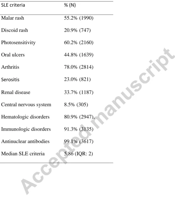
Table 1: ACR-97 SLE criteria distribution
SLE criteria % (N) Malar rash 55.2% (1990) Discoid rash 20.9% (747) Photosensitivity 60.2% (2160) Oral ulcers 44.8% (1639) Arthritis 78.0% (2814) Serositis 23.0% (821) Renal disease 33.7% (1187) Central nervous system 8.5% (305) Hematologic disorders 80.9% (2947), Immunologic disorders 91.3% (3135) Antinuclear antibodies 99.1% (3617) Median SLE criteria 5.86 (IQR: 2)
Table 2: SLE-related treatments At time of RELESSER-T assessment At time of severe infection Cumulative Treatments (any use) N % N % N % Corticosteroids 1918 52.4% 1009 87.2% 3095 84.6% Methotrexate 215 5.9% 39 6.6% 577 15.8% Leflunomide 47 1.3% 1 0.2% 127 3.5% Azathioprine 431 11.8% 196 33.3% 1140 31.2% Cyclophosphamide 53 1.4% 130 22.1% 776 21.2% Mycophenolate mofetil 322 1.4% 117 19.9% 522 14.3% Mycophenolic acid 64 1.7% 16 2.7% 85 2.3% Antimalarials 2021 55.2% NR NR 2882 78.7% Rituximab 71 1.9% 25 4.2% 226 6.1%
Localization N % Respiratory 425 35.5% Urinary tract 180 15.0% Soft tissues 159 13.3% Bacteraemia /sepsis 141 11.8% Gastrointestinal 108 9.0% Septic arthritis 39 3.3% CNS 33 2.8% Endocarditis 11 0.9% Others 102 8.5% Total 1198 100% Agent N % Bacteria 622 51.9% Mycobacterium 42 3.5% Fungus 27 2.3% Virus 143 11.9% Unknown 364 30.4%
Table 4 Rate of infection in presence or absence of factor and relative risks. Bivariate analysis (dependent variable: Density of incidence = number of infections/1000 patients-year)
Independent variables N (%) Present (infections/1000 patients-year) Absent (infections/1000 patients-year) RR (95%CI) P value Age at diagnosis (years) (≤30) 1523 (42) 30.6 27.6 1.12 (1.03-1.22) 0.01 Male sex 353 (9.7) 36.6 28.5 1.69 (1.31-2.16) <0.0001 Latin-American ethnicity 185 (5.1) 48.5 28.5 1.69 (1.31-2.16) < 0.0001 SLEDAI > 2 1239 (34.1) 38.6 25.0 1.54(1.38-1.73) <0.0001 Last SKI > 4 515 (14.2) 68.3 20.9 2.78(2.55-3.04) <0.0001 Renal involvement 1296 (35.7) 45.4 18.3 2.48 (2.21-2.79) 0.0001 Charlson index > 3 727 (20.0) 47.7 23.1 1.98(1.82-2.16) 0.001 Chronic liver disease 38 (1.0) 129.5 28.4 4.56 (3.47-5.99) < 0.0001 HIV/AIDS 9 (0.2) 102.6 29.0 3.54 (1.82-6.20) 0.007 Hepatitis C virus infection 48 (1.3) 79.0 28.3 2.79 (2.16-3.61) < 0.0001 Malignancy 207 (5.7) 40.5 28.3 1.40 (1.17-1.68) 0.0004 COPD 98 (2.7) 65.6 28.0 2.34 (1.89-2.89) <0.0001 Splenectomy 52 (1.4) 53.7 28.8 1.86 (1.33-2.61) 0.0004 Dialysis 106 (2.9) 88.3 27.3 3.31 (2.80-3.92) < 0.0001 Diabetes 179 (4.9) 58.2 27.5 2.12 (1.78-2.53) < 0.0001 Tobacco smoking (any history) 1353 (37.3) 32.8 27.2 1.35 (1.06-1.73) 0.018 Hospitalization by SLE 1954 (53.8) 43.8 7.7 5.67 (4.73-6.80) < 0.0001 Antimalarials use ≤5 years 951 (26.2) 34.4 24.0 1.39(1.11-1.74) 0.004 IS (any use) 394 (10.8) 132.3 13.0 10.20 (9.10-11.44) < 0.0001 RR = relative risk; SLEDAI = Systemic Lupus Erythematosus Disease Activity Index; SKI= Severity Katz index; HIV: Human Immunodeficiency Virus; COPD = Chronic obstructive pulmonary disease; SLE = Systemic Lupus Erythematosus; IS = immunosuppressant; HCV=hepatitis C virus.
β p value HR* 95%CI
Age at diagnosis 0.01566111 0.0000 1.016 1.0091.023
Hispanic ethnicity 0.76575765 0.0000 2.151 1.5393.005
Corticoids (≥10 mg/day), any use 0.23954448 0.0224 1.271 1.0341.561 Time on antimalarials (months) 0.00175786 0.0022 0.998 0.9970.999 Immunosuppressors, any use 0.29879736 0.0085 1.348 1.0791.684
Hospitalization by SLE 0.94264204 0.0000 2.567 1.9053.459
Renal involvement 0.31466771 0.0013 1.370 1.1301.660
SKI 0.14835582 0.0000 1.160 1.1051.217
SDI 0.06663931 0.0003 1.069 1.0311.108
Tobacco any use 0.28698037 0.0011 1.332 1.1211.583
Table 6 Time repeated-events Cox regression. Dependent variable: Time to severe infection
β HR* 95%CI p value
Age at diagnosis (quintiles) 0,1163 1.12 1.07–1.18 0.001
Male sex 0.3962 1.49 1.22–1.81 0.0001
Hispanic ethnicity 0.427 2.40 2.29–2.50 0.001
Corticoids (≥10mg/day), any use 0.2878 1.33 1.15–1.55 0.001
Hospitalization by SLE 1.0049 2.73 2.22–3.35 0.0000
SKI 0.062 1.06 1.03–1.10 0.002
Previous infection (order) 0.8739 2.40 2.29–2.50 0.0000
*HR: hazard ratio. SLE= Systemic Lupus Erythematosus; SKI= Severity Katz Index. Enter method: Forward likelihood-ratio. Variables not included in the model: Charlson (p =0.134), SLEDAI (p =0.103), SLE Damage Index (p= 197), diabetes (p =0.151), malignancy (p =0.145), hepatitis C virus infection (p =0.184), antimalarials (p =0.096).
Captions
Figure 1: Comparative survival analysis / infection-free time graphic.
The time until second infection was lower than time until first infection
HR (95%CI):1.82 (1.59- 2.11) p<0.001