Static posturography : a new perspective in the assessment of lameness in a canine model / Maria E Manera [et al ]
13
0
0
Texto completo
(2) Static Posturography in a Lame Canine Model. derived consequences of changes in paw area, and mean or maximum pressure values, among other parameters. The correct balance and its continuous preservation is a combined process connected with the central nervous system, sight, and muscular system [10]. In quiet stance position, the control of body posture is assumed as a constant action of stabilization of a multilink inverted pendulum [11,12], which corresponds with the attempt of to keep the center of mass (COM) symmetrically to the support base [13]. As posture is being constantly perturbed by internal and external mechanisms, the balance recovery is performed by constant compensatory movements, known as postural sway. In this way, static posturography becomes an objective evaluation method of the balance system, and it is widely used in human medicine [14], rehabilitation [15] or sport fields [16]. Many clinical practitioners assume that the COP position coincides with the projection of the COM on the support surface, although these two parameters are based on different concepts [17]. This sway is registered in both the latero-lateral (X) and craniocaudal (Y) axes of the body. Based on these principles, we could obtain two different graphical recordings: the statokinesiogram, which depicts the movement of COP in an X-Y coordinate system, and the stabilogram, representing the location of the COP as a time function, where movements in the X and Y axes are considered separately. When lameness is present, the associated pain causes loss of balance in the static position and is provoked by the patient transferring weight from the painful limb to the healthy (or less lame) contralateral in an effort to alleviate the pain [18]. In other words, pain can also cause postural (COP) modifications. Clinically, impaired COP balance returns to a more normal value in some cases when an effective treatment is applied; this suggests that changes in COP balance could also be a predictive tool for gait recovery [19], thus meriting evaluation [20]. In addition, theoretically, changes in COP balance in lame dogs should determine changes in paw area, as the pads are elastic structures that spread when ground contact pressure grows [21,22]. The patterns of pressure distribution in the paws might also be assessed to generate useful, objective, and complementary data as location of maximal pressure point and/or limb COP location within the paw to evaluate locomotor system status. The term medial coronoid disease (MCD) encompasses all pathologic changes of articular cartilage and subchondral bone involving the medial coronoid process of the elbow joint [23,24]. MCD is the most common cause of thoracic limb lameness in large and giant breed dogs [25], and MCD lesions have traditionally been evaluated radiographically [26–28]. A presumptive diagnosis of MCD is frequently based on detection of the resultant secondary osteoarthritis (OA), rather than on detection of the primary lesion [26,29]. Different strategies have been proposed for the treatment of OA, and among them, Platelet Rich Plasma (PRP)-based products such as plasma rich in growth factor (PRGF) therapy, has been widely used as a single or co-adjuvant therapy in the treatment of OA in dogs [30]. Based on this, we hypothesized that assessment of COP variations, together with other secondary static parameters, could serve as an objective and quantifiable tool to detect lameness and its variations. The aim of this study was to test static posturography as a potentially reliable, objective method to evaluate lameness in five lame dogs affected by OA in elbow joints. The verification of this objective builds a foundation for the feasibility of using static posturography in the clinical assessment of postural stability in lame dogs.. PLOS ONE | DOI:10.1371/journal.pone.0170692 January 23, 2017. 2 / 13.
(3) Static Posturography in a Lame Canine Model. Materials and Methods Animals A total of 10 client-owned, adult dogs of similar conformation were used in this study. The body weight of enrolled dogs ranged from 30 to 41.8 kg, and ages were 3 to 9 years. The control (sound) group was formed by five dogs (two Labradors and three Rottweilers). As they were sound dogs, a certain asymmetry should be assumed; for that reason, for comparison purposes, limbs with lesser and higher values were specifically considered. The study group contained five lame dogs (three Labradors and two Rottweilers). The lameness was unilateral and clinically classified as “severe” although with presence of weight bearing in all dogs and attributable to OA secondary to MCD of the elbow joints. Dogs did not receive medication of any kind over the 4 weeks before the analysis. To confirm or rule out OA, three standard radiographic views of both elbow joints (a lateral extension, lateral flexion, and a 15˚ oblique craniomedial caudolateral) [24] were taken under sedation with dexmedetomidine 10–20 μg/kg (Dexdomitor, zoetis, Spain)from dogs belonging to both study and control groups. Additional standard radiographs of knee and hip joints were taken in order to ensure that elbow OA was the unique reason for the observed clinical signs. A complete clinical evaluation (physical examination, including vital signs and neurologic and orthopedic exams) assured that general health was otherwise normal. The procedure was revised and authorized by the Ethical Committee of Animal Welfare (CEBA) of the University CEU Cardenal Herrera of Valencia. The owners of each animal gave permission and signed a written consent form.. Obtention-inoculation of PRP PRP was obtained using similar procedure to the PRGF1 (BTI, Vitoria, Spain), but with different materials. This product produces a moderated amount of platelets (double respecting to peripheral blood) and less than 0.3 leukocytes/ μL. The procedure is as follows: whole blood (10 mL) was aseptically extracted from the cephalic vein and collected in two 4.5-mL centrifuge tubes (BD Vacutainer1, Plymouth, UK), each containing 0.5 mL citrate solution, then centrifuged for 8 minutes at 460 × g. Only the inferior third of the obtained plasma (adjacent to the buffy coat) was used to be activated with 5% of its volume with 10% calcium chloride. The resultant ~2 mL solution was injected aseptically into the elbow joint through the conventional arthrocentesis site previous sedation with dexmedetomidine iv. The appearance of joint fluid confirmed proper needle placement. A total of four doses were administered on D0, D7, D14, and D21. After every inoculation, exercise was restricted to a walk of maximum of 30 minutes at leash during the following 2 days.. Static posturography For the recording of data, a pressure platform (EPS/R1, Loran Engineering, Bologne, Italy) was used. The device contains a total of 2096 pressure sensors of 1 cm2 distributed in an area of 48 × 48 cm. The range of pressure was 30–400 kPa, and acquisition frequency 100 Hz. Animals were placed in quiet stance with their thoracic limbs over the pressure platform, perpendicular to the ground, and each dog’s owner remained in front of the animal to attract the dog’s attention at a close distance. Three recordings of 10 seconds were obtained from each animal. Only recordings in which the animal was completely immobile in symmetric position were considered valid. Data acquisition was performed using Biomech software (Loran Engineering). The parameters for static posturography were as follows:. PLOS ONE | DOI:10.1371/journal.pone.0170692 January 23, 2017. 3 / 13.
(4) Static Posturography in a Lame Canine Model. • Distribution of pressure between both limbs measured in kilopascal (Kpa) and expressed in percentage. • Changes in paw area measured in cm2 and expressed in percentage. • Mean and maximum pressure of each thoracic limb measured in Kpa. • Graphic distribution of pressure ranges in paws of lame and sound limbs shown in a 2-dimensional and 3-dimensional (3-D) color scale from blue (low pressure), to red (high pressure). In order to obtain a correct contrast among the colors in the different pressure ranges, calibration was set manually to 212 Kpa to avoid saturation of the sensors. • Statokinesiogram represents the amplitude of the spatial migration of the COP in a 2-D space, estimated by computing ellipse area (measured in mm2), which contains 90% of the data points of the COP trajectory. • Stabilogram recorded independent X and Y oscillations (measured in mm) as a function of time.. Statistical analysis Parameters were estimated by using the free R statistical software (https://www.r-project.org/). Analysis of variance with repeated measures and a Tukey test were used to determine significant differences. Normality and homoscedasticity of residuals was confirmed using Shapiro and Levene tests, respectively.. Results The animals had a mean body weight of 36.88 ± 4.25 kg and a mean age of 5.8 ± 2.04 years. The mean values± SD of all obtained parameters are summarized in Table 1. Pressure distribution between both limbs Differences between lame and sound limbs in the study group diminished significantly from before treatment (day 0) to day 90 post treatment (p < 0.001). Compared with the control group, differences in pressure distribution at day 90 were also significant (p = 0.0044). Data were normal (p = 0.24) and homoscedastic (p = 0.64). Changes of paw area Differences in area between lame and sound limbs in the study group diminished significantly from day 0 to day 90 (p < 0.001). Compared with the control group, differences in paw area at day 90 were not significant (p = 0.072). Data were normal (p = 0.10) and homoscedastic (p = 0.68).. Mean pressure Differences in mean pressure between lame and sound limbs in the study group diminished significantly from day 0 to day 90 (p < 0.001). Compared with the control group, differences at day 90 were also significant (p < 0.001). Data were normal (p = 0.76) and homoscedastic (p = 0.72). PLOS ONE | DOI:10.1371/journal.pone.0170692 January 23, 2017. 4 / 13.
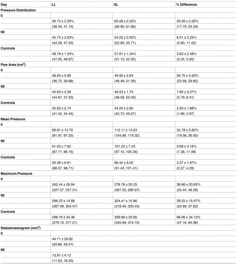
(5) Static Posturography in a Lame Canine Model. Table 1. Posturographic Parameters in Dogs, Expressed as Mean ± SD, and 95% Confidence Intervals. Day. LL. SL. % Difference. 39.72 ± 2.50%. 60.28 ± 2.50%. 20.56 ± 5.00%. (38.34, 41.10). (58.90, 61.66). (17.79, 23.33). Pressure Distribution 0. 90 45.75 ± 2.63%. 54.25 ± 2.63%. 8.51 ± 5.25%. (44.29, 47.20). (52.80, 55.71). (5.60, 11.42). Controls 48.19 ± 1.24%. 51.81 ± 1.24%. 3.63 ± 2.48%. (47.50, 48.87). (51.13, 52.50). (2.25, 5.00). 38.20 ± 2.68. 49.93 ± 2.63. 26.70 ± 5.63%. (36.72, 39.68). (48.48, 51.39). (23.58, 29.82). 45.93 ± 2.28. 49.53 ± 1.73. 7.60 ± 3.27%. (44.67, 47.20). (48.58, 50.49). (5.79, 9.41). 42.93 ± 2.74. 44.20 ± 2.65. 2.93 ± 1.88%. (41.42, 44.45). (42.73, 45.67). (1.89, 3.97). 89.61 ± 13.79. 112.11 ± 13.03. 22.78 ± 5.82%. (81.97, 97.25). (104.89, 119.32). (19.56, 26.00). 91.93 ± 7.62. 101.22 ± 7.43. 9.69 ± 4.16%. (87.71, 96.15). (97.10, 105.34). (7.38, 11.99). 93.39 ± 9.61. 96.42 ± 9.02. 3.27 ± 1.81%. (88.07, 98.71). (91.43, 101.41). (2.27, 4.28). 242.44 ± 26.84. 278.76 ± 20.25. 36.86 ± 20.62%. (227.57, 257.31). (267.55, 289.97). (25.44, 48.28). 296.23 ± 14.88. 324.41 ± 10.86. 29.25 ± 15.47%. (287.99, 304.47). (318.40, 330.43). (20.69, 37.82). 298.18 ± 34.36. 359.89 ± 25.65. 66.06 ± 34.13%. (279.15, 317.21). (345.69, 374.10). (47.16, 84.96). 2. Paw Area (cm ) 0. 90. Controls. Mean Pressure 0. 90. Controls. Maximum Pressure 0. 90. Controls. Statokinesiogram (mm2) 0 44.71 ± 26.82 (29.86, 59.57) 90 13.91 ± 4.12 (11.63, 16.20) (Continued). PLOS ONE | DOI:10.1371/journal.pone.0170692 January 23, 2017. 5 / 13.
(6) Static Posturography in a Lame Canine Model. Table 1. (Continued) Day. LL. SL. % Difference. Controls 2.17 ± 1.10 (1.56, 2.79) Day 0: Before treatment in study group; Day 90: After first application of treatment in study group; SL: Sound limb in study group or limb with higher value in control group; LL: Lame limb in study group or limb with lesser value in control group. doi:10.1371/journal.pone.0170692.t001. Maximum pressure Maximum pressure in sound limbs in the study group increased significantly from day 0 to day 90 (p < 0.001). Compared with the control group, differences at day 90 were also significant (p < 0.001). Data were normal (p = 0.20) and homoscedastic (p = 0.10). In lame limbs, maximum pressure also increased significantly from day 0 to day 90 (p < 0.001). However, compared with the control group, differences at day 90 were not significant (p = 0.99). Data were normal (p = 0.63) and homoscedastic (p = 0.42).. Statokinesiogram Oscillations of the COP were greater in lame dogs during the 10 seconds of recording, as observed in graphics, which sway measure area is greater in lame dogs, and assymetric (Fig 1). The sway area in the study group diminished significantly from day 0 to day 90 (p < 0.001). Compared with the control group, differences at day 90 were also significant (p = 0.005). Data were not normal (p = 0.005) but were homoscedastic (p = 0.14). For that reason, the results are reliable due to the robustness of analysis of variance when data are homoscedastic.. Stabilogram A symmetric latero-lateral oscillation could be seen in sound dogs, while craniocaudal oscillation was almost insignificant. In contrast, lame dogs evidenced greater or/and asymmetric oscillations. Graphical representation of COP sway in X and Y axes showed asymmetry in the study group in all cases during the 10-second recording period (Fig 2).. Graphic pressure distribution in the paws 2-D graphics showed as, when limbs were compared in sound dogs, a similar color (pressure) distribution pattern was evident: In addition, limb COP was symmetrically located within the paw, and the maximal pressure point was located over the 2nd digital pad. In lame group, the pressure distribution pattern when limbs were compared was markedly different, with maximal pressure point deviated laterally in the sound limb and cranially in the lame limb. Limb COP in the sound limb of lame dogs was located in the center, but in the lame limb has migrated craniomedially. The maximal pressure point was found in the sound limb laterally over the 5th digital pad, while in the lame limb, was located cranially, over the 4th digital pad. Body COP deviation towards the sound limb could also be seen (Fig 3). In 3-D color scale graphics, the pressure patterns in sound limbs were similar, while in lame limbs they are asymmetric (Fig 4).. PLOS ONE | DOI:10.1371/journal.pone.0170692 January 23, 2017. 6 / 13.
(7) Static Posturography in a Lame Canine Model. Fig 1. Statokinesiograms of Sound (A) and Lame (B) Dogs, Showing the Ellipse that Contains 90% of the Body COP Migration. In addition to show a greater area, in this case, a left deviation of the ellipse is evident in lame dog. doi:10.1371/journal.pone.0170692.g001. PLOS ONE | DOI:10.1371/journal.pone.0170692 January 23, 2017. 7 / 13.
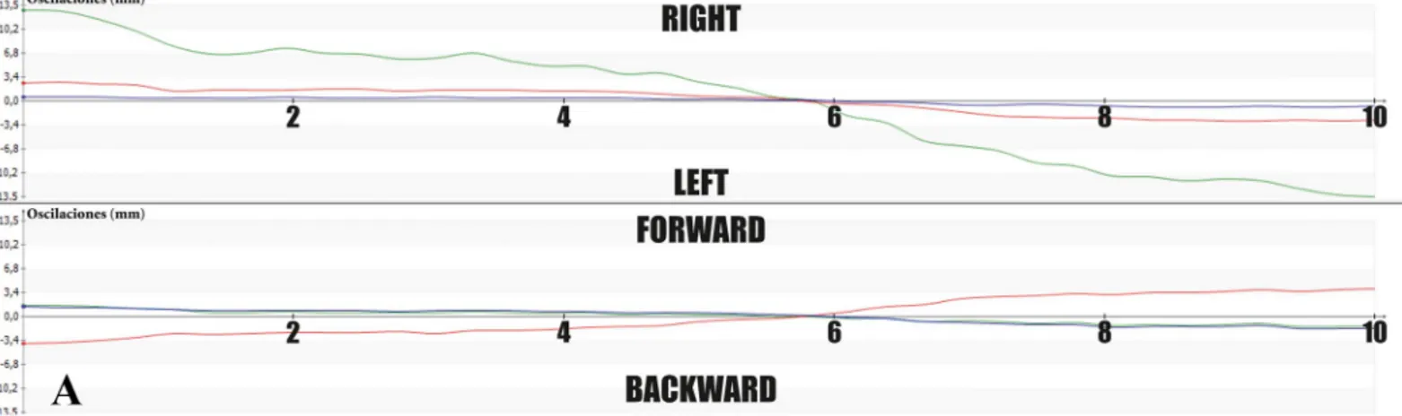
(8) Static Posturography in a Lame Canine Model. Fig 2. Stabilograms of Sound (A) and Lame (B) Dogs, Showing Oscillation of Body COP (Green Lines). Graphic shows that this lame dog had a greater oscillation to the right side. doi:10.1371/journal.pone.0170692.g002. Discussion With static posturography, we were able to determine differences in a set of parameters between lame and sound dogs. Additionally, these differences diminished following treatment, indicating a positive response and thus, effective therapy, as previously published in humans [31]. While the redaction of this paper was conducted, we were unable to find specific studies regarding the use of static posturography and pressure measurements of dog paws other than PVF and VI using a pressure platform. Moreover, this methodology is widely used in humans in different fields [14–16] with excellent results; this should encourage the increase of pressure platform technology use in dogs. Statokinesiograms and stabilograms require the necessary time for recording the spatial variations of the body COP; in humans, it is advisable to calculate parameters as the average of those obtained in three successive recordings of 10–60-second duration between studies. PLOS ONE | DOI:10.1371/journal.pone.0170692 January 23, 2017. 8 / 13.
(9) Static Posturography in a Lame Canine Model. Fig 3. 2-D Color Scale Graphics of Sound (A) and Lame (B) Dogs. In (A) symmetry is found in all measured parameters as pressure distribution, limb COP (black circle) and maximal pressure point (white circle). In (B), the pressure distribution pattern is manifestly different between lame (right) and sound limb (left), and assymetry in the other parameters is evident: limb COP (black and red in sound limb or blue circle in lame limb) maximal pressure point (white circle), body COP (black and grey circle). doi:10.1371/journal.pone.0170692.g003. [32,33]. In agreement with these recommendations, we used three successive recordings of 10 seconds on each animal. This was enough to detect asymmetries in the dislocation of the COP. Regarding the sampling frequency, recent observations in humans [34] suggest to consider an effective sampling rate of about 50 Hz. Considering that COP sway is a pendular movement, the pendulus length is shorter in dogs than in humans; thus, the frequency of oscillation should be, theoretically, higher. For that reason, in order to gain accuracy, we decided to increase the sampling rate to 100 Hz. In this study, the method suitability was assessed by using dogs with forelimb lameness because the evaluation of hind limb lameness has been demonstrated harder to perform [35]. Some authors try to explain this fact arguing that the proximal joints of the hind limb are more able to decrease the supported load during the stance phase when comparing with the forelimb, as occurs in horses [36]. In addition, the lameness should be less noticeable as a lower proportion of body weight is supported by the hind limb [37].. PLOS ONE | DOI:10.1371/journal.pone.0170692 January 23, 2017. 9 / 13.
(10) Static Posturography in a Lame Canine Model. Fig 4. 3-D Color Scale Graphic of a Sound (A) and Lame (B) Dog. In this case, lame dog shows a lateral deviation of the pressure in the sound limb (left) and a craniomedial deviation of pressure in the lame limb (right). doi:10.1371/journal.pone.0170692.g004. Static posturography in dogs not only can contribute to the scientific knowledge, but also can help clinicians to graphically show lameness status to pet owners; however, some limitations were found during the design and development of this study. First, to obtain substantial differences in the parameters derived from COP migration, the use of large breed dogs was necessary. Second, it was absolutely necessary to enroll calm and obedient dogs to stand in quiet stance for the minimum time required to obtain reliable statokinesiograms and stabilograms. In addition, each trial required an adaptation phase, and during this phase, some dogs showed fatigue or lack of attention; for that reason, acquisition time was established at 10 seconds. More than this time was almost impossible to achieve in most cases. Although 6 months is considered the minimum standard for testing the evolution of a medical or surgical treatment, we tested the animals after 3 months; we did this because after 6 months, and based on some clinical evidences, the effect of the treatment could be diminished, possibly making variation in posturographic parameters less evident or undetectable.. PLOS ONE | DOI:10.1371/journal.pone.0170692 January 23, 2017. 10 / 13.
(11) Static Posturography in a Lame Canine Model. This study used a relative low number of animals. Although the study used dogs of similar conformation, the low sample size could result in a potential lack of statistical power. Nevertheless, we believe that this study clearly demonstrates that static posturography is a powerful tool to assess lameness in dogs and advance the objective quantification of pressure distribution anomalies in limbs due to COP sway variations. For example, in the future, the maps of pressure redistribution could help to identify and even characterize specific pathologies like those detectable using other kinematic [38] or kinetic studies [39]. Great disparity of criteria has been published about the regimen of PRP administration for OA, oscillating from one to a series of six injections [40–43]. Based on these, we decided a series of four injections with a 1-week interval between them.. Conclusion Based on our results, static posturography was possible to perform in dogs and provided fairly accurate measurements of various parameters not assessed before in the evaluation of forelimb lameness. These parameters, in addition to complement the classic kinetic PVF and VI, may furnish to veterinary practitioners a useful tool to assess asymmetries simultaneously due to COP sway in lame dogs.. Supporting Information S1 File. ARRIVE guidelines. (PDF). Acknowledgments The authors thank Thomas Michael Oxlee and Misty Bailey for translation and editing, respectively. Also thanks to the dogs’ owners for their collaboration. Thanks also to the Cátedra Garcia Cugat for its technical support.. Author Contributions Conceptualization: JMV JMC MB. Data curation: AS. Formal analysis: AS. Investigation: JS MEM MR. Methodology: JMV. Resources: JMC MC JS. Supervision: JMV. Writing – original draft: MEM. Writing – review & editing: JMV JMC MB.. References 1.. Fanchon L, Grandjean D. Accuracy of asymmetry indices of ground reaction forces for diagnosis of hind limb lameness in dogs. Am J Vet Res. 2007; 68: 1089–1094. doi: 10.2460/ajvr.68.10.1089 PMID: 17916016. PLOS ONE | DOI:10.1371/journal.pone.0170692 January 23, 2017. 11 / 13.
(12) Static Posturography in a Lame Canine Model. 2.. Vilar JM, Batista M, Morales M, Santana A, Cuervo B, Rubio M, et al. Assessment of the effect of intraarticular injection of autologous adipose-derived mesenchymal stem cells in osteoarthritic dogs using a double blinded force platform analysis. BMC Vet Res. 2014; 10: 143. doi: 10.1186/1746-6148-10-143 PMID: 24984756. 3.. Gibert S, Lequang T, Maitre P, Cachon T, Carozzo C, Fau D, et al. Sensitivity and specificity to determine lameness in dogs with a pressure walkway system. Vet Comp Orthop Traumatol. 2012; A21.. 4.. Besancon MF, Conzemius MG, Derrick TR, Ritter MJ. Comparison of vertical forces in normal greyhounds between force platform and pressure walkway measurement systems. Vet Comp Orthop Traumatol. 2003; 16: 153–157.. 5.. Oosterlinck M, Pille F, Back W, Dewulf J, Gasthuys F. A pressure plate study on fore and hindlimb loading and the association with hoof contact area in sound ponies at the walk and trot. Vet J. 2011; 190: 71–76. doi: 10.1016/j.tvjl.2010.08.016 PMID: 20875762. 6.. Marghitu DB, Swaim SF, Rumph PF, Cojonaru D, Gillette RL, Scardino MS. Dynamics Analysis of Ground Contact Pressure of English Pointer Dogs. Nonlinear Dyn. 2003; 33: 253–265,. 7.. Souza AN, Pinto AC, Marvulle V, Matera JM. Evaluation of vertical forces in the pads of German Shepherd dogs. Vet Comp Orthop Traumatol. 2013; 26: 6–11. doi: 10.3415/VCOT-11-07-0100 PMID: 23111688. 8.. Souza AN, Tatarunas AC, Matera JM. Evaluation of vertical forces in the pads of Pitbulls with cranial cruciate ligament rupture. BMC Vet Res. 2014; 10: 51. doi: 10.1186/1746-6148-10-51 PMID: 24581287. 9.. Vassalo FG, Rahal SC, Agostinho FS, Mamprim MJ, Melchert A, Kano WT, et al. Gait analysis in dogs with pelvic fractures treated conservatively using a pressure-sensing walkway. Acta Vet Scand. 2015; 57: 68. doi: 10.1186/s13028-015-0158-3 PMID: 26438541. 10.. Ruhe A, Fejer R, Walker B. The test-retest reliability of centre of pressure measures in bipedal static task conditions–a systematic review of the literature. Gait Posture. 2010, 32: 436–445. doi: 10.1016/j. gaitpost.2010.09.012 PMID: 20947353. 11.. Winter DA, Prince F, Frank JS, Powell C, Zabjek KF. Unified theory regarding A/P and M/L balance in quiet stance. J Neurophysiol. 1996; 75: 2334–2343. PMID: 8793746. 12.. Maurer C, Peterka RJ. A new interpretation of spontaneous sway measures based on a simple model of human postural control. J Neurophysiol. 2005; 93: 189–200. doi: 10.1152/jn.00221.2004 PMID: 15331614. 13.. Błaszczyk JW, Lowe DL, Hansen PD. Ranges of postural stability and their changes in the elderly. Gait Posture. 1994; 2: 11–17.. 14.. Blaszczyk JW. The use of force-plate posturography in the assessment of postural instability. Gait Posture. 2016; 44: 1–6. doi: 10.1016/j.gaitpost.2015.10.014 PMID: 27004624. 15.. Tamburella F, Scivoletto G, Molinari M. Balance training improves static stability and gait in chronic incomplete spinal cord injury subjects: a pilot study. Eur J Phys Rehabil Med. 2013; 49: 353–364. PMID: 23486301. 16.. Whiteside D, Elliott BC, Lay B, Reid M. Coordination and variability in the elite female tennis serve. J Sports Sci. 2015; 33: 675–686 doi: 10.1080/02640414.2014.962569 PMID: 25358037. 17.. Baratto L, Morasso PG, Re C, Spada G. A new look at posturographic analysis in the clinical context: sway-density versus other parameterization techniques. Motor control. 2002; 6: 246–270. PMID: 12122219. 18.. Buchner HH, Obermuller S, Scheidl M. Body centre of mass movement in the lame horse. Equine Vet J Suppl. 2001; 33: 122–127.. 19.. Scivoletto G, Romanelli A, Mariotti A, Marinucci D, Tamburella F, Mammone A, et al. Clinical factors that affect walking level and performance in chronic spinal cord lesion patients. Spine. 2008; 33: 259– 264. doi: 10.1097/BRS.0b013e3181626ab0 PMID: 18303457. 20.. Tamburella F, Scivoletto G, Molinari M. Balance training improves static stability and gait in chronic incomplete spinal cord injury subjects: a pilot study. Eur J Phys Rehabil Med. 2013; 49: 53–364.. 21.. Basher, A. ‘Foot injuries in dogs and cats’, Compendium on Continuing Education Practicing Veterinarian. 1994; 1159–1176.. 22.. Swaim S F. Management and bandaging of soft tissue injuries of dog and cat feet, Journal of the American Animal Hospital Association. 1985; 21: 329–340.. 23.. Moores AP, Benigni L, Lamb CR. Computed tomography versus arthroscopy for detection of canine elbow dysplasia lesions. Vet Surg. 2008; 37: 390–398. doi: 10.1111/j.1532-950X.2008.00393.x PMID: 18564264. PLOS ONE | DOI:10.1371/journal.pone.0170692 January 23, 2017. 12 / 13.
(13) Static Posturography in a Lame Canine Model. 24.. Villamonte-Chevalier A, van Bree H, Broeckx B, Dingemanse W, Soler M, Van Ryssen B, et al. Assessment of medial coronoid disease in 180 canine lame elbow joints: a sensitivity and specificity comparison of radiographic, computed tomographic and arthroscopic findings. BMC Vet Res. 2015; 11: 243. doi: 10.1186/s12917-015-0556-9 PMID: 26407863. 25.. Fitzpatrick N, Yeadon R, Smith T, Schulz K. Techniques of application and initial clinical experience with sliding humeral osteotomy for treatment of medial compartment disease of the canine elbow. Vet Surg. 2009; 38: 261–278. doi: 10.1111/j.1532-950X.2008.00493.x PMID: 19236684. 26.. Cook CR, Cook JL. Diagnostic imaging of canine elbow dysplasia: a review. Vet Surg. 2009; 38: 144– 153. doi: 10.1111/j.1532-950X.2008.00481.x PMID: 19236671. 27.. Temwichitr J, Leegwater PA, Hazewinkel HA. Fragmented coronoid process in the dog: a heritable disease. Vet J. 2010; 185: 123–129. doi: 10.1016/j.tvjl.2009.06.022 PMID: 19640749. 28.. Smith TJ, Fitzpatrick N, Evans RB, Pead MJ. Measurement of ulnar subtrochlear sclerosis using a percentage scale in labrador retrievers with minimal radiographic signs of periarticular osteophytosis. Vet Surg. 2009; 38: 199–208. doi: 10.1111/j.1532-950X.2008.00488.x PMID: 19236678. 29.. Hornof WJ, Wind AP, Wallack ST, Schulz KS. Canine elbow dysplasia. The early radiographic detection of fragmentation of the coronoid process. Vet Clin North Am Small Anim Pract. 2000; 30: 257–266. PMID: 10768233. 30.. Vilar JM, Morales M, Santana A, Spinella G, Rubio M, Cuervo B, et al. Controlled, blinded force platform analysis of the effect of intraarticular injection of autologous adipose-derived mesenchymal stem cells associated to PRGF-Endoret in osteoarthritic dogs. BMC Vet Res. 2013; 9: 131. doi: 10.1186/17466148-9-131 PMID: 23819757. 31.. Hirjaková Z, Šingliarová H, Bzdúšková D, Kimijanová J, Bučková K, Valkovič P, et al. Postural stability and responses to vibrations in patients after anterior cruciate ligament surgical reconstruction. Physiol Res. 2016, 24: S409–S416.. 32.. Pinsault N, Vuillerme N. Test-retest reliability of centre of foot pressure measures to assess postural control during unperturbed stance. Med Eng Phys. 2009; 31: 276–286. doi: 10.1016/j.medengphy. 2008.08.003 PMID: 18835738. 33.. Schmid M, Conforto S, Camomilla V, Cappozzo A, D’Alessio T. The sensitivity of posturographic parameters to acquisition settings. Med Eng Phys. 2002; 24: 623–631. PMID: 12376049. 34.. Bottaro A, Casadio M, Morasso PG, Sanguineti V. Body sway during quiet standing: is it the residual chattering of an intermittent stabilization process? Hum Mov Sci. 2005; 24: 588–615. doi: 10.1016/j. humov.2005.07.006 PMID: 16143414. 35.. Peham C, Licka T, Girtler D, Scheidl M. Hindlimb lameness: clinical judgement versus computerised symmetry measurement. Vet Rec. 2001; 148: 750–752. PMID: 11442235. 36.. Buchner HHF. Gait adaptation in lameness. In: Back W, Clayton HM, editors. Equine Locomotion. London: WB Saunders: London, 2013. p. 251–279.. 37.. Gillette R, Graig T. Canine locomotion analysis. In: Millis D, Levine D, editors. Canine Rehabilitation and Physical Therapy. Amsterdam: Elsevier, 2014. p 201–210.. 38.. Caron A, Caley A, Farrell M, Fitzpatrick N. Kinematic gait analysis of the canine thoracic limb using a six degrees of freedom marker set. Study in normal Labrador Retrievers and Labrador Retrievers with medial coronoid process disease. Vet Comp Orthop Traumatol. 2014; 27: 461–469. doi: 10.3415/ VCOT-14-03-0051 PMID: 25345466. 39.. Kim SE, Pozzi A, Banks SA, Conrad BP, Lewis DD. Effect of cranial cruciate ligament deficiency, tibial plateau leveling osteotomy, and tibial tuberosity advancement on contact mechanics and alignment of the stifle in flexion. Vet Surg. 2010; 39: 363–370. doi: 10.1111/j.1532-950X.2010.00655.x PMID: 20522216. 40.. Moraes VY, Lenza M, Tamaoki MJ, Faloppa F, Belloti JC. Platelet-rich therapies for musculoskeletal soft tissue injuries. Cochrane Database Syst Rev. 2014: 4: CD010071.. 41.. Sánchez M, Anitua E, Azofra J, Aguirre JJ, Andia I. Intra- articular injection of an autologous preparation rich in growth factors for the treatment of knee OA: a retrospective cohort study. Clin Exp Rheumatol. 2008; 26: 910–913 PMID: 19032827. 42.. Franklin SP, Garner BC, Cook JL. Characteristics of canine platelet-rich plasma prepared with five commercially available systems. Am J Vet Res. 2015; 76: 822–827. doi: 10.2460/ajvr.76.9.822 PMID: 26309111. 43.. Cook JL, Smith PA, Bozynski CC, Kuroki K, Cook CR, Stoker AM, et al. Multiple injections of leukoreduced platelet rich plasma reduce pain and functional impairment in a canine model of ACL and meniscal deficiency. Orthop Res. 2016; 34: 607–615.. PLOS ONE | DOI:10.1371/journal.pone.0170692 January 23, 2017. 13 / 13.
(14)
Figure


+3


Documento similar
Fig. Pressure distribution in longitudinal section of breaker plate for two different geometric distributions of homogenization holes: a) not optimized; b) optimized (with x and z
Then renal and ocular resistive and pulsatility indices and systolic blood pressure were obtained in dogs and cats with diseases that can cause hypertension, such as
General thermodynamic prediction asserts the existence of a close stability region (SR) in temperature-pressure plane for the native folded state of a protein.. evidences support
[20] observed for the pyrolysis of rice husk (at a peak temperature of 700 °C in a fixed-bed reactor) that the yields of char and gas increased with elevating the pressure from
The Dwellers in the Garden of Allah 109... The Dwellers in the Garden of Allah
In summary, in a large sample of community-living individuals aged 60 and over in Spain, social support score is associated with lower nocturnal systolic BP and higher BP
In this sense, this paper studies the thermal performance of a forced draft counter-flow wet cooling tower fitted with two water distribution systems (the pressure water
The main goal of this PhD thesis is to experimentally measure pressure drop and heat transfer coefficient in condensation processes inside mini-channel tubes with different
