Trends in Neurosciences, Vol. 22, Issue 3, Pages 109-116
Sight and insight – on the physiological role of nitric oxide in the
visual system
Javier Cudeiro, Casto Rivadulla
Abstract
Research in the fields of cellular communication and signal transduction in the brain has moved very rapidly in recent years. Nitric oxide (NO) is one of the latest discoveries in the arena of messenger molecules. Current evidence indicates that, in visual system, NO is produced in both postsynaptic and presynaptic structures and acts as a neurotransmitter, albeit of a rather unorthodox type. Under certain conditions it can switch roles to become either a neuronal ‘friend’ or ‘foe’. Nitric oxide is a gas that diffuses through all physiological barriers to act on neighbouring cells across an extensive volume on a specific time scale. It, therefore, has the opportunity to control the processing of vision from the lowest level of retinal transduction to the control of neuronal excitability in the visual cortex.
Keywords: Nitric oxide; Vision; Retina; LGN; Visual cortex; NMDA; Development
Visual processing starts in the retina. Here, the image of the world is broken down through visual filters (the receptive fields of individual neurones). In mammals this visual message then moves to an intermediate station in the thalamus, the dorsal lateral geniculate nucleus (dLGN). This is a laminar structure that receives the ganglion-cell axons in an organized manner depending on the eye from which the image originated, the cell type and other species-dependent characteristics. Finally, this information is relayed to the primary visual cortex (V1) from which connections are made with many other visual cortical and sub-cortical structures. At every level of this pathway, including the retina, passage of the visual message involves the activation of members of the family of excitatory-amino-acid receptors. However, besides these specific visual signals, there is also modulation of the visual message by a number of non-specific inputs (for example, dopaminergic, cholinergic and noradrenergic), which control the excitability of the neurones and gate the flow of information. Recently, a newly discovered neurotransmitter has emerged rather spectacularly that is involved in the processing of visual information – nitric oxide (NO).
Nitric oxide: ubiquitous neurotransmitter or ‘saint–sinner’?
Since NO was first recognized as a messenger molecule in the brain that mediates the increased cGMP levels that occur on activation of NMDA receptors1, major efforts have been made to understand the extent of its actions. Nitric oxide is a gas synthesized from l-arginine by the enzyme nitric-oxide synthase (NOS). At least three forms of the enzyme have been characterized: the constitutive endothelial and neuronal types are both Ca2+ dependent and the third is a Ca2+-independent inducible isoform, which is expressed only in the presence of cytokines. In this review, only data related to neuronal NOS (nNOS) will be considered. Available data from immunocytochemistry, in situ hybridization and NADPH-diaphorase histochemistry have given a reasonably comprehensive picture of the anatomical localization of NO-generating cells and their processes throughout the CNS ( Ref. 2). The brain contains the highest level of NOS of any tissue so far examined and the broad distribution of the enzyme suggests that NO could be involved in many aspects of CNS function 1.
to some extent, controversial and confusing. Some laboratories using brain-slice or primary-tissue-culture models of glutamate neurotoxicity have reported that NO is also involved in these pathologies8 and 9, while others, employing similar methods, have shown no obvious role10 and 11. Furthermore, there is also much evidence to suggest that NO might be neuroprotective12, 13 and 14. Differences between the effects of NO have been attributed, at least in part, to different redox-related species of the NO group and their disparate chemical activities. Neurodestruction has been attributed to peroxynitrite alone and not to NO− (the reduced form), and the neuroprotective properties of NO have been attributed to NO+ (the oxidized form), as this species downregulates NMDA-receptor activity by reaction with thiol group(s) in the redox modulatory site of the receptor15, 16 and 17 (Fig. 1).
Table 1. Products and targets of neuronal nitric oxide synthasea
Nitric oxide-related species Principal biological target-reactants
N2O Metals, hydrophobic pockets
NH2OH Oxidants
NO− (MNO; SNO) Thiols, metals, oxygen
NO· (MNO; SNO) Thiols, metals, superoxide, oxygen
NOx–SNO–MNO (NO+) Thiols
OONO− Thiols, metals, tyrosine, methionine
NO2−–NO3− –
a All of the above nitric oxide (NO)-related species have been identified from purified preparations of neuronal nitric oxide synthase
(NOS)94. The cellular availability of substrates and cofactors appears to influence the oxidation state of the NOS product. Nitrous oxide (N2O) and hydroxylamine (NH2OH) are end-reduction products, while nitrite–nitrate (NO2−–NO3−) are end-oxidation
products. The remaining compounds have different biological actions and potential toxicities that reflect their chemistry in different redox milieu95, 96 and 97. Note that different SNO and MNO species might function as NO·, NO+, or NO−
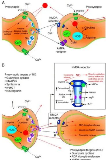
Fig. 1. Presynaptic and postsynaptic locations of nitric-oxide synthase (NOS) and the probable routes of action of nitric oxide (NO).(A) Postsynaptic location of NOS, showing possible presynaptic actions (red arrows). Calcium signal from glutamate-mediated activation of NMDA receptors or voltage-dependent Ca2+ channels (VDCCs) binds calmodulin (CaM) and activates NOS,
producing NO and citrulline from l-arginine (reviewed in Ref. 1). The free diffusion of NO suggests that this presynaptic activity need not necessarily be restricted to the presynaptic boutons directly involved with this postsynaptic element. (B) Presynaptic location of NOS, showing possible postsynaptic actions. Note the direct actions on the NMDA receptor, which is expanded in the inset box and shows the different modulatory sites. Neurotransmitter (NT) release is triggered by the arrival of an action potential in the presynaptic terminal by the opening of VDCCs. The resulting elevation in the internal Ca2+ concentration, is the signal that
causes NO production. This has been shown to occur in the dorsal lateral geniculate nucleus of the cat 18, 19 and 20, where NOS is
co-localized with ACh within the axons arising from the brainstem 21. Again, diffusion of NO can induce actions on sites that are remote from the synapse illustrated here. Therefore, the possibility of combined presynaptic and postsynaptic activities from either NOS location cannot be excluded. Abbreviations: Glu, glutamate; SNAP25, 25 kDa synaptosomal-associated protein.
Thus, in brief, NO seems to be an almost-ubiquitous messenger substance in the CNS that can, under certain conditions (such as its excessive production or the absence of regulatory control mechanisms), be toxic to cells, while possibly also being capable of acting as a neuroprotectant. However, under normal, physiological conditions NO seems to act as a neurotransmitter, albeit of a novel and unusual type. Within the more restricted field of sensory neurobiology there currently exists a lesser but no less significant interest in NO. Although NO has been demonstrated at all levels of the sensory CNS and across many modalities, there is a large amount of evidence for its presence and action in the visual system (see Fig. 2). Data exist that show NO has a role in the visual system from retina to cerebral cortex and it seems appropriate that a review of these studies is made.
Table 2. Neuronal targets of nitric oxide-related speciesa
Targets Effect Refs
Guanylate cyclase LTP, modulation of visual processing at the level of primary visual cortex NMDA receptor Neuroprotection, facilitation of NMDA-mediated responses in dLGN
SNAP25 Synaptic plasticity and transmission 98
Syntaxin 1a Synaptic vesicle docking–fusion 99
n-sec1 Synaptic vesicle docking–fusion 99
Neurogranin LTP, neurotransmitter release 100
H+-ATPase Vesicular glutamate uptake 101
Cyclic nucleotide-gated channel
Olfactory transduction, visual transduction, modulation of retinal
ganglion-cell activity 102
Ca2+ channel (rods) Retinal photoreceptor function
Na+/K+-ATPase Ion homeostasis 103
?VAMP 99
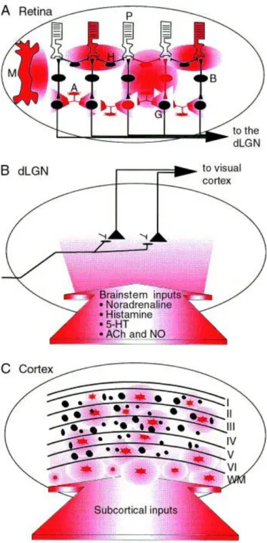
Fig. 2. Distribution of nitric-oxide synthase (NOS) containing cells and NOS positive fibres in the visual system. Summary of the three primary levels of the mammalian visual system. (A) Nitric oxide (NO) is produced in photoreceptors (P), horizontal cells (H), amacrine cells (A), Müller cells (M) and ganglion cells (G) (red) 23, 24, 25, 26, 27, 28, 29, 30, 31, 32, 33, 34, 35 and 36. In the retina it regulates
phototransduction 37 and 38; modulates photoreceptor output 37, 39, 40, 41 and 42, bipolar cells 38 and horizontal cells 43, 44, 45, 46, 47, 48 and 49;
controls ganglion-cell excitability 50 and modulates the electroretinogram 38 and 51. The widespread distribution of NOS-positive cells in these areas gives rise to possible modulation of retinal processes at all three levels by NO. The time course of effects at each level is likely to be similar because the half-life of NO in vitro has been estimated to be around 6 s ( Ref. 52) (although in vivo it could be much longer 53). These effects on retinal processing include: (1) regulation of phototransduction (altering levels of cGMP and by
ADP ribosylation); (2) modulation of output at photoreceptor synapses (by altering Ca2+ currents); (3) activation of ON-bipolar cells
(by acting on its NO-sensitive GC); (4) decreasing the lateral spread of light responses (by decreasing electrical coupling and responsiveness to glutamate in horizontal cells); and (5) controlling ganglion-cell excitability and thereby the retinal output (by acting on cGMP-gated cation channels). (B) In the cat, monkey and human dorsal lateral geniculate nucleus (dLGN) there are no NOS-positive cells 21, 54, 55, 56 and 57, but they are found in small mammals such as rats and tree shrews 58, 59, 60, 61 and 62. Massive
projection of NOS-positive terminals from the brainstem are shown in red 21. In the dLGN, nitric oxide modulates NMDA-receptor
activation, the gating of visual transmission 18, 19 and 20, the control of oscillatory activity 63 and has an important role in development in this region 64, 65, 66, 67, 68, 69, 70 and 71. (C) Scattered NOS-producing neurones are found in all layers of the cortex (red), many in the
white matter (WM) 29, 55, 57, 58, 62, 72, 73 and 74. Here NO modulates visual responses via cGMP ( Ref. 75) and is involved in the regulation
The retina
In a general scheme of vertebrate retinal physiology, visual excitation in photoreceptors is mediated by the light-triggered hydrolysis of intracellular cGMP and is transmitted via bipolar cells to the output, ganglion-cell layer of the retina. The visual signal is laterally modulated by two major classes of neurones: horizontal cells located in the outer plexiform layer and amacrine cells located in the inner retina. Such modulation is carried out via chemical synapses using a number of different neurotransmitters, and also by electrical coupling77. There is now much evidence that demonstrates that NO has a role in the physiological regulation of diverse processes within the retina, from the transduction to the gating of the output signal. These include:
Localization of nitric-oxide synthesis
Nitric-oxide synthase has been reported to have NADPH-diaphorase (NADPH-d) activity78 and 79 and both histochemical detection of NADPH-d activity and immunoreactivity to antibodies raised against NOS are used extensively as methods for identifying nNOS. These methods have revealed that horizontal, amacrine and ganglion cells of different mammals78, 23, 24, 25, 26, 27 and 28 and non-mammals29 and 30 contain nNOS. Moreover, human retinal tissues have been found to express mRNA for constitutive and inducible NOS (Ref. 31). The presence of nNOS activity in photoreceptors has been a matter of controversy. Several studies using immunocytochemical and NADPH-d histochemical staining failed to localize nNOS activity78, 80 and 81. Nevertheless, other studies, including the most recent, claim that nNOS activity to be present in the inner and outer segments of photoreceptor rods25, 26, 32, 33, 34 and 35. Furthermore, using NADPH-d histochemistry to study the cone-dominated retina of the tree shrew it has been possible to reveal several patterns of activity in the cellular subcompartments of the spectral classes of cones, which suggests that NO may be differentially involved in the functioning of different classes of photoreceptors36. Interestingly, there is also evidence that NOS is found in Müller cells of both fish and amphibian species, suggesting yet another route by which NO can modulate retinal function29.
NO affects the metabolism of cGMP in a variety of cells
(1) Available data show that NO is functionally coupled to a soluble guanylate cyclase and might be able to increase cGMP levels in rod photoreceptors thereby increasing the cGMP-gated conductances37, which affect both response amplitude and response kinetics82. Nitric oxide has also been shown to modulate Ca2+ channels and transmission of the photoresponse to second-order cells37, and to increase adenosine diphosphate (ADP) ribosylation of a variety of proteins such as transducin, G proteins and other, as yet unidentified, proteins in the outer segment of photoreceptor rods39, 40 and 41. These alterations of cellular proteins could be a mechanism by which NO modifies the operational mode of enzymes in the visual transduction cascade38. Nitric oxide can also modulate Ca2+ and cyclic-nucleotide-gated channels in both rod and cone photoreceptors, which control exocytosis at cone synapses, thereby altering synaptic efficacy42 and 83.
(2) Work carried out in fish has shown that NO donors, NOS inhibitors and NO-related substances modify electrical coupling in horizontal cells in such a way that the presence of cGMP, l-arginine or the NO donor, sodium nitroprusside, decreases electrical and dye-tracer coupling between the cells43 and 44. This modulation of the gap-junctional conductance seems to be produced by activating the cGMP-dependent protein kinase G (PKG) pathway45. Nitric oxide might also modify horizontal-cell activity by actions on chemical synapses. For example, NO reduces the responsiveness of glutamate receptors on retinal horizontal cells taken from the hybrid bass retina46. Interestingly, while dopamine is known to modify electrical coupling in horizontal cells43 and 47 and NO has been shown to modulate dopamine release in the retina48, these actions seem separate, because the action on dopamine release seem to affect only K+-mediated release and not basal release49. In summary, NO acts to downregulate horizontal-cell activity, which can alter apparent receptive-field size and thus influence the lateral spread of light-responses in the retina, while at the same time protecting horizontal cells from glutamate excitotoxicity.
All the studies mentioned so far have been carried out using in vitro preparations (mainly dissociated cells). Without doubt these have been useful and have made it possible to investigate problems that are otherwise unexaminable by current in vivo techniques. However, the disruption of the functional anatomy of the retina can clearly alter response properties of this complex system, which makes the use of different and complementary experimental approaches necessary. Recent studies that recorded the electroretinogram (ERG) in the intact cat eye 38 or both ERG and compound action potentials from the optic nerve in the intact rabbit retina 51 during application of NO-related compounds suggest that NO contributes physiologically to retinal processing.
The thalamus
In mammals, the dLGN plays a pivotal role in the transmission of visual information to the cerebral cortex. Unlike the retina, this is a site where both processing and gating of information takes place – thalamic transmission can be modulated by a number of inputs that arise from the brainstem84 and are dependent upon the behavioural state of the organism. Moreover, processing and gating in the dLGN are also functions that are intimately associated with, and regulated by, neural functioning in the visual cortex, by virtue of the large descending corticofugal input.
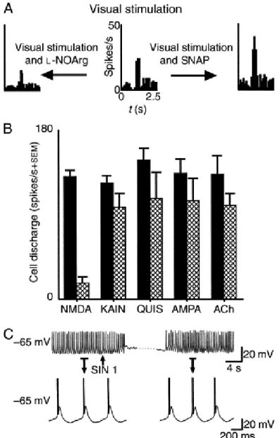
Fig. 3. Summary of the actions of the nitric-oxide system in the feline dorsal lateral geniculate nucleus (dLGN).(A) Control visual responses to small spot of light centred over the receptive field centre (centre). Application of the nitric-oxide (NO) donor, s-nitroso-N-acetyl-(d,l)-penicillamine (SNAP), increases responsiveness (right). Application of l-NOArg, a nitric-oxide synthase (NOS) inhibitor, suppresses visual responses (left). Adapted, with permission, from Ref. 20. (B) Histogram showing that application of the NOS antagonist, l-NOArg, depresses responses to applied NMDA in a highly selective fashion, compared to similar tests with the other drugs KAIN (kainic acid), QUIS (quisqualic acid), AMPA and ACh. Reproduced, with permission, from Ref. 19.(C) Intracellular recordings of a feline geniculate cell in vitro. Example of a neurone that spontaneously generates rhythmic Ca2+
-mediated burst activity. Generation of NO through SIN 1 reversibly inhibits this activity. Indicated segments are expanded for details. Adapted, with permission, from Ref. 63.
A role for NO in the development of subcortical structures
A role for NO in development has been described in different species. In rats, the superior colliculus is a major target for retinal axons and the refinement of retinocollicular connections takes place during the first few postnatal weeks. Given that nNOS is expressed in the retinorecipient layers of the rat superior colliculus during this period, it has been suggested that NO has a role in synaptic refinement64 and 65. Similarly, in the chick, the superficial layers of the optic tectum are the main sites for termination of retinal axons. Prior to the innervation by retinal axons, NADPH-d-stained cells are found only in deep layers. When axons innervate this area, NADPH-d-positive cells appear in the superficial layers, progressively increasing in number until the peak period for remodelling of retinal connections , which coincides with the loss of several transient projections, occurs. This loss is reduced if NO synthesis is inhibited66. Although the exact mechanism by which NO mediates this effect is not clearly understood, it has been suggested that coordinated activity in the major inputs, NMDA-receptor activation and NO production could each have a key role66. Interestingly, in the tadpole explanted retina, application of NO donors results in the collapse of active growth cones of ganglion-cell axons; such a mechanism could explain the termination of axonal growth at the tectal level during development67.
Similar results have been found in the dLGN of the ferret and the cat where NOS is transiently expressed during the period in which projections from the retina are refined68 and 69. In the ferret dLGN, retinal information is segregated into ON–OFF sublaminae, a process that requires NMDA-receptor activation, and application of a NOS inhibitor resulted in an overall pattern of sublamination that was clearly reduced when compared with normal animals70. In the developing kitten (by contrast with the adult cat), NADPH-d staining of dLGN cells suggested that NO might act in a retrograde fashion and perhaps have a role in the maintenance of associative processes that underlie activity-dependent refinement of retinogeniculate connections69. Further indirect results on the putative role of NO on development and plasticity have also been obtained in cats. After monocular lid suturing as kittens, adult cats showed an abnormal presence of NADPH-d-positive cells within the dLGN, which was not seen in normally reared controls, clearly indicating that NOS activity can be induced (or perhaps retained) by visual deprivation71.
Visual cortex
Fig. 4. NADPH-diaphorase staining in macaque primary visual cortex. Photomicrographs of NADPH diaphorase-positive neurones and fibres in the visual cortex (area V1) of a macaque monkey stained according to the protocol of Hope and Vincent89.
Moderately and darkly stained neurones are localized mainly in layers II, the upper half of layer III, the lower third of layer VI, and in the white matter. (A) Low-power photomicrograph from layer I to the white matter (WM). The black arrow indicates a strongly stained nonpyramidal cell that is also shown at higher magnification in (B) and (C). (B) Photomicrograph from layers I to III showing numerous lightly or moderately stained neurones (red arrows) and the plexus of horizontally oriented fibres (f) located in the upper half of layer I. (C) High-power photomicrograph of the darkly stained cell (black arrow) also indicated in (A) and (B). Note the dense network of fibres running in all directions. The red arrow indicates a lightly labeled neurone. Scale bar, 172 μm in
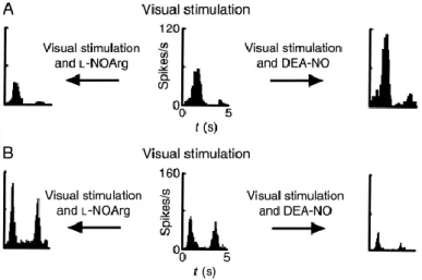
Fig. 5. Peri-stimulus time histograms illustrating the range of activities of nitric oxide (NO) and related compounds in cat visual cortex.(A) Control visual response of a directionally selective complex cell (centre). Application of DEA-NO, a NO donor, increases the response to the stimulus (right). Application of l-NOArg, a NOS inhibitor, decreases the same response (left). (B) Control visual responses of a non-directionally selective complex cell (centre). DEA-NO causes almost complete response suppression (right). l-NOArg application greatly increases responses in both directions equally (left). Drugs were applied by iontophoresis and responses averaged over a number of stimulus trials. Cells were driven monocularly through the dominant eye and the stimulus orientation selected from a quantitatively derived orientation tuning curve. Adapted, with permission, from Ref. 75.
Interestingly, in contrast to studies mentioned above that show a role for NO in the development of subcortical visual structures, there is no evidence to date to suggest a similar developmental role for NO in the visual cortex91 and 92, even though NOS distribution in visual cortex can be altered by manipulation of visual inputs55.
Concluding remarks
One of the most intriguing features of NO, considering the simplicity of the molecule, is that it is involved in so many different regulatory functions and has many other effects. At low concentrations, it can work as a neuromodulator or a retrograde messenger in the CNS; at relatively high concentrations it can be toxic. It is tempting to speculate that NO could have multiple roles in separate regions and circuits, each role related to local physiological functions and not necessarily part of the more general role that NO has in neurotoxicity or neuroprotection. These represent alterations of normal homeostatic function of the CNS and its regulatory mechanisms. It is important to note the significance of in vivo studies and to understand the need for studies of the physiology of whole systems.
Nitric oxide, the gas, the common air pollutant, the suspected carcinogen and the destroyer of ozone could be the archetypal example used to illustrate the concept of ‘parasynaptic’ information transmission in the brain, a domain of versatility and plasticity, as formulated by Schmitt93 ‘…new ways of conceptualization of information–transactional chemical processes as applied to basic concepts of neurobiology’, in this case to the concept of vision.
Acknowledgements
References
1. J. Garthwaite, C.L. Boulton. Annu. Rev. Physiol., 57 (1995), pp. 683–706 2. S.R. Vincent. Prog. Neurobiol., 42 (1994), pp. 129–160
3. J.B. Schulz, T.M. Russell, F. Beal. Curr. Opin. Neurol., 8 (1995), pp. 480–486 4. H.H.H. Schmidt, U. Walter. Cell, 78 (1994), pp. 919–925
5. Beckman, J.S. (1996) in Nitric Oxide. Principles and Actions (Lancaster, J., Jr, ed.), pp. 1–82,
10. C. Demerlé-Pallardy, et al. Biochem. Biophys. Res. Commun., 181 (1991), pp. 456–464 11. G. Garthwaite, J. Garthwaite. Neuropharmacology, 33 (1994), pp. 1431–1438
12. I. Hanbauer, et al. NeuroReport, 3 (1992), pp. 409–412
13. L. Kiedrowski, E. Costa, J.T. Wrobleski. Mol. Pharmacol., 41 (1992), pp. 779–784 14. D.A. Wink, et al. Proc. Natl. Acad. Sci. U. S. A., 90 (1993), pp. 9813–9817 15. S.Z. Lei, et al. Neuron, 8 (1992), pp. 1087–1099
16. S.A. Lipton, et al. Nature, 364 (1993), pp. 626–632
17. Z.-H. Pan, M.M. Segal, S.A. Lipton. Proc. Natl. Acad. Sci. U. S. A., 93 (1996), pp. 15423–15428 18. J. Cudeiro, et al. J. Neurophysiol., 71 (1994), pp. 146–149
19. J. Cudeiro, et al. Neuropharmacol., 33 (1994), pp. 1413–1418 20. J. Cudeiro, et al. Eur. J. Neurosci., 8 (1996), pp. 144–152 21. M.E. Bickford, et al. J. Comp. Neurol., 334 (1993), pp. 410–430 22. J.S. Stamler, et al. Neuron, 18 (1997), pp. 691–696
23. J.H. Sandell. J. Comp. Neurol., 238 (1985), pp. 466–472 24. J. Mitrofanis. Neurosci. Lett., 102 (1989), pp. 165–172
25. N.N. Osborne, N.L. Barnett, A.J. Herrera. Brain Res., 610 (1993), pp. 194–198 26. R. Yamamoto, et al. Neuroscience, 54 (1993), pp. 189–200
27. K.R. Huxlin, M.R. Bennett. Eur. J. Neurosci., 7 (1995), pp. 2226–2239 28. M.T.R. Perez, et al. Exp. Brain Res., 104 (1995), pp. 207–217
29. B.A. Liepe, et al. J. Neurosci., 14 (1994), pp. 7641–7654 30. T. Östholm, et al. Neurosci. Lett., 168 (1994), pp. 233–237
31. C-S. Park, et al. Biochem. Biophys. Res. Commun., 205 (1994), pp. 85–91 32. S.M. Sagar. J. Comp. Neurol., 300 (1990), pp. 309–319
33. C.M. Venturini, et al. Biochem. Biophys. Res. Commun., 180 (1991), pp. 920–925 34. K-W. Koch, et al. EMBO J., 13 (1994), pp. 3312–3320
35. A. Yoshida, et al. Visual Neurosci., 12 (1995), pp. 493–500
36. H.M. Petry, H.A. Murphy. Proc. Natl. Acad. Sci. U. S. A., 92 (1995), pp. 5121–5123 37. D.E. Kurenny, et al. Neuron, 13 (1994), pp. 315–324
38. I.M. Goldstein, P. Ostwald, S. Roth. Vis. Res., 36 (1996), pp. 2979–2994 39. B. Brune, E.G. Lapetina. Arch. Biochem. Biophys., 279 (1990), pp. 286–290 40. S. Ehret-Hilberer, et al. Fed. Eur. Biochem. Soc., 309 (1992), pp. 394–398
41. N. Pozdnyakov, et al. Biochem. Biophys. Res. Commun., 192 (1993), pp. 610–615 42. F. Rieke, E.A. Schwartz. Neuron, 13 (1994), pp. 863–873
43. S.H. DeVries, E.A. Schwartz. J. Physiol., 414 (1988), pp. 351–375
44. E. Miyachi, M. Mukarami, T. Nakaki. NeuroReport, 1 (1990), pp. 107–110 45. C. Lu, D.G. McMahon. J. Physiol., 499 (1997), pp. 689–699
46. D.G. McMahon, L.V. Ponomareva. J. Neurophysiol., 76 (1996), pp. 2307–2315
47. D.G. McMahon, A.G. Knapp, J.E. Dowling. Proc. Natl. Acad. Sci. U. S. A., 86 (1989), pp. 7639– 7643
48. O. Bugnon, N.C. Schaad, M. Schorderet. NeuroReport, 5 (1994), pp. 401–404 49. M. Pottek, K. Schultz, R. Weiler. Vis. Res., 37 (1997), pp. 1091–1102 50. I. Ahmad, et al. Neuron, 12 (1994), pp. 155–165
51. K.I. Maynard, P. Yanez, C.S. Ogilvy. NeuroReport, 6 (1995), pp. 850–852 52. R.E. Furchott, P.M. Vanhoutte. FASEB J, 3 (1989), pp. 2007–2018 53. A. Meulemans. Neurosci. Lett., 171 (1994), pp. 89–93
54. K. Mizukawa, et al. J. Comp. Neurol., 279 (1989), pp. 281–311 55. C. Aoki, et al. Brain Res., 620 (1993), pp. 97–113
58. S.R. Vincent, H. Kimura. Neuroscience, 46 (1992), pp. 755–784 59. S. Agarwala, et al. J. Comp. Neurol., 218 (1992), pp. 267–276 60. J. Mitrofanis. Visual Neurosci., 9 (1992), pp. 211–216
61. P.L.A. Gabbott, S.J. Bacon. J.Comp. Neurol., 350 (1994), pp. 281–301 62. J. Rodrigo, et al. Phil. Trans. R. Soc. London Ser. B, 345 (1994), pp. 175–221 63. H-C. Pape, R. Mager. Neuron, 9 (1992), pp. 441–448
64. T. Gonzalez-Hernandez, M. Conde-Sendin, G. Meyer. Anat. Embryol., 186 (1992), pp. 245–250 65. G. Prusky, M. Hofer, M. Constantine-Paton. Soc. Neurosci. Abstr., 18 (1992), p. 1311
66. Wu, H.H., Waid, D.K. and McLoon, S.C. (1996) in Prog. Brain Res. (Vol. 108) (Mize, R.R. and Erzurumlu, R.S., eds), pp. 273–286, Elsevier
67. R.C. Renteria, M. Constantine-Paton. J. Neurobiol., 29 (1995), pp. 415–428 68. K.S. Cramer, C.I. Moore, M. Sur. J. Comp. Neurol., 353 (1995), pp. 306–316 69. W. Guido, et al. Visual Neurosci., 14 (1997), pp. 1167–1173
70. K.S. Cramer, et al. J. Neurosci., 16 (1996), pp. 7995–8004
71. A.E. Günlük, M.E. Bickford, S.M. Sherman. J. Comp. Neurol., 350 (1994), pp. 215–228 72. S. Kuchiiwa, et al. NeuroReport, 5 (1994), pp. 1662–1664
73. J.H. Sandell. J. Comp. Neurol., 251 (1986), pp. 388–397 74. H-J. Lúth, et al. J. Neurocytol., 23 (1994), pp. 770–782 80. D.I. Vaney, H.M. Young. Brain Res., 474 (1988), pp. 380–385
81. J.M. Provis, J. Mitrofanis. Vis. Neurosci., 4 (1990), pp. 619–623 82. G.N. Noll, et al. Neuropharmacology, 33 (1994), pp. 1407–1412
83. A. Savchenko, S. Barnes, R.H. Kramer. Nature, 390 (1997), pp. 694–698
84. Sherman, S.M. and Koch, C. (1988) in The Synaptic Organization of the Brain (Shepherd, G.M., ed.), pp 289–328, Oxford University Press
85. K.R. Hoyt, et al. Brain Res., 592 (1992), pp. 310–316
86. T. Akira, D. Henry, C.G. Wasterlain. Brain Res., 652 (1994), pp. 190–194 87. M.E. Bickford, et al. J. Comp. Neurol., 348 (1994), pp. 481–510
88. C. Iadecola. Trends Neurosci., 16 (1993), pp. 206–214
89. B.T. Hope, S.R. Vicent. J. Histochem. Cytochem., 37 (1989), pp. 653–661 90. C. Estrada, J. DeFelipe. Cereb. Cortex, 8 (1998), pp. 193–203
91. S.N.M. Reid, et al. J. Physiol., 494 (1996), pp. 511–517 92. E.S. Ruthazer, et al. J. Physiol., 494 (1996), pp. 519–527 93. F.O. Schmitt. Neuroscience, 13 (1984), pp. 991–1001
94. H.H.H.W. Schmidt, et al. Proc. Natl. Acad. Sci. U. S. A., 93 (1996), pp. 14492–14497
95. Feelisch, M. and Stamler, J.S. (1996) Methods in Nitric Oxide Research, pp. 19–28, John Wiley and Sons
96. A.J. Gow, D.D. Buerk, H. Ischiropoulos. J. Biol. Chem., 272 (1997), pp. 2841–2845 97. J.S. Stamler, D.J. Singel, J. Loscalzo. Science, 258 (1992), pp. 1898–1902
98. D.T. Hess, et al. Nature, 366 (1993), pp. 562–565 99. M.K. Meffert, et al. Neuron, 16 (1996), pp. 1229–1236
100. C.W. Mahoney, J.H. Pak, K-P. Huang. J. Biol. Chem., 271 (1996), pp. 28798–28804 101. H. Wolosker, et al. J. Neurochem., 66 (1996), pp. 1943–1948
102. M-C. Broillet, S. Firestein. Neuron, 16 (1996), pp. 377–385 103. T. Sato, et al. J. Neurochem., 68 (1997), pp. 1312–1318