Effects of ions releasing restorative materials on the dentine bonding longevity of modern universal adhesives after load cycle and prolonged artificial saliva aging
14
0
0
Texto completo
(2) Materials 2019, 12, 722. 2 of 14. Keywords: adhesion; cycling mechanical stress; dentine; longevity; glass-ionomer cements; universal adhesives. 1. Introduction Direct restorations in modern operative dentistry are frequently accomplished using conventional resin composites due to their excellent mechanical and aesthetic properties [1,2]. Nevertheless, such restorative materials are still characterized by important downsides associated to polymerization shrinkage; a phenomenon that may induce stress at resin–dentine interfaces during the light-curing procedures and jeopardize their longevity [3–5]. Indeed, it has been widely demonstrated that the volumetric contraction of conventional resin composites can transfer polymerization stress directly to the adhesive-bonded interface, causing its innermost deformation due to a lack of proper bonding performance of some adhesive systems [3,6,7]. Consequently, the sealing between composite and dental hard tissues (i.e., dentine and enamel) can be seriously compromised. This will result in gaps and marginal leakage formation, which are pathways for microleakage of oral fluids, bacteria, and enzymes penetration [3,8–10]. Such issues may translate into important clinical problems such as post-operative sensitivity, marginal discoloration, recurrent caries, and advanced pulp pathology in all those cases that are seriously compromised by the caries process [11,12]. The recently introduced universal adhesive systems are currently very popular in general dental practices, as well as in dental hospitals, due to the fact that they can be applied both in self-etching (SE) and etch-and-rinse (ER) modes. Considering their compositions, universal adhesives can be also classified as simplified systems because all ingredients, including acidic functional monomers and solvents, are incorporated into one bottle. They are similar to one-step self-etching systems, so that they might still present issues related to bonding performance, degradation, and longevity [9,13]. However, application in self-etching mode minimizes recontamination of the dentine by blood and saliva during etch washing and drying. This makes SE a less technique-sensitive procedure compared to ER application mode. Moreover, SE systems present further benefits such as less post-operative sensitivity due to residual smear plugs, which are usually only partially removed from inside the dentinal tubules because of the mild acidic nature of SE systems. Indeed, the tubules remain occluded and the dentinal fluid movement is less evident compared to that usually experienced with ER systems [9,11]. On the other hand, great attention has been given to improve the effectiveness and longevity of resin–dentine bonds through several clinical strategies that may abate stress concentration at the resin–dentine interface during polymerization [14]. For instance, the use of flowable composites or resin-modified glass-ionomer cements (RMGIC) as liners or as dentine substitute materials may represent a suitable method to provide a sort of “stress-absorption” effect at the bonding interface [15,16]. This has been advocated to prevent stress development at the dentine-bonded interface and reduce gap formation, microleakage, and degradation over time [14,17,18]. Although RMGIC are self-adhesive materials, they are also often applied in dentine after etching and adhesive application, especially in those situations where the structure of the dental crown is highly compromised and a lack of mechanical retention is encountered [19–21]. It is also important to consider that occlusal stress during mastication, swallowing, as well as in cases of parafunctional habits, can affect the integrity of the bonding interface, making such a structure more susceptible to “quicker” degradation in the oral environment [22]. This seems to be of particular interest in modern, minimally invasive therapeutic restorative dentistry since it has been demonstrated that cyclic mechanical stress can promote gap formation at the margins along the composite restorations; bacteria penetration into narrow marginal gaps might ultimately promote secondary caries formation [23]. Recently, it has been advocated that ion-releasing resin-based.
(3) Materials 2019, 12, 722. 3 of 14. restorative materials can reduce such biofilm penetration into marginal gaps of simulated tooth restorations; the risk for development and propagation of secondary caries is also reduced [24]. It is widely accepted that glass-ionomer cement (GIC)-based materials have a bioactive ability to release therapeutic ions such as fluoride. The presence of such ions has been associated with long-term caries inhibition when GIC-based materials are applied as a dentine substitute [25–27]. Moreover, GIC-based materials are an ideal dentine substitute as their physical properties, such as the coefficient of thermal expansion, dimensional stability, optical properties (i.e., opacity), and microhardness, are very close to that of dentine [28]. ACTIVA BioActive Restorative is a new type of restorative, bioactive, flowable, resin-based composite comparable to RMGICs. It contains fluoro-aluminum silicate particles and polyacid components of glass ionomer that undergo the acid-base setting reaction. Moreover, a bioactive ionic resin matrix is also contained in ACTIVA, which confers both light and chemical polymerization. According to the manufacturer, ACTIVA release calcium, phosphate, and fluoride when in contact with saliva. It has been advocated that restorative materials able to release specific “therapeutic” ions (e.g., calcium, phosphates, fluoride, strontium, and other minerals) into the dental hard tissues may buffer the constant assault of day-to-day ingestion of acidic food and beverages and encourage remineralization along the margins of the restoration with the tooth [29]. However, it is of great relevance that the use of ion-releasing materials in restorative dentistry may contribute to the reduced activity of proteases such as metalloproteinases (MMPs) and cathepsins involved in collagen degradation. Such enzymes are considered one of the main causes for reduction of bonding longevity when simplified bonding systems are applied in dentine with self-etching or etch-and-rinse protocols [30,31]. Moreover, there is a lack of knowledge about the effects of modern ion-releasing materials based on glass ionomer cements on resin–dentine interfaces created using current universal adhesives after mechanical load cycling and prolonged storage in artificial saliva. Thus, the aim of this study was to evaluate, after short-term load-cycle aging or after load-cycle stress followed by prolonged aging (8 months) in artificial saliva (AS), the microtensile bond strength (MTBS) of resin–dentine bonded specimens created using universal adhesives applied in an etch-and-rinse or self-etching mode in combination with modern ion-releasing RMGIC-based materials. Fractographic analysis was also performed using field-emission scanning electron microscopy (FE-SEM). The hypothesis tested was that compared to conventional resin composite, the use of modern ion-releasing materials would preserve the bonding performance of modern universal adhesives, applied in etch-and-rinse or self-etching, after mechanical load cycling and/or prolonged storage in artificial saliva (8 months). 2. Materials and Methods 2.1. Preparation of Dentine Specimens and Experimental Design Sound human molars were extracted for periodontal or orthodontic reasons (ethical approval number: LEC No 11.18, 05/12/2018) and stored in distilled water at 5 ◦ C for no longer than 3 months. The roots were removed 1 mm beneath the cemento–enamel junction using a diamond blade (XL 12205; Benetec, London, UK) mounted on a low-speed microtome (Remet evolution, REMET, Bologna, Italy). A second parallel cut was made to remove the occlusal enamel and expose mid-coronal dentine. Three main groups (n = 72 specimens/group) were created based on the restorative materials used in this study: (i) RC: Resin composite (Aura SDI, Bayswater Victoria, Australia), applied in 2 mm increment layers up to 6 mm, and light-cured as per manufacturer’s instructions; (ii) RMGIC: Resin-modified glass-ionomer cement (Ionolux; VOCO GmbH, Cuxhaven, Germany) mixed for 10 s in a trituration unit and applied in bulk. Two capsules of RMGIC were used and each one was light-cured as per manufacturer’s instructions to obtain 6 mm build-ups; (iii) ACTIVA (ACTIVA BioActive Restorative, PULPDENT, Watertown, MA, USA) applied in 2 mm increment layers up to 6 mm and light-cured as per manufacturer’s instructions. Light-curing was performed using an.
(4) Materials 2019, 12, 722. 4 of 14. light-emitting diode (LED) light source ( >1000 mW/cm2 ) (Radii plus, SDI Ltd., Bayswater Victoria, Australia). The experimental design of this study required that the specimens in each main group were subsequently subdivided into four sub-groups (n = 18 specimens/group) based on the protocol employed for bonding procedures. Two modern universal adhesives were employed in this study: SCU (Scotchbond Universal, 3M Oral Care, St. Paul, MN, USA); FTB: (Futurabond M+, VOCO, Cuxhaven, Germany). These adhesives were applied as per manufacturer’s instructions in self-etching (SE) or in etch-and-rinse (ER) mode (Table 1). In groups SCU–ER and FTB–ER, dentine was etched with 37% orthophosphoric acid for 15 s and subsequently rinsed with distilled water (15 s) and blotted, leaving the substrate moist. Adhesives were light-cured for 10 s. In groups SCU–SE and FTB–SE, the adhesives were applied with a microbrush for 20 s and air dried for 5 s to evaporate the solvent. These were finally light-cured for 10 s using am LED light source ( >1000 mW/cm2 ) (Radii plus, SID Ltd., Bayswater VIC, Australia). The specimens were finally restored with the selected restorative materials as aforementioned in the main groups. At this point, the specimens in each sub-group were furtherly divided into three groups (n = 6 specimens/group) based on the aging protocol: CTR: no aging (control, 24 h in deionized water); LC: Load cycling (350,000 cycles in artificial saliva); LC–AS: Load cycling (350,000 cycles in artificial saliva), followed by prolonged water storage (8 months in artificial saliva). A detailed description of the test groups can be found in Table 2 (Experimental design). The composition of the artificial saliva was AS: 0.103 g L−1 of CaCl2 , 0.019 g L−1 of MgCl2 ·6H2 O, 0.544 g L−1 of KH2 PO4 , 30 g L−1 of KCl, and 4.77 g L−1 HEPES (acid) buffer, pH 7.4] [32]. The specimens in the subgroup LC and LC–AS were mounted in plastic rings with acrylic resin for load cycle testing. A compressive load was applied to the flat surface (3 Hz; 70 N) using a 5 mm diameter spherical stainless-steel plunger attached to a cyclic-load machine (model S-MMT-250NB; Shimadzu, Tokyo, Japan) while immersed in AS [18,33]. Table 1. Adhesive system, composition, and application procedures. Name. Scotchbond Universal, 3M Oral Care, USA (lot: 627524). FuturaBond M+, VOCO, Germany (lot: 1742551). Composition. Application. 10-MDP, HEMA, silane, dimethacrylate resins, Vitrebond™ copolymer, filler, ethanol, water, initiators, and catalysts (pH 2.7). 1. Apply the adhesive on the surface and rub it for 20 s. 2. Gently air-dry the adhesive for approximately 5 s for the solvent to evaporate. 3. Light cure for 10 s (>500 mW/cm2 ).. HEMA, BIS-GMA, ethanol, Acidic adhesive monomer (10-MDP), UDMA, catalyst ethanol, water, initiators, and catalysts (pH 2.8). 1. Apply the adhesive homogenously to the surface. 2. Rub for 20 s. 3. Dry off the adhesive layer with dry, oil-free air for at least 5 s. 4. Light cure for 10 s (>500 mW/cm2 ).. Abbreviations: 10-MDP 10-methacryloxydecyl dihydrogen phosphate, Bis-GMA bisphenol A diglycidyl methacrylate, HEMA 2-hydroxyethyl methacrylate, UDMA urethane dimethacrylate.. 2.2. Micro-Tensile Bond Strength and Failure/Fractographic Analysis The specimens were cut after the aging period using a hard-tissue microtome (Remet evolution, REMET, Bologna, Italy) across the resin–dentine interface, obtaining approximately 15–18 matchstick-shaped specimens from each tooth (Ø 0.9 mm2 ). These were submitted to microtensile bond strength tests using a device with a stroke length of 50 mm, peak force of 500 N, and a displacement resolution of 0.5 mm. Modes of failure were evaluated at 50× magnification using stereoscopic microscopy and conveyed in a percentage of adhesive (A), mixed (M), or cohesive (C) bonding fracture. Five representative fractured specimens from each sub-group were mounted on aluminum stubs with carbon glue after the critical-point drying process. The specimens were gold-sputter-coated and.
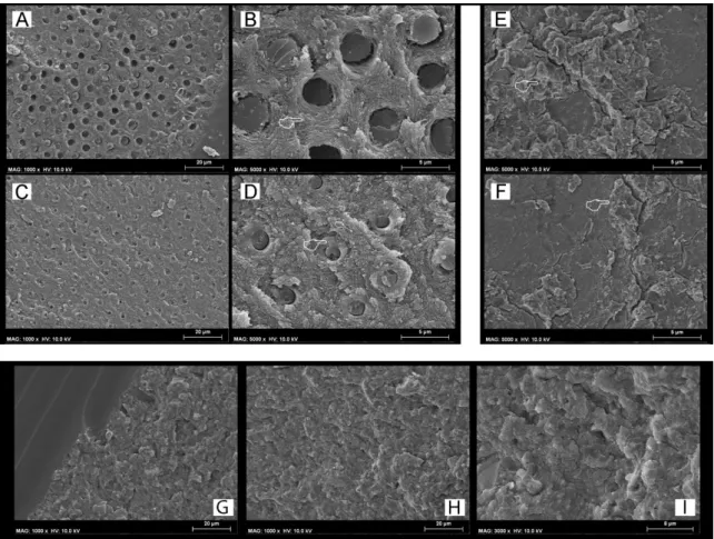
(5) Materials 2019, 12, 722. 5 of 14. analyzed using field-emission scanning electron microscopy (FE-SEM S-4100; Hitachi, Wokingham, UK) at 10 kV and a working distance of 15 mm. Bond strength values in MPa were initially assessed for normality distribution and variances homogeneity using Kolmogorov–Smirnov and Levene’s tests, respectively. Data were then analyzed using a three-way Analysis of Variance (ANOVA Factors: restorative material, adhesive, and aging protocol) and Newman–Keuls multiple-comparison test (α = 0.05). SPSS V16 for Windows (SPSS Inc., Chicago, IL, USA) was used. Table 2. Experimental design. Distribution of specimens in groups and sub-groups for evaluation via microtensile bond strength (MTBS), interface confocal microscopy, and SEM fractographic analysis. CTR = control, no aging; LC = load-cycling; AS = artificial saliva. Total Number of Specimens in Main Groups. RESIN COMPOSITE (72 Specimens). Number of specimens in sub-groups (18/group). RMGIC (72 Specimens). ACTIVA (72 Specimens). Number of specimens in aging sub-groups (6/ group). SCU–ER: Scotchbond Etch and rinse. CTR 6 spec. LC 6 spec. LC+AS 6 spec. CTR 6 spec. LC 6 spec. LC+AS 6 spec. CTR 6 spec. LC 6 spec. LC+AS 6 spec. FTB–ER Futurabond M+ Etch and rinse. CTR 6 spec. LC 6 spec. LC+AS 6 spec. CTR 6 spec. LC 6 spec. LC+AS 6 spec. CTR 6 spec. LC 6 spec. LC+AS 6 spec. SCU–SE: Scotchbond Self-etch. CTR 6 spec. LC 6 spec. LC+AS 6 spec. CTR 6 spec. LC 6 spec. LC+AS 6 spec. CTR 6 spec. LC 6 spec. LC+AS 6 spec. FTB–SE: Futurabond M+ Self-etch. CTR 6 spec. LC 6 spec. LC+AS 6 spec. CTR 6 spec. LC 6 spec. LC+AS 6 spec. CTR 6 spec. LC 6 spec. LC+AS 6 spec. 3. Results Micro-Tensile Bond Strength (MTBS) and Failure Mode Analysis There were no pre-test failures before the microtensile bond strength assessment. Three-way ANOVA revealed a significant effect of adhesive (F = 28.75, p < 0.001) and restorative material (F = 6.68, p < 0.001) on the bond strength, whereas the aging protocol was not statistically significant (F = 8.17; p = 0.125). The interactions between the three variables were significant (p < 0.001). The results of the microtensile bond strength test (mean and ± SD) are depicted in Table 3. It was observed that there was no significant difference (p > 0.05) at 24 h testing between the two adhesives when applied in etch-and-rinse (ER) or self-etching (SE) mode and then restored using the conventional RC or the two RMGIC-based materials (IONOLUX and ACTIVA). Conversely, the specimens created with the conventional RMGIC presented no significant differences (p > 0.05) when bonded using the two adhesives applied in ER or SE mode. However, all the specimens created with the conventional RMGIC showed a significant lower bond strength compared to those created with RC or ACTIVA. The failure mode showed that all the specimens restored with the RMGIC failed mainly in the cohesive mode, leaving a clear presence of the material still bonded to the dentine. The specimens created with RC or ACTIVA failed mainly in the cohesive in composite and mixed mode, leaving part of the dentine still covered by the restorative material and the other part exposed. The fractographic analysis showed that the restorative materials employed in this study had no influence on the outcomes in the control storage period (24 h), but all those specimens created with SCU in ER mode presented less resin infiltration within exposed acid-etched dentine collagen fibrils (Figure 1A,B), while the specimens bonded using the FTB applied in ER mode presented fractures mainly underneath the hybrid layer (Figure 1C). Moreover, in this latter case, there was mineralized peri-tubular dentine around the lumen of the dentine tubules and no demineralized and exposed collagen fibrils (Figure 1D). Conversely, all the specimens bonded with the two adhesives applied in SE or ER mode and then restored with RMGIC showed a surface still covered by the restorative material.
(6) Materials 2019, 12, 722. 6 of 14. (cohesive mode within RMGIC) with no exposure of the dentine (Figure 1E,F) after microtensile bond strength testing. Furthermore, the fractographic analysis showed that the specimens created both with SCU (Figure 1F) and FTB (Figure 1G) applied in SE, and that failed in mixed or adhesive mode, presented a dentine surface still covered by a smear layer with no presence of collagen fibrils and/or exposed dentinal tubules (Figure 1I). Materials 2018, 11, x FOR PEER REVIEW 7 of 14. Figure1.1.SEM SEMfractographic fractographic analysis analysis of of the the control control specimens. specimens. (A) Figure (A) SEM SEM fractography fractographyof ofaaspecimen specimen created with SCU applied in ER mode and restored with resin composite (RC) showing the presenceof created with SCU applied in ER mode and restored with resin composite (RC) showing the presence of exposed dentine and several resin tags still in the dentinal tubules. (B) At higher magnification, exposed dentine and several resin tags still in the dentinal tubules. (B) At higher magnification, ititis is possible to note presence of resin inside demineralized dentine tubules collagen fibrils possible to note the the presence of resin tagstags inside demineralized dentine tubules andand collagen fibrils not not infiltrated well infiltrated theadhesive SCU adhesive (pointer). Thismorphological latter morphological characteristic may well by theby SCU (pointer). This latter characteristic may indicate indicate that such resin–dentine interface would be by degradation over timedrop and in would that such resin–dentine interface would be affected by affected degradation over time and would bond drop in bond strength. (C) SEM fractography of a specimen created with FTB applied in ER mode strength. (C) SEM fractography of a specimen created with FTB applied in ER mode and restored and with restoredshowing with ACTIVA showing presence of exposed dentine and several resin tags still inside theof ACTIVA the presence of the exposed dentine and several resin tags still inside the small lumen small lumen of the dentinal tubules. (D) At higher magnification it is possible to observe a typical the dentinal tubules. (D) At higher magnification it is possible to observe a typical failure occurred at failure occurred the bottom of thecharacterized hybrid layer by (HL) by the presence of mineralized the bottom of the at hybrid layer (HL) thecharacterized presence of mineralized peritubular dentine peritubular dentine (pointer), with tubules totally obliterated by resin tags and with no presence of (pointer), with tubules totally obliterated by resin tags and with no presence of demineralized exposed demineralized exposed collagen fibrils. Conversely, the dentine specimens bonded with SCU (E) and collagen fibrils. Conversely, the dentine specimens bonded with SCU (E) and FTB (F) applied in ER FTB (F) applied in ER mode restored with RMGIC the presence of thethat remaining mode and restored with the and RMGIC show thethe presence ofshow the remaining RMGIC totallyRMGIC covered that totally covered the dentine surface. (G) SEM fractography of a specimen created the dentine surface. (G) SEM fractography of a specimen created with SCU applied in SEwith modeSCU and appliedwith in SEACTIVA mode and ACTIVA (H) FTB applied in SE and arestored with restored andrestored (H) FTBwith applied in SE and mode and restored with RCmode showing characteristic RC showing a characteristic in mixed Note theresin presence of the remaining resin and failure in mixed mode. Note failure the presence of mode. the remaining (G) and smear layer on the(G) dentine smear layer on the dentine surface; the latter was even more evident at higher magnification (I). surface; the latter was even more evident at higher magnification (I).. After submitting the specimens to load-cycle aging, the only group that showed a significant bond strength drop (p < 0.05) was that created with the SCU applied in ER mode and restored using the conventional RC. In this group, an important change in the failure mode was also observed; only 15% of the specimens failed in cohesive mode, while failure in mixed and adhesive modes were 55% and 30%, respectively (Table 3). This situation was not evident in the specimens bonded with the same adhesive but restored using IONOLUX (RMGIC) or ACTIVA; no significant bond strength drop (p > 0.05) and no radical change in failure mode was observed. The SEM fractography showed no important ultra-morphological changes in most of the fractured resin–dentine interfaces of these.
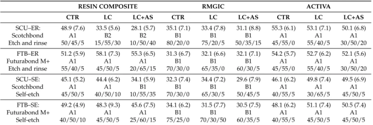
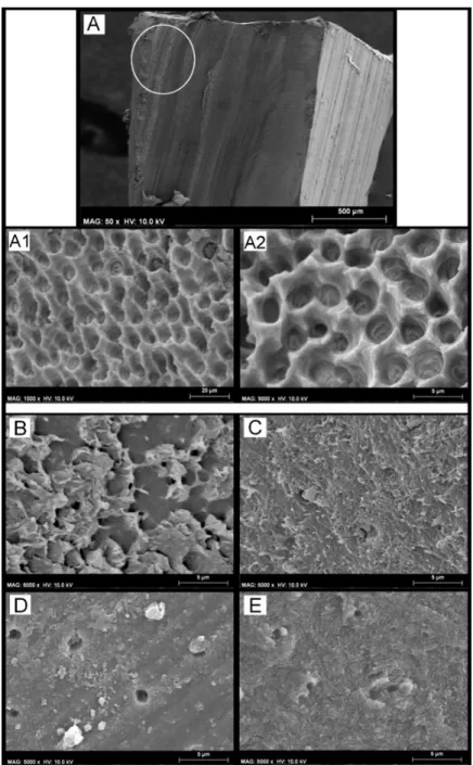
(7) Materials 2019, 12, 722. 7 of 14. Table 3. The results show the mean (± SD) of the MTBS (MPa) to dentine and the percentage (%) of the failure mode analysis. RESIN COMPOSITE. RMGIC. ACTIVA. CTR. LC. LC+AS. CTR. LC. LC+AS. CTR. LC. LC+AS. SCU–ER: Scotchbond Etch and rinse. 48.9 (7.6) A1 50/45/5. 33.5 (5.6) B2 15/55/30. 28.1 (5.7) B2 10/50/40. 35.1 (7.1) B1 80/20/0. 33.4 (7.8) B1 75/20/5. 31.1 (8.8) B1 50/35/15. 55.3 (6.1) A1 45/55/0. 53.1 (7.1) A1 55/40/5. 50.1 (6.8) A1 30/50/20. FTB–ER Futurabond M+ Etch and rinse. 51.2 (5.9) A1 55/40/5. 58.1 (7.3) A1 45/50/5. 55.3 (6.5) A1 20/65/15. 31.3 (6.7) B1 70/30/0. 32.1 (6.6) B1 65/35/0. 32.1 (7.1) B1 60/30/5. 54.2 (5.7) A1 45/55/0. 52.7 (6.2) A1 55/40/5. 52.1 (5.6) A1 30/50/20. SCU–SE: Scotchbond Self-etch. 45.1 (5.2) A1 45/50/5. 44.4 (6.2) A1 40/50/10. 34.1 (5.9) B1 10/55/35. 32.3 (7.4) B1 70/30/0. 34.4 (7.2) B1 65/30/5. 29.6 (7.9) B1 50/45/5. 46.1 (6.2) A1 40/55/5. 49.8 (7.4) A1 30/65/5. 49.5 (6.9) A1 45/50/5. FTB–SE: Futurabond M+ Self-etch. 49.2 (4.9) A1 40/50/10. 48.3 (9.3) A1 45/50/5. 45.6 (7.5) A1 25/60/15. 34.1 (6.2) B1 75/25/0. 31.5 (7.7) B1 70/30/50. 30.5 (7.5) B1 60/35/5. 48.1 (6.2) A1 40/55/5. 51.1 (7.4) A1 45/50/5. 50.5 (7.4) A1 45/50/5. Failure mode [Cohesive/Mixed/Adhesive]. The same number indicates no significance in column, while the same letter indicates no significance in row (p > 0.05).. After submitting the specimens to load-cycle aging, the only group that showed a significant bond strength drop (p < 0.05) was that created with the SCU applied in ER mode and restored using the conventional RC. In this group, an important change in the failure mode was also observed; only 15% of the specimens failed in cohesive mode, while failure in mixed and adhesive modes were 55% and 30%, respectively (Table 3). This situation was not evident in the specimens bonded with the same adhesive but restored using IONOLUX (RMGIC) or ACTIVA; no significant bond strength drop (p > 0.05) and no radical change in failure mode was observed. The SEM fractography showed no important ultra-morphological changes in most of the fractured resin–dentine interfaces of these groups compared to the control group. Conversely, the specimens created with the SCU applied Materials 2018, 11, x FOR PEER REVIEW 8 of 14 in ER mode (Figure 2A) and restored with the conventional RC, which failed prevalently in mixed and adhesive mode, showed fracture occurred underneath hybrid layer with signofof adhesive mode, showed thatthat the the fracture occurred underneath thethe hybrid layer with nonosign demineralized poorly infiltrated infiltratedcollagen collagenfibrils fibrils(Figure (Figure2B 2B,C). demineralized and/or and/or poorly and 2C).. Figure 2.2. SEM SEM Fractographic Fractographic analysis analysis after Figure after load-cycle load-cycle aging. aging. (A) (A)SEM SEMfractography fractographyofofaaspecimen specimen created with SCU applied in ER mode and restored with RC showing a characteristic failure in created with SCU applied and restored with RC showing a characteristic failure inmixed mixed mode. The finger pointer indicates a brighter mode. The finger brighter area area of ofgreater greaterand andmore moreevident evidentaging aging(pointer), (pointer),which which wasprobably probably induced induced by the cycling load. was load. However, However, when whenwe weobserved observedthat thatspecific specificarea areaatathigher higher magnifications (B), (B), it was possible to observe magnifications observe that that the the fracture fractureoccurred occurredunderneath underneaththe thehybrid hybridlayer layer (pointer),which which is is characterized characterized by the presence (pointer), presence of of mineralized mineralizeddentine dentine(pointer), (pointer),with withtubules tubulestotally totally obliterated by by resin resin tags tags and and with with no no presence presence of obliterated of demineralized demineralized exposed exposed collagen collagenfibrils fibrils(C). (C).. The load-cycling stress stressinduced induced Theprolonged prolongedaging aginginin artificial artificial saliva saliva performed performed subsequent subsequent load-cycling important changes on microtensile bond strength as well as on the ultramorphology of the fracture important changes microtensile bond strength as well as on the ultramorphology of the fracture of the specimens specimens bonded bondedwith withSCU SCUapplied appliedboth bothininER ERand andSE SE ofsome some specific specific groups. groups. In particular, the mode composite had hadaa significant significantdrop dropininbond bondstrength strength mode and and then then restored restored with the conventional conventional composite compared specimensin inthe thegroups groupsCTR CTRand andLC LC < 0.05). Moreover, number of failures compared to to the specimens (p(p < 0.05). Moreover, thethe number of failures in in mixed adhesive modes increased the aforementioned compared to the (CTR) control mixed andand adhesive modes increased in theinaforementioned groupsgroups compared to the control group. The SEM fractography showed evident signs of dentine degradation in the group of specimens created with SCU applied in ER mode and then restored with the conventional composite (Figures A1 and A2). The SEM fractography showed that specimens created with SCU applied in SE mode and then restored with the RC presented degradation both of the adhesive (Figure 3B) and dentine hybrid layers (Figure 3C). Conversely, the same specimens restored with the RMGIC or.
(8) Materials 2019, 12, 722. 8 of 14. (CTR) group. The SEM fractography showed evident signs of dentine degradation in the group of specimens created with SCU applied in ER mode and then restored with the conventional composite (Figure 3(A1,A2)). The SEM fractography showed that specimens created with SCU applied in SE mode and then restored with the RC presented degradation both of the adhesive (Figure 3B) and dentine hybrid layers (Figure 3C). Conversely, the same specimens restored with the RMGIC or ACTIVA presented a stable bond strength with no significant drop (p < 0.05), and the type of failure remained quite similar to the control group. The SEM fractography showed no drastic changes in all those groups for the ultramorphology of fractured resin–dentine interfaces compared to the control group (Figure 3D,E). In particular, the SEM fractography of a specimen created with SCU applied in ER mode and restored with ACTIVA and RMGIC showed the presence of dentine that was well mineralized with no sign of demineralized collagen fibrils, but with the presence of mineral debrides as a possible result of the bioactivity of such GIC-based materials (Figure 3D). Materials 2018, 11, x FOR PEER REVIEW. 9 of 14. Figure SEMFractographic Fractographic analysis analysis after after load load cycling cycling and SEM Figure 3. 3.SEM and aging aging in in artificial artificialsaliva. saliva.(A)(A) SEM fractography of a specimen created with SCU applied in ER mode and restored with RC showing fractography of a specimen created with SCU applied in ER mode and restored with RC showing a characteristic failure in adhesive mode. Note that the white circle indicates no physical difference in the material; it was added to show the reader that images (A1) and (A2) were obtained by higher magnification in that zone. Indeed, in (A1) and (A2) it is possible to see severe collagen degradation without the presence of any resin residual. (B) SEM fractography of a specimen created with SCU applied in SE mode where it is possible to see a failure between composite and adhesive, probably due to degradation induced by excessive water sorption upon mechanical stress and prolonged AS.
(9) Materials 2019, 12, 722. 9 of 14. a characteristic failure in adhesive mode. Note that the white circle indicates no physical difference in the material; it was added to show the reader that images (A1,A2) were obtained by higher magnification in that zone. Indeed, in (A1,A2) it is possible to see severe collagen degradation without the presence of any resin residual. (B) SEM fractography of a specimen created with SCU applied in SE mode where it is possible to see a failure between composite and adhesive, probably due to degradation induced by excessive water sorption upon mechanical stress and prolonged AS storage. However, it was also possible to see, in those specimens that failed in mixed mode, signs of degradation of the collagen fibrils underneath the hybrid/interdiffusion layer (C). (D) SEM fractography of a specimen created with SCU applied in ER mode and restored with ACTIVA showing that the failure occurred underneath the hybrid layer, but the exposed dentine is well mineralized with no sign of exposed demineralized collagen fibrils. Note also the presence of mineral debrides that are a possible result of the bioactivity of ACTIVA, which released ions and diffused through the resin-bonded dentine. (E) SEM fractography of a specimen created with FTB applied in SE mode and restored with RMGIC. The specimens of this group failed mainly in cohesive and mixed mode; this latter zone is characterized by a fracture occurring underneath the hybrid layer, leaving behind a dentine surface completely mineralized with no sign of exposed, denatured, or demineralized collagen fibrils. Please note the presence of a well mineralized intratubular dentine inside the lumen of the dentine tubules.. 4. Discussion This study showed that the use of modern ion-releasing materials such as conventional RMGIC or RMGIC-based composite (ACTIVA) preserved the bonding performance of only one (SCU) of the two modern universal adhesives bonded to dentine in etch-and-rinse or self-etching mode, after the two aging protocols employed in the experimental design. Conversely, the dentine-bonded specimens created with the FTB universal bonding system applied in etch-and-rinse or self-etching showed no significant drop in bonding performance after aging, regardless the restorative material employed or the aging protocol. Hence, the hypothesis tested in this study needs to be partially accepted as the use of a specific new generation universal bonding systems may confer a stable dentine-bonded interface over time. Nevertheless, the use of modern ion-releasing restorative materials such as RMGIC or ACTIVA may preserve the bonding performance of those universal adhesives that are more prone to degradation after aging. The effects of the load-cycle aging protocol on the bonding performance of the SCU system applied in ER mode and restored with the conventional RC were relevant; the bond strength of this group of specimens dropped significantly (p < 0.05). Moreover, only the specimens bonded using SCU applied both in ER and SE mode and then restored with the conventional composite showed a significant drop in bond strength compared to the specimens in the control (CTR, 24 h) group after prolonged aging in artificial saliva. The ultramorphology analysis performed in the specimens of the control group (24 h), created using the SCU system applied in ER mode and restored with RC showed the presence of demineralized-acid-etched dentine collagen that was not well resin-infiltrated (Figure 1A,B). While the same specimens submitted to LC aging showed no exposed collagen, but mineralized dentine with resin tags that obliterated the dentinal tubules (Figure 2). This was an interesting result, so we hypothesize that a possible explanation to the difference in bonding performance observed between these two latter situations (LC-only aging vs. CTR) may be attributed to the fact that the hybrid layer created using simplified adhesives applied in etch-and-rinse mode can represent the critical part of the resin–dentine interface, as it probably remains only partially polymerized [33–35]. Indeed, it has been advocated that during cycling loading such un-polymerized monomers within the hybrid layer, created with simplified, highly hydrophilic etch-and-rinse adhesives, may be mechanically “intruded” into the demineralized dentine causing a more compact and performant hybrid layer. However, such a morphological change within the resin–dentine interface may favor higher stress concentrations during the cycling load at the bottom of the hybrid layer, causing an accelerated mechanically-induced degradation phenomenon in this specific zone that often remains partially.
(10) Materials 2019, 12, 722. 10 of 14. demineralized and poorly infiltrated by adhesive monomers [34,35]. Indeed, the absence of a proper, partially demineralized bottom of the hybrid layer may explain why the dentine bonded with SCU or FTB in SE mode showed no bond-strength drop after load-cycle aging, regardless of the restorative material and the protocol employed for aging [33,34]. Our results seem to be in accordance with those of Dorfer et al. [36] who demonstrated water diffusion within the resin–dentine interface and hybrid layer during flexure; this promoted chemical/mechanical degradation and washout of “poorly” polymerized water-soluble monomers. Apparently, such type of degradation mentioned above was improbable in dentine etched with phosphoric acid and bonded using the same simplified adhesive (SCU), but restored with RMGIC or ACTIVA. Indeed, such restorative materials may have absorbed some of the stress generated by the load-cycle aging due to their lower modulus of elasticity, thereby reducing the risk for degradation at the bonding interface [14,15]. The fact that the two GIC-based materials with lower moduli of elasticity may have distributed stresses within their bulk structure lowering the tension concentration at the interface created with the SCU adhesive, applied both in ER and SE mode and subsequently submitted to a cycling load followed by prolonged storage in AS. This observation was supported by the absence of reduction in bonding performance compared to those specimens restored with the conventional composite; this latter group presented a significant bond strength drop (p < 0.05) after such a prolonged aging protocol. In addition to the significant bond strength reduction (Table 3), the results of this current study also showed the presence of funneled dentinal tubules, with no presence of collagen fibrils and no residual of restorative material on the dentine surface (Figure 1), which are all typical morphological signs that indicate collagen hydrolysis and proteolytic denaturation caused by the activity of proteases such as MMPs and cathepsins [34,35,37]. Conversely, the SCU adhesive applied in ER mode and restored with ACTIVA failed mainly in mixed mode or in cohesive/mixed mode when restored with the RMGIC. The SEM fractographic analysis highlighted in those specimens the presence of exposed dentine due to a fracture that occurred underneath the hybrid layer, which left behind a well mineralized dentine with no sign of collagen degradation. Indeed, in this latter case, mineralized peri-tubular dentine around the lumen of the dentine tubules and with no demineralized and exposed collagen fibrils was often observed; this is a typical ultramorphological aspect of failure occurring away from the hybrid layer in resin–dentine interface characterized by high bonding stability [37]. Furthermore, mineral debris were detected as a possible result of the bioactivity of ACTIVA and RMGIC (Figure 3D). Indeed, glass-ionomer materials are considered the main bioactive ion-releasing restorative materials currently available in clinics, since they may be able to induce mineral growth within the bonded-dentine interface [18]. We speculate that the results of this study may be somehow correlated to the those hypothesized by Toledano et al. [22,33], who showed that when bioactive materials are submitted to mechanical cycling load, they may promote diffusion of ions through the adhesive-bonded dentine due to the permeable nature of simplified all-in-one bonding systems [37], increasing the mineral–matrix ratio, and reduce nanoleakage and permeability at the resin–dentine interface. Moreover, it has been demonstrated that fluoride ions may inhibit both pro- and active metalloproteinases (MMP-2 and MMP-9) [38], thus reducing the enzymatic degradation at the bonding interface. It may be also possible that in the case of diffusion of calcium and phosphate ions through permeable hybrid layers, these may precipitate and crystallize in complex calcium-phosphates and inhibit MMPs through the formation of a Ca-PO/MMP complex [39]. On the other hand, a possible explanation for the differences in bonding performance attained in this study with the two simplified universal adhesives when restored with a conventional RC may be related to their different chemical compositions. Unlike FTB, the SCU system, which was the only adhesive that both when applied in ER and SE mode in combination with RC presented a significant bond strength drop after prolonged aging protocol, contains a polyalkenoic acid copolymer (PAC). It has been shown that PAC contained in adhesives tends to accumulate primarily on the outer surface of the hybrid layer and creates “isles” between dentine and the adhesive layer [39]. It is also well known that PAC has multiple pendent carboxylic acids along a linear backbone that bind water, which.
(11) Materials 2019, 12, 722. 11 of 14. causes important water sorption and solubility. Moreover, the high molecular weight of PAC [40] precludes its penetration into interfibrillar spaces within the acid-etched dentine. Several reports indicated that simplified adhesives containing relatively high amount of bisphenol A diglycidyl methacrylate (Bis-GMA) in combination with PAC and 2-hydroxyethyl methacrylate (HEMA) do not infiltrate well into acid-etched dentine, so creating HEMA-rich/Bis-GMA-poor hybrid layers. It is also believed that HEMA, mixing with water within the hybrid layer, may produce hydrogels able to absorb water, which in turn enable hydrolytic and enzymatic degradation processes that jeopardize the longevity of resin–dentine interfaces [41–43]. Furthermore, it is generally well known that water-containing and acidic, single-bottle, pre-hydrolyzed silane coupling agents have a relatively short shelf life because both water and lower pH media can cause silane to degrade over time [44]. A modern, universal adhesive such as SCU contains both free silane and silaned nanofillers. Thus, we believe that water sorption at the adhesive layer may have accelerated polymer hydrolysis and filler debonding, reducing the durability of its bonding performance [44,45]. The information obtained in this study, along with all the observations discussed above, may also be relevant to the contemporary philosophy in atraumatic restorative dentistry. This is based on the preparation of minimally invasive cavities in order to preserve as much sound dental tissue as possible. However, such an ultraconservative intervention should always be followed by restorative treatments performed using therapeutic restorative approaches that protect the resin–dentine interface from degradation processes and prevent the reoccurrence of secondary carious lesions [46,47]. It is well known that the bonding performance of adhesive systems applied to caries-affected dentine (CAD) is not as strong as that attained when such materials are used in sound dentine; the bonding performance seems correlated to the low biomechanical properties of CAD (e.g., modulus of elasticity) [47]. Therefore, such a situation leads to failure of the restoration over time, so that improvements and suitable alternative restorative procedures are necessary in order to improve the durability of the bonding between adhesives and CAD. Wang et al [48] demonstrated distinct differences in the depth of dentine demineralization and degree of adhesive infiltration in non-carious and CAD. Because of the structural alteration and porosities in CAD, deeper, demineralized layers occurred. The deeper the demineralized collagen, the poorer the resin infiltration into the deepest part of the CAD. This resulted in phase separation of resin adhesives and “weak” bond strength. However, Tekçe et al. [49] showed that in such circumstances, the use of flowable resin-based composites, RMGICs, and compomers may provide stronger dentine-bond strength and better margin sealing than conventional glass-ionomer cement and resin composites due to the ability of such materials to dissipate the occlusal stress and the therapeutic effect of ions released over time. In conclusion, within the limitations of this study, it is possible to affirm that the choice of appropriate materials from a chemical and mechanical point of view can make a difference on the bonding performance/durability of dentine-bonded interfaces. Indeed, the application of well-formulated modern adhesive systems in combination with ion-releasing dentine-replacement materials might offer to clinicians the possibility to perform more long-lasting adhesive restorations. However, these concepts must be corroborated by future in vivo and/or clinical trial studies in order to evaluate their true suitability in a clinical scenario. Author Contributions: Conceptualization, S.S. and V.F.-L.; Data curation, F.F., M.E., and M.M.; Formal Analysis, A.A.N. and M.G.; Investigation, P.M.P. and I.M.; Methodology, M.G. and S.S.; Resources I.M.; Project Administration, V.F.-M.; Supervision, S.S.; Validation, M.G.; Writing–Original Draft Preparation, S.S. and F.F.; Writing–Review and Editing, S.S., F.F., and M.G. Funding: This research received no external funding. Acknowledgments: In this section you can acknowledge any support given which is not covered by the author contribution or funding sections. This may include administrative and technical support, or donations in kind (e.g. materials used for experiments). Conflicts of Interest: The authors declare no conflict of interest..
(12) Materials 2019, 12, 722. 12 of 14. References 1. 2. 3. 4.. 5. 6. 7. 8. 9.. 10.. 11.. 12. 13. 14.. 15. 16. 17.. 18.. 19. 20. 21. 22.. Arhun, N.; Celik, C.; Yamanel, K. Clinical evaluation of resin-based composites in posterior restorations: Two-year results. Oper. Dent. 2010, 35, 397–404. [CrossRef] [PubMed] Ferracane, J.L. Resin composite–state of the art. Dent. Mater. 2011, 27, 29–38. [CrossRef] [PubMed] Kakaboura, A.; Rahiotis, C.; Watts, D.; Silikas, N.; Eliades, G. 3D-marginal adaptation versus setting shrinkage in light-cured microhybrid resin composites. Dent. Mater. 2007, 23, 272–278. [CrossRef] [PubMed] Boaro, L.C.; Froes-Salgado, N.R.; Gajewski, V.E.; Bicalho, A.A.; Valdivia, A.D.; Soares, C.J.; Miranda Junior, W.G.; Braga, R.R. Correlation between polymerization stress and interfacial integrity of composites restorations assessed by different in vitro tests. Dent. Mater. 2014, 30, 984–992. [CrossRef] [PubMed] Van Dijken, J.W.; Lindberg, A. A 15-year randomized controlled study of a reduced shrinkage stress resin composite. Dent. Mater. 2015, 31, 1150–1158. [CrossRef] [PubMed] He, Z.; Shimada, Y.; Sadr, A.; Ikeda, M.; Tagami, J. The effects of cavity size and filling method on the bonding to Class I cavities. J. Adhes. Dent. 2008, 10, 447–453. [PubMed] Sakaguchi, R.L.; Peters, M.C.; Nelson, S.R.; Douglas, W.H.; Poort, H.W. Effects of polymerization contraction in composite restorations. J. Dent. 1992, 20, 178–182. [CrossRef] Davidson, C.L.; de Gee, A.J.; Feilzer, A. The competition between the composite-dentin bond strength and the polymerization contraction stress. J. Dent. Res. 1984, 63, 1396–1399. [CrossRef] [PubMed] De Munck, J.; Van Landuyt, K.; Coutinho, E.; Poitevin, A.; Peumans, M.; Lambrechts, P.; Van Meerbeek, B. Micro-tensile bond strength of adhesives bonded to Class-I cavity-bottom dentin after thermo-cycling. Dent. Mater. 2005, 21, 999–1007. [CrossRef] [PubMed] Fleming, G.J.; Cara, R.R.; Palin, W.M.; Burke, F.J. Cuspal movement and microleakage in premolar teeth restored with resin-based filling materials cured using a ‘soft-start’ polymerisation protocol. Dent. Mater. 2007, 23, 637–643. [CrossRef] [PubMed] Bernardo, M.; Luis, H.; Martin, M.D.; Leroux, B.G.; Rue, T.; Leitao, J.; DeRouen, T.A. Survival and reasons for failure of amalgam versus composite posterior restorations placed in a randomized clinical trial. J. Am. Dent. Assoc. 2007, 138, 775–783. [CrossRef] [PubMed] He, Z.; Shimada, Y.; Tagami, J. The effects of cavity size and incremental technique on micro-tensile bond strength of resin composite in Class I cavities. Dent. Mater. 2007, 23, 533–538. [CrossRef] [PubMed] Chen, C.; Niu, L.N.; Xie, H.; Zhang, Z.Y.; Zhou, L.Q.; Jiao, K.; Chen, J.H.; Pashley, D.H.; Tay, F.R. Bonding of universal adhesives to dentine–Old wine in new bottles? J. Dent. 2015, 43, 525–536. [CrossRef] [PubMed] Nikolaenko, S.A.; Lohbauer, U.; Roggendorf, M.; Petschelt, A.; Dasch, W.; Frankenberger, R. Influence of c-factor and layering technique on microtensile bond strength to dentin. Dent. Mater. 2004, 20, 579–585. [CrossRef] [PubMed] Irie, M.; Suzuki, K.; Watts, D.C. Immediate performance of self-etching versus system adhesives with multiple light-activated restoratives. Dent. Mater. 2004, 20, 873–880. [CrossRef] [PubMed] Irie, M.; Suzuki, K.; Watts, D.C. Marginal gap formation of light-activated restorative materials: Effects of immediate setting shrinkage and bond strength. Dent. Mater. 2002, 18, 203–210. [CrossRef] Sampaio, P.C.; de Almeida Junior, A.A.; Francisconi, L.F.; Casas-Apayco, L.C.; Pereira, J.C.; Wang, L.; Atta, M.T. Effect of conventional and resin-modified glass-ionomer liner on dentin adhesive interface of Class I cavity walls after thermocycling. Oper. Dent. 2011, 36, 403–412. [CrossRef] [PubMed] Sauro, S.; Faus-Matoses, V.; Makeeva, I.; Nunez Marti, J.M.; Gonzalez Martinez, R.; Garcia Bautista, J.A.; Faus-Llacer, V. Effects of Polyacrylic Acid Pre-Treatment on Bonded-Dentine Interfaces Created with a Modern Bioactive Resin-Modified Glass Ionomer Cement and Subjected to Cycling Mechanical Stress. Materials 2018, 11, 1884. [CrossRef] [PubMed] Boksman, L.; Jordan, R.E.; Suzuki, M.; Charles, D.H. A visible light-cured posterior composite resin: Results of a 3-year clinical evaluation. J. Am. Dent. Assoc. 1986, 112, 627–631. [CrossRef] [PubMed] Boksman, L.; Jordan, R.E.; Suzuki, M. Posterior composite restorations. Compend. Contin. Educ. Dent. 1984, 367, 372–373. Jordan, R.E.; Suzuki, M.; Gwinnett, A.J. Conservative applications of acid etch-resin techniques. Dent. Clin. N. Am. 1981, 25, 307–336. [PubMed] Toledano, M.; Cabello, I.; Aguilera, F.S.; Osorio, E.; Osorio, R. Effect of in vitro chewing and bruxism events on remineralization, at the resin-dentin interface. J. Biomech. 2015, 48, 14–21. [CrossRef] [PubMed].
(13) Materials 2019, 12, 722. 23.. 24. 25. 26. 27. 28. 29. 30.. 31.. 32.. 33. 34.. 35.. 36.. 37.. 38.. 39.. 40. 41. 42. 43.. 13 of 14. Khvostenko, D.; Salehi, S.; Naleway, S.E.; Hilton, T.J.; Ferracane, J.L.; Mitchell, J.C.; Kruzic, J.J. Cyclic mechanical loading promotes bacterial penetration along composite restoration marginal gaps. Dent. Mater. 2015, 31, 702–710. [CrossRef] [PubMed] Khvostenko, D.; Hilton, T.J.; Ferracane, J.L.; Mitchell, J.C.; Kruzic, J.J. Bioactive glass fillers reduce bacterial penetration into marginal gaps for composite restorations. Dent. Mater. 2016, 32, 73–81. [CrossRef] [PubMed] Browning, W.D. The benefits of glass ionomer self-adhesive materials in restorative dentistry. Compend. Contin. Educ. Dent. 2006, 27, 308–314. [PubMed] Forsten, L. Resin-modified glass ionomer cements: Fluoride release and uptake. Acta Odontol. Scand. 1995, 53, 222–225. [CrossRef] [PubMed] Forss, H.; Jokinen, J.; Spets-Happonen, S.; Seppa, L.; Luoma, H. Fluoride and mutans streptococci in plaque grown on glass ionomer and composite. Caries Res. 1991, 25, 454–458. [CrossRef] [PubMed] Cho, S.Y.; Cheng, A.C. A review of glass ionomer restorations in the primary dentition. J. Can. Dent. Assoc. 1999, 65, 491–495. [PubMed] Fuss, M.; Wicht, M.J.; Attin, T.; Derman, S.H.M.; Noack, M.J. Protective Buffering Capacity of Restorative Dental Materials In Vitro. J. Adhes. Dent. 2017, 19, 177–183. [PubMed] Tezvergil-Mutluay, A.; Agee, K.A.; Hoshika, T.; Tay, F.R.; Pashley, D.H. The inhibitory effect of polyvinylphosphonic acid on functional matrix metalloproteinase activities in human demineralized dentin. Acta Biomater. 2010, 6, 4136–4142. [CrossRef] [PubMed] Osorio, R.; Yamauti, M.; Sauro, S.; Watson, T.F.; Toledano, M. Experimental resin cements containing bioactive fillers reduce matrix metalloproteinase-mediated dentin collagen degradation. J. Endod. 2012, 38, 1227–1232. [CrossRef] [PubMed] Sauro, S.; Watson, T.; Moscardo, A.P.; Luzi, A.; Feitosa, V.P.; Banerjee, A. The effect of dentine pre-treatment using bioglass and/or polyacrylic acid on the interfacial characteristics of resin-modified glass ionomer cements. J. Dent. 2018, 73, 32–39. [CrossRef] [PubMed] Toledano, M.; Aguilera, F.S.; Sauro, S.; Cabello, I.; Osorio, E.; Osorio, R. Load cycling enhances bioactivity at the resin-dentin interface. Dent. Mater. 2014, 30, e169–e188. [CrossRef] [PubMed] Sauro, S.; Osorio, R.; Watson, T.F.; Toledano, M. Assessment of the quality of resin-dentin bonded interfaces: An AFM nano-indentation, muTBS and confocal ultramorphology study. Dent. Mater. 2012, 28, 622–631. [CrossRef] [PubMed] Sauro, S.; Toledano, M.; Aguilera, F.S.; Mannocci, F.; Pashley, D.H.; Tay, F.R.; Watson, T.F.; Osorio, R. Resin-dentin bonds to EDTA-treated vs. acid-etched dentin using ethanol wet-bonding. Part II: Effects of mechanical cycling load on microtensile bond strengths. Dent. Mater. 2011, 27, 563–572. [CrossRef] [PubMed] Dorfer, C.E.; Staehle, H.J.; Wurst, M.W.; Duschner, H.; Pioch, T. The nanoleakage phenomenon: Influence of different dentin bonding agents, thermocycling and etching time. Eur. J. Oral Sci. 2000, 108, 346–351. [CrossRef] [PubMed] Sauro, S.; Pashley, D.H.; Mannocci, F.; Tay, F.R.; Pilecki, P.; Sherriff, M.; Watson, T.F. Micropermeability of current self-etching and etch-and-rinse adhesives bonded to deep dentine: A comparison study using a double-staining/confocal microscopy technique. Eur. J. Oral Sci. 2008, 116, 184–193. [CrossRef] [PubMed] Tezvergil-Mutluay, A.; Seseogullari-Dirihan, R.; Feitosa, V.P.; Cama, G.; Brauer, D.S.; Sauro, S. Effects of Composites Containing Bioactive Glasses on Demineralized Dentin. J. Dent. Res. 2017, 96, 999–1005. [CrossRef] [PubMed] Makowski, G.S.; Ramsby, M.L. Differential effect of calcium phosphate and calcium pyrophosphate on binding of matrix metalloproteinases to fibrin: Comparison to a fibrin-binding protease from inflammatory joint fluids. Clin. Exp. Immunol. 2004, 136, 176–187. [CrossRef] [PubMed] Larraz, E.; Deb, S.; Elvira, C.; Roman, J.S. A novel amphiphilic acrylic copolymer based on Triton X-100 for a poly(alkenoate) glass-ionomer cement. Dent. Mater. 2006, 22, 506–514. [CrossRef] [PubMed] Wang, Y.; Spencer, P. Quantifying adhesive penetration in adhesive/dentin interface using confocal Raman microspectroscopy. J. Biomed. Mater. Res. 2002, 59, 46–55. [CrossRef] [PubMed] Wang, Y.; Spencer, P. Effect of acid etching time and technique on interfacial characteristics of the adhesive-dentin bond using differential staining. Eur. J. Oral Sci. 2004, 112, 293–299. [CrossRef] [PubMed] Sattabanasuk, V.; Vachiramon, V.; Qian, F.; Armstrong, S.R. Resin-dentin bond strength as related to different surface preparation methods. J. Dent. 2007, 35, 467–475. [CrossRef] [PubMed].
(14) Materials 2019, 12, 722. 44.. 45. 46. 47.. 48. 49.. 14 of 14. Yoshihara, K.; Nagaoka, N.; Sonoda, A.; Maruo, Y.; Makita, Y.; Okihara, T.; Irie, M.; Yoshida, Y.; Van Meerbeek, B. Effectiveness and stability of silane coupling agent inc’orporated in universal adhesives. Dent. Mater. 2016, 32, 1218–1225. [CrossRef] [PubMed] Van Landuyt, K.L.; De Munck, J.; Mine, A.; Cardoso, M.V.; Peumans, M.; Van Meerbeek, B. Filler debonding & subhybrid-layer failures in self-etch adhesives. J. Dent. Res. 2010, 89, 1045–1050. [PubMed] Eick, J.D.; Robinson, S.J.; Chappell, R.P.; Cobb, C.M.; Spencer, P. The dentinal surface: Its influence on dentinal adhesion. Part III. Quintessence Int. 1993, 24, 571–582. [PubMed] Erhardt, M.C.; Toledano, M.; Osorio, R.; Pimenta, L.A. Histomorphologic characterization and bond strength evaluation of caries-affected dentin/resin interfaces: Effects of long-term water exposure. Dent. Mater. 2008, 24, 786–798. [CrossRef] [PubMed] Wang, Y.; Spencer, P.; Walker, M.P. Chemical profile of adhesive/caries-affected dentin interfaces using Raman microspectroscopy. J. Biomed. Mater. Res. A 2007, 81, 279–286. [CrossRef] [PubMed] Tekçe, N.; Tuncer, S.; Demirci, M.; Pashaev, D. The bonding effect of adhesive systems and bulk-fill composites to sound and caries-affected dentine. J. Adhes. Sci. Technol. 2016, 30, 171–185. [CrossRef] © 2019 by the authors. Licensee MDPI, Basel, Switzerland. This article is an open access article distributed under the terms and conditions of the Creative Commons Attribution (CC BY) license (http://creativecommons.org/licenses/by/4.0/)..
(15)
Figure
+2
Documento similar
Although some public journalism schools aim for greater social diversity in their student selection, as in the case of the Institute of Journalism of Bordeaux, it should be
In the “big picture” perspective of the recent years that we have described in Brazil, Spain, Portugal and Puerto Rico there are some similarities and important differences,
The main goal of this work is to extend the Hamilton-Jacobi theory to different geometric frameworks (reduction, Poisson, almost-Poisson, presymplectic...) and obtain new ways,
The paper is structured as follows: In the next section, we briefly characterize the production technology and present the definition of the ML index as the geometric mean of
Modern irrigation is transforming traditional water irrigation norms and therefore it is critical to understand how the access mechanisms to assets (natural, financial, social),
Keywords: Metal mining conflicts, political ecology, politics of scale, environmental justice movement, social multi-criteria evaluation, consultations, Latin
In the previous sections we have shown how astronomical alignments and solar hierophanies – with a common interest in the solstices − were substantiated in the
teriza por dos factores, que vienen a determinar la especial responsabilidad que incumbe al Tribunal de Justicia en esta materia: de un lado, la inexistencia, en el
