Reduced thymic output, cell cycle abnormalities, and
increased apoptosis of T lymphocytes in patients with
cartilage-hair hypoplasia
Miguel A. de la Fuente, MD, PhD,a* Mike Recher, MD,a* Nicholas L. Rider, DO,cKevin A. Strauss, MD,c,h D. Holmes Morton, MD,c,hMargaret Adair, MD,dFrancisco A. Bonilla, MD, PhD,aHans D. Ochs, MD,e
Erwin W. Gelfand, MD,dItai M. Pessach, MD, PhD,aJolan E. Walter, MD, PhD,aAlejandra King, MD,fSilvia Giliani, PhD,g Sung-Yun Pai, MD,band Luigi D. Notarangelo, MDa Boston, Mass, Strasburg and Lancaster, Pa, Denver, Colo, Seattle, Wash, Santiago, Chile, and Brescia, Italy
Background: Cartilage-hair hypoplasia (CHH) is characterized by metaphyseal dysplasia, bone marrow failure, increased risk of malignancies, and a variable degree of immunodeficiency. CHH is caused by mutations in the RNA component of the mitochondrial RNA processing(RMRP)endoribonuclease gene, which is involved in ribosomal assembly, telomere function, and cell cycle control. Objectives: We aimed to define thymic output and characterize immune function in a cohort of patients with molecularly defined CHH with and without associated clinical immunodeficiency.
Methods: We studied the distribution of B and T lymphocytes (including recent thymic emigrants),in vitrolymphocyte proliferation, cell cycle, and apoptosis in 18 patients with CHH compared with controls.
Results: Patients with CHH have a markedly reduced number of recent thymic emigrants, and their peripheral T cells show defects in cell cycle control and display increased apoptosis, resulting in poor proliferation on activation.
Conclusion: These data confirm thatRMRPmutations result in significant defects of cell-mediated immunity and provide a link
between the cellular phenotype and the immunodeficiency in CHH. (J Allergy Clin Immunol 2011;128:139-46.)
Key words: Cartilage-hair hypoplasia, RMRP, immunodeficiency, T lymphocytes, thymus, cell cycle, apoptosis
Cartilage-hair hypoplasia (CHH) is a rare autosomal recessive syndrome, characterized by metaphyseal dysplasia, hypoplastic hair, bone marrow failure, immunodeficiency, and an increased risk of Hirschsprung disease and malignancies.1,2A variable de-gree of immunodeficiency has been reported in CHH that predom-inantly affects T-lymphocyte number and function. A minority of patients with CHH present with combined immunodeficiency.3,4 Hematopoietic cell transplantation is the treatment of choice in these cases.5,6Recently, a case of antibody deficiency resembling common variable immune deficiency has been identified in an adult patient with CHH.7
Cartilage-hair hypoplasia is caused by mutations of theRMRP
gene,8which encodes for the RNA component of the mitochondrial RNA processing(RMRP)endoribonuclease complex that cleaves the precursor ribosomal RNA and contributes to ribosome biogen-esis.9In yeast, RMRP regulates cell cycle.RMRPmutations in hu-man beings result in decreased levels of cyclin B2 mRNA10and may result in chromosomal instability. Finally, recent data indicate that RMRP also forms a complex with the telomerase reverse tran-scriptase catalytic subunit,11raising the possibility that telomere dysfunction is part of the cellular phenotype of the disease.
The cellular mechanisms underlying the immunodeficiency of CHH have remained poorly characterized, also because of the lack of animal models. It has been demonstrated that T cells show reduced secretion of IL-2 and IFN-gafter activationin vitroand enhanced apoptosis, associated with increased expression of proapoptotic molecules.12,13Furthermore, severe abnormalities of thymic architecture have been described in patients with com-bined immunodeficiency (CID) caused byRMRPmutations4,14; however, no data are available on thymic function in patients with CHH who do not have clinical features of immunodeficiency. Here, we present data on recent thymic emigrants (RTEs),
in vitroT-lymphocyte proliferation, cell cycle, and apoptosis in 18 patients with typical features of CHH, well definedRMRP muta-tions, and a variable degree of immunodeficiency. Our data indi-cate that lymphocyte abnormalities are an integral component of CHH, reflecting the role of RMRP in cell metabolism and function.
Fromathe Division of Immunology and the Manton Center for Orphan Disease Research
andbthe Division of Hematology, Children’s Hospital Boston;cthe Clinic for Special
Children, Strasburg;dthe Department of Pediatrics, National Jewish Health, Denver; ethe Department of Pediatrics, University of Washington School of Medicine and
Se-attle Children’s Research Institute;fthe Immunology Unit, Hospital Luis Calvo
Mack-enna, Santiago;g‘‘Angelo Nocivelli’’ Institute for Molecular Medicine and Department of Pediatrics, University of Brescia; andhthe Department of Biology, Franklin and
Marshall College, Lancaster.
*These authors contributed equally to this work.
Supported by the Manton Foundation. M. Recher was supported by the Swiss National Science Foundation (SSMB; grant PASMP3-127678/1). M. Adair was supported by a fellowship from Talecris Biotherapeutics.
Disclosure of potential conflict of interest: F. A. Bonilla receives research support from Talecris Biotherapeutics, has consultant arrangements with ENTRA Pharmaceuticals and CSL Behring, receives royalties from UpToDate, and is an advisor for the Immune Deficiency Foundation. H. D. Ochs has served on the advisory board for Baxter and receives research support from the NIH, the Jeffrey Modell Foundation, and CSL Behr-ing. The rest of the authors have declared that they have no conflict of interest. Received for publication November 17, 2010; revised February 21, 2011; accepted for
publication March 17, 2011. Available online May 13, 2011.
Reprint requests: Luigi D. Notarangelo, MD, Division of Immunology, Children’s Hos-pital Boston, Karp Research Building, Room 9210, 1 Blackfan Circle, Boston, MA 02115. E-mail:[email protected].
0091-6749/$36.00
Ó2011 American Academy of Allergy, Asthma & Immunology doi:10.1016/j.jaci.2011.03.042
Abbreviations used
7-AAD: 7-Aminoactinomycin D AnnV: Annexin V
BrdU: Bromodeoxyuridine
CFSE: Carboxyfluorescein diacetate succinimidyl ester CHH: Cartilage-hair hypoplasia
CID: Combined immunodeficiency cpm: Counts per minute
EdU: 5-Ethynyl-2-deoxyuridine HCT: Hematopoietic cell transplantation hrIL-2: Human recombinant IL-2
RMRP: RNA component of the mitochondrial RNA processing RTE: Recent thymic emigrant
SI: Stimulation index
METHODS Patients
Eighteen subjects with typical features of CHH (skeletal and hair abnor-malities) were included in the study (mean age, 10.9 years; range, 1.0-21.0 years). Of these, 13 belonged to the Amish population in Pennsylvania. Deidentified information on clinical history was obtained for all patients from the referring physicians. Blood was collected from patients and controls by venipuncture. Informed consent was obtained from patients and parents in accordance with the local Institutional Review Board at Hershey Medical Center and Children’s Hospital Boston, Mass.
A 21-year-old patient was included in the group 15 to 18 years old because no normal values were available for ages >18 years.15
Patients were classified into different clinical subsets as shown inTable I: (1) CHH without a history of infections, (2) CHH with infections, and (3) CHH with CID.
All patients had a confirmed mutation in theRMRPgene. For this purpose, genomic DNA was extracted from blood samples in EDTA by using standard procedures and was analyzed by PCR amplification and direct sequencing of theRMRPgene as previously described.3In the case of patient 18, who was a compound heterozygote for a genomic insertion, the PCR fragments were cloned into a TOPO-TA cloning vector (Invitrogen, Paisley, United Kingdom). At least 10 single colonies were picked and sequenced.
Fluorescence-activated cell sorting analysis of lymphocyte subpopulations
PBMCs were separated by Ficoll gradient (Ficoll Histopaque 1077; Sigma-Aldrich, St Louis, Mo), counted, and stained with combinations of the following mAbs: CD3-APC/CD19-PERCP-CY5.5/IgD-fluorescein isothiocyanate/CD27-phycoerythrin, CD4 PERCPCY5.5/CD45RA fluores-cein isothiocyanate/CD8 APC/CD31 phycoerythrin (all from BD Biosciences, San Jose, Calif). Analysis of lymphocyte subsets was performed by 4-color flow cytometry using FACS Calibur (BD Biosciences). Data were analyzed by using FlowJo software (Tree Star, Ashland, Ore).
Lymphocyte proliferation
PBMCs were labeled with carboxyfluorescein diacetate succinimidyl ester (CFSE; Molecular Probes, Eugene, Ore), 5mmol/L final concentration in PBS10.1% BSA for 5 minutes at 37 degrees. Cells were then washed twice with RPMI 10% FCS and cultured for 72 hours in the absence or presence of 100 ng/mL anti-CD3 (clone OKT3; eBioscience, San Diego, Calif) with or without 20 ng/mL human recombinant IL-2 (hrIL-2; Roche Applied Biosci-ence, Mannheim, Germany). Fluorescence-activated cell sorting analysis was performed by 4-color analysis on a FACS Calibur (BD Biosciences). Proliferation to PHA was assessed by culturing PBMCs with PHA (10mg/ mL final concentration) for 72 hours, followed by measuring tritiated thymidine (3[H]TdR) incorporation as counts per minute (cpm). Results were expressed as the stimulation index (SI), as follows:
SI5½cpmðPHAÞ2½cpmðmediumÞ=½cpmðmediumÞ:
Cell cycle and cell death analysis
PBMCs (prepared and stimulated for 96 hours as described with anti-CD3, with or without IL-2) were labeled with bromodeoxyuridine (BrdU; BD Biosciences) or with 5-ethynyl-2-deoxyuridine (EdU Click-It kit; Invitrogen, Carlsbad, Calif), according to the manufacturers’ instructions.
For analysis of cell death,in vitrocultured PBMCs were stained for Annexin V (AnnV; eBioscience, San Diego, Calif; or BD Biosciences) and 7-aminoac-tinomycin D (7-AAD; eBioscience) in AnnV buffer for 30 minutes at room temperature.
Samples were analyzed by 4-color flow cytometry using a FACS Calibur (BD Biosciences).
RESULTS
Description of the clinical phenotype and genotype
We analyzed a cohort of 18 patients with clinical diagnosis of CHH and genetically confirmedRMRPmutations. Clinical fea-tures of a subgroup of the patients analyzed here were previously described in an Amish CHH cohort.16Individual data on the clin-ical phenotypes are reported inTable I. Eleven patients had a his-tory of severe and/or recurrent infections (n 5 11), chronic diarrhea (n 5 2), cytopenias (n 5 3), and/or autoimmunity (n51). Three of these 11 patients had a clinical phenotype of CID (Table I). Seven patients did not have a significantly in-creased incidence of infections. All patients from the Amish com-munity (patients 1-13) were homozygous for the common g.A70>G mutation. In addition, the g.C217>T and g.A118>G mu-tations (identified in patients 14 and 15, respectively) had also been previously reported in patients with CHH.10In contrast, the g.G18>C, the g.G27>A, and the g.G61>A nucleotide changes (identified in patients 15, 17, and 18, respectively) represent novel mutations. Moreover, in patient 18, we found a duplication lo-cated in the region between the TATA box and the transcription initiation site (-23-8dupTACTCTGTGAAGCTGAG).
Analysis of lymphocyte subsets
Analysis of the immunologic phenotype showed that patients’ absolute total CD31, CD41, and CD81T lymphocyte numbers were low or at the lower range of normal compared with published control values for an age-matched healthy pediatric population15 (Fig 1; mean age, 10.9 years; range, 1.0-21.0 years). In most pa-tients, the absolute number of circulating CD191B lymphocytes was at the lower range of normal, but the relative proportions of IgD-CD271 switched memory CD191 B cells and of IgD1CD271(mostly marginal zone–like) B cells appeared com-parable to what was observed in healthy controls (Fig 1). To define whether the observed T-cell lymphopenia was secondary to defec-tive thymopoiesis, we evaluated the absolute and reladefec-tive number of CD41CD45RA1CD311RTE cells. As shown inFig 2, both the absolute and relative numbers of RTEs were significantly re-duced in the patients with CHH, regardless of their age, compared with published normal values.15
Analysis of T-cell activation and proliferation
in vitro
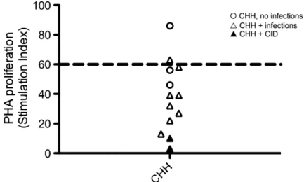
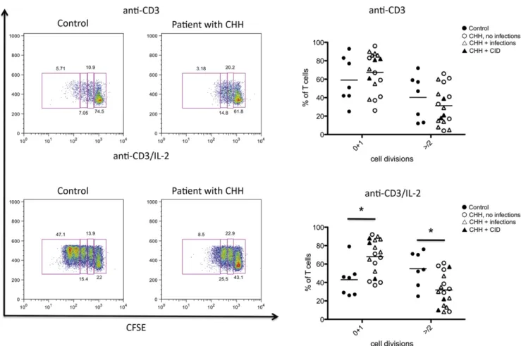
analyzed the response to PHA and anti-CD3in vitro. As shown in Fig 3, the majority of individuals with CHH tested had a dimin-ished response to PHA, with SI values that were below the lower range for normal controls (SI560). PHA responses in patients with CHH and infection were significantly lower than in patients without infections. We also measured proliferation of
CFSE-labeled PBMCs on stimulationin vitrowith anti-CD3 ag-onistic antibodies in the presence or absence of hrIL-2. As shown inFig 4, proliferation of CD31 lymphocytes from pa-tients with CHH was impaired. In particular, under stronger (anti-CD31IL-2) activating conditions, the percentage of CD31 lymphocytes that did not initiate cell division was TABLE I.Clinical and molecular features of the patients
Patient no. Age (y) Sex Mutation Clinical history Diagnosis
1 10.0 F g.70A>G; g.70A>G No infections CHH, no infections
2 12.0 F g.70A>G; g.70A>G Parvovirus infection, juvenile arthritis CHH1infections
3 2.5 F g.70A>G; g.70A>G Interstitial pneumonia CHH1infections
4 1.0 M g.70A>G; g.70A>G Recurrent URTI CHH1infections
5 3.9 F g.70A>G; g.70A>G No infections CHH, no infections
6 5.8 M g.70A>G; g.70A>G No infections CHH, no infections
7 7.7 M g.70A>G; g.70A>G No infections CHH, no infections
8 3.7 M g.70A>G; g.70A>G No infections CHH, no infections
9 21.0 F g.70A>G; g.70A>G Recurrent URTI CHH1infections
10 15.0 F g.70A>G; g.70A>G Sepsis CHH1infections
11 6.7 F g.70A>G; g.70A>G No infections CHH, no infections
12 5.0 F g.70A>G; g.70A>G Pneumonia CHH1infections
13 9.0 F g.70A>G; g.70A>G No infections CHH, no infections
14 7.0 F g.70A>G; g.217C>T Recurrent URTI, chronic diarrhea CHH1CID
15 5.0 M g.18G>C; g.118A>G Recurrent URTI, molluscum CHH1infections
16 2.0 F g.70A>G; g.70A>G Sepsis CHH1infections
17 11.7 M g.27G>A; g.27G>A Aspergillusand CMV pneumonia,
EBV-related lymphoproliferation
CHH1CID
18 2.0 M g.61G>A; g.-23_8dup
TACTCTGTGAAGCTGAG
Sepsis, recurrent pneumonia CHH1CID
CMV,Cytomegalovirus;F,female;M,male;URTI,upper respiratory tract infection.
significantly higher among patients with CHH (mean 6 SD, 69.4 6 18.7) than in controls (45.3 6 19.3; P < .05). Some but not absolute correlation was observed between the degree of impairment of T-cell proliferation and the severity of the clin-ical phenotype.
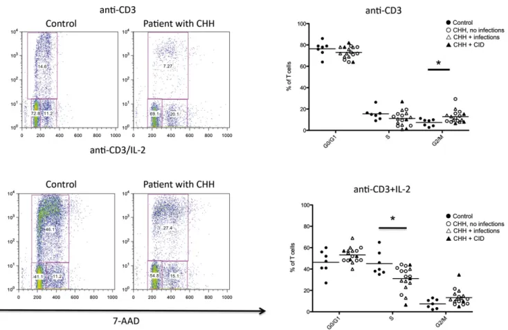
Analysis of cell cycle
RMRP has been shown to play a key role in cell cycle control. Deficiency of the RMRP homolog non-metastatic cells 1 (nme1) gene in yeast causes accumulation of cyclin B2 mRNA and delay in cell-cycle progression.17Similar findings have been reported in fibroblasts from patients with CHH, along with reduced levels of cyclin A2.10We evaluated the impact ofRMRPmutations on cell cycle in T lymphocytes as a possible contributory factor to the im-munodeficiency in CHH. For this purpose, we stimulated PBMCs
in vitrowith agonistic anti-CD3 mAb (in the presence or absence of hrIL-2), followed by addition of BrdU/EdU. To assess the dis-tribution of cells at various phases during the cell cycle, we used flow cytometry and analyzed BrdU/EdU incorporation in combi-nation with staining for the DNA binding dye 7-AAD, which per-mits definition of cell ploidy. Using this method, it was possible to
distinguish cells that were in G0/G1 phase (BrdU–7-AADlo), syn-thesis (S phase; BrdU17-AADlo/hi), or G2/M phase (BrdU– 7-AADhi). As shown in Fig 5, on in vitro stimulation with anti-CD3, patients with CHH had an increased proportion of cells in the G2/M phase (mean6SD, 12.865.7 vs 7.466.7 in pa-tients vs controls;P< .05). An increase of cells in the G2/M phase was also observed in lymphocytes of patients with CHH on stim-ulation with anti-CD31IL-2, although it did not reach statistical significance, and was associated with a marked decrease of lym-phocytes in S phase (mean6SD, 31.1611.2 vs 45.1611.1 for patients vs controls;P< .05). The reduced proportion of cells entering S phase and the prolongation of the cell cycle (with a higher proportion of cells in the G2/M phase) are consistent with the reduced number of cell divisions and the defective T-cell proliferation reported.
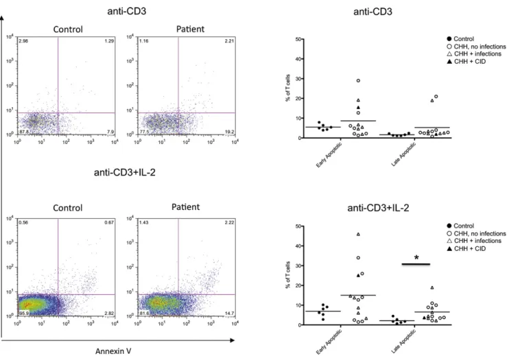
Increased activation-induced cell death in patients CHH
Increased T-cell apoptosis has been previously reported in 1 patient with CHH13but has not been further investigated. We have analyzed activation-induced apoptosis in lymphocytes FIG 2.RTEs. The absolute(A)and relative(B)number of RTEs (CD41CD45RA1CD311) in patients with CHH
was plotted against the value range of healthy pediatric controls for various age groups.15
from patients with CHH and controls afterin vitroculture with anti-CD3 mAb in the presence or absence of IL-2. We used flow cytometry by simultaneously staining the cells for AnnV and for incorporation of 7-AAD. This method allows identifica-tion of viable (AnnV– 7-AAD–), early apoptotic (AnnV1 7-AAD–), and late apoptotic/necrotic (AnnV17-AAD1) cells. As shown inFig 6, the percentage of both early and late apoptotic cells was increased in patients with CHH versus healthy controls, reaching a statistically significant difference for late apoptotic/ne-crotic cells on culture with anti-CD3 mAb1IL-2 (mean6SD in patients with CHH vs controls, 6.564.6 vs 2.161.4;P< .05).
Correction of T-cell proliferation after hematopoietic cell transplantation
Patients with CHH and severe immunodeficiency may require hematopoietic cell transplantation (HCT), and this treatment often allows prolonged survival with immune reconstitution.5,6 We have shown thatRMRPmutations cause abnormal progres-sion along the cell cycle and defective T-cell proliferation after stimulation. Because of severe infections, EBV-related lympho-proliferation, and chronic lung disease, patient 17 received HCT from his HLA-phenotypically identical father after
reduced-intensity conditioning with fludarabine 150 mg/m2, mel-phalan 140 mg/m2, and alemtuzumab 1 mg/kg. This treatment re-sulted in mixed/split chimerism, with 90% donor T cells and 1% donor myeloid cells, respectively, at 11 months after HCT. We compared the ability of the patient’s T lymphocytes to undergo multiple rounds of cell division in response toin vitrostimulation with anti-CD31IL-2 before and after HCT (Fig 7). As expected, before HCT, most of the patient’s CD31T cells failed to divide or underwent a single round of proliferation; in contrast, after HCT, the majority of the cells underwent multiple rounds of cell divi-sion. These results confirm the cell-intrinsic role ofRMRPfor normal T-lymphocyte proliferation.
DISCUSSION
Significant variability in the clinical phenotype has been reported in patients withRMRPmutations. There is some evi-dence that the type and level of RMRP functional impairment may correlate with the severity of the clinical spectrum. In par-ticular, mutations that affect ribosomal RNA cleavage and ribo-somal assembly are associated with more severe bone dysplasia, whereas mutations that reduce mRNA cleavage have a more significant impact on cell cycle regulation and are associated FIG 4. T-cell proliferation in response to agonistic anti-CD3 antibody.Left, Representative examples of
proliferation of control-derived or patient-derived PBMCs in response to anti-CD3(top)or anti-CD31IL-2
(bottom), as measured by CFSE dilution of CD31cells.Boxesidentify different rounds of proliferation, and the proportion of cells within each box is annotated. Gating was done on viable cells.Right,Proportion of PBMCs undergoing 0 to 1 or>_2 rounds of cell division in response to anti-CD3(top)or anti-CD31IL-2
with a higher incidence of immunologic and hematologic abnor-malities and cancer.10 Although imprecise, this reported genotype-phenotype correlation may contribute to significant variability in the degree of immunodeficiency among patients with CHH.2-6,16,18 The majority of the patients included in this study belonged to an Amish population and were homozy-gous for the g.A70>G mutation. The observation thatin vitro– activated lymphocytes from the patients’ cohort showed accumulation of cells in the G2/M phase and decreased propor-tions of cells in the S phase, associated with impaired prolifer-ation, is consistent with the demonstration that transient transfection of normal human fibroblasts with the g.A70>G
RMRP mutant construct causes very severe impairment of cyclin B2 mRNA cleavage.10
Our data indicate thatRMRPmutations have a drastic effect on lymphocyte maturation and function, and that they interfere with thymic generation of T lymphocytes. This is not unexpected, be-cause thymic generation of T lymphocytes requires robust prolif-eration of thymocyte progenitors. Consistent with a potential role of RMRPin supporting thymopoiesis, we and others have re-ported profound abnormalities of thymic architecture in some pa-tients with RMRP mutations.4,14 Furthermore, we previously reported low to undetectable levels of T-cell receptor excision circles (another marker to assessde novothymic generation of
T lymphocytes) in 6 of 7 patients with CHH3; however, all 7 of these patients had significant clinical features suggestive of CID. In this article, we demonstrate that a low number of RTEs may also be seen in patients with CHH without significant clinical immunodeficiency. Along with defects in cell cycle progression and increased apoptosis, these data suggest that at least certain
RMRPmutations are consistently associated with severe cellular immunodeficiency. It will be important to perform similar studies in a larger cohort of patients with different mutations to verify whether RMRP mutations known to interfere with cyclin B2 mRNA processing and cell cycle progression consistently cause abnormalities of lymphocyte function.
Our results also imply that in contrast with T-cell maturation, B-cell maturation was less affected byRMRPmutations. B-cell function was not formally addressed in the current article; how-ever, we collected data on serum immunoglobulins in 14 of the 18 patients with CHH. With the exception of the patients with CHH and CID, all patients with CHH tested had normal immuno-globulin levels. Nine of them were evaluated for tetanus-toxoid specific antibodies, and 7 had protective (>0.15 IU/mL) titers. Pa-tient 16 was also immunized with bacteriophage ØX174 and showed a clearly diminished IgG response suggestive of a func-tional TH-cell defect reminiscent of patients with CD40 ligand
gene (CD40LG) mutations.
FIG 5.Cell cycle analysis.Left,Representative examples of cell cycle analysis in control-derived or patient-derived PBMCs onin vitrostimulation with anti-CD3(top)or anti-CD31IL-2(bottom).Boxesidentify cells in G0/G1 (bromodeoxyuridine[BrdU]27-AAD2), S (BrdU17-AAD2/1), and G2/M (BrdU27-AAD1) phases of the cell cycle. The proportion of cells within each of these is annotated.Right,Proportion of PBMCs in G0-G1, S, and G2/M phases onin vitroculture with anti-CD3(top)or anti-CD31IL-2(bottom).Individual symbols
Various mechanisms may contribute to the cell cycle abnor-malities and increased apoptosis observed in our series of patients. In particular, CHH belongs to a family of diseases, collectively called ribosomopathies, in which well defined com-ponents of the ribosome RNA/protein complex are defective.9
These diseases mostly affect the hematopoietic lineage. Evidence from animal models indicates that when ribosomal function is dis-turbed, the levels of master regulator p53 are increased, because the p53 ubiquitin ligase murine double minute 2 binds to free ribosomal proteins.19Increased p53 is associated with cell cycle arrest and apoptosis.20 The possibility that p53 levels are increased in CHH and may contribute to the immunodeficiency remains to be investigated.
Finally, we have confirmed significant heterogeneity of the CHH clinical phenotype (including variable degree of immu-nodeficiency), even among patients who share the sameRMRP
genetic defect. Given the large number of patients from the same Amish community, there are limitations to the genetic var-iability seen in the present cohort. The basis for heterogeneity of the clinical phenotype of CHH remains unknown, because it cannot be easily attributed to either genetic or environmental factors.
In summary, we show that in CHH, despite individual varia-bility, there is a clear defect in the thymic generation of T lymphocytes and in peripheral T-cell proliferation, cell cycle control, and activation-induced cell death. These data provide a common ground for understanding the functional basis of the underlying immunodeficiency in CHH.
FIG 6.Analysis of apoptosis and cell death.Left,Representative example of fluorescence-activated cell sort-ing plots after stainsort-ing of PBMCs for AnnV and 7-AAD in healthy controls and patients with CHH onin vitro
stimulation with anti-CD3 (top) or anti-CD3 1 IL-2 (bottom). Right, Proportion of early apoptotic (AnnV17-AAD
-) and late apoptotic/necrotic (AnnV17-AAD1) PBMCs onin vitroculture with anti-CD3(top)
or anti-CD31IL-2(bottom).Individual symbolsidentify single subjects. In each diagram, thehorizontal barrepresents the mean value. *P< .05.
FIG 7.Correction of T-cell proliferation defect after HCT in a patient with CHH. The proportion of T lymphocytes undergoing 0, 1, 2 or >2 rounds of proliferation (as indicated by CFSE dilution) after 72 hours ofin vitroculture with anti-CD31IL-2 is shown for patient 17 before(black bars)or 11 months after(white bars)HCT from his HLA phenotypically identical father.Tx,
Clinical implications: Defects of thymic function and of T-lym-phocyte cell cycle control, survival, and proliferation likely con-tribute to the increased frequency of severe infections in patients with CHH.
REFERENCES
1. Notarangelo LD, Roifman CM, Giliani S. Cartilage-hair hypoplasia: molecular basis and heterogeneity of the immunological phenotype. Curr Opin Allergy Clin Immunol 2008;8:534-9.
2. M€akitie O, Kaitila I. Cartilage-hair hypoplasia–clinical manifestations in 108 Finn-ish patients. Eur J Pediatr 1993;152:211-7.
3. Kavadas FD, Giliani S, Gu Y, Mazzolari E, Bates A, Pegoiani E, et al. Variability of clinical and laboratory features among patients with ribonuclease mitochondrial RNA processing endoribonuclease gene mutations. J Allergy Clin Immunol 2008; 122:1178-84.
4. Roifman CM, Gu Y, Cohen A. Mutations in the RNA component of RNase mito-chondrial RNA processing might cause Omenn syndrome. J Allergy Clin Immunol 2006;117:897-903.
5. Guggenheim R, Somech R, Grunebaum E, Atkinson A, Roifman CM. Bone mar-row transplantation for cartilage-hair-hypoplasia. Bone Marmar-row Transplant 2006; 38:751-6.
6. Bordon V, Gennery AR, Slatter MA, Vandecruys E, Laureys G, Veys P, et al. Clin-ical and immunologic outcome of patients with cartilage hair hypoplasia after he-matopoietic stem cell transplantation. Blood 2010;116:27-35.
7. Horn J, Schlesier M, Warnatz K, Prasse A, Superti-Furga A, Peter HH, et al. Fatal adult-onset antibody deficiency syndrome in a patient with cartilage hair hypopla-sia. Hum Immunol 2010;71:916-9.
8. Ridanp€a€a M, van Eenennaam H, Pelin K, Chadwick R, Johnson C, Yuan B, et al. Mutations in the RNA component of RNase MRP cause a pleiotropic human dis-ease, cartilage-hair hypoplasia. Cell 2001;104:195-203.
9. Narla A, Ebert BL. Ribosomopathies: human disorders of ribosome dysfunction. Blood 2010;115:196-205.
10. Thiel CT, Mortier G, Kaitila I, Reis A, Rauch A. Type and level of RMRP func-tional impairment predicts phenotype in the cartilage hair hypoplasia-anauxetic dysplasia spectrum. Am J Hum Genet 2007;81:519-29.
11. Maida Y, Yasukawa M, Furuuchi M, Lassmann T, Possemato R, Okamoto N, et al. An RNA-dependent RNA polymerase formed by TERT and the RMRP RNA. Na-ture 2009;461:230-5.
12. Castigli E, Irani AM, Geha RS, Chatila T. Defective expression of early activation genes in cartilage-hair hypoplasia (CHH) with severe combined immunodeficiency (SCID). Clin Exp Immunol 1995;102:6-10.
13. Yel L, Aggarwal S, Gupta S. Cartilage-hair hypoplasia syndrome: increased apoptosis of T lymphocytes is associated with altered expression of Fas (CD95), FasL (CD95L), IAP, Bax, and Bcl2. J Clin Immunol 1999;19: 428-34.
14. Poliani PL, Facchetti F, Ravanini M, Gennery AR, Villa A, Roifman CM, et al. Early defects in human T-cell development severely affect distribution and matu-ration of thymic stromal cells: possible implications for the pathophysiology of Omenn syndrome. Blood 2009;114:105-8.
15. van Gent R, van Tilburg CM, Nibbelke EE, Otto SA, Gaiser JF, Janssens-Korpela PL, et al. Refined characterization and reference values of the pediatric T- and B-cell compartments. Clin Immunol 2009;133:95-107.
16. Rider NL, Morton DH, Puffenberger E, Hendrickson CL, Robinson DL, Strauss KA. Immunologic and clinical features of 25 Amish patients with RMRP 70 A–>G cartilage hair hypoplasia. Clin Immunol 2009;131:119-28.
17. Cai T, Aulds J, Gill T, Cerio M, Schmitt ME. The Saccharomyces cerevisiae RNase mitochondrial RNA processing is critical for cell cycle progression at the end of mitosis. Genetics 2002;161:1029-42.
18. M€akitie O, Kaitila I, Savilahti E. Susceptibility to infections and in vitro immune functions in cartilage-hair hypoplasia. Eur J Pediatr 1998;157:816-20. 19. Fumagalli S, Di Cara A, Neb-Gulati A, Natt F, Schwemberger S, Hall J, et al.
Ab-sence of nucleolar disruption after impairment of 40S ribosome biogenesis reveals an rpL11-translation-dependent mechanism of p53 induction. Nat Cell Biol 2009; 11:501-8.