TítuloHuman amniotic mesenchymal stromal cells as favorable source for cartilage repair
16
0
0
Texto completo
(2) Decades of research in tissue engineering for effective treatment of articular cartilage have led to a variety of therapeutic approaches.1,2 However, to date, none of these approaches has been yet successful in overcoming the poor intrinsic repair response of articular cartilage. Articular cartilage is an avascular, aneural, and aliphatic tissue with particular low cell density.3 On the other hand, cartilage has high demanding functional requirements even at the time of treatment, which could determine reparation failure.4 Hence, the complete functional recovery of affected cartilage remains an unsolved problem.5,6 The development of engineered grafts for cartilage regeneration has tried the creation of a favorable microenvironment for native cells harbored on the cartilage and for new implanted cells. This strategy could assist in the function of implanted cells to restore lost tissue, avoiding the progressive degeneration of the articular cartilage to osteoarthritis (OA). 7 However, one of the main problems in tissue engineering is the lack of integration with the surrounding native cartilage, resulting in the absence of long-term functional implants. To enhance integration, there are several strategies centered in the use of natural scaffolds.8 One of the most promising tissues, used for ages as natural scaffold, is the human amniotic membrane (HAM). HAM or amnion is an embryonic tissue created from the extra-embryonic ectoderm.9 Numerous intrinsic properties allow its use as a biomaterial. It has low immunogenicity, anti-inflammatory properties, and capacity to act as an antimicrobial agent. In addition, HAM secretes a variety of growth factors and presents an important therapeutic value reducing fibrotic healing.10 Besides, HAM has special characteristics that may permit articular cartilage regeneration. It shares components with the extracellular matrix (ECM) of articular cartilage, with the presence of glycosaminoglycans (GAG), different types of collagen, or fibronectin.11 From a materials point of view, HAM ECM is elastic, flexible, and resistant to the traction forces. Furthermore, it holds an economic advantage over other biological materials because of its availability and easy harvest.12 All these properties increase the interest of the amnion as a substrate for the transfer of both autologous and allogeneic cells. For tissue engineering purposes another important point is to define the most appropriate and effective cell source to treat specific diseases. Bone marrow mesenchymal stromal cells (BMSCs) are the most widely studied type of cells and considered the gold standard when other mesenchymal cell sources have been studied.13 Nevertheless, BMSCs have several disadvantages such as low cell yield during isolation, decrease differentiation capacity associated at the age of the donor, or morbidity derived from the harvest method. 14 All these reasons boost the search for alternative cell sources. Apart from its scaffold properties, HAM is one of the extra-embryonic tissues with significant potential as source of stem cells.15 It is an embryonic-derived noncontroversial tissue, its size secures good yield of cells, and is obtained without invasive or expensive procedures.16 Two different populations of stromal cells could be harvested, human amniotic mesenchymal stromal cells (hAMSCs) and human amniotic epithelial cells (hAECs).17 Additionally, another important benefit is its immunological privileges. As a tissue related to the fetomaternal tolerance during pregnancy, it owns great capacity for autologous and allogeneic transplantation when compared with other cell sources.18 The objective of this study was to develop an in vitro model for focal repair of human articular cartilage using HAM as biomaterial assessing the therapeutic potential of different cell sources..
(3) Materials and Methods Harvest and preparation of HAMs This study was approved by the Ethics Committee for Clinical Research of Galicia (Spain). Human placentas (n = 30) from healthy donors were obtained from selected caesarean sections at the Hospital Materno-Infantil Teresa Herrera (A Coruña, Spain). All the donors gave written informed consent before tissue and cell collection. Under stringent sterile conditions, the harvested placentas were placed in 199 Medium (Thermo Fisher Scientific, Waltham, MA) with an antibiotic cocktail composed of the following: cotrimoxazol 50 µg/mL (Soltrim®; Almirall-Prodesfarma S.A., Barcelona, Spain), vancomycin 50 µg/mL (Vancomicina Hospira®; Laboratorio Hospira S.L., Madrid, Spain), amykacin 50 µg/mL (Amikacina Normon®; Laboratorios Normon S.A., Madrid, Spain), and B amphotericin 5 µg/mL (Fungizona®; Bristol-Myers Squibb, New York, NY) for 6 to 20 h at 4ºC. The HAM was carefully separated from the chorion and washed three to five times with 0.9% NaCl solution to remove blood cells. Cryopreservation and thawing of amnion for its use as scaffold TheHAMused as scaffold (n = 13)was placed on a supportive sterile nitrocellulose filter (Merk Millipore, Darmstadt, Germany) and cut into 6 x 6 cm patches in 20mL of 199 Medium (Thermo Fisher Scientific) without antibiotics but with a cryoprotectant, 10% dimethyl sulfoxide (DMSO; Sigma-Aldrich, St Louis, MO). Each patch of HAM was cryopreserved following a protocol of controlled freezing using CM 2000 (Carburos Metálicos, Barcelona, Spain). Freezing rates were −1ºC/min to a temperature of −40ºC, −2ºC/min to −60ºC, and −5ºC/min to −150ºC. All HAMs were stored in the gas phase of liquid nitrogen at −150ºC. Thawing was carried out for 5 min at room temperature followed by 37ºC until thawing was complete. To reduce cell damage due to osmotic changes, the DMSO was removed by sequential washing and progressive dilution with 0.9% NaCl at 4ºC. Isolation and culture of human amnion-derived cells The HAMs used as source of stromal cells (n = 17) were processed following the protocol of Soncini et al.17 Briefly, HAM was cut into ~2 x 2 cm2 pieces and transferred into an enzymatic digestion buffer containing 2.4 U/mL of dispase (Thermo Fisher Scientific) in phosphate-buffered saline (MP Biomedicals, Inc., Santa Ana, CA) and incubated at 37ºC for 7 min. The digested tissue was centrifuged and the supernatant was discarded. The tissue was then subjected to a second enzymatic digestion containing 0.75 mg/mL of type I clostridial collagenase (Thermo Fisher Scientific) and 20 µg/mL of deoxyribonuclease I (Sigma-Aldrich) in RPMI 1640 (Lonza, Basel, Switzerland) culture medium for 3 h at 37ºC. Following this digestion the resulting cell suspension was filtered through a sterile 70 µm filter (BD Biosciences, Franking Lakes, NJ). The collected cells were designated as hAMSCs. Nondigested amnion fragments were incubated with a solution containing 0.25% trypsin-EDTA (Sigma-Aldrich) at 37ºC for 2–3 min. The resulting cell suspension, containing hAECs, was centrifuged at 600 g for 10 min. The cells were recovered in Dulbecco’s modified Eagle medium (DMEM; Sigma-Aldrich Quimica) with 20% fetal bovine serum (FBS; Lonza) and 1% penicillin-streptomycin (P/S) (Thermo Fisher Scientific) and seeded into 162 cm2 culture flasks (Corning, New York, NY). Nonadherent cells were removed after 48 h of culture. Both hAECs and hAMSCs cells were cultured in a humidified 5% CO2 atmosphere at 37ºC until 70% confluency was reached. The cells were expanded to obtain enough number of cells for further experiments..
(4) Harvest of human BMSCs Immediately after hip joint surgery for prosthetic replacement, femoral heads were obtained aseptically from five patients with OA (mean age 65 years, range 50–70 years) after signing informed consent. The human bone marrow-derived mesenchymal stromal cell (hBMSC) were obtained after repeated washing of the bone marrow with DMEM supplemented with 5% FBS and 1% P/S. Then, cells were centrifuged at 600 g for 8–10 min and seeded into 162 cm2 culture flasks in a humidified 5% CO2 atmosphere at 37ºC. After 48 h of culture nonadherent cells were removed changing the medium two to three times a week. Harvest and isolation of human articular chondrocytes The Autopsy Service of the Hospital Universitario A Coruña supplied cartilage samples derived from patients who underwent leg amputations due to peripheral vascular disease, diabetes, or chronic vascular inflammation. The Orthopedics Surgery Department provided cartilage samples from patients with partial or total knee/hip replacement. Cartilage samples, from femoral heads or knees, comprised 25 donors (15 male and 10 female) with a mean age of 67 years (from 25 to 85 years). Cartilage biopsies were aseptically removed, sliced full thickness (excluding the mineralized cartilage and subchondral bone), and washed in DMEM as previously described by Rendal-Vázquez et al.19 The excised cartilage slivers were placed in a Petri dish, minced, and transferred to 50mL falcon tubes where chondrocytes were isolated by sequential enzymatic digestion. First, minced cartilage slivers were incubated in 10mL of basic medium, containing 0.25% Trypsin-EDTA (Sigma-Aldrich) for 10 min at 37ºC under agitation. The supernatant, without chondrocytes, was discarded and the trypsinized cartilage was subjected to a second digestion. Twenty milliliters of extraction medium, containing collagenase (2 mg/mL; Sigma-Aldrich), were added. After an overnight incubation on a shaker at 37ºC, digested dilution was filtered with a 70mm cell strainer (Corning) transferred to a 50 mL falcon tube. Then, cells were subjected to a centrifugation for 10 min at 600 g, supernatant was removed and chondrocyte number determined using a hemocytometer. Cells were expanded in DMEM with 10% FBS and 1% P/S into 162 cm2 culture flasks. Cells were cultured in a humidified 5% CO2 atmosphere at 37ºC until 80% confluence was reached. In vitro differentiation studies Adipogenesis and Osteogenesis of hBM-MSCs (n = 5), hAECs (n = 11) and hAMSCs (n = 11) were done between passages 2 to 4. The different cells were detached using 0.25% trypsin-EDTA solution and seeded at 2.2 x 10⁴ cells/cm2 into an eight-well chamber slide (Thermo Fisher Scientific) in growth medium until confluence. Adipogenesis was induced maintaining the cells in Bullekit Adipogenic Differentiation Medium (Lonza) for 21 days following the manufacturer’s instructions. In the case of osteogenesis induction, cells were cultured for 21 days using hMSC Bullekit Osteogenic Differentiation Medium (Lonza). Culture mediums were changed every 2–3 days. Chondrogenesis was assessed by micropellet formation (hBM-MSCs [n = 5], hAECs [n = 11], and hAMSCs [n = 11]) as previously described by Yoo and Johnstone with some modifications.20 HAM-derived cells (2.5 x 10⁵ cells/cm2) from passages 2 to 4 were detached using trypsin-EDTA and centrifuged at 600 g for 10 min. The resulting pellets were cultured in chondrogenic differentiation medium, DMEM containing 15% FBS, supplemented with 5 mg/mL ascorbic acid (AA), 1/1000 monotioglycerol (Sigma), and 1% P/S during the first 48 h to promote the induction of chondrogenesis. After 48 h, the medium was replaced by DMEM with 15% knockout serum (Gibco), 1% P/S and supplemented with 1 µL/mL AA, 10 µM dexamethasone, 6 µL/mL Transferrin, 1 x 10⁷ Mretinoic acid (all from Sigma), and 10 ng/mL recombinant human transforming growth factor-β3 (Prospec-Tany Technogene Ltd., Rehovot, Israel) for 21 days. Medium was changed every 2–3 days..
(5) For control samples, each differentiation assay with their respective control cultivating hBMSCs, hAECs, and hAMSCs were cultured inDMEMmedium, containing 20%FBS and1% P/S for 21 days. Both kind of samples, control and differentiated, were evaluated using the same histological and immunohistochemical analyses. Analysis of in vitro differentiation. For these analyses, all samples were fixed using 4% paraformaldehyde. To evaluate adipogenesis, the presence of cytoplasmic lipid droplets in the cultures was revealed directly on the eight-well chamber slides by Oil-red O stain (OR). The same was done for osteogenesis, but analyzing the presence of mineral deposits using Alizarin Red (AR) stain according to a standard protocol. Quantification of positive histological staining for AR and OR was performed using the analiSIS® software (version D) (Olympus, Melville, NY). Micropellets from chondrogenic differentiation were embedded in paraffin to carry out histological and immunohistological evaluation for the presence of hyaline cartilage-characteristic molecules. Histological and immunohistological analyses. For histological evaluation, 4 µm-thick paraffin sections were deparaffinized in xylol and rehydrated in a graded series of ethanol. Sections were stained with hematoxylin and eosin (HE), toluidine blue (TB), safranin O (SO), and Masson’s modified trichrome (MT). For immunohistochemical studies, sections were incubated with monoclonal antibodies to detect type I (COLI) (Abcam, Cambridge, UK), COLII (Merk Millipore) collagens and proliferating cell nuclear antigen (PCNA, Calbiochem; Merk Millipore). Sections were pretreated with proteinase K and the peroxidase/DAB ChemMateTM DAKO EnVisionTM detection kit (Dako, Santa Clara, CA) was also used. Samples were examined using an optical microscope and the COLI and COLII levels were quantified using a plugin developed for Fiji ImageJ (GNU, General Public Licence). The International Cartilage Research Society (ICRS) visual histological score21 was the quantitative system selected to grade the damage cartilage repair. With this scoring, more reliable data were obtained to evaluate objectively the harvested results. Development of in vitro model for articular cartilage repair The in vitro model was developed in three steps as detailed on Figure 1A. Amnion as scaffold. The HAM used as scaffold (n = 16) was placed on CellCrownTM (Scaffdex, Finland) disposable inserts with the stromal side of the HAM exposed to seed the different type of cells on it. All the cell types, chondrocytes, hBM-MSCs, hAECs, or hAMSCs, were seeded in a density of 1 x 10⁶ cells/cm2 (Fig. 1B). It was maintained in culture until the total confluence of the cells on the stromal side (max for 3 weeks) with DMEM plus 10% FBS and 1% P/S (Fig. 1D, d1). Harvesting of cartilage biopsies. Cartilage samples were both OA (n = 12) and healthy (n = 2) subjects. Cartilage selected to develop the in vitro focal repair model was harvested in slicers. The samples included male and female equally with a mean age of 77 ± 9 years. Then, they were cut with a biopsy punch (Biopsy Punch Stiefel, España) into 6mm diameter discs. For the focal injury, a lesion of 2mm diameter with a dental drill was created in the superficial zone of each cartilage (Fig. 1D, d2)..
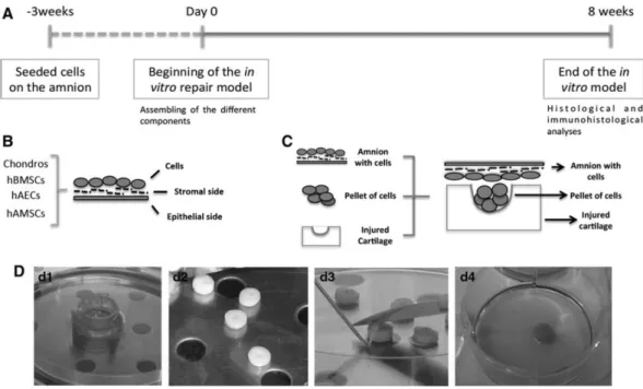
(6) FIG. 1. Schematic representation of the timeline and development of the in vitro model. (A) Chronogram of the main steps of the model. (B) Design of the implanted cells over the stromal layer of the amnion. (C) Assembly of all the components, membrane-laden and cell pellet with the different type of cells are implanted into the injured cartilage plug. (D) Life images of the components of the in vitro system and their assembly: (d1) CellCrownTM system; (d2) drilled human cartilage plugs; (d3) assembly of the system with the membrane covering the cartilage lesion; (d4) assembled plug in culture.. Assembling of the in vitro repair model. Each patch of HAM with the cells on it was dismantled from the Cell-Crown system. Just before putting the HAM over the cartilage biopsies, a pellet of 6 x 10⁵-same-type-cells that were on the HAM, were deposited inside the drilled defect. For it, each cell type was put under a 600 g centrifugation for 8 min. The formed pellet was broken with two short pulses in the shaker and placed in the cartilage lesion. Cartilage plugs with cells inside of the lesion were placed in a six-well plate and submitted a short centrifugation. This step allowed concentrating the cells on the deep part of the lesion. Then, HAM was placed over the superficial zone of the 6 mm-cartilage biopsies in such a way that the stromal layer of the HAM, where the cells were grown, was in direct contact with the superficial surface of the cartilage (Fig. 1C, D, d3). In vitro models for articular cartilage repair were developed for chondrocytes (n = 6), hBMSCs (n = 5), hAECs (n = 5), and hAMSCs (n = 6). Cartilage biopsies with the focal defect covered with the amniotic membrane, but without the cellular pellet inside the lesion were used as controls. All cartilage biopsies, loaded with the pellets or unloaded (Supplementary Fig. S1; Supplementary Data are available online at www.liebertpub.com/tea), and covered with the HAM were placed in six-well culture plates (Costar®, Washington, DC) and cultured for 8 weeks (Fig. 1, d4). Two milliliters of DMEM culture medium supplemented with 10% FBS and 1% P/S (for chondrocytes) and chondrogenic medium (for the models with hBM-MSCs, hAECs, and hAMSCs) was placed in each well and replaced twice weekly. The culture plates were incubated in a 5% CO2 humidified atmosphere at 37ºC. All procedures were performed under stringent sterile conditions. After 8 weeks in culture, the in vitro cartilage repair model was retrieved from the culture plate, embedded in paraffin, and processed for histological and immunohistochemical analyses..
(7) Statistical analysis Statistical analyses for the repair percentage of the lesion area, ICRS scoring scale, and the quantification of COLI and COLII for the different models were performed using SPSS 16.0 software for Windows. All the data are represented as mean ± standard error. When normal distribution of the samples was confirmed, two-tailed Student’s t test was applied. When normal distribution was not detected Mann–Whitney test was applied. In all cases, a p < 0.05 was considered statistically significant.. Results As we previously published, hAMSCs and hAECs showed similar differentiation capacity into the three mesenchymal lineages than hBMSCs (Fig. 2).22 To determine the best source of cells to regenerate articular cartilage, we developed an in vitro model of articular cartilage repair comparing the differences in cartilage regeneration of HAM loaded with different sources of cells (chondrocytes, hBMSCs, hAMSCs, and hAECs). Chondrocytes and hBMSCs were considered as gold standards. Initially, cells were expanded (Fig. 3) and then cultured over the stromal layer of the HAM. Masson’s trichrome staining was used to visualize the cells growing over the HAM (Fig. 4A, C, E, G). All cell types were grown mainly on monolayer over the amnion and were able to synthetize some ECM, especially chondrocytes (Fig. 4B). In these conditions, chondrocytes maintained their phenotype, verified by COLII immunohistochemistry (Fig. 4B), ruling out that the dedifferentiation process was taking place. Also hBMSCs, hAMSCs, and hAECs showed positive presence of COLII after cultured time in chondrogenic medium (Fig. 4D, F, H). When cells reached confluence over the amnion, the amnion was implanted in vitro filling a focal defect performed in human-derived condral plugs. The model was maintained for 8 weeks and then subjected to histology and immunohistochemistry evaluation..
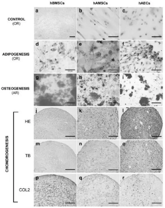
(8) FIG. 2. In vitro differentiation capacity of three progenitor cells tested in the model. (a–c) After 21 days in culture without differentiation factors the indicated progenitor cells did not show specific adipogenic or osteogenic staining. When exposed to specific differentiation factors, all kind of progenitor cells presented capacity to differentiate into adipocytes (d–f), osteoblast (g–i), and chondrocytes (j–r). OR, Oil-red O staining; AR, Alizarin Red staining; HE, hematoxylin and eosin staining; TB, toluidine blue staining; COL2, immunohistochemistry for type II collagen. Scale bar: (a, h–j, l, m) 200 µm, (b–d, f, g, k, n–r) 100 µm, and (e) 50 µm..
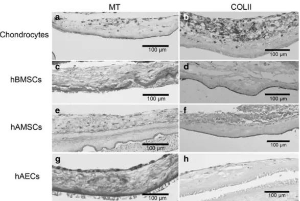
(9) FIG. 3. Morphology of human chondrocytes, hBMSCs, hAMSCs, and hAECs during in vitro expansion. Scale bar: 200 µm. hAEC, human amniotic epithelial cell; hAMSC, human amniotic mesenchymal stromal cell; hBMSC, human bone marrow-derived mesenchymal stromal cell.. FIG. 4. Representative images, of the different cell-types cultivated over the stromal layer of the amniotic membrane before plug assembly. (a, b) Chondrocytes. (c, d) hBMSCs. (e, f) hAMSCs. (g, h) hAECs. COLII, immunoshistochemistry for type II collagen; MT, Masson’s trichrome staining. Scale bar: 100 µm.. Histological study Implanted chondrocytes synthesized ECM and were able to integrate with the native cartilage. In the newly synthesized tissue, the cells showed a rounded morphology (Fig. 5A, a). hBMSCs also synthetized high amounts of new tissue that appear more abundant than when chondrocytes were implanted (Fig. 5A, b), though the ECM synthetized by hAMSCs was even superior (Fig. 5A, c). In addition, the appearance of the repair tissue in hAMSCs was hyaline and contained some cells with a rounded morphology. On the other hand, hAECs showed the lowest amount of synthesized ECM and presented a poor morphological appearance with high nuclear size and reduced cytoplasm. Due to the pyknotic appearance of these cells, the presence of PCNA was tested by immunohistochemistry (Supplementary Fig. S2). In all cases, the repaired tissue showed a high cellularity than the remaining native cartilage..
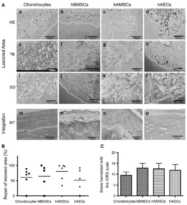
(10) The presence of proteoglycans was visualized by SO and TB staining. Chondrocytes and hAMSCs showed metachromasia for both stains when compared with hBMSCs. In defects treated with hAECs SO staining was not detected, while TB staining exhibited slight positivity (Fig. 5A, e–l). When the cells were grown over HAM, HAM adapted itself to the irregular superficial layer of OA cartilage, even in OA cartilage and in areas of normal cartilage (Fig. 5, Integration). When chondrocytes were included as the cell population, HAM showed the best quality integration (Fig. 5A, m). When HAM containing hBMSCs, hAMSCs, or hAECs were applied a reduction in the superficial imperfections in OA cartilage was also observed (Fig. 5A, n–p). The extension of repaired tissue was quantified in the defect using Image J (Fig. 5B). When chondrocytes were used, the repair tissue filled between 54% and 83% of the injured area. For hBMSCs, these values varied between 50% and 90%. For hAMSCs the injury filling varied between 40% to completely full defect. Finally, for hAECs a variation between 30% and 90% of filled injuries was calculated. ICRS scoring was used to evaluate the general grade of reparation in the model with the different cell types (Fig. 5C). Chondrocytes, hBMSCs, hAMSCs, and hAECs did not show significant differences in the scoring with values of 9.5 ± 3.7, 12.8 ± 5.0, 12.5 ± 6.2, and 11.8 ± 6.0, respectively. Immunohistochemical analyses The presence of specific components of the ECM in the repaired tissue was visualized with specific immunostainings for COLII and COLI. Controls with healthy human skin and cartilage were used to optimize the immunohistochemistry techniques (Supplementary Fig. S3). Positive staining for COLII was only detected in the cytoplasm of the different cell types, but not in the newly synthetized ECM of any cell type (Fig. 6A). The quantification of the presence of COLII showed higher signal for hAMSCs when compared to other cells, this difference was statistically significant when compared with the chondrocyte-laden group (Fig. 6B). The presence of COLI in hBMSCs was significantly higher than with other type of cells, none of them showing negligible levels of COLI staining (Fig. 6C). Therefore, most of the cell types presented an acceptable relation of new synthesized tissue and fibrotic tissue..
(11) FIG. 5. (A) Histological analysis of the repair tissue present in the defect (a–d). The presence of proteoglicans was visualized by toluidin blue staining (e–h) and safranin O staining (i–l). The integration capacity of the membrane with the superficial layer of the native cartilage was analyzed with Masson’s trichrome staining (m–p). Yellow dotted line indicates the edges of native cartilage and new synthesized tissue in the lesion (a–l) and the integration of the native cartilage with the amnion (m–p). Scale bar: (o) 50 µm and (a–n, p) 100 µm. (B) Histomorphometric quantification of the repaired area. (C) Scoring of the repair tissue using ICRS scale. HE, hematoxylin-eosin staning; ICRS, International Cartilage Research Society; SO, safranin O staining. Graphics expressed the mean ± standard error..
(12) Discussion The use of HAM as a source of mesenchymal progenitors cells and a scaffold for tissue engineering has grown in interest in the last few years. From a materials point of view, HAM is the eldest natural biomaterial.23–26 It has been applied in tissue engineering approaches in ophthalmology, dermatology, odontology, cardiology, and gynecology.27–31 In addition, HAM has been used in preclinical and clinical studies for the repair of corneal tissue, spinal cord injuries, cerebral stroke, and Parkinson’s disease.32–35 All these studies have demonstrated little risk of infection and high versatility as biomaterial. Trying to understand the diverse potential of HAM, some studies have focused on the mechanism underlying these wide applications. One of the most important properties of HAM is the low immunogenicity, enabling maternal-fetal tolerance during pregnancy.36 The two types of cells isolated from this tissue, hAMSCs and hAECs, lack the human leukocyte antigens (HLA)-A, -B, -C from class I major histocompatibility complex, HLA-DR or -DQ immunological response molecules.37,38 On the other hand, it was demonstrated that epithelial amniotic cells express high amount of HLA-G, a protein related to immune tolerance, which activates the apoptosis of cytotoxic T cell through Fas ligand.39 Furthermore, this immune privilege could facilitate allogeneic transplantation, preventing the use of immune suppression therapy for patients after HAM implantation. Another valuable point is that HAM is a discarded tissue after delivery, with reduced ethical concerns to its harvest. Different methods of preservation have been developed that facilitate their long-term storage and use.40–42 First reports on HAM were centered in its use as carrier supporting cells growth. In this aspect, it has demonstrated to be not only a good scaffold for different cells, but also to contain properties that allow the maintenance of the original phenotype of cells.43,44 Wound protection and pain reduction are also qualities suggested for HAM. HAM-derived cells, hAECs and hAMSCs, showed synthesis of anti-inflammatory factors, as tissue inhibitor of metalloproteases (TIMP)-1, TIMP-2, TIMP-3, TIMP-4, and IL-10; a variety of growth factors, EGF, bFGF, or TGF-β1, TGF-β2, and TGF–b3; and antimicrobial substances/agents, like βdefensin or secretory leukocyte protease inhibitor.16,45,46 In addition, on the stromal side of the HAM there is a suppression of the profibrotic TGF-β1 signaling system minimizing the scar tissue and promoting the reconstruction of the native tissue.47 Also, HAM is a biodegradable and permeable avascular structure. All these benefits have supported the easy translation for clinical purposes. There are some specific properties on the HAM that reinforced the idea of its use in cartilage regeneration; the simple but solid structure of HAM showed enough strength to overcome the high demanding mechanical environment on the joint.48–51 Furthermore, HAM shares numerous components of its matrix with the ECM of native articular cartilage as GAG, different types of collagen, or fibronectin.11 The natural synthesis of TGF-β1, -β2, and -β3 would facilitate the chondrogenic differentiation of MSCs without exogenous addition of differentiation factors favoring an easy clinical translation.45 The stratification of the HAM might facilitate integration with the native cartilage seen in our results. Also, the plasticity and easy handling of the HAM would allow perfect fit into the inherent cracks and fissures present at the OA cartilage. This integration could potentially delay the evolution of OA disease or, in the best scenario, stop the degeneration of the cartilage, and recover the treated OA surface.52 Lastly, despite the promising clinical results with autologous chondrocyte implantation, there are some limitations associated to the use of a periosteal flap. As detachment of the flap, delamination and hypertrophy, generated by the cells and factors of the periosteum, has been described.53 The replacement of the periosteal flap by HAM would prevent these limitations..
(13) Our group has demonstrated the utility of HAM as a source of mesenchymal progenitors cells for a variety of cell-based therapies.54 Here, the central idea of this report is to evaluate the specific application of HAM for cartilage regeneration. Even when this is not the first approximation to evaluate the superficial reparation of osteoarthritic cartilage with HAM as scaffold,55 it is the first study analyzing hAECs and hAMSCs for cartilage repair. But, in our view, the strength of this study is the comparison of HAM-derived progenitor cells with the two principal type of cells, hBMSCs and chondrocytes, used extensively in the clinic for cartilage repair. Between the benefits of HAM as source of cells, the possibility of harvesting two different cell types from the same tissue is attractive.56 We detected that all type of cells assessed exhibited good integration and adhesion with the native cartilage. Thus, the immobilization of the cell pellet inside the lesion contributed to fill the injured cartilage. Chondrocytes, hBMSCs, hAECs, and hAMSCs infiltrated the superficial zone (closer to the damaged area) and the edges of the lesion, decreasing the small fissures or irregularities characteristic of OA cartilage. Also, it was possible to detect a thickening of the stromal part of the HAM when implanted with chondrocytes, which could be derived of the growth and infiltration of cells that synthesize ECM, as reported when a chondrocytes implantation model is used.57 Of note, there were no significant differences between the cells used in the clinic and the HAM-derived cells. However, a deeper analysis through the presence of chondrogenic markers showed that hBMSCs presented a reduced level in the synthesis of COLII when compared with hAMSCs. Progenitor cells isolated from extra-embryonic tissues, as the amnion, have emerged as a potential alternative sharing properties of embryonic stromal cells and adult stem cells.15 There is the assumption that hAMSCs remain in a previous differentiation step than hBMSCs, thus, retaining a higher differentiation potential. Moreover, the repaired tissue by hBMSCs displayed the highest COLI content, an important marker of fibrotic tissue, confirming that the stimulus with chondrogenic factors was not enough for these cells. In this regard, the age of the donors is an important factor, and BMSCs were harvested from patients with a mean age of 65 years. Taking into account chondrocytes, even this source of cells does not seem the most appropriate, especially when lower levels of COLII were detected when comparing with the rest of nondifferentiated cells. These low competence of chondrocytes could be explained by ex vivo chondrocyte expansion, where chondrocytes lose their phenotype58 or because of the pathology background of chondrocytes. Overall, HAM is an optimal source of progenitor cells for cartilage repair purposes; progenitors cells could be obtained with high yields, without invasive and/or expensive procedures, being a noncontroversial source of stromal cells. 59 Limitations of the study This is an in vitro reparation model with important limitations. The main limitation is the lack of mechanical stimulus with recognized influence on the biochemical constitution and biosynthesis of cartilage molecules.3 The absence of these forces could explain the high content of COLII in the cellular cytoplasm not being delivered to the matrix. In addition to this, the cartilage used for these experiments was mainly derived from patients affected by OA, thus, the predominance of catabolic processes in OA cartilage may hamper some reparative properties of the assessed cells.60,61 Finally, as an in vitro model, important biochemical signals derived from the proximal cartilage tissues, synovial membrane, bone, and synovial liquid cannot be evaluated.62.
(14) Conclusions In this study we explored the use of HAM in the regeneration of articular cartilage, not only as scaffold but also as a source of progenitor cells. HAM demonstrated good capacity adhering to the superficial zone of the native cartilage as well as integrating into the surface structure. Furthermore, HAM-derived hAMSCs showed better reparation scores even when compared with BMSCs and chondrocytes, cells widely used in preclinical and clinical procedures.. Acknowledgments The authors would like to thank E. Rendal-Vázquez, PhD from Tissue Bank and Criobiology Department, INIBIC Hospital Universitario A Coruña, for the HAM samples. We thank P. Filgueira, M.J. Sánchez, and N. Goyanes-Rey from INIBIC-CHUAC for their technical assistance. We thank Froilán Granero-Moltó for the critical reading of the article. Emma Muiños-López was beneficiary of a fellowship from the Rheumatology Spanish Foundation (FER), Spain during the development of this work. This study was supported by grants: Servizo Galego de Saúde, Xunta de Galicia (PS07/84), Cátedra Bioibérica de la Universidade da Coruña, Instituto de Salud Carlos III, CIBERBBN,REDICENT (Rede de Investigación en Células Nai e Terapia Celular) and GPC (Grupos con Potencial de Crecemento) both from Xunta de Galicia (CN2012/142, R2014/050, and GPC2014/048); Rheumatology Spanish Foundation (FER 2014 grant). The results of this work have been funded by the Project No. PI16/02124, integrated in the National Plan for Scientific Research, Development and Technological Innovation 2013–2016 and funded by the ISCIII— General Subdirection of Assesment and Promotion of the Research—European Regional Development Fund (FEDER) ‘‘A way of making Europe.’’. Disclosure Statement No competing financial interests exist.. References 1. Musumeci, G., et al. New perspectives for articular cartilage repair treatment through tissue engineering: a contemporary review. World J Orthop 5, 80, 2014. 2. Benders, K.E.M., et al. Extracellular matrix scaffolds for cartilage and bone regeneration. Trends Biotechnol 31, 169, 2013. 3. Ng, K.W., et al. Duty cycle of deformational loading influences the growth of engineered articular cartilage. Cell Mol Bioeng 2, 386, 2009. 4. Mastbergen, S.C., Saris, D.B., and Lafeber, F.P. Functional articular cartilage repair: here, near, or is the best approach not yet clear? Nat Rev Rheumatol 9, 277, 2013. 5. Mollon, B., Kandel, R., Chahal, J., and Theodoropoulos, J. The clinical status of cartilage tissue regeneration in humans. Osteoarthritis Cartilage 21, 1824, 2013. 6. Chevalier, X., Eymard, F., and Richette, P. Biologic agents in osteoarthritis: hopes and disappointments. Nat Rev Rheumatol 9, 400, 2013. 7. Muzzarelli, R.A.A., Greco, F., Busilacchi, A., Sollazzo, V., and Gigante, A. Chitosan, hyaluronan and chondroitin sulfate in tissue engineering for cartilage regeneration: a review. Carbohydrate Polymers 89, 723, 2012. 8. Ye, K., et al. Bioengineering of articular cartilage: past, present and future. Regen Med 8, 333, 2013. 9. Ferner, K., and Mess, A. Evolution and development of fetal membranes and placentation in amniote vertebrates. Respir Physiol Neurobiol 178, 39, 2011. 10. Wolbank, S., van Griensven, M., Grillari-Voglauer, R., and Peterbauer-Scherb, A. Alternative sources of adult stem cells: human amniotic membrane. Adv Biochem Eng. Biotechnol 123, 1, 2010. 11. Niknejad, H., et al. Properties of the amniotic membrane for potential use in tissue engineering. Eur Cell Mater 15, 88, 2008. 12. Mamede, A.C., et al. Amniotic membrane: from structure and functions to clinical applications. Cell Tissue Res 349, 447, 2012. 13. Beane, O.S., and Darling, E.M. Isolation, characterization, and differentiation of stem cells for cartilage regeneration. Ann Biomed Eng 40, 2079, 2012..
(15) 14. Motaln, H., Schichor, C., and Lah, T.T. Human mesenchymal stem cells and their use in cellbased therapies. Cancer 116, 2519, 2010. 15. Abdulrazzak, H., Moschidou, D., Jones, G., and Guillot, P.V. Biological characteristics of stem cells from foetal, cord blood and extraembryonic tissues. J R Soc Interface 7(Suppl 6), S689, 2010. 16. Toda, A., Okabe, M., Yoshida, T., and Nikaido, T. The potential of amniotic membrane/amnion-derived cells for regeneration of various tissues. J Pharmacol Sci 105, 215, 2007. 17. Soncini, M., et al. Isolation and characterization of mesenchymal cells from human fetal membranes. J Tissue Eng Regen Med 1, 296, 2007. 18. Parolini, O., et al. Concise review: isolation and characterization of cells from human term placenta: outcome of the first international Workshop on Placenta Derived Stem Cells. Stem Cells 26, 300, 2008. 19. Rendal-Vázquez, M.E., et al. Effect of cryopreservation on human articular chondrocyte viability, proliferation, and collagen expression. Cryobiology 42, 2, 2001. 20. Yoo, J.U., and Johnstone, B. The role of osteochondral progenitor cells in fracture repair. Clin Orthop Relat Res S73, 1998. 21. van den Borne, M.P.J., et al. International Cartilage Repair Society (ICRS) and Oswestry macroscopic cartilage evaluation scores validated for use in autologous chondrocyte implantation (ACI) and microfracture. Osteoarthr Cartil 15, 1397, 2007. 22. Díaz-Prado, S., et al. Multilineage differentiation potential of cells isolated from the human amniotic membrane. J Cell Biochem 111, 846, 2010. 23. Davis, J.S. Skin transplantation with a review of 550 cases at the Johns Hopkins Hospital. Johns Hopkins Med J 15, 307, 1910. 24. Stern, W. The grafting of preserved amniotic membrane to burned and ulcerated surfaces, substituting skin grafts. JAMA 13, 973, 1913. 25. Sabella, N. Use of fetal membranes in skin grafting. Med Rec NY 83, 478, 1913. 26. Meller, D., Pauklin, M., Thomasen, H., Westekemper, H., and Steuhl, K.-P. Amniotic membrane transplantation in the human eye. Dtsch Arztebl Int 108, 243, 2011. 27. Tejwani, S., Kolari, R.S., Sangwan, V.S., and Rao, G.N. Role of amniotic membrane graft for ocular chemical and thermal injuries. Cornea 26, 21, 2007. 28. Mohammadi, A.A., Johari, H.G., and Eskandari, S. Effect of amniotic membrane on graft take in extremity burns. Burns 39, 1137, 2013. 29. Rinastiti, M., Harijadi, Santoso, A.L.S., and Sosroseno, W. Histological evaluation of rabbit gingival wound healing transplanted with human amniotic membrane. Int J Oral Maxillofac Surg. 35, 247, 2006. 30. Cargnoni, A., et al. Amniotic membrane patching promotes ischemic rat heart repair. Cell Transplant 18, 1147, 2009. 31. Morton, K.E., and Dewhurst, C.J. Human amnion in the treatment of vaginal malformations. Br J Obstet Gynaecol 93, 50, 1986. 32. Shimmura, S., and Tsubota, K. Ocular surface reconstruction update. Curr Opin Ophthalmol 13, 213, 2002. 33. Sankar, V., and Muthusamy, R. Role of human amniotic epithelial cell transplantation in spinal cord injury repair research. Neuroscience 118, 11, 2003. 34. Sakuragawa, N., et al. Human amnion mesenchyme cells express phenotypes of neuroglial progenitor cells. J Neurosci Res 78, 208, 2004. 35. Kakishita, K., Elwan, M.A., Nakao, N., Itakura, T., and Sakuragawa, N. Human amniotic epithelial cells produce dopamine and survive after implantation into the striatum of a rat model of Parkinson’s disease: a potential source of donor for transplantation therapy. Exp Neurol 165, 27, 2000. 36. Banas, R.A., et al. Immunogenicity and immunomodulatory effects of amnion-derived multipotent progenitor cells. Hum Immunol 69, 321, 2008. 37. Kronsteiner, B., et al. Human mesenchymal stem cells from adipose tissue and amnion influence t-cells depending on stimulation method and presence of other immune cells. Stem Cells Dev 20, 2115, 2011. 38. Magatti, M., et al. Human amnion mesenchyme harbors cells with allogeneic T-cell suppression and stimulation capabilities. Stem Cells 26, 182, 2008. 39. Insausti, C.L., et al. The amniotic membrane as a source of stem cells. Histol Histopathol 25, 91, 2010. 40. Thomasen, H., Pauklin, M., Steuhl, K.-P., and Meller, D. Comparison of cryopreserved and air-dried human amniotic membrane for ophthalmologic applications. Graefe’s Arch Clin Exp Ophthalmol 247, 1691, 2009. 41. Ab Hamid, S.S., Zahari, N.K., Yusof, N., and Hassan, A. Scanning electron microscopic assessment on surface morphology of preserved human amniotic membrane after gamma sterilisation. Cell Tissue Bank 15, 15, 2014. 42. Uhlig, C.E., et al. Long-term efficacy of glycerineprocessed amniotic membrane transplantation in patients with corneal ulcer. Acta Ophthalmol 93, e481, 2015..
(16) 43. Rendal-Vázquez, M.E., et al. Culture of limbal stem cells on human amniotic membrane. Cell Tissue Bank 13, 513, 2012. 44. Madhira, S.L., et al. Culture and characterization of oral mucosal epithelial cells on human amniotic membrane for ocular surface reconstruction. Mol Vis 14, 189, 2008. 45. Koizumi, N.J., et al. Growth factor mRNA and protein in preserved human amniotic membrane. Curr Eye Res 20, 173, 2000. 46. Stock, S.J., Kelly, R.W., Riley, S.C., and Calder, A.A. Natural antimicrobial production by the amnion. Am J Obstet Gynecol 196, 255.e1, 2007. 47. Ilancheran, S., et al. Stem cells derived from human fetal membranes display multilineage differentiation potential. Biol Reprod 77, 577, 2007. 48. Buerzle, W., et al. Multiaxial mechanical behavior of human fetal membranes and its relationship to microstructure. Biomech Model Mechanobiol 12, 747, 2013. 49. Reyna-Villasmil, E., Torres-Montilla, M., Reyna-Villasmil, N., and Mejias-Montilla, J. Estructura y función de la matriz extracelular de las membranas fetales humanas. Rev Obstet Ginecol Venez 63, 19, 2003. 50. Chua, W.K., and Oyen, M.L. Do we know the strength of the chorioamnion?: a critical review and analysis. Eur J Obstet Gynecol Reprod Biol 144(Suppl 1), S128, 2009. 51. Zhang, S.Z.S., Zhang, C.Z.C., Gao, L.G.L., Sun, M.S.M., and Xu, Q.X.Q. The Simulation of Mechanical States of Repaired Articular Cartilage. Bioinformatics and Biomedical Engineering iCBBE, 2010 4th International Conference 2–5, 2010. DOI:10.1109/ICBBE.2010.5514834. 52. Lu, H.H., Subramony, S.D., Boushell, M.K., and Zhang, X. Tissue engineering strategies for the regeneration of orthopedic interfaces. Ann Biomed Eng 38, 2142, 2010. 53. Mobasheri, A., Kalamegam, G., Musumeci, G., and Batt, M.E. Chondrocyte and mesenchymal stem cell-based therapies for cartilage repair in osteoarthritis and related orthopaedic conditions. Maturitas 78, 188, 2014. 54. Díaz-Prado, S., et al. Isolation and characterization of mesenchymal stem cells from human amniotic membrane. Tissue Eng Part C Methods 50, 73, 2010. 55. Krishnamurithy, G., et al. Human amniotic membrane as a chondrocyte carrier vehicle/substrate: in vitro study. J Biomed Mater Res A 99, 500, 2011. 56. Kang, N.-H., et al. Potential antitumor therapeutic strategies of human amniotic membrane and amniotic fluid-derived stem cells. Cancer Gene Ther 19, 517, 2012. 57. Jin, C.Z., et al. Human amniotic membrane as a delivery matrix for articular cartilage repair. Tissue Eng 13, 693, 2007. 58. Nazempour, A., and Van Wie, B.J. Chondrocytes, mesenchymal stem cells, and their combination in articular cartilage regenerative medicine. Ann Biomed Eng 44, 1325, 2016. 59. Pratama, G., et al. Changes in culture expanded human amniotic epithelial cells: implications for potential therapeutic applications. PLoS One 6, e26136 2011. 60. Goldring, M.B., and Goldring, S.R. Articular cartilage and subchondral bone in the pathogenesis of osteoarthritis. Ann N Y Acad Sci 1192, 230, 2010. 61. Cillero-Pastor, B., et al. Mitochondrial dysfunction activates cyclooxygenase 2 expression in cultured normal human chondrocytes. Arthritis Rheum 58, 2409, 2008. 62. Henrotin, Y., Pesesse, L., and Sanchez, C. Subchondral bone in osteoarthritis physiopathology: state-of-the art and perspectives. Biomed Mater Eng 19, 311, 2009..
(17)
Figure
Documento similar
An in vitro comparison of the incorporation, growth, and chondrogenic potential of human bone marrow versus adipose tissue mesenchymal stem cells in clinically relevant cell
Human Bone Marrow- and Adipose Tissue-derived Mesenchymal Stromal Cells are Immunosuppressive In vitro and in a Humanized Allograft Rejection Model.. Stem
BMMSCs: Bone marrow-derived mesenchymal stem cells; ADSCs: Adipose-derived stem cells; DPSC: Dental pulp stem cells; WJMSCs: Umbilical cord Wharton's Jelly mesenchymal stem
Background aims: After recent observations that intrathecal administration of autologous bone marrow mesenchymal stromal cells (MSCs) increases cerebral metabolism in patients
Human bone marrow mesenchymal stem cells can express insulin and key transcription factors of the endocrine pancreas developmental pathway upon genetic and/or
AMSCs: adipose mesenchymal stem cells; ASCs: adult stem cells; b-FGF: basic fibroblast growth factor; BMMSCs: bone marrow mesenchymal stem cells; BMP: bone morphogenetic
[25], who compared human MSCs isolated from bone marrow, syn- ovium, periosteum, skeletal muscle, and adipose tissue and expanded them by similar processes, synovium-derived cells
Nurse’s A- phase material enhance adhesion, growth and differentiation of human bone marrow-derived stromal mesenchymal stem cells... Impact of a porous Si-Ca-P monophasic
