Otras secciones de este sitio:
☞ ☞ ☞ ☞
☞ Índice de este número ☞
☞ ☞ ☞
☞ Más revistas
Others sections in this web site:
☞ ☞ ☞ ☞
☞ Contents of this number ☞
☞ ☞ ☞
☞ More journals Artículo:
Cervical pain and dysphagia: Is it hyperostosis or anterior cervical HNP?
Derechos reservados, Copyright © 2005: Colegio Mexicano de Anestesiología, AC
Revista Mexicana de Anestesiología
Número
Number 2
Abril-Junio
April-June2 0 0 5
Volumen
edigraphic.com
ARTÍCULO ORIGINALVol. 28. No. 2 Abril-Junio 2005 pp 74-79
C C OLE
GIO ÍAA.C
.
ANTE
S SOC
IED
AD MEXICANA DE ANES
TESIO
LOGÍ
A
Anestesiología
Anestesiología
Revista
Anestesiología
Anestesiología
SUMMARY
Background: Dysphagia along with cervical pain may be due either to large osteophytes of the cervical vertebrae or to an anteriorly herniated cervical disk. Objectives: Identifying the clinical and radiological features of cervical osteophytosis and anterior disk herniation severe enough to cause dysphagia. Methods: Seventy nine patients with neck pain and dysphagia were studied, 49 women (62%) and 30 men (38%). Twenty two of them (28%) were diag-nosed with skeletal hyperostosis, 21 (26%) had isolated osteophytosis and 36 (45%) had an anterior herniated cervical disk between C2 and C7. A medical history and physical exam were performed, plus an MRI, 3D or regular CAT scan and a cervical spine x-RAY series. Barium swallow was done in patients with painful swallowing (odynophagia). Nerve conduction studies plus evoked potentials were performed in patients with brachial radiculopathy. Results: Out of 79 patients, 62 (65.8%) had foreign body sensation in the pharynx and 27 (39%) had difficulty swallowing solid foods; 14 (51%) of the latter had pain on swallowing. Of these 52 patients, 49 (94%) had hypertension with moderate obesity. Out of the latter, 41 (78.8%) had moderate obesity (between 20 and 39% over the ideal body weight [BW]) and 11 (21%) had severe obesity (> 40% over the ideal BW). Twenty seven patients below their ideal BW had difficulty swallowing, out of which 12 (33.7%) had odynophagia. Those with radiculopa-thy usually had lateral foramen impingement of nerve roots. Conclusions: The association of neck pain and dysphagia needs a complete work-up to determine its origin and extent and to identify the predominant symptom, which may require surgical fusion or conservative interventional therapy.
Key words: Dysphagia, odynophagia, neck pain, osteophytosis, cervical disk herniation.
RESUMEN
Introducción: La disfagia junto con el dolor cervical pueden ser secundarios
a grandes osteófitos de las vértebras cervicales o a una hernia discal cervical.
Objetivos: Identificar las manifestaciones clínicas y los hallazgos
radiológi-cos de la osteofitosis cervical y de la herniación discal anterior como causa de disfagia. Métodos: Se estudiaron 79 pacientes con dolor cervical y disfagia. Cuarenta y nueve mujeres (62%) y 30 hombres (38%). Veintidós fueron diag-nosticados con hiperostosis esquelética, 21 tenían osteofitosis aislada y 36 tenían un disco cervical anterior herniado entre C2 y C7. Se realizó historia clínica y examen físico, resonancia magnética nuclear, tomografía computada regular o tridimensional y radiografías simples de la columna cervical. En pacientes con odinofagia se practicó trago baritado. En pacientes con radicu-lopatía del plexo braquial se practicó velocidad de conducción y potenciales
Cervical pain and dysphagia:
Is it hyperostosis or anterior cervical HNP?
Rhamsis F Ghaly, M.D.,* J Antonio Aldrete, M.D., MS**
* Ghaly Neurosurgical Associates. Rush Copley Medical Center, Aurora, IL.
* * Arachnoiditis Foundation, Inc. and Sunshine Medical Center, Chipley, FL.
Address Correspondence to: Rhamsis F. Ghaly, M.D. Ghaly Neurosurgical Associates Rush Copley Medical Center 1900 Ogden Avenue Aurora, IL 60504 Tel: (630) 978-6793 Fax: (630) 518-3599 e-mail: [email protected]
Ghaly RF et al. Cervical pain and dysphagia
edigraphic.com
INTRODUCTION
The association of pain in the neck and difficulty in swallo-wing was first recognized by Zahn(1) in 1905; this symbiosis is not uncommon in patients with degenerative cervical spi-ne disease with radiculopathy and in presence of osteophyto-sis; nevertheless, they are not frequently considered as ha-ving a common origin(2). We studied 79 patients in whom these associated symptoms were present in cases of severe osteophytosis and degenerative cervical disc disease (in one or more intervertebral spaces) of the cervical spine.
MATERIAL AND METHODS
In a five-year period, out of 634 adult patients found to have cervical radiculopathy in our clinic, 79 (12.4%) complai-ned of pain at swallowing and/or dysphagia. In addition to their usual work-up including MRI, plain cervical spine se-ries, and a complete history and physical exam were perfor-med. Three-dimensional and regular CAT scans of the cervi-cal spine were done whenever MRI was contraindicated. Barium swallow studies were done in those patients with pain at swallowing (phagodynia).
There were 49 (62%) women and 30 (38%) men with a mean age of 54.3 (SD ± 3.4) and 61.4 (SD ± 2.6) years, res-pectively. The diagnosis according to the criteria stipulated by Camisa et al.(3) and Marks et al.(4) included 22 (28%) patients who could be diagnosed as having diffuse idiopa-thic skeletal hyperostosis, 21 (26%) who had isolated osteo-phytosis and 36 (45.5%) had anteriorly herniated cervical nucleus pulposus. These lesions were noted anywhere from C2 to C7.
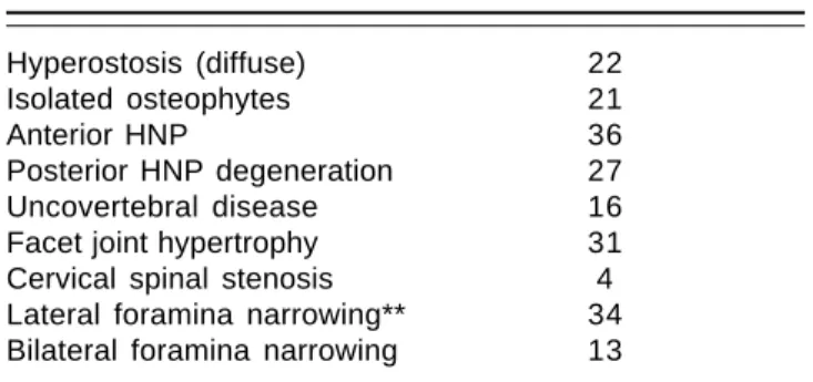
The concomitant lesions in the cervical spine are depic-ted in table I. The previous diagnostic interventions perfor-med on the patients as well as other spinal illnesses, confir-med by plain roentgenograms, CAT scan, or MRI and any other pathology, other surgical and diagnostic intervention are shown in table II. Barium swallows were performed in
evocados. Resultados: De los 79 pacientes, 62 (65.8%) tenían sensación de cuerpo extraño en la faringe y 27 (39%) dificultad para deglutir sólidos; 14 de éstos tenían dolor a la deglución. De estos 52 pacientes, 49 (94%) tenían hipertensión y obesidad. Cuarenta y uno (78.8%) tenían obesidad moderada (entre 20 y 39% del peso ideal) y 11 (21%) tenían obesidad grave (> 40% del peso ideal). Veintisiete pacientes por debajo de su peso ideal tenían dificultad en la deglución, de los cuales 12 tenían odinofagia. Los enfermos con radiculopatía cursaban con com-presión de las raíces a nivel del foramen lateral. Conclusiones: La asociación de dolor de cuello y disfagia requiere de una revisión completa para determinar su origen, identificar el síntoma predominante y valorar su extensión. El tratamiento puede requerir cirugía o terapia intervencionista conservadora.
Palabras clave: Disfagia, odinofagia, cervicalgia, osteofitosis, hernia discal
cervical.
the cases with severe obstructive symptoms. Sleep apnea studies were conducted when indicated.
RESULTS
Of the 79 patients evaluated, 52 (65.8%) had a sporadic sensation of having a foreign body (FB) in their throat, whi-le 27 (34%) others also had difficulty in swallowing solid foods and of these, 14 (51%) had pain upon swallowing. Of fifty-two patients having FB sensation, 49 (94.2%) had arte-rial hypertension with obesity of either moderate degree (> 20-39% of their expected body weight), in 41 patients (78.8%) or severe degree (> 40% of their expected body weight) in 11 others (21%). Forty-four patients (84.6%) had
Table II. Previous related surgical interventions.
Anterior cervical fusion 21
Posterior cervical decompression 4
Endoscopies of pharynx and esophagus 36
Dilatation of esophagus 9
Table I. Radiologic findings in patients with dysphagia
and neck pain*.
Hyperostosis (diffuse) 22
Isolated osteophytes 21
Anterior HNP 36
Posterior HNP degeneration 27
Uncovertebral disease 16
Facet joint hypertrophy 31
Cervical spinal stenosis 4
Lateral foramina narrowing** 34
Bilateral foramina narrowing 13
edigraphic.com
:rop odarobale FDP
VC ed AS, cidemihparG
arap
acidémoiB arutaretiL :cihpargideM
sustraídode-m.e.d.i.g.r.a.p.h.i.c sustraídode-m.e.d.i.g.r.a.p.h.i.c
cihpargidemedodabor
developed late onset diabetes. Of these, 15 (34%) received insulin therapy and 29 others (65%) managed their diabetes with diet and oral antiglycemic medications. The 27 pa-tients who were below their expected body weight had diffi-culty swallowing solid food, frequently cleared their throat, and 12 (33.7%) had pain at swallowing. All of these patients were found to have extrinsic esophageal obstruction at ba-rium swallow. The radiologic findings listed in table I, in-cluded severe osteophytosis (Figure 1) and multiple disc herniations at various levels (Figures 2 and 6).
In one severe case of extensive multiple osteophytosis, the barium swallow revealed almost 80% esophageal ex-trinsic occlusion (Figure 3) with considerable weight loss experienced by this patient. This same patient presented difficulty for orotracheal intubation requiring a fiber optic bronchoscope to achieve it. Six patients with sleep apnea were found to have partial upper airway obstruction at sleep. In these patients, nerve conduction studies showed that the signs and symptoms of radiculopathy were present in pa-tients with lateral foramina compromise.
Treatment: Multiple level surgical intervention (Figures 4 and 5) was needed in only thirteen patients with severe multiple osteophytosis, consisting of excision of the osteo-phytes by an anterior exposure route followed by a vertical wedge multiple level cervical spine fusion with complete re-lief of the symptoms (Figure 5). Six of the patients with one level osteophytes and 19 with a herniated nucleus pulposus had anterior fusions. Other measures consisted of a soft cervi-cal collar worn when traveling in an automobile, and for two hours in the morning and two hours in the evening; isometric exercises of the neck and shoulders, lozenges, and non-steroi-dal anti-inflammatories. In the patients with degenerative disc disease with minimal radiculopathy, treatment consisted of a series of cervical epidural blocks with either steroids or with indomethacin(5) in diabetic patients. Of these 25 patients, eight (31.6%) had temporary relief of their symptoms with recu-rrence of their neck pain within 6 months, but not of the dys-phagia. Four of them, who also had brachial radiculopathy had anterior cervical fusions.
DISCUSSION
Severe spondylosis, multiple osteophytes and anterior her-niation of intervertebral cervical discs (AHICD) have usua-lly been considered irrelevant. However, we have found the-se patients to be frequently symptomatic with complaints of dysphagia without anatomical or functional abnormalities of the pharynx or esophagus; some of them also had pain at swallowing, headaches and metabolic disturbances deser-ving special attention. Not uncommonly patients undergo unnecessary procedures (pharyngoscopy, esophagoscopy in search for non-existent intrinsic, pathology.
Cervical osteophytosis has a peculiar course; it usually begins with the sensation of having a foreign body in the throat that cannot be cleared, coughed up, or swallowed. A common denominator is a prior traumatic event to the cervi-cal spine(2,4), but repeated microtrauma may also cause it(6). Pain may occur when swallowing solids then progressing to mostly soft foods, and then even when liquids are ingested, being worse during neck extension(7).
Some of these patients may also have dysphonia(8), hoar-seness, throat irritation, stridor, choking, limited range of motion of the neck; depending on the location of the osteo-phytes (at what intervertebral space are they located). Signs and symptoms of cervical radiculopathy(9,10) may also be pre-sent with some acromegalic features(7,8), vertebrobasilar insu-fficiency, obesity, arcus senilis, xantomas, arterial hyperten-sion and peripheral edema. Hypothyroidism, adult-onset diabetes mellitus(11) and sleep apnea(12) are less common.
Differential diagnosis include:
a) Diffuse idiopathic skeletal hyperostosis or Forestier di-sease(13) as seen in middle-aged and elderly individuals who develop ligament ossification, periarticular osteo-phytosis and bone production on tendon insertions(12,16) occurring in different locations of the spine
b) Cervical disc disease with intervertebral discs herniating anteriorly
c) Esophageal disease from strictures or tumors that can pro-duce progressive dysphagia
d) Carcinoma of the posterior tongue or pharynx
e) Soft tissue tumors extrinsically compressing the upper esophagus which are usually unilateral
f) Spine tumors such as chondromas of the vertebral bodies
Other laboratory studies that could assist in the specific diagnosis include calcium, phosphorus, magnesium and uric acid determinations with rheumatology profile, erythrocyte sedimentation rate and growth hormone.
Ghaly RF et al. Cervical pain and dysphagia
edigraphic.com
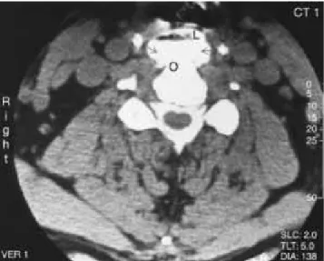
Figure 1. Axial CT-scan at C3-C4 level showing calcificationof the anterior longitudinal ligament (L) as a part of a skele-tal hyperostotic mass (O) (between arrow heads). There is marked compression to the aero-digestive tract.
Figure 2. Sagittal view
of an MRI of the cervi-cal spine demonstra-ting considerable cer-vical spondylosis with osteophytic formation from C1 to C7. There is severe esophageal compression by the spondylytic formation.
Figure 3. Barium
swa-llow depicting an al-most complete block of the esophagus (E) at the C3-C4 level where the osteophy-te formation was the largest.
Figure 4. Transoperative lateral view of the cervical spine
showing an endotracheal tube (T) in place and a metal sur-gical probe (P) displacing anteriorly a large osteophyte in-denting the endotracheal tube and compressing the eso-phagus.
Figure 5.
Postopera-tive lateral view of the cervical spine without the osteophyte with a triangular bone graft fusion (f).
Figure 6. Sagittal
edigraphic.com
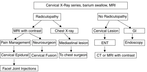
In chronic severe cases, to rule out pharyngeoesophageal pathology, a barium swallow (Figure 3) and esophageal moti-lity studies are indicated; endoscopy of the pharynx and up-per esophagus are indicated only if needed. When soft tissue tumors are suspected, a CAT scan of the neck and thorax may be helpful. Bone scans with scintigraphy may reveal the in-volvement of other tendinous, osseous structures(17). Kiss et al.(18) found frequent hyperuricemia questioning the term “idiopathic” that has been given to this disease(7). The patho-physiology is usually characterized by a progressive wearing of the anterior intervertebral cartilage that is gradually repla-ced by new bone (osteophytes). As the water content of the cartilage decreases (desiccation), it becomes brittle. In the cervical spine, uncovertebral joint disease occurs mostly from C3 to C7(19), but may also affect the apophyseal joints with deterioration of the cartilage and narrowing of the joint surfa-ces. Calcification of the anterior and/or posterior longitudi-nal ligament may take place. Rosenbloom and Silverstein(20) proposed that formation of advanced glycation-end products as a result of non-enzymatic reaction of glucose with proteins may cause stiffening and induration favoring calcium depo-sition. The clinical syndrome may include headaches, neck pain, occipital neuralgia or cervical brachioradiculopathy, from nerve root compression by osteophytes located at diffe-rent points of the lateral foramina with irregular spur forma-tion(2,17). For guidance in making the diagnosis, an algorithm was constructed (Figure 7).
Since a metabolic disorder has been suspected, Shengy et al.(7) and Julkunem et al.(21), related this disease to
hyper-glycemia, hyperinsulinemia(15,21), obesity and adrenal gland disorders, indicating a complete endocrinological work-up. When several intervertebral levels are involved a vertical bone wedge may be needed after excision of the osteophytes. In the case with a large osteophyte that nearly obstructed the esophagus (Figures 2, 3, 4 and 5) tracheal intubation by direct laryngoscopy was difficult, requiring fiberoptic en-doscopy to insert the endotracheal tube. Patients with this condition may also have sleep apnea with partial airway obstruction requiring special precautions(8,22-24), when pa-tients undergo anesthesia for other operative procedures or in the perioperative period of the corrective operation.
Other diagnosis might have to be ruled out, such as ossi-fication of the anterior longitudinal ligament(25) or the so-called “pivoting larynx”(26) as a cause of dysphagia. Since anterior HNP and severe osteophytosis may be concurrently present in the same patients; it is important to identify them specifically so an operative plan can be devised for each intervertebral space. The frequency (44%) of lateral forami-na forami-narrowing predisposes to the high recurrence of radiculo-pathy symptoms in this group of patients(8). Airway com-promise can occur manifesting itself during deep sedation, endoscopies, tracheal intubation27,28.
Recognition of this pathology focuses attention to this hazardous complication of cervical osteophytosis and de-generative disc disease, requiring specific diagnosis of the cervical spine pathology plus cardiovascular and/or metabolic derangements and specific treatment of the above.
REFERENCES
1 . Zahn H. Ein Fall von Abnickung der Speiseröhre durch verte-brale Eckondrose. Munch Med Wochenschr 1905;52:1680-2. 2 . Mecks LW, Renshaw TS. Vertebral osteophytosis and
dyspha-gia. J Bone Joint Surg 1973;1:197-201.
3 . Cammisa M, De Seria A, Guglielmi G. Diffuse idiopathic skeletal hyperostosis. Eur J Radiol 1998;27:7-11.
4 . Marks B, Schober E, Swoboda H. Diffuse idiopathic skeletal hyperostosis causing obstructing laryngeal edema. Eur Arch Otor-hinolaryngol 1998;255:256-8.
5 . Aldrete JA: Epidural injections of indomethacin for postlami-nectomy syndrome: a preliminary report. Anesth Analg 2003;96:463-8.
Figure 7. Algorithm to
Ghaly RF et al. Cervical pain and dysphagia
edigraphic.com
6 . Peppone N, Del Puente A, Scarpa R, Oriente P. Diffuse idiopathic skeletal hyperostosis (DISH): a retrospective analysis. Clin Rheu-matol 1996;15:121-4.
7 . Shengy WJ, Nunley JA, Caldwell DS. Dysphagia due to diffuse idiopathic skeletal hyperostosis. Am Fam Physician 1989;39:149-52.
8 . Valadka AB, Kubal WS, Smith MM. Updated management stra-tegy for patients with cervical osthophytic dysphagia. Dysphagia 1995;10:167-71.
9 . Kodama M, Sarvada H, Udaka F, Kameyama M, Koyama T. Dysphagia caused by an anterior cervical osteophyte: case re-port. Neuroradiology 1995;37:58-9.
10. Stuart D. Dysphagia due to cervical osteophytes: a description of five patients and a review of the literature. Int Orthop 1989;13:95-9. 11. Mosher HP. Exostoses of the cervical vertebrae as a cause for
difficulty in swallowing. Laryngoscope 1926;36:181-2. 12. Davies RP, Sage MR, Brophy BP. Cervical osteophyte induced
dysphagia. Australas Radiol 1989;33:223-5.
13. Forestier J, Rotes-Querol J. Senile ankylosing hyperostosis of the spine. Am Rheum Dis 1950;9:321-30.
14. Yec C, Wang HY, Fewer HD, Rogers AG. Two cases of dyspha-gia due to cervical osteophytes successfully treated surgically. Canad Med Assoc J 1985;132:810-2.
15. Krause P, Castro WH. Cervical hyperostosis: a rare cause of dys-phagia. Case description and bibliographical survey. Eur Spine J 1994;3:56-8.
16. Littlejohn GO, Smythe HA. Marked hyperinsulinemia after glu-cose challenge in patients with diffuse idiopathic skeletal hype-rostosis. J Rheumatol 1981;8:965-8.
17. Mata S, Chhem RK, Fortin PR, Joseph L, Esdaile JM. Compre-hensive radiographic evaluation of diffuse idiopathic skeletal
hyperostosis: development and interrater reliability of a scoring system. Semin Arthritis Rheum 1998;28:88-96.
18. Kiss C, Szilagyi M, Mituszova M, Poor G. A diffuz idiopathias skeltalis hyperostosis prevalenciaja es rizikotenyezoi presentativ hazai populacioban. Orv Hetil 1997;138:619-23.
19. Gordon GV. Arthritis of the cervical spine. MT Sinai J Med 1994;61:204-11.
20. Rosenbloom AA, Silverstine JH. Connective tissue and joint di-sease in diabetes mellitus. Endocrinol Metab Clin North Am 1996;25:473-83.
21. Julkunem H, Heinomen OP, Pyorala K. Hyperostosis of the spine in an adult population its relation to hyperglycemia and obesity. Am Rheum Dis 1971;30:605-12.
22. Utsinger PD, Resnick D, Shapiro R. Diffuse skeletal abnormali-ties in Forestier disease. Arch Int Med 1976;136:763-8. 23. Venga RP, Rowbottom JR. A nine-year retrospective review of
posto-perative airway-related problems in patients following multilevel an-terior cervical corpectomy. Anesthesiology 2001:95 (suppl 3) B14. 24. Fujiwara H, Nakayama H, et al. Postoperative respiratory
distur-bance after anterior cervical fusion. Jap J Anesth 1998;47:475-8. 25. Khalifa MC, Amr F, El Fouly S, Aouf M. Ossification of the anterior longitudinal ligament of the spine as a cause of dyspha-gia. J Laryngol Otol 1981;95:527-8.
26. Charters P, Jones AS. Pivoting larynx – an unusual observation of laryngoscopy. Br J Anaesth 1990;65:424-6.
27. Ansari A, Rockswold G. Cervical disk herniation. N Eng J Med 1998;341:1358.
28. Young W. Cervical spondylitic myelopathy: a common cause of spinal cord dysfunction in older persons. Amer Fam Phys 2000;62:1064-70.