www.elsevier.com.mx
medicina
universitaria
* Corresponding author: “Dr. José Eleuterio González” University Hospital, Universidad Autónoma de Nuevo León. Gonzalitos 235 North Avenue, Mitras Centro, Z.P. 64020, Monterrey, N. L., Mexico. Telephone: (+52) (81) 8348 2015. Fax: (+52) (81) 8348 2065. E-mail address: [email protected] (I. J. Colunga-Pedraza).
OrIGINAL ArTICLE
Similarities between the lipid proile of Mexican patients with
lupus and the general population
I. J. Colunga-Pedraza
a,*, D. Á. Galarza-Delgado
a,b, F. Góngora-Rivera
c, J. A.
Esquivel-Valerio
a, R. A. Carrillo-Palacios
d, S. Segarra-Linares
d, A. L. Sánchez-Núñez
d, P. R.
Colunga-Pedraza
b, D. Vega-Morales
a, M. A. Garza-Elizondo
aa Service of Rheumatology, “Dr. José Eleuterio González” University Hospital, Universidad Autónoma de Nuevo León,
Monterrey, N. L., Mexico
b Department of Internal Medicine, “Dr. José Eleuterio González” University Hospital, Universidad Autónoma de Nuevo
León, Monterrey, N. L., Mexico
c Service of Neurology, “Dr. José Eleuterio González” University Hospital, Universidad Autónoma de Nuevo León,
Monterrey, N. L., Mexico
d Faculty of Medicine, Universidad Autónoma de Nuevo León, Monterrey, N. L., Mexico
received: October 2013; Accepted: January 2014
KEYWORDS
Lipids; Systemic lupus erythematosus; Atherosclerosis; risk factors; Mexico.
Abstract
Introduction: Premature cardiovascular events have been observed in systemic lupus erythema-tosus (SLE) patients, but the reason for this accelerated process is still debatable; although traditional risk factors are more prevalent in such patients than in the general population, they do not seem to fully explain that enhanced risk. One of the most important conditions is a
pro-atherogenic lipid proile. There is not enough data about it in Mexican SLE patients.
Objective: To establish the differences in the lipid proiles between Mexican patients with SLE
and the general population.
Material and methods: Observational, transversal, descriptive and comparative study, between SLE patients and age-sex-matched healthy volunteers. We performed a full lipid proile (by spectrophotometry) 14 hours of fast. The results obtained were analyzed by the statistical pro -gram SPSS® Statistics version 17.
Results: We studied the full lipid proiles of 138 subjects, 69 with a diagnosis of SLE and 69 age-sex-matched healthy volunteers; 95.7% were females and 4.3% males. Average age was 30 years; average body mass index (BMI) 25.96 ± 5.96 kg/m² in SLE patients and 26.72 ± 4.36 kg/m² in the
Introduction
Premature cardiovascular events have been observed in sys-temic lupus erythematosus (SLE) patients, but the reason for this accelerated process is still debatable.1
In addition to traditional cardiovascular risk factors in SLE patients, there are factors inherent to the disease such as: immune complex-induced endothelial damage, vasculitis,
thrombosis associated with antiphospholipid antibodies,
Lib-man Sacks´ endocarditis, renovascular hypertension, glo-merulonephritis and corticosteroid therapy used as part of the disease treatment.1
A greater prevalence of dyslipidemia has been reported in
these patients, inding a marked pro-atherogenic lipid proi
-le, i.e., elevation low density lipoprotein (LDL), lipoprotein (a), triglycerides and free fatty acids, as well as reduction of
high density lipoproteins (HDL).2 Amongst traditional
cardio-vascular risk factors, dyslipidemia is considered to represent a greater impact on the development of cardiovascular disea-se, in addition to being related to a higher renal morbidity and a higher mortality in SLE patients.3
Chronic use of glucocorticoids, as part of the treatment of this pathology, also favours the development of a pro-
atherogenic lipid proile in this population. Additionally, fre -quently used immunosuppressants for SLE treatment
positi-vely correlate with the antioxidant and antiinlammatory
ability, reporting an oxidized LDL reduction (LDLox) in
pa-tients who receive them.4
About 75% of SLE patients develop hypercholesterolemia 3
years after diagnosis; these patients report the highest ra-tes of cardiovascular events.5 In patients with more than
5-year of diagnosis progression, the risk of acute myocardial infarction has been reported to increase up to 52 times.6
For the above mentioned reasons, we decided to analyze and establish the differences in the lipid proiles between Mexican patients with SLE and the general population, mat-ched by age and gender.
Material and methods
We performed an observational, transversal, descriptive
and comparative study. We included 69 patients diagnosed with SLE by a rheumatologist, meeting at least 4 classiica -tion criteria of the American College of rheumatology,
mo-diied in 1997.7 The patients were 18 years of age or older, had no known cardiovascular history (myocardial infarction,
angina, stroke, transient ischemic attack), attended by a rheumatologist in “Dr. José Eleuterio González” University Hospital and agreed to participate through a signed
infor-med consent. This study was approved by our local Ethics Committee (MI09-007).
Subsequently, the patients were matched with 69 healthy volunteers by age and gender; subjects without any autoim
-mune diseases and no known cardiovascular pathologies at the randomization. All subjects (138) were required to com
-ply with a 14-hour fast, following which a venipuncture was performed to obtain a full lipid proile, using the spec
-trophotometry technique. In addition, the following tests were done: erythrocyte sedimentation rate using the Win -trobe method, ultra-sensitive C-reactive protein (CrP), Complete Blood Count in the same lab.
We compared the total cholesterol value, LDL, HDL and
triglycerides; then, we identiied pro-atherogenic characte
-ristics in the lipid proile, establishing as a cut-off point in accordance with that described in the literature as a cardio
-vascular risk factor: total cholesterol > 200 mg/dl, triglyce
-rides > 150 mg/dl, HDL < 45 mg/dl, LDL > 100 mg/dl. We
obtained the BMI of all patients.
Results were analyzed using the statistical program SPSS
v17, performing a descriptive analysis for demographic and clinical variables.
For the binominal variable contrast, we used chi-square test or Fisher’s exact test, with 2 x 2 contingency tables; for non-parametric variables we used the Mann-Whitney U test. p ≤ 0.05 was taken as a signiicant value.
Results
We analyzed 69 SLE patients and 69 healthy patients, 66 women (95.7%) and 3 men (4.7%) in each group matched by sex and age, with a median age of 30 years, and an inter -quartile range of 14 (Table 1).
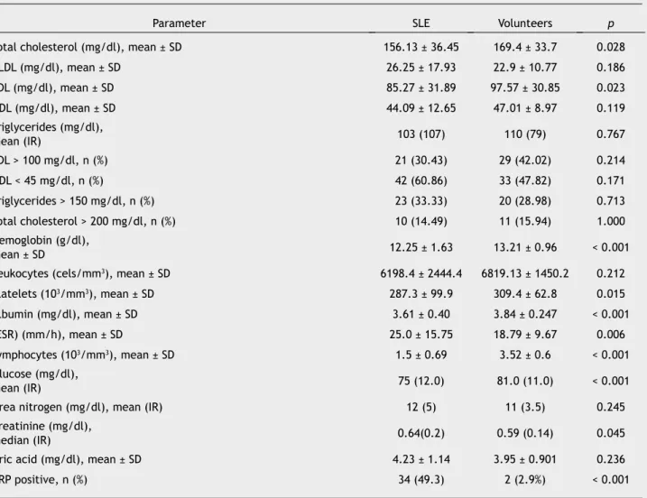
In the lipid proile, we found a total cholesterol average of 156 mg/dl in the group of SLE patients compared to 169.4 mg/dl in the control group (p = 0.028). The higher mean is
in the group of healthy individuals, with a statistically signi
-icant difference. As far as LDL cholesterol levels, we found an average of 85.27 mg/dl in the group of SLE patients while in the group of healthy subjects the mean was 97.57 mg/dl
(p = 0.023), also with a statistically signiicant difference. Despite inding statistically signiicant differences for abso
-lute igures of total cholesterol and LDL cholesterol; when we performed the analysis for pro-atherogenic characteris
-tics we did not ind any statistically signiicant differences between both groups (Table 2).
In addition, we analyzed other clinical and biochemical
parameters (BMI, erythrocyte sedimentation rate, albumin,
CRP, uric acid, glucose and hemoglobin), inding that data in
the group of patients regarding the erythrocyte
sedimenta-tion rate, CRP and serum creatinine were higher with statisti-cal signiicance. On the other hand, the healthy volunteers
85.27 mg/dl in the SLE patients and 97.57 mg/dl in the control group (p = 0.023).
Conclusions: We did not ind statistical differences in the lipid proiles among patients and healthy volunteers, which could explain increased cardiovascular morbidity and mortality ob -served in SLE patients.
1665-5796 © 2014 Revista Medicina Universitaria. Facultad de Medicina UANL. Publicado por Elsevier México. Todos
group had higher hemoglobin, lymphocytes, platelets,
al-bumin and glucose, also with statistical signiicance (Table
2).
In the 69 SLE patients, we also analyzed the disease’s cha
-racteristics; inding that the average number of years of SLE diagnosis at the time of entering the study was 5 years; with
an interquartile range of 8, this population had an average
diagnostic age of 25.8 ± 7.41 years. The disease activity in
-dex used was the MEX-SLEDAI, inding an average of 1.69 ± 2.71, which places the average of the population in the low
activity group.
Concerning medication use, we found that 68 patients (97.1%) used antimalarials at the time of the study; 20 pa
-tients (29.4%) were taking chloroquine at an average dosage of 150 mg/day, while 48 patients (70.5%) were taking hydro
-xychloroquine at an average dosage of 200 mg/day. Sixty patients (88.2%) had been taking antimalarials routinely for
over a year.
Discussion
We found the difference in total cholesterol and LDL
avera-ges to be statistically signiicant; however, it was signii
-cantly greater in the healthy group, which may be explained
by the genetics that our Mexican population carries for
dys-lipidemia. Thus, we compared BMI between both groups and lipid proiles, inding no statistically signiicant differences.
When we analyzed atherogenic lipid levels, thus identifying cardiovascular risk, we found no differences between the li
-pid proiles from the SLE patients and the healthy volunteers.
This suggests that there may be qualitative differences rather than quantitative ones in lipids of SLE patients.
In SLE patients, an increase in hydroperoxidized lipids
as-sociated with endothelial dysfunction has been reported, as well as a decrease in antioxidizing ability.8 High oxidized
HDL levels (HDLox), not only eliminate the protecting effect
of HDL, but also are associated with pro-inlammatory ac
-tion and accelerated atherogenesis.2 IgG-type antibodies
against LDLox are also linked with the early development of
atherosclerosis observed in SLE patients.8
One of the weaknesses in our work is the fact that we did
not measure LDLox levels. Increased cardiovascular risk,
which in recent years has been found in SLE patients, in this studied population is not consistent with a pro-atherogenic lipid proile; however, further studies including larger num -ber of patients are required to determine the causes that
lead to an increase in cardiovascular morbimortality in this
speciic population.
In our Mexican population with SLE, we were not able to ind a marked atherogenic proile, contrary with the recent
published.3 However, in this last publication they studied
Caucasian, Black and Asian races but did not include the Latin American population. Additionally, in this group
cha-racteristics associated with total cholesterol increase were age over 30 years and use of prednisone, with dosages grea
-ter than 10 mg/day. In the population studied by Petri et al., 35% of the patients used hydroxichloroquine on a regu
-lar basis, unlike our population in which 97.1% were taking
anti-malarials. This is relevant because the use of hydro-xychloroquine has been linked to an improvement in lipid
proiles of SLE patients.9
In a study performed in India including 30 SLE patients
paired by sex and age with 30 control subjects, dyslipide
-mia was found in 63% of SLE patients; however, a statisti
-cally signiicant difference was only found in triglycerides, which turned out to be greater in the SLE group.10 In the same study, 63% of SLE patients had been diagnosed with lupus nephropathy, which could have altered lipid charac
-teristics in that population in contrast with our population, in which they found only 12% of lupus nephropathy (none with proteinuria in nephrotic range nor undergoing renal replacement therapy, when subjects were included in the
study).
In a trial including Mexican subjects, in which 16 SLE pa
-tients were studied, no difference was found in the lipid
profile of SLE patients, compared to a healthy control
group, paired by age, sex and BMI. However, one advantage of our study was that did include a larger number of pa -tients and controls.11
The few studies performed in a Mexican population regar
-ding the lipid proile in SLE patients, make it indispensable
to conduct further studies on the issue. The differences found about hemoglobin, erythrocite sedimentation rate, albumin, CrP, albumin, creatinine, platelets and lympho-cytes, may be explained as expected changes in SLE pa-tients.
Another important consideration is that the SLE patient
sample had a low MEX-SLEDAI (under 2, on average), which places them in the subgroup of low-activity patients, the
disease activity being another variable frequently
associa-ted with acceleraassocia-ted atherosclerosis; however, in our popu
-lation did not keep a re-lationship with the disease activity.12 Table 1 Clinical characteristics of the studied population. Monterrey, N. L., Mexico, 2011.
Characteristic SLE Volunteers p
Gender
• Male n (%) • Female n (%)
3 (4.3)
66 (95.7)
3 (4.3)
66 (95.7)
1.000
Median age in years (interquartile range) 30 (14.0) 30 (14.0) 1.000
Framingham’s Score ± SD 1.63 ± 1.48 1.43 ± 0.977 0.771
Body mass index ± SD 25.96 ± 5.93 26.72 ± 4.36 0.396
As for other traditional cardiovascular risk factors (obesi-ty, hypertension, hyperglycemia after fasting),12 no link with lipid proile alterations was found. However, this could be
related to our patients’ age at the time of inclusion in the study (mean 30 years); at this age, cardiovascular comorbi-dities are less prevalent.
Conclusions
We did not find statistical differences in the lipid profiles among patients and healthy volunteers to explain the increa-sed cardiovascular morbimortality observed in SLE patients.
Conlicts of interest
The authors have no conlicts of interest to declare.
Funding
No inancial support was provided.
References
1. Esdaile JM, Abrahamowicz M, Grodzicky T, et al. Traditional
Framingham risk factors fail to fully account for accelerated atherosclerosis in systemic lupus erythematosus. Arthritis rheum 2001;44:2331-2337.
2. Thomas GN, Tam LS, Tomlinson B, et al. Accelerated
atheros-clerosis in patients with systemic lupus erythematosus: a re
-view of the causes and possible prevention. Hong Kong Med J 2002;8:26-32.
3. Petri M, Spence D, Bone Lr, et al. Coronary artery disease risk factors in the Johns Hopkins Lupus Cohort: prevalence, recognition by patients, and preventive practices. Medicine
(Baltimore) 1992;71:291-302.
4. Leong KH, Koh ET, Feng PH, et al. Lipid proiles in patients with systemic lupus erythematosus. J Rheumatol 1994;21:1264-1267.
5. Páramo JA, rodríguez JA, Orbe J. Aterosclerosis en las
enfer-medades inlamatorias. Med Clin (Barc) 2007;128:749-756.
6. Manzi S, Meilahn EN, Rairie JE, et al. Age-speciic incidence ra
-tes of myocardial infarction and angina in women with systemic lupus erythematosus: Comparison with the Framingham Study. Am J Epidemiol 1997;145:408-415.
Table 2 Comparison between the lipid proile and biochemical parameters of patients with SLE and healthy volunteers, of the same age and gender. Monterrey, N. L., Mexico, 2011.
Parameter SLE Volunteers p
Total cholesterol (mg/dl), mean ± SD 156.13 ± 36.45 169.4 ± 33.7 0.028
VLDL (mg/dl), mean ± SD 26.25 ± 17.93 22.9 ± 10.77 0.186
LDL (mg/dl), mean ± SD 85.27 ± 31.89 97.57 ± 30.85 0.023
HDL (mg/dl), mean ± SD 44.09 ± 12.65 47.01 ± 8.97 0.119 Triglycerides (mg/dl),
mean (Ir) 103 (107) 110 (79) 0.767
LDL > 100 mg/dl, n (%) 21 (30.43) 29 (42.02) 0.214
HDL < 45 mg/dl, n (%) 42 (60.86) 33 (47.82) 0.171
Triglycerides > 150 mg/dl, n (%) 23 (33.33) 20 (28.98) 0.713
Total cholesterol > 200 mg/dl, n (%) 10 (14.49) 11 (15.94) 1.000
Hemoglobin (g/dl),
mean ± SD 12.25 ± 1.63 13.21 ± 0.96 < 0.001 Leukocytes (cels/mm3), mean ± SD 6198.4 ± 2444.4 6819.13 ± 1450.2 0.212
Platelets (103/mm3), mean ± SD 287.3 ± 99.9 309.4 ± 62.8 0.015
Albumin (mg/dl), mean ± SD 3.61 ± 0.40 3.84 ± 0.247 < 0.001 (ESR) (mm/h), mean ± SD 25.0 ± 15.75 18.79 ± 9.67 0.006
Lymphocytes (103/mm3), mean ± SD 1.5 ± 0.69 3.52 ± 0.6 < 0.001
Glucose (mg/dl),
mean (Ir) 75 (12.0) 81.0 (11.0) < 0.001
Urea nitrogen (mg/dl), mean (IR) 12 (5) 11 (3.5) 0.245
Creatinine (mg/dl),
median (Ir) 0.64(0.2) 0.59 (0.14) 0.045
Uric acid (mg/dl), mean ± SD 4.23 ± 1.14 3.95 ± 0.901 0.236
CRP positive, n (%) 34 (49.3) 2 (2.9%) < 0.001 SLE: systemic lupus erythematosus; LDL: low density lipoproteins; VLDL: very low density lipoproteins; HDL: high density lipoproteins;
7. Hochberg MC. Updating the American College of rheumatology
revised criteria for the classiication of systemic lupus erythe
-matosus. Arthritis Rheum 1997;40:1725.
8. López Lr, Salazar-Paramo M, Palafox-Sánchez C, et al. Oxidized
low-density lipoprotein and beta2-glycoprotein I in patients with systemic lupus erythematosus and increased carotid
intima-media thickness: implications in autoimmune-mediated atherosclerosis. Lupus 2006;15:80-86.
9. Pons-Estel GJ, Alarcón GS, Hachuel L, et al. Anti-malarials
exert a protective effect while Mestizo patients are at increa -sed risk of developing SLE renal disease: data from a
Latin-American cohort. Rheumatology (Oxford) 2012;51:1293-1298.
10. Kakati S, Doley B, Deve A, et al. Serum lipid proiles in patients with systemic lupus erythematosus. J Indian Rheumatol Assoc
2003;11:5-7.
11. Díaz-González O, Andrade-Ortega L, Irazoque-Palazuelos F, et al. El lupus eritematoso sistémico como factor de riesgo inde-pendiente en el desarrollo de dislipidemias. reumatol Clin 2008;4 Supl.