O R I G I N A L A R T I C L E
In vitro anti-canine distemper virus activity of fucoidan extracted
from the brown alga
Cladosiphon okamuranus
Laura M. Trejo-Avila• Maria Elena Morales-Martı´nez•
Denis Ricque-Marie• L. Elizabeth Cruz-Suarez•Pablo Zapata-Benavides• Karla Mora´n-Santiban˜ez•Cristina Rodrı´guez-Padilla
Received: 16 June 2014 / Accepted: 10 September 2014
ÓIndian Virological Society 2014
Abstract Canine distemper virus (CDV) is a morbillivi-rus related to measles vimorbillivi-rus that infects dogs and other carnivores. CDV has a significant global impact on animal health; however, there is no current antiviral treatment for CDV infection. In recent years, it has been demonstrated that sulfated polysaccharides exhibit antiviral properties both in vivo and in vitro, despite their low cytotoxicity to host cells. Fucoidan is a sulfated polysaccharide found in the cell wall matrix of brown algae. In this study, we evaluated in vitro anti-CDV activity of fucoidan, which was derived from Cladosiphon okamuranus. Fucoidan actively inhibited CDV replication in Vero cells at a 50 % inhibitory concentration (IC50) of 0.1lg/ml. The derived
selectivity index (SI50) was[20,000. This polysaccharide
likely inhibits viral infection by interference in the early steps and by inhibiting CDV-mediated cell fusion. Fucoi-dan may be useful in development of pharmacological strategies to treat and control CDV infection.
Keywords FucoidanCDV AntiviralCladosiphon okamuranusMorbillivirus
Introduction
Canine distemper virus (CDV) is a negative-stranded RNA morbillivirus related to measles virus. CDV infection
causes severe morbidity and mortality in dogs and other carnivores and significantly affects animal health world-wide [22]. Despite an increasing demand from veterinari-ans and pet owners, an antiviral treatment for CDV infection is not available; but research is in progress. Ribavirin inhibits CDV replication in vitro in a dose- and time-dependent manner [7]. However, ribavirin exhibited low selectivity indexes, so it is unsuitable for clinical treatment of CDV-infected animals.
Marine algae produce metabolites that have been rec-ognized as promising biologically active compounds for use as antiviral drugs. Fucoidans are complex sulfated fu-cosylated polysaccharides produced by brown seaweeds and have a wide spectrum of pharmacological properties (e.g., anti-inflammatory, anti-angiogenic, anti-coagulant, and antiviral) [2,5,14,19]. Because fucoidans are present in large quantities in dietary brown seaweed products consumed in Asian countries, these compounds may already have an important role in disease prevention [6].
Cladosiphon okamuranusis an edible brown alga that is commercially cultured around Okinawa Island, Japan. Fu-coidan is prepared from this alga on an industrial scale and is used as an additive for health foods, drinks, and cos-metics. Fucoidan extracted fromC. okamuranuspossesses
a-1-3-linkedL-fucosyl residues that are substituted withD
-glucuronic acid at C-2 and with sulfate groups at C-4 of the
L-fucosyl residues. The average branched chain structure
consists of one sulfate group for every two molecules of fucose and one glucuronic residue for every six molecules of fucose [16]. The polysaccharide also contains xylose as a minor monosaccharide constituent. Using a room tem-perature extraction, Tako et al. reported an acetylfucoidan yield of 2.3 % (w/w) based on the wet alga weight. The total contents of carbohydrates,D-glucuronic acid, sulfuric
acid, ash, and moisture were 69, 13.5, 13.6, 23 and 3.2 %,
L. M. Trejo-Avila (&)M. E. Morales-Martı´nez
D. Ricque-MarieL. E. Cruz-SuarezP. Zapata-Benavides
K. Mora´n-Santiban˜ezC. Rodrı´guez-Padilla
respectively, and the molar ratio of L-fucose:D-xylose:D
-glucuronic acid:acetic acid:sulfuric acid was estimated to be 4.0:0.03:1.0:2.0:2.0 [24]. In contrast with other sulfated polysaccharides,Cladosiphon fucoidan does not promote an inflammatory response in vitro [21] and lacks anti-angiogenic and anti-coagulant activities [4]. Cladosiphon okamuranusfucoidan is not toxic to hepatocytes and has no adverse effects on the serum electrolytes of Wistar rats [10]. However, similar doses potently inhibit dengue virus type 2 (DEN2) replication [12] and significantly inhibit the proliferation of HTLV-1-infected T-cells [11]. In the present study, we examined the in vitro anti-CDV activity of fucoidan derived from C. okamuranus, and compared this activity to ribavirin.
Materials and methods
Fucoidan and ribavirin
Fucoidan was purchased as a dried powder from Kadoya & Co., Kobe, Japan (lot A03012) and extracted from cultured kelp C. okamuranus harvested off the coast of Okinawa Island, Japan [24]. The polysaccharide preparation was cer-tified to contain 90.4 % fucoidan (anthrone-sulfuric acid method) and to have a mean molecular weight of 92.1 kDa (HPLC method). The fucose and sulfate contents of fucoidan were 38.6 and 15.9 %, respectively, with ash comprising 19.6 % of the content and other sugars comprising 23 % (glucuronic acid and traces of xylose). Dried samples of fu-coidan were suspended in Dulbecco’s modified Eagle’s medium (DMEM) at a concentration of 2.5 mg/ml and filtered through a 0.22 mm membrane filter. Ribavirin (Vilonape-diatrica, Valeant, Me´xico) was used as an antiviral control. Cells culture and virus
African green monkey kidney cells (Vero cells) were grown as monolayers in Dulbecco’s modified Eagle’s medium (DMEM) supplemented with 10 % fetal calf serum (FCS) and 1 % penicillin and streptomycin. Vero cells were cultured at 37°C in a 5 % CO2 incubator.
Canine distemper virus (CDV; Onderstepoort strain) adapted to Vero cells was titrated by plaque forming units (PFU). Virus aliquots were stored at -70°C until use. Cytotoxicity assay
The conventional MTT (3-(4,5-dimethylthiazol-2-yl)-2,5-diphenyltetrazolium bromide) method [15], with some modifications, was used to evaluate cytotoxicity. Vero cells were seeded in 96-well plates at an initial density of 59103cells per well. After the cells were incubated for
20 h at 37°C, various concentrations of the antiviral compounds fucoidan (1, 10, 100, 500 and 1,000lg/ml) or ribavirin (0.1, 1, 10 and 100lg/ml) were added to the growth medium, with six wells for each concentration. The cultures were then incubated for an additional 72 h. MTT solution (20ll well) at a concentration of 5 mg/ml was added to each well and cells were incubated at 5 % CO2
and 37°C for 4 h. The medium and excess MTT were removed from each well, and 100ll dimethyl sulfoxide was added to dissolve the formazan crystals. The optical density was measured at 570 and 690 nm using a Micro-plate Autoreader EL311 (BIOTEK Instruments Inc., USA). Each experiment was repeated three times. Cytotoxicity was expressed as 50 % cytotoxic concentration (CC50),
which was the concentration of the test substances that inhibited up to 50 % of the growth of Vero cells.
Virus plaque reduction assays
Fucoidan was tested for anti-viral activity against CDV with a plaque reduction assay using Vero cell monolayers in six-well culture plates. The assay was performed by adding the compound during all assay (72 h). For a typical assay, 100 plaque forming units (PFUs) were incubated with increasing concentrations of fucoidan (0.1, 1 and 10lg/ml) for 1 h at room temperature. Virus was allowed to adsorb to the cells for 1 h at 37°C. The residual inoc-ulum was then discarded and infected cells were overlaid with medium containing 0.6 % agar (MP Biomedicals, Inc) and the indicated concentrations of the sulfated polysac-charide. Each concentration was performed in triplicate. After incubation of 72 h at 37°C to allow for plaque for-mation, the cells were fixed with methanol and stained with 1 % crystal violet. Plaques in the Vero cell monolayer were counted. Using dose–response curves, plaque reduction by the test compounds was determined by, and expressed as, the test compound concentration that inhibited plaque formation by 50 % (IC50).
Time of addition assays
In order to analyze the inhibitory mechanism of fucoidan against CDV, infected cells were treated with increasing concentrations (0.001–10lg/ml) for various periods of time prior to, during, and after infection with CDV. Fu-coidan antiviral activity was compared with ribavirin in post-adsorption assays.
Drug treatment before virus infection or concurrent with infection
infection. After incubation, the cells were washed three times with phosphate buffered saline to ensure the com-plete removal of fucoidan from the media, then infected with 100 PFU per well of CDV and incubated at 37°C for 1 h. The infected cell monolayer was then washed three times with phosphate buffered saline, overlaid with med-ium containing 0.6 % agar (MP Biomedicals, Inc.), and incubated for 72 h at 37°C. A second assay was performed with cells infected at the same time as fucoidan treatment. The virus suspension was mixed with the indicated con-centration of fucoidan before adding it to the cell mono-layer. Vero cells were treated with the mix for 1 h at 37°C, then removed from cell media by extensively washed and the monolayer was cultured for plaque assay as previously described.
Drug treatment after virus adsorption
Cells were infected with CDV for 1 h at 37°C. After incubation, the infected cell monolayer was then washed three times with phosphate buffered saline and increasing concentrations of fucoidan or ribavirin were then added and cells were incubated at 37°C for 1 h. The infected cell monolayer was then washed three times with phosphate buffered saline, overlaid with medium containing 0.6 % agar, and incubated for 72 h at 37°C.
Viral syncytium inhibition assay
Virus-induced cell fusion assays were used to examine the ability of fucoidan to inhibit CDV induced syncytia for-mation. Vero cells were cultured in 6-well plates in DMEM, supplemented with 10 % FCS. Cell monolayers were infected with CDV at 100 PFU per well. After adsorption for 1 h, the inoculum was replaced by DMEM with 2 % FCS and dilutions of fucoidan in medium without FCS were added. At 24 h post infection, the cells were fixed and stained with 1 % (w/v) crystal violet in 100 %
methanol. The number of nuclei was counted for 20 syn-cytia per well, and inhibition of syncytium size was determined.
Data analysis
Data were analyzed using SPSS 10 software (IBM, Ar-monk, New York, USA). Probit regression analysis was used to determine CC50, which was the concentration of the
test substance that reduced the optical density by 50 %, compared with the controls. The IC50 was the
concentra-tion that reduced PFU number by 50 %. The selectivity index (SI) was the ratio CC50/IC50. The CC50 and IC50
values were estimated from the mean and standard devia-tions or standard errors (time of addition assays) of the mean values from three independent experiments.
Results
Cytotoxicity assay
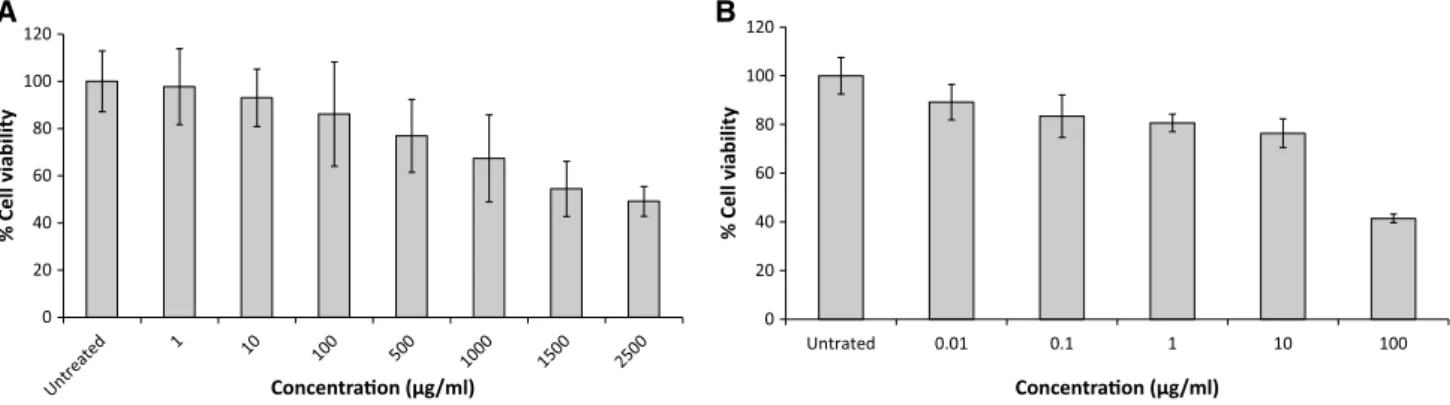
The cytotoxicity of fucoidan and ribavirin to Vero cells was evaluated. As shown in Fig.1, although the no effect on cytotoxicity was detected for fucoidan at concentrations up to 10 lg/ml (CC50=2,089lg/ml±6 SD). However,
10lg/ml ribavirin was toxic to Vero cells (CC50 = 88.9lg/ml±6 SD). For the remainder of the studies, the antiviral activity of these compounds was assessed at concentrations below 10lg/ml to rule out non-specific inhibition due to cell cytotoxicity.
Fucoidan inhibits CDV replication
To assess fucoidan inhibition of CDV replication, Vero cell monolayers were treated with increasing concentrations of fucoidan that was added at the same time, each well received 100 PFUs of CDV. The same concentration of
A B
Fig. 1 Evaluation of fucoidan (A) and ribavirin (B) cytotoxicity to Vero cells using the MTT assay. Control: cells without treatment.Bars
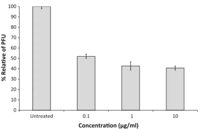
fucoidan was maintained in the plaque assay overlay medium throughout the infection. Antiviral activity was evaluated by observing a reduction in the number of CDV plaques. The results demonstrated significant and repro-ducible antiviral activity of fucoidan against CDV (Fig.2). Fucoidan treatment reduced not only the number but also the size of plaques (data not shown). Fucoidan inhibition resulted in an IC50\0.1lg/ml. The resulting selectivity
index (SI; [20,000 CC50/IC50) indicated that fucoidan
potently inhibited CDV at a very low drug concentration, and was not cytotoxic.
Effect of fucoidan on virus adsorption and entry
Vero cells were pretreated with various concentrations of fucoidan (0.001–10lg/ml), which was removed before
incubation with CDV by extensively washed. The results indicated that they were protected from CDV infection in a manner that was not strictly dose-dependent (Fig.3a). Additional assays were performed with fucoidan added directly to virus suspensions. Under these conditions, the apparent efficacy of fucoidan was slightly increased.
After virus adsorption, the unadsorbed virus was removed and Vero cells were treated with various con-centrations of fucoidan (0.001–10lg/ml) or ribavirin (0.01–10lg/ml) as described in methodology, then washed to ensure the complete removal of test compounds. Virus yield was determined by plaque assay. Fucoidan displayed anti-CDV activity when the cells were treated after virus adsorption (Fig.3b). However, this activity was slightly lower than the inhibition that occurred when cells were treated at the time of infection (Fig. 3a). Fucoidan-treated Vero cells were much better protected from CDV infection than ribavirin-treated cells (Fig. 3b). These experimental results suggest that fucoidan has antiviral activity via the inhibition of virus binding and/or penetration.
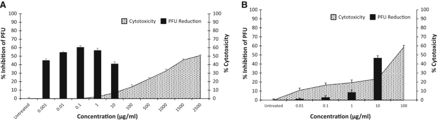
Antiviral efficacy versus cytotoxicity
Assessment of cytotoxicity is clearly an important part of the evaluation of any potential antiviral agent. As shown in Fig.4, fucoidan effectively inhibits the replication of CDV even at drug concentrations that showed no cytotoxicity. In contrast, ribavirin was cytotoxic at drug concentrations that inhibit CDV replication (SI\8.9, a[2000-fold differ-ence; Fig. 4).
Fucoidan reduces cell-to-cell spread of CDV
F and H proteins of CDV are necessary for fusion of adjacent cell membranes. The expression of CDV F protein Fig. 2 Evaluation of antiviral activity of fucoidan against canine
distemper virus in Vero cells by plaque reduction. Infected cells were cultured in the absence of drug, or with the indicated concentration of fucoidan.Barsrepresent mean values andvertical linesrepresent the standard deviations from the mean (n=9, *p\0.05)
A B
Fig. 3 Effect of fucoidan on adsorption and entry of canine distemper virus (CDV). The fucoidan was added to and incubated for 1 h with cells either ‘‘before infection (1 h b.i.)’’ or ‘‘during infection (0 h)’’ of cell infection with CDV (100 PFU) (A). The fucoidan or ribavirin was added and incubated for 1 h with cells after
on the cell surface results in cell–cell fusion (syncytia) and subsequent cell-to-cell spread of progeny viruses [23]. Vero cells infected with CDV were incubated in increasing concentrations (0.01–10lg/ml) of fucoidan. Syncytia size was markedly reduced by fucoidan treatment in a dose-dependent manner (Fig.5).
Discussion
The aim of the present study was to test and evaluate the antiviral activity of fucoidan against CDV. Fucoidan is a polysaccharide isolated from the brown seaweed Cladosi-phon okamuranus, since other similar polysaccharides have been shown to have a wide array of pharmacological activities. This study also sought to determine the mecha-nism(s) by which fucoidan blocked CDV infection. Understanding the mechanism of antiviral activity of this algal polysaccharide is important for its future develop-ment as antiviral drugs for clinical or veterinary use.
The major undesirable side effect of sulfated polysac-charides is their well-known anticoagulant activity. This
adverse effect can be avoided by selecting sulfated poly-mers, such as fucoidan fromC. okamuranus, which exhibit virtually no anticoagulant activity [4]. Cumashi et al. suggested that the high presence of glucuronic acid bran-ches is the most likely feature responsible for the lack of anticoagulant activity by C. okamuranusfucoidan, as the less active compounds are characterized by a low degree of sulfation and a high presence of 2-O-a-D-glucuronyl
sub-stituents along the linear polysaccharide backbone (char-acteristics fucoidan fromC. okamuranus) [4]. By contrast, it has been proposed that the antiviral activity of fucoidan is related to the concentration of fucose and uronic acids. Hidari et al. found that the antiviral properties of fucoidan fromC. okamuranusagainst dengue virus type 2 vanished when the glucuronic acid was carboxyl-reduced [12]. In the present study, we evaluated the antiviral activity of fucoi-dan fromC. okamuranusextracted by the method of Tako et al. with a significant degree of uronic acids, and inves-tigated its possible mechanism of action in Vero cells [24]. Sulfated polysaccharides act primarily through inhibi-tion of the entry of enveloped viruses into host cells [5,9, 14]. Marine polysaccharides from different sources can
A B
Fig. 4 Fucoidan and ribavirin antiviral efficacy. Percent inhibition of plaque forming units versus percent cytotoxicity. Test results based on treatment with fucoidan, concurrent with infection (A) and test
results based on ribavirin treatment after adsorption (B). Bars
represent mean values andvertical linesrepresent the standard error from the mean (n=9, *p\0.05)
B A
Fig. 5 Effect of fucoidan on syncytium formation. (A) Circles
represent mean values andvertical linesrepresent standard deviation from the mean. The mean±standard deviation (%) for 20 syncytia
block virus infection by interfering with the virus adsorp-tion process. The antiviral acadsorp-tion of fucoidan seems to stem from inhibiting the binding of the virus particles to the host cell and interferes with the adsorption process [1]. Certain sulfated marine polysaccharides, can interfere with virus internalization and the subsequent uncoating by blocking the allosteric process of virus particles [26]. Our results support the ability of fucoidan to block the early stages of CDV infection by blocking viral adsorption and maybe one or more post binding penetration steps.
Fucoidan inhibited the first steps of the viral infection cycle and strongly suppressed the formation of CDV-induced syncytia in infected cells perhaps by direct action on the proteins responsible for CDV fusion (F or H) or by inhibiting its expression. We previously found that fucoi-dan ofC. okamuranusboth blocking of post-binding entry steps of NDV to cells and strongly suppresses syncytia formation when fucoidan was added before cleavage of the fusion protein, feasibly indicating a specific interaction between fucoidan and the F0 protein [8]. These apparent multifaceted mechanisms of fucoidan action are similar to the mechanisms of action of other sulfated polysaccharides [3, 18, 20, 25]. Our results agree with the results of Hoshino et al. and Nyberg et al., who demonstrated that the mechanisms of antiviral activity of low molecular weight sulfated polysaccharides include inhibition of the entry of enveloped virions into host cells and inhibition of cell-to-cell spread of the virus [13,17]. Cytolytic viruses, such as CDV, spread through a cell culture by producing infectious particles and by lateral cell-to-cell fusion. Thus, virus production observed in our studies with fucoidan might reflect predominantly on secondary spread rather than rapid lateral transmission, resulting in a greatly reduced syncytia number and plaque size.
Elia et al. reported that ribavirin was active against CDV in vitro [7]. In this study, ribavirin showed lower antiviral activity than fucoidan, (ribavirin SI\9, fucoidan SI[20,000). Assessment of cytotoxicity is clearly an important part of the evaluation of a potential antiviral agent since a useful compound should not show acute or long-term toxicity against the host. In the study by Gideon and Rengasamy, rats treated withC. okamuranusexhibited no necropsy or other pathological changes in organs or changes in histopathological morphology, consistent with the lower toxicity possessed by sulfated polysaccharides, specifically fucoidan fromC. okamuranus[10].
This is the first report of a sulfated polysaccharide with antiviral activity against CDV. The high selectivity of antiviral action reported for fucoidan[20,000 is clinically promising.
Acknowledgments We greatly appreciate the gift of Canine dis-temper virus (Onderstepoort strain) provided by Dr. Raymundo Iturbe
(Departamento de Microbiologı´a, Facultad de Medicina Veterinaria of Universidad Nacional Auto´noma de Me´xico). This work was supported by Grants from Consejo Nacional de Ciencia y Tecnologı´a (CONACYT) Me´xico (No. 99862).
References
1. Baba M, Snoeck R, Pauwels R, de Clercq E. Sulfated polysac-charides are potent and selective inhibitors of various enveloped viruses, including herpes simplex virus, cytomegalovirus, vesic-ular stomatitis virus, and human immunodeficiency virus. Anti-microb Agents Chemother. 1988;32:1742–5.
2. Berteau O, Mulloy B. Sulfated fucans, fresh perspectives: struc-tures, functions, and biological properties of sulfated fucans and an overview of enzymes active toward this class of polysaccha-ride. Glycobiology. 2003;13:29R–40R.
3. Chen MZ, Xie HG, Yang LW, Liao ZH, Yu J. In vitro anti-influenza virus activities of sulfated polysaccharide fractions fromGracilaria lemaneiformis. Virol Sin. 2010;25:35–341. 4. Cumashi A, Ushakova NA, Preobrazhenskaya ME, D’Incecco A,
Piccoli A, Totani L, Tinari N, Morozevich GE, Berman AE, Bilan MI, Usov AI, Ustyuzhanina NE, Grachev AA, Sanderson CJ, Kelly M, Rabinovich GA, Lacobelli S, Nifantiev NE, Consorzio Interuniversitario Nazionale per la Bio-Oncologia, Ital. A com-parative study of the anti-inflammatory, anticoagulant, antian-giogenic, and antiadhesive activities of nine different fucoidans from brown seaweeds. Glycobiology. 2007;17:541–52.
5. Damonte EB, Matulewicz MC, Cerezo AS. Sulfated seaweed polysaccharides as antiviral agents. Curr Med Chem. 2004;11: 2399–419.
6. Doh-Ura K, Kuge T, Uomoto M, Nishizawa K, Kawasaki Y, Iha M. Prophylactic effect of dietary seaweed fucoidan against ent-eral prion infection. Antimicrob Agents Chemother. 2007;51: 2274–7.
7. Elia G, Belloli C, Cirone F, Lucente MS, Caruso M, Martella V, Decaro N, Buonavoglia C, Ormas P. In vitro efficacy of ribavirin against canine distemper virus. Antiviral Res. 2008;77:108–13. 8. Elizondo-Gonzalez R, Cruz-Suarez LE, Ricque-Marie D,
Men-doza-Gamboa E, Rodriguez-Padilla C, Trejo-Avila LM. In vitro characterization of the antiviral activity of fucoidan from
Cladosiphon okamuranusagainst newcastle disease virus. Virol J. 2012;9:307.
9. Ghosh T, Pujol CA, Damonte EB, Sinha S, Ray B. Sulfated xy-lomannans from the red seaweedSebdenia polydactyla: structural features, chemical modification and antiviral activity. Antivir Chem Chemother. 2009;19:235–42.
10. Gideon TP, Rengasamy R. Toxicological evaluation of fucoidan fromCladosiphon okamuranus. J Med Food. 2008;11:638–42. 11. Haneji K, Matsuda T, Tomita M, Kawakami H, Ohshiro K,
Uchihara JN, Masuda M, Takasu N, Tanaka Y, Ohta T, Mori N. Fucoidan extracted fromCladosiphon okamuranusTokida indu-ces apoptosis of human T-cell leukemia virus type 1-infected T-cell lines and primary adult T-cell leukemia cells. Nutr Cancer. 2005;52:189–201.
12. Hidari KI, Takahashi N, Arihara M, Nagaoka M, Morita K, Su-zuki T. Structure and anti-dengue virus activity of sulfated polysaccharide from a marine alga. Biochem Biophys Res Commun. 2008;376:91–5.
13. Hoshino T, Hayashi T, Hayashi K, Hamada J, Lee JB, Sankawa U. An antivirally active sulfated polysaccharide fromSargassum horneri (TURNER) C. AGARDH. Biol Pharm Bull. 1998;21: 730–4.
15. Mosmann T. Rapid colorimetric assay for cellular growth and survival: application to proliferation and cytotoxicity assays. J Immunol Methods. 1983;65:55–63.
16. Nagaoka M, Shibata H, Kimura-Takagi I, Hashimoto S, Kimura K, Makino T, Aiyama R, Ueyama S, Yokokura T. Structural study of fucoidan from Cladosiphon okamuranus TOKIDA. Glycoconj J. 1999;16:19–26.
17. Nyberg K, Ekblad M, Bergstro¨m T, Freeman C, Parish CR, Ferro V, Trybala E. The low molecular weight heparan sulfate-mimetic, PI-88, inhibits cell-to-cell spread of herpes simplex virus. Anti-viral Res. 2004;63:15–24.
18. Preeprame S, Hayashi K, Lee JB, Sankawa U, Hayashi T. A novel antivirally active fucan sulfate derived from an edible brown alga,
Sargassum horneri. Chem Pharm Bull. 2001;49:484–5.
19. Pomin VH, Moura˜o PA. Structure, biology, evolution, and medical importance of sulfated fucans and galactans. Glycobi-ology. 2008;18:1016–27.
20. Schaeffer DJ, Krylov VS. Anti-HIV activity of extracts and compounds from algae and cyanobacteria. Ecotoxicol Environ Saf. 2000;45:208–27.
21. Shibata H, Kimura-Takagi I, Nagaoka M, Hashimoto S, Aiyama R, Iha M, Ueyama S, Yokokura T. Properties of fucoidan from
Cladosiphon okamuranus tokida in gastric mucosal protection. BioFactors. 2000;11:235–45.
22. Silin D, Lyubomska O, Ludlow M, Duprex WP, Rima BK. Development of a challenge-protective vaccine concept by modification of the viral RNA-dependent RNA polymerase of canine distemper virus. J Virol. 2007;81:13649–58.
23. Singethan K, Hiltensperger G, Kendl S, Wohlfahrt J, Plattet P, Holzgrabe U, Schneider-Schaulies J.N -(3-cyanophenyl)-2-phen-ylacetamide, an effective inhibitor of morbillivirus-induced membrane fusion with low cytotoxicity. J Gen Virol. 2010;91: 2762–72.
24. Tako M, Yoza E, Tohma S. Chemical characterization of acetyl fucoidan and alginate from Commercially CulturedCladosiphon okamuranus. Bot Mar. 2000;43:393–8.
25. Tong XK, Qiu H, Zhang X, Shi LP, Wang GF, Ji FH, Ding HY, Tang W, Ding K, Zuo JP. WSS45, a sulfated alpha-D-glucan,
strongly interferes with Dengue 2 virus infection in vitro. Acta Pharmacol Sin. 2010;31:585–92.