Antitumoral properties of epidermal growth factor derivatives
147
0
0
Texto completo
(2) . PhD Thesis . ANTITUMORAL PROPERTIES OF EPIDERMAL GROWTH FACTOR DERIVATIVES Clara Panosa Roqueta 2015 . . .
(3) . .
(4) . . PhD Thesis . ANTITUMORAL PROPERTIES OF EPIDERMAL GROWTH FACTOR DERIVATIVES Clara Panosa Roqueta 2015 Ciències Experimentals i Sostenibilitat . Vist-‐i-‐plau . Vist-‐i-‐plau . El co-‐director de Tesi . La co-‐directora de Tesi . . Dr. Rafael de Llorens i Duran . Dra. Anna Massaguer i Vall-‐llovera . Catedràtic de Bioquímica i Biologia Molecular . Professora Agregada de Bioquímica i Biologia Molecular . Memòria presentada per optar al títol de doctora per la Universitat de Girona .
(5) . .
(6) Agraïments . Gràcies al grup de recerca de Bioquímica del Càncer, que em van donar la oportunitat d’iniciar aquesta tesi. També donar les gràcies als grups de recerca externs que em van acollir, ensenyar i donar tot el que vaig necessitar per completar aquest treball, gràcies Siscu, gràcies Reilly. Gràcies a tots aquells qui m’heu acompanyat durant aquest temps i que m’heu donat suport en els moments difícils, he tingut la gran sort d’estar envoltada de becaris genials. Aquesta tesi ha estat realitzada amb el suport de la Universitat de Girona (beques BR08/19 i PUG2007A/10). . Dedicatòria . Júlia, aquest treball te’l dedico a tu, perquè puguis arribar a gaudir també algun dia de les mateixes oportunitats d’aprendre i estudiar que he tingut jo. .
(7) . .
(8) Publications arising from this thesis Panosa C, Tebar F, Ferrer-‐Batallé M, Fonge H, Seno M, Reilly RM, Massaguer A, De Llorens R. Development of an epidermal growth factor derivative with EGFR blocking activity. PLoS One; 2013; 8 (7): e69325. Panosa C, Fonge H, Ferrer-‐Batallé M, Menéndez JA, Massaguer A, De Llorens R, Reilly RM. A comparison of non-‐biologically active truncated EGF (EGFt) and full-‐length hEGF for delivery of Auger electron-‐emitting 111In to EGFR-‐positive breast cancer cells and tumor xenografts in athymic mice. Nucl Med Biol; 2015; Epub ahead of print. .
(9) .
(10) Abbreviations 111. In In-‐DTPA-‐EGFt . Indium-‐111 Truncated form of EGF 111 labeled with In 111 In-‐DTPA-‐hEGF Human EGF labeled with 111 In 131I Iodine-‐131 186Re Rhenium-‐186 211At Astatine-‐211 111. 225Ac 231Bi 99mTc Aa AIDS Akt AR Arg Asn Asp ATP AUC BC BSA BTC CBL cDTPAA CL CR CTL Da DMEM DMSO DNA DTPA DUBs E.coli EC EDTA. Actinium-‐225 Bismuth-‐231 Technetium-‐99m Amino acid Acquired immune deficiency syndrome Protein kinase B Amphiregulin Arginine Asparagine Aspartic acid Adenosine Triphosphate Area under the curve Breast cancer Bovine serum albumin Betacellulin Ubiquitin ligase Bicyclic anhydride of DTPA Clearance Cysteine-‐rich domain Control Daltons Dulbecco’s modified eagle medium Dimethyl sulfoxide Deoxyribonucleic acid Diethylenetriamine pentaacetate De-‐ubiquitylating enzymes Escherichia coli Electron capture Ethylenediaminetetraacetic acid . EGF EGFR EGFt ELISA EPG EPR ER ErbB 1/HER1 ErbB 2/HER2 ErbB 3/HER3 ErbB 4/HER4 FBS FWHM Gln HB-‐EGF hEGF HIV HPLC HRP i.v. ID Ig G Ile IPTG ITLC-‐SG Kd KDa keV Kvp . Epidermal growth factor Epidermal growth factor receptor Epidermal growth factor truncated form Enzyme-‐linked immunosorbent assay Epigen Epiregulin Endoplasmatic reticulum Epidermal growth factor receptor Receptor tyrosine-‐protein kinase erbB-‐2 Receptor tyrosine-‐protein kinase erbB-‐3 Receptor tyrosine-‐protein kinase erbB-‐4 Fetal bovine serum Full width at half maxiumum Glutamine Heparin-‐binding EGF-‐like growth factor Human epidermal growth factor Human immunodeficiency virus High performance liquid chromatography Horseradish peroxidase Intravenous Injected dose Immunoglobulin G Isoleucine Isopropyl β-‐D-‐1-‐ thiogalactopyranoside Instant thin layer-‐silica gel chromatography Dissociation constant Kilodaltons Kiloelectronvolt Peak kilovoltage .
(11) LB LET Leu mAbs MALDI-‐TOF MS . MAPK mBq MBq Met MeV Micro SPECT/CT . ml mM mm MTT . . MVB NLS NPC NRG NSCLC OmpA OSEM p p.i. PBS PBS-‐T PCI . . pCi PCNA . . Luria-‐Bertani medium Linear energy transfer Leucine Monoclonal antibodies Matrix-‐assisted laser desorption/ionization mass spectometry Mitogen-‐activated protein kinase millibeckerels megabeckerels Metionine Megaelectronvolts Micro-‐single-‐photon emission computed tomography/computed tomography milliliters millimolar millimeters 3-‐(4,5-‐dimethylthiazol-‐2-‐yl)-‐ 2,5-‐diphenyltetrazolium bromide . Phe PI3K PKC PLCγ PSA PTB PVDF Ras RIT RITC RP-‐HPLC s.c. SD SDS-‐PAGE . SE Ser SF SFKs SH2 Src . . STAT . PDRT PE . . . Multi vesicular body Nuclear localization signal Nuclear pore copmplex Neuregulin Non-‐Small Cell Lung Cancer Outer membrane protein A Ordered subset expectation statistical significance post injection phosphate buffered saline PBS plus Tween-‐20 buffer Potato Carboxypeptidase inhibitor picoCurie Proliferating cell nuclear antigen Peptide-‐directed radiotherapy Plating efficiency . T1/2 α T1/2 β TGFα . . TK TKIs TMB . . Tyr UIM V1 Val Vss . . x g Y mg μm . . . . Phenilalanine Phosphoinositide 3-‐kinase Protein kinase C Phospholipase C gamma Prostate-‐specific antigen Phospho-‐tyrosine binding Polyvinylidene difluoride Small GTPase proteins Radioimmunotherapy Rhodamine isothiocyanate Reverse phase HPLC Subcutaneous Standard deviation Dodecyl sulfate polyacrylamide gel electrophoresis Standard error Serine Surviving fraction Src family kinases Src homology 2 Proto-‐oncogene tyrosine kinase Signal transducer and activator of transcription Distribution half-‐life Elimination half-‐life Transforming growth factor alpha Tirosine Kinase Tyrosine Kinase inhibitors 3,3’,5,5’-‐ Tetramethylbenzidine Tyrosine Ubiquitin-‐interacting motif Initial volume of distribution Valine Volume of distribution at steady state Relative centrifuge force Tyrosine milligrams micrometers .
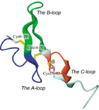
(12) List of figures Figure 1. The hallmarks of cancer ................................................................................. 30 Figure 2. Ten leading cancer types for the estimated new cancer cases and deaths by sex, USA, 2015 .......................................................................................................... 31 Figure 3. Clinical events in cancer therapy .................................................................... 33 Figure 4. Therapeutic targeting of the Hallmarks of cancer ......................................... 34 Figure 5.Basic Structure of EGFR demonstrating relevant domains ............................. 36 Figure 6. EGF structure .................................................................................................. 38 Figure 7. Mapping the interaction sites onto ribbon representations of EGFR and EGF ................................................................................................................................ 39 Figure 8. Model for EGF-‐induced dimerization of the EGFR extracellular region ......... 41 Figure 9. Ligand binding to EGFR causes receptor homodimerization or heterodimerization ....................................................................................................... 43 Figure 10. Endocytosis of ligand-‐activated EGFR molecules ......................................... 44 Figure 11. Trafficking of EGFR and routes for nuclear import ....................................... 46 Figure 12. Structural comparison between EGF and PCI structures ............................. 53 Figure 13. Ribbon diagram of the crystal structure of an EGFR homodimer in complex with two EGF ligands ...................................................................................... 84 Figure 14. Purification and characterization of hEGF and EGFt .................................... 86 Figure 15. EGFR expression profile in MDA-‐MB-‐468, MCF-‐7 and Caco-‐2 cells ............. 87 Figure 16. Effect of EGFt on the phosphorylation of EGFR ........................................... 89 Figure 17. Comparative effect between hEGF and EGFt on MAPK and Akt activation in MCF-‐7, Caco-‐2 and MDA-‐MB-‐468 cells ..................................................... 90 Figure 18. Effect of EGFt on the dimerization of EGFR ................................................. 92 Figure 19. Effect of EGFt on the internalization and localization of EGFR compared to hEGF .......................................................................................................................... 94 Figure 20. Confocal fluorescence microscopy analysis of the cellular localization of RITC-‐hEGF and RITC-‐EGFt after 3 h of exposure ........................................................... 96 .
(13) Figure 21. Effect of EGFt treatment on EGFR degradation ........................................... 97 Figure 22. Assessment of the optimal cell concentration for cell proliferation assays ............................................................................................................................ 98 Figure 23. Effect of EGFt on the growth of two human cancer cells ............................. 99 Figure 24. Representative binding curve for 111In-‐DTPA-‐EGFt to MDA-‐MB-‐468 cells . 100 Figure 25. Cellular uptake and intracellular distribution of 111In-‐DTPA-‐EGFt and 111. In-‐DTPA-‐hEGF .......................................................................................................... 101 . Figure 26. Surviving fraction measured in a clonogenic assay for (A) MDA-‐MB-‐468 or (B) MCF-‐7 cells ........................................................................................................ 103 Figure 27. Elimination of 111In-‐DTPA-‐hEGF or 111In-‐DTPA-‐EGFt from the blood of athymic mice .............................................................................................................. 105 Figure 28. Tumor and normal tissue localization ........................................................ 106 Figure 29. Subcellular distribution of . 111. In in tumor cells isolated from . subcutaneous MDA-‐MB-‐468 human breast cancer xenografts in athymic mice ........ 107 Figure 30. Micro SPECT/CT images illustrating the whole-‐body transport of 111In-‐ DTPA-‐hEGF and 111In-‐DTPA-‐EGFt ................................................................................. 108 .
(14) List of tables Table 1. EGFR overexpression in different tumor types ............................................... 49 Table 2. Different types of agent used to target EGFR ................................................. 50 Table 3. Pharmacokinetic parameters ........................................................................ 104 . . .
(15)
(16) Summary The members of the epidermal growth factor (EGF) / ErbB family are prime targets for cancer therapy. However, the therapeutic efficiency of the existing anti-‐ErbB agents is limited. Thus, identifying new molecules that inactivate the ErbB receptors through novel strategies is an important goal on cancer research. In this thesis work we have developed a shorter form of human EGF (EGFt) with a truncated C-‐terminal as a novel EGFR inhibitor. EGFt was designed based on the superimposition of the three-‐dimensional structures of EGF and the Potato Carboxypeptidase Inhibitor (PCI), an EGFR blocker previously described by our group. The peptide was produced in E. coli with a high yield of the correctly folded peptide. EGFt induced poor EGFR homodimerization and phosphorylation. Interestingly, EGFt promoted EGFR internalization and translocation to the cell nucleus although it did not stimulate the cell growth. In addition, EGFt competed with EGFR native ligands, inhibiting the proliferation of cancer cells. . The lack of EGFR-‐mediated growth-‐stimulatory activity prompted us to evaluate EGFt for targeted delivery of 111In, an Auger electron emitter, into EGFR-‐positive cancer cells. An 111In-‐DTPA-‐EGFt radioconjugate was developed and its properties were analyzed and compared to those of 111In-‐DTPA-‐hEGF. First we determined that 111In-‐ DTPA-‐EGFt displays high specificity and affinity for EGFR. However, the cellular uptake of 111In-‐DTPA-‐EGFt resulted to be lower than that of 111In-‐DTPA-‐hEGF. Once internalized, 111In-‐DTPA-‐EGFt showed a high efficiency to accumulate into the cell nucleus, where the radioactivity emitted by . 111. In may damage the DNA. In . accordance, 111In-‐DTPA-‐EGFt showed to be cytotoxic in vitro against breast cancer cells, although its cytotoxicity was lower compared to . 111. In-‐DTPA-‐hEGF. In vivo . studies revealed a longer half-‐life in blood for 111In-‐DTPA-‐EGFt than for 111In-‐DTPA-‐ hEGF and higher uptake in the kidney, with minor accumulation in other normal .
(17) tissues. 111In-‐DTPA-‐EGFt accumulated in MDA-‐MB-‐468 tumors where, interestingly, 111. In-‐DTPA-‐EGFt was detected in a great proportion in the cell nucleus. . All the data obtained from this work indicate that EGFt may be a potential EGFR blocker for cancer therapy and also an attractive ligand for delivery of cytotoxic agents into the nucleus of EGFR-‐positive cancer cells.. . .
(18) Resum Els receptors i lligands de la família del factor de creixement epidèrmic (EGF) / ErbB són dianes molt importants en el desenvolupament de teràpies contra el càncer. No obstant, l’eficàcia terapèutica dels fàrmacs dirigits a atacar aquesta via i que són utilitzats actualment en clínica és limitada. Per aquest motiu la recerca de noves molècules que inactivin els receptors d’aquesta família mitjançant noves estratègies és avui dia una de les vies més explorades. En aquesta tesi s’ha desenvolupat un pèptid idèntic al factor de creixement epidèrmic EGF que li manca la seva part C-‐ terminal (EGFt) com a nou inhibidor de EGFR. El disseny d’aquest pèptid truncat s’ha basat en la superposició tridimensional de l’estructura de l’EGF i de l’inhibidor de la carboxipeptidasa de patata (PCI), un bloquejador de la via de l’EGFR descrit prèviament pel nostre grup. El pèptid ha estat produït en E.coli i s’ha aconseguit obtenir un alt rendiment de la proteïna i amb la seva conformació estructural correcta. Hem observat que l’EGFt in vitro té una capacitat molt menor per induir dímers del receptor i també la seva fosforilació si la comparem amb l’activitat que té l’hEGF natiu. Per altra banda, l’EGFt promou la internalització del receptor i la seva translocació al nucli cel·∙lular tal i com ho fa l’hEGF, tot i que no estimula el creixement cel·∙lular. A més, l’EGFt competeix amb els lligands natius de la família i inhibeix la proliferació cel·∙lular. La manca d’activitat estimuladora del creixement cel·∙lular d’aquest pèptid quan s’uneix a l’EGFR ens va portar a provar la utilització de l’EGFt com a vehicle de toxines dirigit a cèl·∙lules tumorals que sobreexpresessin EGFR. Concretament, es va produir un radioconjugat de EGFt amb l’isòtop radioactiu emissor d’electrons Auger Indi-‐111. Les propietats d’aquest radioconjugat es van analitzar i es van comparar amb el radioconjugat produït amb hEGF natiu. En primer lloc es va determinar que 111In-‐ DTPA-‐EGFt té una alta especificitat i afinitat per EGFR. No obstant, la captació cel·∙lular de 111In-‐DTPA-‐EGFt va resultar ser menor que la de 111In-‐DTPA-‐hEGF. Un cop internalitzat, 111In-‐DTPA-‐EGFt va mostrar una alta eficiència per acumular-‐se en el .
(19) nucli de la cèl·∙lula, on la radioactivitat emesa per 111In danya l'ADN. 111In-‐DTPA-‐EGFt va mostrar ser citotòxic in vitro contra cèl·∙lules de càncer de mama, encara que la seva citotoxicitat va ser menor en comparació amb 111In-‐DTPA-‐hEGF. Els estudis in vivo van revelar una vida mitjana més llarga en sang per 111In-‐DTPA-‐EGFt que per 111. In-‐DTPA-‐hEGF, una major captació en el ronyó i una menor acumulació en altres . teixits normals. 111In-‐DTPA-‐EGFt es va detectar en els tumors de cèl·∙lules MDA-‐MB-‐ 468 on el radiocompost es va acumular preferentment en el nucli de la cèl·∙lula. Les dades recollides en aquest treball indiquen que l’EGFt pot tenir un gran potencial com a bloquejador en teràpia pel càncer i a més pot ser un bon lligand per utilitzar com a vehicle d’agents citotòxics dirigits al nucli de cèl·∙lules tumorals positives en EGFR. .
(20) Resumen Los receptores y ligandos de la familia del factor de crecimiento epidérmico (EGF) / ErbB son dianas muy importantes en el desarrollo de terapias contra el cáncer. Sin embargo, la eficacia terapéutica de los fármacos dirigidos a atacar esta vía y que son utilizados actualmente en clínica es limitada. De ahí que la búsqueda de nuevas moléculas que inactiven los receptores de esta familia mediante nuevas estrategias es hoy en día una de las vías más exploradas. En esta tesis se ha desarrollado un péptido idéntico al factor de crecimiento epidérmico EGF que le falta su parte C-‐ terminal (EGFt) como nuevo inhibidor de EGFR. El diseño de este péptido truncado se ha basado en la superposición tridimensional de la estructura del EGF y del inhibidor de la carboxipeptidasa de patata (PCI), un bloqueador de la vía del EGFR descrito previamente por nuestro grupo. El péptido ha sido producido en E.coli y se ha logrado obtener un alto rendimiento de la proteína y con su conformación estructural correcta. Hemos observado que el EGFt in vitro tiene una capacidad mucho menor para inducir dímeros del receptor y también su fosforilación si la comparamos con la actividad que tiene el hEGF nativo. Por otra parte, el EGFt promueve la internalización del receptor y su translocación al núcleo celular tal como lo hace el hEGF, aunque no estimula el crecimiento celular. Además, el EGFt compite con los ligandos nativos de la familia e inhibe la proliferación celular. La falta de actividad estimuladora del crecimiento celular de este péptido cuando se une al EGFR nos llevó a probar la utilización del EGFt como vehículo de toxinas dirigido a células tumorales que sobreexpresaran EGFR. Concretamente, se produjo un radioconjugado de EGFt con el isótopo radiactivo emisor de electrones Auger Indio-‐111. Las propiedades de este radioconjugado se analizaron y se compararon con el radioconjugado producido con EGF nativo. En primer lugar se determinó que 111. In-‐DTPA-‐EGFt tiene una alta especificidad y afinidad por EGFR. No obstante, la . captación celular de 111In-‐DTPA-‐EGFt resultó ser menor que la de 111In-‐DTPA-‐hEGF. Una vez internalizado, 111In-‐DTPA-‐EGFt mostró una alta eficiencia para acumularse en .
(21) el núcleo de la célula, donde la radiactividad emitida por 111In daña el ADN. 111In-‐ DTPA-‐EGFt mostró ser citotóxico in vitro contra células de cáncer de mama, aunque su citotoxicidad fue menor en comparación con 111In-‐DTPA-‐hEGF. Los estudios in vivo revelaron una vida media más larga en sangre para 111In-‐DTPA-‐EGFt que para 111In-‐ DTPA-‐hEGF, una mayor captación en el riñón y una menor acumulación en otros tejidos normales. 111In-‐DTPA-‐EGFt se detectó en los tumores de células MDA-‐MB-‐468 donde el radiocompuesto se acumuló preferentemente en el núcleo de la célula. Los datos recogidos en este trabajo indican que el EGFt puede tener un gran potencial como bloqueador en terapia para el cáncer y además puede ser un buen ligando para utilizar como vehículo de agentes citotóxicos dirigidos al núcleo de células tumorales positivas en EGFR . . . .
(22) CONTENTS CONTENTS . 21 . INTRODUCTION . 27 . 1. Cancer overview. 29 . 1.1. Molecular basis of cancer. 29 . 1.2. Classification of human cancers. 30 . 1.3. Cancer statistics. 31 . 1.4. Cancer treatment today. 32 . 1.5. Targeted cancer therapy. 33 . 2. The Epidermal Growth Factor Receptor (EGFR). 35 . 2.1. EGFR and its family. 35 . 2.2. EGFR ligand: Epidermal Growth Factor (EGF). 38 . 2.3. Receptor dimerization. 40 . 2.4. Activation of EGFR signaling pathways. 42 . 2.5. Internalization and endosomal sorting of EGFR. 43 . 2.6. EGFR nuclear localization. 45 . 3. EGFR in cancer. 47 . 3.1. EGFR signaling network in human cancers. 47 . 3.2. EGFR-‐targeted therapies. 49 .
(23) 4. Potato carboxypeptidase inhibitor: an EGF peptide mimetic with EGFR blocking activity . 52 . 5. Targeted radiotherapy and molecular imaging . 54 . 5.1. EGFR-‐targeted auger-‐electron radiotherapy . 56 . OBJECTIVES . 59 . METHODS . 63 . 1. Production of the recombinant human EGF and its truncated derivative EGFt . 65 . 1.1. Molecular cloning and expression of hEGF and EGFt on small batch culture conditions. . 65 . 1.2. Production of hEGF and EGFt in a 10 liter fermentor . 66 . 1.3. Protein purification . 66 . 1.4. Protein analysis . 67 . 2. In vitro characterization of hEGF and EGFt . 68 . 2.1. Cells and cell culture . 68 . 2.2. Immunofluorescence analysis by flow citometry . 68 . 2.3. Western blot analysis . 69 . 2.4. Dimerization analysis . 69 . 2.5. Analysis of EGFR activation and degradation . 70 . 2.6. EGFR internalization analysis . 71 . 2.7. Immunofluorescence staining of EGFR and cellular localization analysis by confocal microscopy . 72 .
(24) 2.8. Fluorescent staining of hEGF and EGFt and cellular localization analysis by confocal microscopy . 72 . 2.9. Cell proliferation assay . 73 . 3. Production and in vitro and in vivo characterization of the radiopeptides 111In-‐ DTPA-‐hEGF and 111In-‐DTPA-‐EGFt . 74 . 3.1. Radiolabeling of EGFt and hEGF with 111In . 74 . 3.2. Receptor binding assay . 75 . 3.3. Cellular uptake and intracellular distribution of 111In-‐DTPA-‐EGFt and 111In-‐DTPA-‐ hEGF . 76 . 3.4. Determination of cytotoxicity in vitro of 111In-‐DTPA-‐hEGF and 111In-‐DTPA-‐EGFt . 76 . 3.5. Tumor xenograft mouse model . 77 . 3.6. Pharmacokinetic studies . 78 . 3.7. Biodistribution . 78 . 3.8 MicroSPECT imaging . 79 . 4. Statistical analysis RESULTS 1. Production of the recombinant human EGF and its truncated derivative EGFt . 79 81 83 . 1.1. Design of an EGF analogue . 83 . 1.2. Production, purification and analysis of recombinant hEGF and EGFt . 84 . 2. In vitro charachterization of EGFt 2.1. EGFR expression profile in three EGFR-‐positive human tumor cell lines . 87 87 .
(25) 2.2. EGFR activation . 87 . 2.3. EGFR dimerization . 91 . 2.4. Effect of hEGF and EGFt on EGFR internalization . 92 . 2.5. Effect of hEGF and EGFt on EGFR localization . 93 . 2.6. EGFt cellular localization analysis by confocal microscopy . 95 . 2.7. Effect of hEGF and EGFt on EGFR degradation . 96 . 2.8. Effect of hEGF and EGFt on cell proliferation . 97 . 3. In vitro and in vivo characterization of the radiopeptides 111In-‐DTPA-‐hEGF AND111In-‐DTPA-‐EGFt 3.1. Characterization of 111In-‐DTPA-‐EGFt 3.2. EGFR binding affinity of EGFt . 99 99 100 . 3.3. Cellular uptake and intracellular distribution of 111In-‐DTPA-‐EGFt and 111In-‐DTPA-‐ hEGF . 101 . 3.4. In vitro cytotoxicity of 111In-‐DTPA-‐EGFt and 111In-‐DTPA-‐hEGF . 102 . 3.5. Pharmacokinetic studies . 104 . 3.6. Tissue biodistribution and intratumoral distribution of 111In-‐DTPA-‐EGFt . 105 . 3.7. MicroSPECT imaging . 107 . DISCUSSION . 109 . 1. Production and characterization of an Epidermal Growth factor derivative with EGFR blocking activity . 111 . 2. Evaluation of truncated human epidermal growth factor (EGFt) as a vehicle for the delivery of Auger electron-‐emitting 111In into the nucleus of EGFR-‐positive breast cancer cells . 116 .
(26) 121 . 3. Potential future directions CONCLUSIONS . 123 . REFERENCES . 129 . . .
(27) .
(28) INTRODUCTION .
(29)
(30) ESCRIBA EL TÍTULO DEL DOCUMENTO Introduction. 1. Cancer overview . . 1.1. Molecular basis of cancer Cancer is a genetic disease that occurs when the information in cellular DNA is corrupted leading to abnormal patterns of gene expression. As a result, the effects of normal genes that control cell growth, survival and spread are enhanced and those of genes that suppress these effects are repressed. The main mechanism by which this corruption of the genetic code occurs is through the accumulation of mutations, although there is increasing recognition of the role of non-‐mutational (epigenetic) changes in the process. Aberrant gene expression leads to a number of key changes in fundamental biological processes within cancer cells, the so-‐called ‘hallmarks’ of cancer (Hanahan et al., 2011). The hallmarks of cancer comprise six biological capabilities acquired during the multistep development of human tumours. They include sustaining proliferative signalling, evading growth suppressors, resisting cell death, enabling replicative immortality, inducing angiogenesis, and activating invasion and metastasis. Underlying these hallmarks are two important characteristics that enable the acquisition of these mentioned function capabilities: genome instability, which generates the genetic diversity that expedites their acquisition, and inflammation, which fosters multiple hallmark functions. Conceptual progress in the last decade has added two emerging hallmarks of potential generality to this list— reprogramming of energy metabolism and evading immune destruction (Figure 1). In addition to cancer cells, tumors exhibit another dimension of complexity: they contain a repertoire of recruited, ostensibly normal cells that contribute to the acquisition of hallmark traits by creating the “tumor microenvironment.” . 29.
(31) ESCRIBA EL TÍTULO DEL DOCUMENTO Introduction Figure 1. The hallmarks of cancer. The illustration encompasses the six biological capabilities acquired during the multistep development of human tumors, the two new emerging hallmark capabilities and two enabling characteristics crucial to the acquisition of these hallmark capabilities. Extracted from Hanahan and Weinberg, 2011. . 1.2. Classification of human cancers There are many different types of cancer. Cancer can be classified according to the tissue that the malignant cells originate from; the main categories include: . Ø Adenocarcinoma – tumours derived from secretory epithelial cells (e.g. prostate, breast and colorectal cancers). Ø Squamous cell carcinoma – tumours derived from epithelial cells (in skin or in tissues covering some inner organs). Ø Sarcoma – tumours derived from connective or supportive tissue (bone, cartilage, fat, muscle and blood vessels). Ø Leukaemia – derived from blood-‐forming tissue (bone-‐marrow). Ø Lymphoma and Myeloma – cancers that originate in the immune system. Ø Glioma – tumours derived from brain and spinal cord. . 30.
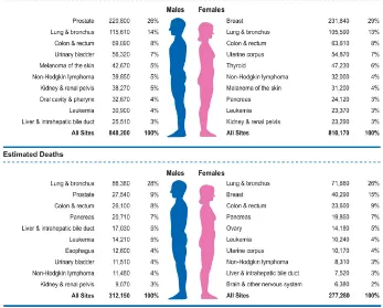
(32) ESCRIBA EL TÍTULO DEL DOCUMENTO Introduction . The two first groups represent the most common human cancers – the carcinomas. Carcinomas are responsible for more than 80% of the cancer-‐related deaths in the Western world (Weinberg, 2007). . 1.3. Cancer statistics Nowadays, cancer accounts for one in every eight deaths worldwide – more than HIV/AIDS, tuberculosis, and malaria combined. In EU a total of 1,359,100 Europeans are projected to die of cancer in 2015 (Malvezzi et al., 2015). And in United States the expected numbers of deaths from cancer in 2015 is estimated in 589,430 Americans, corresponding to about 1600 deaths per day (Siegel et al., 2015). . Figure 2. Ten leading cancer types for the estimated new cancer cases and deaths by sex, USA, 2015. Estimates are rounded to the nearest 10 and cases exclude basal cell and squamous cell skin cancers and in situ carcinoma except urinary bladder. Extracted from Siegel et al., 2015. . 31.
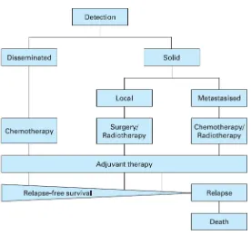
(33) ESCRIBA EL TÍTULO DEL DOCUMENTO Introduction Figure 2 indicates the most common cancers expected to occur and deaths in men and women in 2015 in United States (Siegel et al., 2015). Among men, cancers of the prostate, lung and bronchus, and colorectum will account for about half of all newly diagnosed cancers. The 3 most commonly diagnosed types of cancer among women in 2015 will be breast, lung and bronchus, and colorectum, accounting for the half of the estimated cancer cases in women. Breast cancer alone is expected to account for 29% (231,840) of all new cancer cases among women. Cancers of the lung and bronchus, prostate, and colorectum in men and cancers of the lung and bronchus, breast, and colorectum in women account for almost half of the total cancer deaths (Siegel et al., 2015). . 1.4. Cancer treatment today Cancer therapy involves several types of treatment. The principal therapeutic modalities are surgery, radiotherapy and chemotherapy. In the case of solid tumors, the first step is usually surgical removal of the primary cancer together with a margin of normal tissue; as such cancers are commonly irregular in shape. Then, the area around the site may be irradiated to destroy any possible remnants of the tumor. At the same time, cytotoxic drugs can be given to kill residual cancer cells and possible metastases and nowadays, beside standard chemotherapy have emerged the targeted therapy drugs, which inhibit a more specific target in cells. This is the usual pattern of treatment, but there are some situations where alternative approaches are used. In regions of anatomical complexity, such as the head and neck, and in regions of vital biological function, such as the brain and spine, surgery would cause many problems, and so radiotherapy is sometimes the preferred modality. In disseminated cancers such as leukaemia, the only modality that can be used is chemotherapy. The time after the treatment that patient survives without any signs or symptoms of cancer are called the relapse-‐free survival. The return of the disease or the signs and symptoms after a period of improvement is known as relapse (Figure 3) (King and Robins, 2006). . 32.
(34) ESCRIBA EL TÍTULO DEL DOCUMENTO Introduction Figure 3. Clinical events in cancer therapy. Extracted from King and Robins, 2006. . . 1.5. Targeted cancer therapy Traditional chemotherapy and radiotherapy will destroy not only proliferating cancer cells but also proliferating normal cells; so side effects are almost inevitable. The term targeted therapy refers to drugs or other substances that block the growth and spread of cancer by interfering with specific molecules (molecular targets) that are involved in the growth, progression, and spread of cancer. These, targeted therapies are deliberately selected or designed to interact with their target and are often cytostatic (they block tumor cell proliferation), whereas many standard chemotherapies were identified because they kill cells (they are cytotoxic). As more is understood about the molecular defects that lead to malignancy, more agents targeted towards specific molecules or specific survival functions of cancer cells . 33.
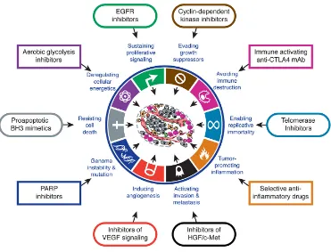
(35) ESCRIBA EL TÍTULO DEL DOCUMENTO Introduction are being developed and are demonstrating effective therapeutic benefit while limited toxicity to normal cells (Zahorowska et al., 2009). Targeted cancer therapies approved for use against specific cancers include agents that prevent cell growth signalling, interfere with tumor blood vessel development, promote the death of cancer cells, stimulate the immune system to destroy cancer cells, and deliver toxic drugs to cancer cells. In addition, new fields have emerged lately as promising targets, including cancer metabolism and epigenetic modulation. Cancer immunotherapy is also a promising field for targeted cancer discovery (Huang et al., 2014). . Figure 6. Therapeutic Targeting of the Hallmarks of Cancer Drugs that interfere with each of the acquired capabilities necessary for tumor growth and progression have been developed and are in clinical trials or in some. cases approved for clinical use in treating certain forms of human cancer. Additionally, the investigational drugs are being developed to target each of the Figure 4. Therapeutic targeting of the Hallmarks of cancer. Drugs that interfere with each of enabling characteristics and emerging hallmarks depicted in Figure 3, which also hold promise as cancer therapeutics. The drugs listed are but illustrative examples; there is a deep pipeline of candidate drugs with different molecular targets and modes of action in development have for most of these hallmarks. the acquired capabilities necessary for tumor growth and progression been developed and are in clinical trials or in some cases approved for clinical use in treating certain forms of therapies. For example, the deployment of apoptosis-inducing The six acquired capabilities—the hallmarks of cancer—have human cancer. xtracted Hanahan mitogenic et al., 2011. drugs may induce Ecancer cells from to hyperactivate stood the test of time as being integral components of most enabling them to compensate for the initial attrition forms of cancer. Further refinement of these organizing princi signaling, triggered by such treatments. Such considerations suggest ples will surely come in the foreseeable future, continuing the. that drug development and the design of treatment protocols will benefit from incorporating the concepts of functionally discrete hallmark capabilities and of the multiple biochemical pathways involved in supporting each of them. Thus, in particular, we can envisage that selective cotargeting of multiple core and emerging hallmark capabilities and enabling characteristics (Figure 6) in mechanism-guided combinations will result in more effective and durable therapies for human cancer. CONCLUSION AND FUTURE VISION. remarkable conceptual progress of the last decade. Looking ahead, we envision significant advances during the coming decade in our understanding of invasion and metastasis. Similarly, the role of aerobic glycolysis in malignant growth will be elucidated, including a resolution of whether this metabolic reprogramming is a discrete capability separable from the core hallmark of chronically sustained proliferation. We remain perplexed as to whether immune surveillance is a barrier that virtually all tumors must circumvent, or only an idiosyncrasy of an especially immunogenic subset of them; this issue too will be resolved in one way or another.. 34.
(36) ESCRIBA EL TÍTULO DEL DOCUMENTO Introduction Targeted therapies include monoclonal antibodies (mAbs), small molecules (such as ATP analogues), peptide mimetics, and antisense oligonucleotides (Stoffel, 2010; Imai et al., 2006). Among the targets for therapy, growth factor receptors and downstream signaling molecules continue to be the most actively explored targets for cancer drug discovery. Figure 4 illustrates some examples of targeted therapeutics categorized according to their respective effects on one or more hallmark capabilities. . 2. The Epidermal Growth Factor Receptor (EGFR) 2.1. EGFR and its family The epidermal growth factor receptor (EGFR) belongs to the ErbB family of proteins, which consists of ErbB1/HER1/EGFR, ErbB2/HER2/Neu, ErbB3/HER3 and ErbB4/ HER4 (Stern, 2010). They are transmembrane glycoproteins with molecular weights ranging from 170 to 185 KDa. Structurally, ErbB family members consist of i) a cysteine-‐rich variable extracellular N-‐terminal domain, ii) a hydrophobic transmembrane domain and iii) an intracellular, highly conserved, cytoplasmic C-‐terminal tyrosine kinase domain with several phosphorylation sites. (Seshacharyulu et al., 2012). The extracellular region of EGFR is subdivided into four domains arranged as a tandem repeat of two types of domains (Figure 5). The first and third domains are homologous and have been designated as domains I and III or L1 and L2, respectively. These domains are responsible for binding to different ligands. The second and four domains, are also homologous and have been designated as domains II and IV or CR1 and CR2, respectively. Domains II an IV are involved in the dimerization of ErbB receptors (Leahy, 2004; Normanno et al., 2006; Flynn et al., 2009; Lemmon, 2009). . 35.
(37) ESCRIBA EL TÍTULO DEL DOCUMENTO Introduction Figure 5.Basic Structure of EGFR demonstrating relevant domains. (I) The extracellular domains: (1) domain I: L1; (2) domain II: CR1; domain III: L2; domain IV: CR2. (II) Transmembrane domains. (III) The intracellular domains (1) juxtamembrane domain; (2) tyrosine kinase domain; (3) regulatory region domain. Adapted from Flynn et al., 2009. . ErbB family members can be activated by 13 known ligands, which include EGF, transforming growth factor alpha (TGFα), amphiregulin (AR), betacellulin (BTC), heparin-‐binding EGF-‐like growth factor (HB-‐EGF), epiregulin (EPR), epigen (EPG) and neuregulins 1 -‐-‐ 6 (NRG) (Citri and Yarden, 2006; Yarden and Sliwkowski, 2001). Among these ligands, EGF, TGFα, AR, BTC and EPR have been demonstrated to bind EGFR. . 36.
(38) ESCRIBA EL TÍTULO DEL DOCUMENTO Introduction Among them, EGF and TGFα are considered the main ligands of EGFR (Linggi et al., 2006; Wells, 1999; Köstler et al., 2010). Although, HER2 is closely related to EGFR, no ligands have been described for HER2 to date (Coussens et al., 1980). EGFR and HER2 share 82% homology in the tyrosine kinase domain, (Prigent and Lemoine, 1992) and cross-‐interactions between EGFR and HER2 receptors are frequent. In fact, HER2 is the preferred binding partner of EGFR (Harari and Yarden, 2000; Yarden and Sliwkowski, 2001). The ligand-‐binding domain of HER3 demonstrates approximately 40% homology to EGFR and binds neuregulin-‐1 and neuregulin-‐2. HER3 has a distinctive feature; its tyrosine kinase domain is functionally defective. Finally, HER4, which is the most recently described family member, is a receptor for HB-‐EGF, betacellulin, epiregulin, and neuregulins 1-‐4 (Campbell et al., 2010; Carpenter, 2003). EGFR is expressed on normal cells at levels ranging from 20,000 to 200,000 receptors per cell (Chow, 1997). Ligand binding induces major conformational changes in the receptor, leading to the exposition of the dimerization arm in domain II. This allows the receptor homodimerization or heterodimerization with other ErbB receptors in the plasma membrane followed by the activation of the receptor tyrosin kinase activity, causing the transphosphorylation of the cytoplasmic tails of each dimer pair. ErbB receptors activation stimulates many complex intracellular signaling pathways that are involved in numerous biological processes such us proliferation (Ullrich and Schlessinger, 1990; Carpenter and Cohen, 1990), apoptosis (Cao et al., 2000; Armstrong et al., 1994), migration, angiogenesis and differentiation (Yarden, 2001; Roskoski, 2004; Wheeler et al., 2010; Bublil et al., 2007). EGFR plays a role during embryogenesis in the morphogenesis of organs like teeth, brain, reproductive tracts, gastrointestinal tracts and cardiovascular system (Goldman et al., 1996; Kato et al., 1995; Thesleff et al., 1995). Physiologically, EGFR has an essential role in wound healing and normal epithelial regeneration of organs such as gastrointestinal, genitourinary, respiratory, skin and corneal epithelia (Schneider and Wolf, 2009; Repertinger, et al., 2011). . 37.
Figure

+7

Documento similar
(2015) Human Epidermal Growth Factor Receptor 2 (HER2)–specific chimeric antigen receptor–modified T cells for the Immunotherapy of HER2- positive sarcoma.. Journal of
Our results show that in cultured tubular epithelial cells, the VDRA agonist paricalcitol inhibited the EGFR pathway activated by Aldo by
Effects of basic fibroblast growth factor on early revascularization and epithelial regeneration in rabbit tracheal orthotopic transplantation. Comparative anatomy, physíology,
- Their capability to inhibit the proliferation of human liver carcinoma cells - Antioxidant content has a crucial effect on growth inhibition and cytostaticity -
In CCN2(IV)-injected mice, renal MCP- 1 and RANTES gene expression was significantly increased in response
This study analyzes whether the release of nitric oxide (NO) and thromboxane A 2 (TXA 2 ) depends on the time lapsed since gonadal function is lost, and their correlation with
Taken together, these pharmacological data likely indicate the presence of two functional P2 receptors in the human neuroblastoma SK-M-MC cell line: one metabotropic,
concept suggests that TSLP, secreted by keratinocytes upon barrier defects, establishes a tumor protective environment by activating T cells (Demehri et al., 2012; Di Piazza et al.,
