ISSN: 1524-4539
Copyright © 2005 American Heart Association. All rights reserved. Print ISSN: 0009-7322. Online
72514
Circulation is published by the American Heart Association. 7272 Greenville Avenue, Dallas, TX
DOI: 10.1161/CIRCULATIONAHA.105.166561
http://circ.ahajournals.org/cgi/content/full/112/24_suppl/IV-89
located on the World Wide Web at:
The online version of this article, along with updated information and services, is
http://www.lww.com/reprints
Reprints: Information about reprints can be found online at
[email protected]
410-528-8550. E-mail:
Fax:
Kluwer Health, 351 West Camden Street, Baltimore, MD 21202-2436. Phone: 410-528-4050.
Permissions: Permissions & Rights Desk, Lippincott Williams & Wilkins, a division of Wolters
http://circ.ahajournals.org/subscriptions/
Coronary Syndromes
A
cute myocardial infarction (AMI) and unstable angina
(UA) are part of a spectrum of clinical disease
collec-tively identified as
acute coronary syndromes
(ACS). The
pathophysiology common to this spectrum of disease is a
ruptured or eroded atheromatous plaque.
1–5The
electrocar-diographic (ECG) presentation of these syndromes
encom-passes ST-segment elevation myocardial infarction (STEMI),
ST-segment depression, and nondiagnostic ST-segment and
T-wave abnormalities. A non–ST-elevation myocardial
in-farction (NSTEMI) is diagnosed if cardiac markers are
positive with ST-segment depression or with nonspecific or
normal ECGs. Sudden cardiac death may occur with any of
these conditions. ACS is the most common proximate cause
of sudden cardiac death.
6 –10Effective interventions for patients with ACS, particularly
STEMI, are extremely time-sensitive. The first healthcare
providers to encounter the ACS patient can have a big impact
on patient outcome if they provide efficient risk stratification,
initial stabilization, and referral for cardiology care. It is
critical that basic life support (BLS) and advanced
cardiovas-cular life support (ACLS) healthcare providers who care for
ACS patients in the out-of-hospital, emergency department
(ED), and hospital environments be aware of the principles
and priorities of assessment and stabilization of these
patients.
These guidelines target BLS and ACLS healthcare
provid-ers who treat patients with ACS within the first hours after
onset of symptoms, summarizing key out-of-hospital, ED,
and some initial critical-care topics that are relevant to
stabilization. They also continue to build on
recommenda-tions from the ACC/AHA Guidelines,
11,12which are used
throughout the United States and Canada.
13As with any
medical guidelines, these general recommendations must be
considered within the context of local resources and
applica-tion to individual patients by knowledgeable healthcare
providers.
The primary goals of therapy for patients with ACS are to
●
Reduce the amount of myocardial necrosis that occurs in
patients with MI, preserving left ventricular (LV) function
and preventing heart failure
●
Prevent major adverse cardiac events (MACE): death,
nonfatal MI, and need for urgent revascularization
●
Treat acute, life-threatening complications of ACS, such as
ventricular fibrillation (VF)/pulseless ventricular
tachycardia (VT), symptomatic bradycardias, and unstable
tachycardias (see Part 7.2: “Management of Cardiac
Ar-rest” and Part 7.3: “Management of Symptomatic
Brady-cardia and TachyBrady-cardia”)
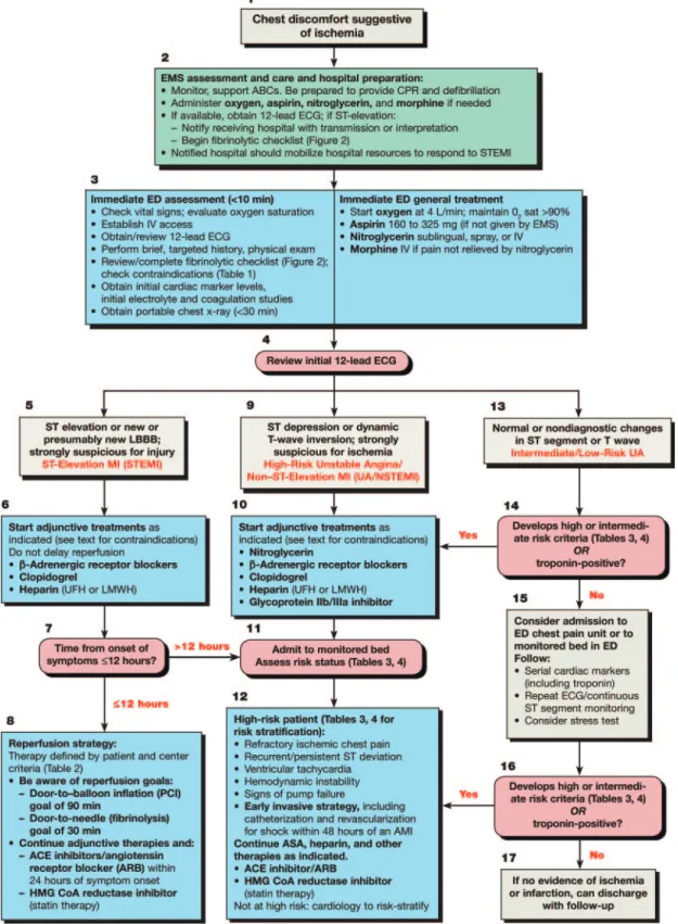
An overview of recommended care for the ACS patient is
illustrated in Figure 1, the Acute Coronary Syndromes
Algorithm. Part 8 provides details of the care highlighted in
the numbered algorithm boxes. Box numbers in the text
correspond to the numbered boxes in the algorithm.
In this part the abbreviation AMI refers to acute
myocar-dial infarction, whether associated with STEMI or NSTEMI.
The diagnosis and treatment of AMI, however, will often
differ for patients with STEMI versus NSTEMI. Note
care-fully which is being discussed.
Out-of-Hospital Management
Recognition (Figure 1, Box 1)
Treatment offers the greatest potential benefit for myocardial
salvage in the first hours of STEMI. Thus, it is imperative that
healthcare providers evaluate, triage, and treat patients with
ACS as quickly as possible. Delays to therapy occur during 3
intervals: from onset of symptoms to patient recognition,
during out-of-hospital transport, and during in-hospital
eval-uation. Patient delay to symptom recognition often constitutes
the longest period of delay to treatment.
14The classic symptom associated with ACS is chest
discom-fort, but symptoms may also include discomfort in other areas
of the upper body, shortness of breath, sweating, nausea, and
lightheadedness. The symptoms of AMI are characteristically
more intense than angina and last
⬎
15 minutes. Atypical
symptoms or unusual presentations of ACS are more
com-mon in elderly, female, and diabetic patients.
15–19Public education campaigns increase public awareness and
knowledge of the symptoms of heart attack but have only
transient effects.
20For patients at risk for ACS (and for their
families), physicians should discuss the appropriate use of
nitroglycerin and aspirin, activation of the emergency
medi-cal services (EMS) system, and location of the nearest
hospital that offers 24-hour emergency cardiovascular care.
Recent ACC/AHA guidelines recommend that the patient or
family members activate the EMS system rather than call
their physician or drive to the hospital if chest discomfort is
unimproved or worsening 5 minutes after taking 1
nitroglyc-erin tablet or using nitroglycnitroglyc-erin spray.
12Initial EMS Care (Figure 1, Box 2)
Half of the patients who die of AMI do so before reaching the
hospital. VF or pulseless VT is the precipitating rhythm in
most of these deaths,
21–23and it is most likely to develop
during the first 4 hours after onset of symptoms.
24 –27Com-munities should develop programs to respond to
out-of-hospital cardiac arrest that include prompt recognition of
symptoms of ACS, early activation of the EMS system, and
(Circulation.2005;112:IV-89-IV-110.)© 2005 American Heart Association.
This special supplement to Circulation is freely available at http://www.circulationaha.org
DOI: 10.1161/CIRCULATIONAHA.105.166561
if needed, early CPR (see Part 4: “Adult Basic Life Support”)
and early access to an automated external defibrillator (AED)
through community AED programs (see Part 5: “Electrical
Therapies”).
28EMS and dispatch system personnel should be
trained to respond to cardiovascular emergencies.
Dispatchers and EMS providers must be trained to
recog-nize symptoms of ACS. Dispatchers should advise patients
with no history of aspirin allergy or signs of active or recent
gastrointestinal bleeding to chew an aspirin (160 to 325 mg)
while awaiting the arrival of EMS providers (Class IIa).
29EMS providers should be trained to determine the time of
onset of symptoms and to stabilize, triage, and transport the
patient to an appropriate facility and to provide prearrival
notification. EMS providers should monitor vital signs and
cardiac rhythm and be prepared to provide CPR and
defibril-lation if needed.
EMS providers may administer oxygen to all patients. If
the patient is hypoxemic, providers should titrate therapy
based on monitoring of oxyhemoglobin saturation (Class
I).
30 – 44If the patient has not taken aspirin and has no history
of aspirin allergy and no evidence of recent gastrointestinal
bleeding, EMS providers should give the patient nonenteric
aspirin (160 to 325 mg) to chew (Class I).
45– 48EMS providers should administer up to 3 nitroglycerin
tablets (or spray) for ongoing symptoms at intervals of 3 to 5
minutes if permitted by medical control and if the patient
remains hemodynamically stable (systolic blood pressure
[SBP]
⬎
90 mm Hg [or no more than 30 mm Hg below
baseline], heart rate between 50 and 100 beats per minute
[bpm]).
49,50EMS providers can administer morphine for chest
pain unresponsive to nitroglycerin if authorized by protocol
or medical control. Additional information about
out-of-hospital stabilization and care is included in the following
sections.
Out-of-Hospital ECGs
Out-of-hospital 12-lead ECGs and advance notification to the
receiving facility speed the diagnosis, shorten the time to
fibrinolysis, and may be associated with decreased mortality
rates.
51– 64The reduction in door-to–reperfusion therapy
in-terval in most studies ranges from 10 to 60 minutes. EMS
providers can efficiently acquire and transmit
diagnostic-quality ECGs to the ED
53,58,65,66with a minimal increase (0.2
to 5.6 minutes) in the on-scene time interval.
52,56,65– 68Qualified and specially trained paramedics and prehospital
nurses can accurately identify typical ST-segment elevation
(
⬎
1 mm in 2 or more contiguous leads) in the 12-lead ECG
with specificity ranging from 91% to 100% and sensitivity
ranging from 71% to 97% when compared with emergency
medicine physicians or cardiologists.
69,70Using radio or cell
phone, they can also provide advance notification to the
receiving hospital of the arrival of a patient with ACS.
56,61– 64We recommend implementation of out-of-hospital 12-lead
ECG diagnostic programs in urban and suburban EMS
systems (Class I). Routine use of 12-lead out-of-hospital ECG
and advance notification is recommended for patients with
signs and symptoms of ACS (Class IIa). A 12-lead
out-of-hospital ECG with advance notification to the ED may be
beneficial for STEMI patients by reducing time to reperfusion
therapy. We recommend that out-of-hospital paramedics
acquire and transmit either diagnostic-quality ECGs or their
interpretation of them to the receiving hospital with advance
notification of the arrival of a patient with ACS (Class IIa). If
EMS providers identify STEMI on the ECG, it is reasonable
for them to begin to complete a fibrinolytic checklist (Figure
2).
Out-of-Hospital Fibrinolysis
Clinical trials have shown the benefit of initiating fibrinolysis
as soon as possible after onset of ischemic-type chest pain in
patients with STEMI or new or presumably new left bundle
branch block (LBBB).
67,71Several prospective studies (LOE
1)
72–74have documented reduced time to administration of
fibrinolytics and decreased mortality rates when
out-of-hospital fibrinolytics were administered to patients with
STEMI and no contraindications to fibrinolytics.
Physicians in the Grampian Region Early Anistreplase
Trial (GREAT)
73administered fibrinolytic therapy to patients
at home 130 minutes earlier than to patients at the hospital
and noted a 50% reduction in hospital mortality rates and
greater 1-year and 5-year survival rates in those treated
earlier.
75,76Delaying fibrinolytic treatment by 1 hour
in-creased the hazard ratio of death by 20%, which is equivalent
to the loss of 43 lives per 1000 patients over 5 years.
A meta-analysis of out-of-hospital fibrinolytic trials found
a relative improvement of 17% in outcome associated with
out-of-hospital fibrinolytic therapy, particularly when therapy
was initiated 60 to 90 minutes earlier than in the hospital.
71A
meta-analysis of 6 trials involving 6434 patients (LOE 1)
72documented decreased all-cause hospital mortality rates
among patients treated with out-of-hospital fibrinolysis
com-pared with in-hospital fibrinolysis (odds ratio [OR]: 0.83;
95% confidence interval [CI]: 0.70 to 0.98) with a number
needed to treat of 62 to save 1 extra life with out-of-hospital
fibrinolysis. Results were similar regardless of the training
and experience of the provider.
The
ECC Guidelines 2000
77recommended consideration
of out-of-hospital fibrinolysis for patients with a transport
time
⬎
1 hour. But in a recent Swiss study (LOE 1),
74prehospital administration of fibrinolytics significantly
de-creased the time to drug administration even in an urban
setting with relatively short transport intervals (
⬍
15
minutes).
74interpretation or direct transmission of ECG) instead of
out-of-hospital delivery of fibrinolysis.
Triage and Transfer
Out-of-Hospital Triage
Hospital and EMS protocols should clearly identify criteria
for transfer of patients to specialty centers and conditions
under which fibrinolytics should be initiated before transfer.
When transfer is indicated, the ACC/AHA guidelines
recom-mend a door-to-departure time
ⱕ
30 minutes.
12It may be
appropriate for the EMS medical director to institute a policy
of out-of-hospital bypass of hospitals that provide medical
therapy only, particularly for patients who require
interven-tional therapy. Patients who require interveninterven-tional therapy
may include those with cardiogenic shock, pulmonary edema,
large infarctions, and contraindications to fibrinolytic
therapy.
At present no randomized studies have directly compared
triage with an experienced percutaneous coronary
interven-tion (PCI) center with medical management at the local
hospital. Extrapolation from several randomized trials on
interfacility transfer
78 – 80suggests that STEMI patients
tri-aged directly to a primary PCI facility may have better
outcomes related to the potential for earlier treatment. A
cost-efficacy substudy of the Comparison of Angioplasty and
Prehospital Thrombolysis in Acute Myocardial Infarction
(CAPTIM) trial
81suggests that direct transport to a primary
PCI facility may be more cost-effective than out-of-hospital
fibrinolysis when transport can be completed in
ⱕ
60 minutes
with a physician in a mobile intensive care unit. There is no
direct evidence, however, to suggest that these strategies are
safe or effective. Patients judged to be at highest risk for a
complicated transfer were excluded from some of these
studies.
In summary, at this time there is inadequate evidence to
recommend out-of-hospital triage to bypass non–PCI-capable
hospitals to bring patients to a PCI center (Class
Indetermi-nate). Local protocols for EMS providers are appropriate to
guide the destination of patients with suspected or confirmed
STEMI.
Interfacility Transfer
All patients with STEMI and symptom duration of
ⱕ
12 hours
are candidates for reperfusion therapy with either fibrinolysis
or PCI (Class I). When patients present directly to a facility
capable of providing only fibrinolysis, 3 treatment options are
available: administering fibrinolytics with admission to that
hospital, transferring the patient for primary PCI, or giving
fibrinolytics and then transferring the patient to a specialized
center. The decision is guided by a risk-benefit assessment
that includes evaluation of duration of symptoms,
complica-tions, contraindicacomplica-tions, and the time delay from patient
contact to fibrinolysis versus potential delay to PCI balloon
inflation.
In 2 prospective studies (LOE 2)
78 – 80and a
meta-analy-sis,
82patients with STEMI who presented 3 to 12 hours after
onset of symptoms to a hospital without capability for
primary PCI had better outcome (improved 30-day combined
incidence of death, reinfarction, or stroke) when they were
transferred to a skilled PCI center (interventionalist
perform-ing
⬎
75 procedures per year) rather than receiving
fibrino-lytics at the presenting hospital. In these studies balloon
inflation occurred
ⱕ
93 minutes after decision to treat.
80,83– 85Thus, interfacility transfer is indicated for patients with
STEMI presenting
⬎
3 hours from onset of symptoms from
hospitals that lack primary PCI capability to centers capable
of providing primary PCI when the transfer can be
accom-plished as soon as possible. The ACC/AHA guidelines
recommend a treatment delay of no more than 90 minutes.
12In patients with STEMI presenting
⬍
3 hours from onset of
symptoms, the superiority of immediate administration of
fibrinolytics in the hospital or transfer for primary PCI is not
established (Class Indeterminate).
In-Hospital Fibrinolytics and Interfacility Transfer for
PCI
Data from the 1980s to 1990s did not support a strategy of
fibrinolytic therapy combined with transfer for facilitated PCI
(LOE 1
86 – 88and meta-analyses
89 –91). But all of the studies
involved in-hospital administration of fibrinolytics, and most
were completed before the era of coronary stenting and
without use of contemporary pharmacologic therapies or PCI
techniques. Three small randomized trials (LOE 1)
92–94sup-ported the strategy of fibrinolytics plus transfer for PCI;
however, the timing of PCI after administration of
fibrinolyt-ics, the inclusion of patients who required transfer for PCI,
the use of coronary stents, and the control group interventions
differ considerably among these trials. The most recent
study
79was fairly small and showed a benefit of early PCI
with 1-year follow-up.
94At present there is inadequate evidence to recommend the
routine transfer of patients for early PCI (ie, within 24 hours)
after successful administration of fibrinolytics in a
commu-nity hospital. The use of out-of-hospital administration of
fibrinolytics followed by early PCI has not been specifically
studied.
Special Transfer Considerations
Special transfer considerations are appropriate for patients
with signs of shock (pulmonary congestion, heart rate
⬎
100
bpm,
and
SBP
⬍
100 mm Hg). The Second National Registry
of Myocardial Infarction found that the mortality rate in
patients with AMI and shock was lower in those treated with
PCI as a primary strategy than in those treated with
fibrino-lysis.
95In the SHOCK (Should We Emergently Revascularize
Occluded Coronaries for Cardiogenic Shock) trial, 152
pa-tients with cardiogenic shock were randomly assigned to an
early revascularization (ERV) strategy, 150 patients were
assigned to a strategy of initial medical stabilization that
included fibrinolytics, and 25% had delayed
revasculariza-tion.
96Although there was no difference in the 30-day
mortality rate, the mortality rate at 6 months was significantly
lower in the ERV group (50.3% versus 63.1%). In a
prespeci-fied subgroup analysis for patients
⬍
75 years of age, early
revascularization was associated with a 15.4% reduction in
30-day mortality and improvement in 1-year survival rates.
97A direct comparison of the outcome of primary or early PCI
patients with patients who received fibrinolytic therapy only
was not reported.
There is inadequate evidence to recommend routine
trans-fer of stable patients for early PCI after successful
adminis-tration of fibrinolytics in community hospitals or the
out-of-hospital setting. Patients
⬍
75 years of age and selected
patients
⬎
75 years of age who develop cardiogenic shock or
persistent ischemic symptoms within 36 hours of STEMI
should be transferred to experienced facilities capable of
ERV if ERV can be performed within 18 hours of onset of
shock.
12ED Evaluation and Risk Stratification
(Figure 1, Boxes 3 and 4)
Focused Assessment and ECG Risk Stratification
ED providers should quickly assess patients with possible
ACS. Ideally within 10 minutes of ED arrival, providers
should obtain a targeted history while a monitor is attached to
the patient and a 12-lead ECG is obtained (if not done in the
prehospital setting).
98The evaluation should focus on chest
discomfort, associated signs and symptoms, prior cardiac
history, risk factors for ACS, and historical features that may
preclude the use of fibrinolytics or other therapies. This initial
evaluation must be efficient because if the patient has
STEMI, the goals of reperfusion are to administer
fibrinolyt-ics within 30 minutes of arrival (30-minute interval
“door-to-drug”) or to provide PCI within 90 minutes of arrival
(90-minute interval “door-to– balloon inflation” in the
cathe-terization suite).
Potential delay during the in-hospital evaluation period
may occur from
door to
data, from data (ECG) to
decision,
and from decision to
drug (or PCI). These 4 major points of
in-hospital therapy are commonly referred to as the “4 D’s.”
99All providers must focus on minimizing delays at each of
these points. Out-of-hospital transport time constitutes only
5% of delay to treatment time; in-hospital evaluation
consti-tutes 25% to 33% of this delay.
100,101The physical examination is performed to aid diagnosis,
rule out other causes of the patient’s symptoms, and evaluate
the patient for complications related to ACS. Although the
use of clinical signs and symptoms may increase suspicion of
ACS, evidence does not support the use of any single sign or
combination of clinical signs and symptoms alone to confirm
the diagnosis.
102–105When the patient presents with signs of ACS, the clinician
uses ECG findings (Figure 1, Box 4) to classify the patient
into 1 of 3 groups:
1. ST-segment elevation or presumed new LBBB (Box 5) is
characterized by ST-segment elevation
⬎
1 mm (0.1 mV) in
2 or more contiguous precordial leads or 2 or more adjacent
limb leads and is classified as
ST-elevation MI (STEMI)
.
2. Ischemic ST-segment depression
ⱖ
0.5 mm (0.05 mV) or
3. Normal or nondiagnostic changes in ST segment or T
waves (Box 13) are inconclusive and require further risk
stratification. This classification includes patients with
normal ECGs and those with ST-segment deviation of
⬍
0.5 mm (0.05 mV) or T-wave inversion of
ⱕ
0.2 mV.
Serial cardiac studies (and functional testing) are
appropriate.
Cardiac Biomarkers
New cardiac biomarkers, which are more sensitive than the
myocardial muscle creatine kinase isoenzyme (CK-MB), are
useful in diagnosis, risk stratification, and determination of
prognosis. An elevated level of
troponin
correlates with an
increased risk of death, and greater elevations predict greater
risk of adverse outcome.
106Patients with increased troponin
levels have increased thrombus burden and microvascular
embolization.
Cardiac biomarkers should be obtained during the initial
evaluation of the patient, but therapeutic decisions and
reperfusion therapy for patients with STEMI should not be
delayed pending the results of these tests. Important
limita-tions to these tests exist because they are insensitive during
the first 4 to 6 hours of presentation unless continuous
persistent pain has been present for 6 to 8 hours. For this
reason cardiac biomarkers are not useful in the prehospital
setting.
107–112Serial marker testing (CK-MB and cardiac troponin) over
time improves sensitivity for detection of myocardial
infarc-tion but remains insensitive in the first 4 to 6 hours.
113,114ST-Segment Elevation MI (Figure 1, Boxes 5
Through 8)
Patients with STEMI usually have complete occlusion of an
epicardial coronary vessel. The mainstay of treatment is
reperfusion therapy through administration of fibrinolytics
(pharmacologic reperfusion) or primary PCI (mechanical
reperfusion). Providers should rapidly identify patients with
STEMI and quickly screen them for indications and
contra-indications to fibrinolytic therapy and PCI.
The first physician who encounters a patient with AMI
should be able to determine the need for reperfusion therapy
and direct its administration (see Tables 1 and 2). If the
patient meets the criteria for fibrinolytic therapy, a
door-to-needle time (door-to-needle time is the beginning of infusion of a
fibrinolytic agent)
ⱕ
30 minutes is desired. Results of cardiac
biomarkers do not delay the administration of fibrinolytic
therapy or referral for PCI. They are normal in a significant
percentage of patients who present early with STEMI.
Con-sultation with a cardiologist or the patient’s personal
physi-cian delays therapy, is associated with increased hospital
mortality rates, and is recommended only in equivocal or
uncertain cases.
115Hospitals with capabilities for
angiogra-phy and PCI should have a clear protocol directing ED triage
and initial management. Confusion about the method of
reperfusion, eg, fibrinolysis or PCI, delays definitive therapy.
UA and NSTEMI (Figure 1, Boxes 9 Through 17)
In the absence of ST-segment elevation, patients with
ische-mic-type chest pain can present with ST-segment depression
or nondiagnostic or normal ECGs. ST-segment depression
identifies a population at increased risk for MACE. Patients
with ischemic-type pain and ECGs consistent with NSTEMI
or normal or nondiagnostic ECGs do not benefit from
fibrinolytic therapy, and fibrinolysis may be harmful.
116Although many patients will not have ACS (ie, the ECG
change is due to an alternative diagnosis, such as LV
hypertrophy), initial triage and therapy appropriately includes
antiplatelet, antithrombin, and antianginal therapy. These
patients usually have a partially or intermittently occluding
thrombus. Clinical features can correlate with the dynamic
nature of clot formation and degradation, eg, waxing and
waning clinical symptoms.
Serial cardiac markers are often obtained during
evalua-tion, including CK-MB and cardiac troponins. At any point
during evaluation, elevation of cardiac troponin places a
patient at increased risk for MACE. Studies have shown that
patients with increased troponin are best managed with a
strategy of small-molecule glycoprotein (GP) IIb/IIIa
inhibi-tor therapy and an early invasive strategy (cardiac
catheter-ization with possible revascularcatheter-ization). Troponin serves as
an additional and incremental adjunct to the ECG. Physicians
TABLE 1.
Fibrinolytic Therapy: Contraindications and
Cautions for Fibrinolytic Use in STEMI From ACC/AHA 2004
Guideline Update*
Absolute Contraindications •Any prior intracranial hemorrhage
•Known structural cerebral vascular lesion (eg, AVM)
•Known malignant intracranial neoplasm (primary or metastatic) •Ischemic stroke within 3 months EXCEPT acute ischemic stroke within
3 hours
•Suspected aortic dissection
•Active bleeding or bleeding diathesis (excluding menses) •Significant closed head trauma or facial trauma within 3 months Relative Contraindications
•History of chronic, severe, poorly controlled hypertension
•Severe uncontrolled hypertension on presentation (SBP⬎180 mm Hg or DBP⬎110 mm Hg)†
•History of prior ischemic stroke⬎3 months, dementia, or known intracranial pathology not covered in contraindications
•Traumatic or prolonged (⬎10 minutes) CPR or major surgery (⬍3 weeks)
•Recent (within 2 to 4 weeks) internal bleeding •Noncompressible vascular punctures
•For streptokinase/anistreplase: prior exposure (⬎5 days ago) or prior allergic reaction to these agents
•Pregnancy •Active peptic ulcer
•Current use of anticoagulants: the higher the INR, the higher the risk of bleeding
AVM indicates arteriovenous malformation; SBP, systolic blood pressure; DBP, diastolic blood pressure; and INR, International Normalized Ratio.
*Viewed as advisory for clinical decision making and may not be all-inclusive or definitive.
need to appreciate that other disorders can increase cardiac
troponin, eg, myocarditis, congestive heart failure, and
pul-monary embolism.
Risk Stratification
Braunwald Stratification
There are many ways to risk-stratify patients with chest pain.
A well-recognized approach is the one initially proposed and
later refined by Braunwald and colleagues on the ACC/AHA
Task Force on the Management of Patients With Unstable
Angina.
11,117–120This approach is based on a combination of
historical, clinical, laboratory, and ECG variables.
Table 3 is a modified version of what has been a work in
progress by Braunwald and colleagues over several
publica-tions.
118,120,121Patients are initially risk-stratified according to
the likelihood that symptoms are due to unstable coronary
artery disease (CAD). Patients at intermediate or high risk for
CAD are further classified by their risk of MACE. This
second classification is useful for prospectively identifying
patients at intermediate or high risk who can benefit from an
invasive strategy and more aggressive pharmacology with
antiplatelet and antithrombin agents.
TIMI Risk Score
The risk of MACE has been further studied and refined.
Researchers who derived the important Thrombolysis in
Myocardial Ischemia (TIMI) risk score used data from the
TIMI-11B and ESSENCE (Efficacy and Safety of
Subcuta-neous Enoxaparin in Non–Q-Wave Coronary Events) trials
for UA/NSTEMI
122,123and from the In-TIME trial for
STEMI.
124The TIMI risk score comprises 7 independent
prognostic variables (Table 4). These 7 variables were
sig-nificantly associated with the occurrence within 14 days of at
least one of the primary end points: death, new or recurrent
MI, or need for urgent revascularization. The score is derived
from complex multivariate logistic regression and includes
variables that seem counterintuitive. It is useful to note that
traditional cardiac risk factors are only weakly associated
with MACE. Use of aspirin within the previous 7 days, for
example, would not seem to be an indicator of a bad outcome.
But aspirin use was in fact found to be one of the most
powerful predictors.
122It is possible that aspirin use identified
a subgroup of patients at higher risk or on active but failed
therapy for CAD.
The creators of the TIMI risk score validated it with 3
groups of patients, and 4 clinical trials showed a significant
interaction between the TIMI risk score and outcome.
124 –128These findings confirm the value of the TIMI risk score as a
guide to therapeutic decisions. A PDA download of this risk
assessment is available at
www.TIMI.org
.
By classifying patients into 1 of 3 risk strata, the
Braun-wald (Table 3) and TIMI (Table 4) risk scores serve as the
dominant clinical guides for predicting the risk of MACE in
patients with ACS. Risk stratification is applicable to patients
at intermediate or high risk of symptoms due to CAD and not
the larger general population of patients presenting with chest
pain or symptoms possibly due to anginal equivalents. Risk
stratification enables clinicians to direct therapy to those
patients at intermediate or high risk of MACE and avoids
unnecessary therapy and the potential for adverse
conse-quences in patients who are at lower risk.
The TIMI risk score has become the primary tool for
evaluating therapeutic recommendations. Incrementally
greater benefit from some of the newer therapies may be
gained for patients with higher risk scores.
One additional product of the TIMI trials is the TIMI
grading system of coronary artery blood flow. Investigators
from the TIMI study developed and validated a coronary
artery perfusion scoring system, characterizing the degree of
reperfusion of a coronary artery on a scale of 0 (no flow) to
3 (normal, brisk flow). This TIMI grading system is now used
as an outcome measure in many studies of ACS interventions.
Indicators for Early Invasive Strategies
Risk stratification (Figure 1, Box 12) helps the clinician
identify patients with NSTEMI and UA who should be
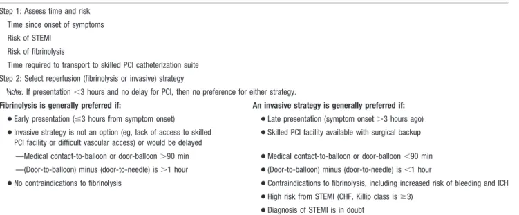
TABLE 2.
ST-Segment Elevation or New or Presumably New LBBB: Evaluation for Reperfusion
Step 1: Assess time and risk
Time since onset of symptoms
Risk of STEMI
Risk of fibrinolysis
Time required to transport to skilled PCI catheterization suite
Step 2: Select reperfusion (fibrinolysis or invasive) strategy
Note:If presentation⬍3 hours and no delay for PCI, then no preference for either strategy.
Fibrinolysis is generally preferred if: An invasive strategy is generally preferred if:
●Early presentation (ⱕ3 hours from symptom onset) ●Late presentation (symptom onset⬎3 hours ago)
●Invasive strategy is not an option (eg, lack of access to skilled PCI facility or difficult vascular access) or would be delayed
●Skilled PCI facility available with surgical backup
—Medical contact-to-balloon or door-balloon⬎90 min ●Medical contact-to-balloon or door-balloon⬍90 min —(Door-to-balloon) minus (door-to-needle) is⬎1 hour ●(Door-to-balloon) minus (door-to-needle) is⬍1 hour
●No contraindications to fibrinolysis ●Contraindications to fibrinolysis, including increased risk of bleeding and ICH
●High risk from STEMI (CHF, Killip class isⱖ3)
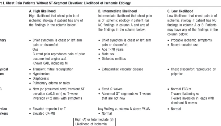
TABLE 3.
Likelihood of Ischemic Etiology and Short-Term Risk
Part I. Chest Pain Patients Without ST-Segment Elevation: Likelihood of Ischemic Etiology
A. High likelihood
High likelihood that chest pain is of ischemic etiology if patient hasanyof the findings in the column below:
B. Intermediate likelihood Intermediate likelihood that chest pain is of ischemic etiology if patient has NO findings in column A andanyof the findings in the column below:
C. Low likelihood
Low likelihood that chest pain is of ischemic etiology if patient has NO findings in column A or B. Patients may have any of the findings in the column below:
History • Chief symptom is chest or left arm pain or discomfort
plus
Current pain reproduces pain of prior documented anginaand
Known CAD, including MI
• Chief symptom is chest or left arm pain or discomfort
• Age⬎70 years • Male sex • Diabetes mellitus
• Probable ischemic symptoms • Recent cocaine use
Physical exam
• Transient mitral regurgitation • Hypotension
• Diaphoresis
• Pulmonary edema or rales
• Extracardiac vascular disease • Chest discomfort reproduced by palpation
ECG • New (or presumed new) transient ST deviation (ⱖ0.5 mm)orT-wave inversion (ⱖ2 mm) with symptoms
• Fixed Q waves
• Abnormal ST segmentsorT waves that are not new
• Normal ECGor T-wave flatteningor T-wave inversion in leads with dominant R waves
Cardiac markers
• Elevated troponin I or T • Elevated CK-MB
Any finding in column B above PLUS • Normal
• Normal
High (A) or Intermediate (B) Likelihood of Ischemia
Part II. Risk of Death or Nonfatal MI Over the Short Term in Patients With Chest Pain With High or Intermediate Likelihood of Ischemia (Columns A and B in Part I)
High risk:
Risk is high if patient hasanyof the following findings:
Intermediate risk:
Risk is intermediate if patient hasany of the following findings:
Low risk:
Risk is low if patient has NO high- or intermediate-risk features; may have any of the following:
History
Character of pain
• Accelerating tempo of ischemic symptoms over prior 48 hours
• Prolonged, continuing (⬎20 min) rest pain
• Prior MIor
• Peripheral-artery diseaseor • Cerebrovascular diseaseor • CABG, prior aspirin use
• Prolonged (⬎20 min) rest angina is now resolved (moderate to high likelihood of CAD)
• Rest angina (⬍20 min) or relieved by rest or sublingual nitrates
• New-onset functional angina (Class III or IV) in past 2 weeks without prolonged rest pain (but with moderate or high likelihood of CAD)
Physical exam
• Pulmonary edema secondary to ischemia
• New or worse mitral regurgitation murmur
• Hypotension, bradycardia, tachycardia
• S3gallop or new or worsening rales • Age⬎75 years
• Age⬎70 years
ECG • Transient ST-segment deviation (ⱖ0.5 mm) with rest angina • New or presumably new bundle
branch block • Sustained VT
• T-wave inversionⱖ2 mm • Pathologic Q waves or T waves that
are not new
• Normal or unchanged ECG during an episode of chest discomfort
Cardiac markers
• Elevated cardiac troponin I or T • Elevated CK-MB
Any of the above findings PLUS • Normal
• Normal
managed with an invasive strategy. Coronary angiography
then allows the clinician to determine whether patients are
appropriate candidates for revascularization with PCI or
coronary artery bypass grafting (CABG).
The
2005 AHA Guidelines for CPR and ECC
define
high-risk patients with indicators that overlap to a
consider-able degree with the more rigorously validated TIMI risk
score
122:
●
New ST-segment depression and positive troponins
●
Persistent or recurrent symptoms
●
Hemodynamic instability or VT
●
Depressed LV function (ejection fraction
⬍
40%)
●
ECG or functional study that suggests multivessel CAD
Normal or Nondiagnostic ECG Changes (Boxes 13
to 17)
The majority of patients with normal or nondiagnostic ECGs
do not have ACS. Patients in this category with ACS are most
often at low or intermediate risk. The physician’s goal
involves risk stratification (see above) to provide appropriate
diagnostic or treatment strategies for an individual patient.
These strategies then target patients at increased risk for
benefit while avoiding risk (eg, anticoagulation therapy and
invasive cardiac catheterization) in patients with low or
minimal risk.
Initial General Therapy for ACS
Several initial measures are appropriate for all patients with
suspected ACS in both the out-of-hospital and ED setting.
These include immediate oxygen therapy, continuous cardiac
monitoring, establishment of intravenous (IV) access, and
several medications discussed below.
Oxygen
Administer oxygen to all patients with overt pulmonary
congestion or arterial oxygen saturation
⬍
90% (Class I). It is
also reasonable to administer supplementary oxygen to all
patients with ACS for the first 6 hours of therapy (Class IIa).
Supplementary oxygen limited ischemic myocardial injury in
animals,
31and oxygen therapy in patients with STEMI
reduced the amount of ST-segment elevation.
35Although a
human trial of supplementary oxygen versus room air failed
to show a long-term benefit of supplementary oxygen therapy
for patients with MI,
30short-term oxygen administration is
beneficial for the patient with unrecognized hypoxemia or
unstable pulmonary function. In patients with severe chronic
obstructive pulmonary disease, as with any other patient,
monitor for hypoventilation.
Aspirin
Early administration of aspirin (acetylsalicylic acid [ASA]),
including administration in the out-of-hospital setting,
47has
been associated with decreased mortality rates in several
clinical trials.
47,129 –131Multiple studies support the safety of
aspirin administration. Therefore, unless the patient has a
known aspirin allergy, nonenteric aspirin should be given as
soon as possible to all patients with suspected ACS.
Aspirin produces a rapid clinical antiplatelet effect with
near-total inhibition of thromboxane A2
production. It reduces
coronary reocclusion and recurrent ischemic events after
TABLE 4. TIMI Risk Score for Patients With Unstable Angina and Non–ST-Segment Elevation MI: Predictor Variables
Predictor Variable
Point Value
of Variable Definition
Ageⱖ65 years 1
ⱖ3 risk factors for CAD 1 Risk factors
• Family history of CAD • Hypertension • Hypercholesterolemia • Diabetes
• Current smoker
Aspirin use in last 7 days 1
Recent, severe symptoms of angina 1 ⱖ2 anginal events in last 24 hours
Elevated cardiac markers 1 CK-MB or cardiac-specific troponin level
ST deviationⱖ0.5 mm 1 ST depressionⱖ0.5 mm is significant; transient ST elevation⬎0.5 mm for⬍20 minutes is treated as ST-segment depression and is high risk; ST elevationⱖ1 mm for more than 20 minutes places these patients in the STEMI treatment category
Prior coronary artery stenosisⱖ50% 1 Risk predictor remains valid even if this information is unknown
Calculated TIMI Risk Score
Risk of>1 Primary End
Point* in<14 Days Risk Status
0 or 1 5% Low
2 8%
3 13% Intermediate
4 20%
5 26% High
6 or 7 41%
fibrinolytic therapy. Aspirin alone reduced death from AMI
in the Second International Study of Infarct Survival (ISIS-2),
and its effect was additive to that of streptokinase.
129In a
review of 145 trials, aspirin was found to substantially reduce
vascular events in all patients with AMI, and in high-risk
patients it reduced nonfatal AMI and vascular death.
132Aspirin is also effective in patients with UA. The standard
dose (160 to 325 mg) is recommended, although higher doses
may be used. Chewable or soluble aspirin is absorbed more
quickly than swallowed tablets.
133,134The early administration of a single chewed dose of aspirin
(160 to 325 mg) is recommended in either the out-of-hospital
or ED setting for patients with suspected ACS (Class I). Other
formulations of ASA (soluble, IV) may be as effective as
chewed tablets. Aspirin suppositories (300 mg) are safe and
can be considered for patients with severe nausea, vomiting,
or disorders of the upper gastrointestinal tract.
Nitroglycerin (or Glyceryl Trinitrate)
Nitroglycerin is an effective analgesic for ischemic chest
discomfort. It also has beneficial hemodynamic effects,
in-cluding dilation of the coronary arteries (particularly in the
region of plaque disruption), the peripheral arterial bed, and
venous capacitance vessels. The treatment benefits of
nitro-glycerin are limited, however, and no conclusive evidence
has been shown to support
routine
use of IV, oral, or topical
nitrate therapy in patients with AMI.
135With this in mind,
these agents should be carefully considered, especially when
low blood pressure precludes the use of other agents shown to
be effective in reducing morbidity and mortality (eg,

-blockers and angiotensin-converting enzyme [ACE]
inhibitors).
IV nitroglycerin is indicated in the following clinical
situations (Class I):
●
Ongoing ischemic chest discomfort
●
Management of hypertension
●
Management of pulmonary congestion
Patients with ischemic discomfort may receive up to 3
doses of sublingual or aerosol nitroglycerin at 3- to 5-minute
intervals until pain is relieved or low blood pressure limits its
use (Class I). IV nitroglycerin is indicated for ongoing chest
discomfort, control of hypertension, or management of
pul-monary congestion in patients with STEMI associated with
LV failure (Class I). In patients with recurrent ischemia,
nitrates are indicated in the first 24 to 48 hours. IV rather than
long-acting preparations should be used acutely to enable
titration.
Do not use nitrates (Class III) in patients with hypotension
(SBP
⬍
90 mm Hg or
⬎
30 mm Hg below baseline), extreme
bradycardia (
⬍
50 bpm), or tachycardia (
⬎
100 bpm).
Admin-ister nitrates with extreme caution if at all to patients with
suspected inferior wall MI with possible right ventricular
(RV) involvement because these patients require adequate
RV preload. Do not administer nitrates (Class III) to patients
who have received a phosphodiesterase inhibitor for erectile
dysfunction within the last 24 hours (longer for some
preparations).
Morphine Sulfate
Morphine sulfate is the analgesic of choice for continuing
pain unresponsive to nitrates, and it is also effective in
patients with pulmonary vascular congestion complicating
ACS. Morphine is a venodilator that reduces ventricular
preload and oxygen requirements. For this reason it should
not be used in patients who may have hypovolemia. If
hypotension develops, elevate the patient’s legs, administer
volume, and monitor for signs of worsening pulmonary
vascular congestion. Start with a 2 to 4 mg IV dose, and give
additional doses of 2 to 8 mg IV at 5- to 15-minute intervals.
Reperfusion Therapies (Figure 1, Box 8)
Perhaps the most significant advance in the treatment of
cardiovascular disease in the last decade is reperfusion
therapy for AMI. Many clinical trials have established early
fibrinolytic therapy as a standard of care for patients with
AMI who present within 12 hours of the onset of symptoms
with no contraindications.
136 –140Reperfusion reduces
mortal-ity, and the shorter the time to reperfusion, the greater the
benefit: a 47% reduction in mortality was noted when
fibrinolytic therapy was provided within the first hour after
onset of symptoms.
139,140The major determinants of myocardial salvage and
long-term prognosis are
●
Short time to reperfusion
136,140●
Complete and sustained patency of the infarct-related
artery with normal (TIMI grade 3) flow
141,142●