Artículo Especial
Repolarización ventricular en la terapia oncológica
Dra. Margarita Dorantes Sánchez
1y Dr. Manuel Bazán Milián
21 Servicio de Arritmias y Estimulación Cardíaca, Instituto de Cardiología y Cirugía Cardiovascular. La Habana, Cuba. 2 Departamento de Cardiología, Instituto Nacional de Oncología y Radiobiología. La Habana, Cuba.
Full English text of this article is also available
INFORMACIÓN DEL ARTÍCULO
Recibido: 11 de enero de 2019 Aceptado: 21 de febrero de 2019
Conflictos de intereses
Los autores declaran que no existen conflictos de intereses
Abreviaturas IQT: intervalo QT IQTL: intervalo QT largo QTL: QT largo
TdP: torsión de puntas Tp-Tf: TPICO-TFINAL
M Dorantes Sánchez Instituto de Cardiología y Cirugía Cardiovascular.
17 N° 702, Vedado, CP 10400. Plaza de la Revolución. La Habana, Cuba. Correo electrónico:
RESUMEN
Se discuten los procesos de despolarización y repolarización ventriculares, con su falta de uniformidad y su heterogeneidad, tanto en pacientes con corazón sano como en aquellos enfermos, cuestión de rangos. Se analizan las mediciones que expresan las características de la repolarización ventricular: el intervalo QT y otras mediciones incluso más fidedignas como el intervalo TPICO-TFINAL, su
disper-sión y otras. Se precisa la existencia del signo y del síndrome de QT largo, así co-mo los tres procesos básicos de la arritco-mogenia: la heterogeneidad, la alternancia y la dispersión, con las diferencias de los potenciales de acción en las tres zonas del miocardio ventricular. Se precisan los factores de riesgo del QT largo (común con esta terapia), de las arritmias ventriculares (en especial la torsión de puntas, extremadamente rara en estos casos) y se discute la necesidad de valorar datos clínicos, eléctricos, comorbilidades, conflictos agregados y las medidas a tomar en estos pacientes.
Palabras clave: Repolarización ventricular, Terapia oncológica, Antineoplásicos, Arritmias cardíacas, Intervalo QT
Ventricular repolarization in cancer therapy
ABSTRACT
Ventricular depolarization and repolarization processes are discussed, including their differences and heterogeneity both in patients with a healthy/sick heart, a matter of ranges. Measurements expressing the characteristics of ventricular repo-larization are analyzed: the QT interval and other even more reliable measure-ments such as the TPEAK-TEND interval, its dispersion and others. We emphasize on
the existence of the long QT syndrome (and sign) and the three basic processes of arrhythmogenesis: heterogeneity, alternation and dispersion, with differences in action potentials in the three zones of the ventricular myocardium. The risk factors of long QT (common in this therapy) and ventricular arrhythmias (especially tor-sades de pointes, extremely rare in these cases) are highlighted. The need to as-sess clinical and electrical features, comorbidities, aggregate conflicts, and manage-ment of these patients is also discussed.
Keywords: Ventricular repolarization, Cancer therapy, Antineoplastic agents, Car-diac arrhythmias, QT interval
GENERALIDADES DE LAS ARRITMIAS VENTRICULARES
Dorantes Sánchez M. y Bazán Milián M.
CorSalud 2019 Abr-Jun;11(2):146-152 147
prescindible que exista una relación armoniosa tre los oncólogos y los cardiólogos a la hora de en-frentar estos conflictos y tomar las decisiones más adecuadas. En este trabajo se enfocan los problemas relacionados con la terapia oncológica y las altera-ciones de la repolarización ventricular.
El segundo autor trabaja en el funcionamiento del primer grupo de Cardio-Oncología en el Instituto Nacional de Oncología y Radiobiología, con planes futuros de crear unidades en otros hospitales del país. Lo cual redundaría en beneficio de estos pa-cientes, al enfocar la estratificación de riesgo cardio-vascular, su temprana detección, prevención y tra-tamiento. Todo ello en estrecha relación con los oncólogos, quienes guían y trazan las estrategias a seguir.
Un concepto básico previo
En los procesos de despolarización y repolarización ventriculares, no existe uniformidad ni homogenei-dad, sino todo lo contrario, heterogeneidad y falta de uniformidad en la formación y conducción del impulso a nivel de todas las estructuras del sistema eléctrico del corazón, tanto en pacientes con car-diopatía estructural como en sujetos normales. Es cuestión de rangos1.
Intervalo QT y otros
El intervalo QT (IQT) evidencia la duración total de la despolarización y de la repolarización; el TPICO
-TFINAL (Tp-Tf) expresa la dispersión de la
repolariza-ción ventricular, es decir, la no uniformidad de la recuperación. No sólo es importante el IQT sino otras medidas, incluso mejores, para evaluar la re-polarización ventricular1-3.
Intervalo QT largo
El intervalo QT largo (IQTL) adquirido es más fre-cuente que el congénito, con el cual presenta algu-nas semejanzas. Debe recordarse que puede haber signo de QT largo (QTL) y síndrome de QTL, el pri-mero es la mera presentación del signo eléctrico; en el segundo se presentan episodios sincopales o eventos de muerte súbita. Aunque durante la evolu-ción clínica, el signo puede transformarse en sín-drome y la conducta a seguir, por supuesto, será diferente4.
Arritmogénesis y las células M
Existen tres procesos fundamentales en relación con la arritmogenia: la alternancia, la heterogeneidad, y la dispersión espacial y temporal. La fisiopatología
de estas alteraciones tiene su base en los potencia-les de acción y sus diversas características en las tres zonas del miocardio ventricular: el epicardio, el endocardio y el miocardio medio. De tal manera que la duración del potencial de acción epicárdico ex-presa el QT pico en tanto la del miocardio medio representa la repolarización completa, hasta el final de la onda T1,5-8.
El orden de inicio del potencial de acción es: en-docárdico, epicárdico y miocárdico medio; en tanto el final del potencial sigue este orden: epicardio, endocardio y miocardio medio. Las células M son un híbrido entre las de Purkinje y el tejido ventricu-lar, con diferencias iónicas, electrofisiológicas y far-macológicas entre los tres tipos celulares. La hetero-geneidad eléctrica de la repolarización ventricular se presenta a nivel transmural y transeptal, en en-fermos y en sanos. La zona M tiene un potencial de acción más prolongado que el del endocardio y el del epicardio, aún más si la frecuencia cardíaca es baja o si se emplean ciertos fármacos antiarrítmi-cos1,5-7.
La zona M es una subpoblación de células con propiedades electrofisiológicas únicas, que mejoran la eficiencia de bomba pero aumentan la inestabili-dad eléctrica, que resulta compensada por las zonas epicárdica y endocárdica1.
Factores de riesgo
Recuérdense algunos factores de riesgo que propi-cian el aumento del IQT y pueden originar torsión de puntas (TdP) por fármacos9,10:
- Genéticos: Susceptibilidad genética, mutaciones.
- Congénitos: IQTL congénito subclínico.
- Sexo y edad: Sexo femenino (dos veces más ries-go), mayor edad.
- Alteraciones electrolíticas: Hipomagnesemia, hi-pocaliemia, hipocalcemia.
- Fármacos: Empleo de diuréticos (independiente-mente de las alteraciones electrolíticas) y digitáli-cos, alta concentración de algunos medicamentos (excepto la quinidina), empleo de más de un fár-maco antiarrítmico, administración endovenosa rápida de un medicamento.
- Enfermedad cardíaca asociada: hipertrofia ven-tricular, insuficiencia cardíaca con baja fracción de eyección, insuficiencia mitral, prolapso valvu-lar mitral, miocardiopatías, valvulopatías.
- Comorbilidades: Enfermedad renal o hepática.
aurículo-ventricular, bradicardia.
- Marcadores electrocardiográficos: IQTL, posdes-polarizaciones tempranas, reciente reversión de un episodio de fibrilación auricular, Tp-Tf anor-mal, muescas de la onda T, cambios del IQT o TU postextrasistólica, morfología TU, T bifásica, U prominente, gran onda U postextrasistólica, TU aberrante o gigante, inestabilidad latido a latido, aberrancia de la onda T luego de un RR largo.
También existen factores de riesgo para el origen de la TdP, que se muestran en el recuadro 1.
Recuadro 1. Factores de riesgo para el origen de la
torsión de puntas9,10.
- Inicio de la arritmia con TU gigante - TU anormal
- Posdespolarizaciones tempranas
- Ascenso lento del QRS de la extrasístole ventricular - Ciclo corto-largo-corto
- Pausas
- Mayor duración del QRS del primer latido de la tor-sión de puntas
- Menor ángulo del QRS - U prominente
- Alternancia de QT-T (en duración, configuración, polaridad, amplitud);
- Fragmentación del QRS - QT largo
- TPICO-TFINAL anormal
Dispersión y heterogeneidad
La dispersión espacial y temporal puede presentarse entre base y ápex, septum y paredes libres, pared miocárdica y entre ambos ventrículos (circunferen-cial). En corazones normales puede existir disper-sión hasta de 55 ms y refleja la no uniformidad de la repolarización ventricular, dentro de ciertos límites. La heterogeneidad se presenta en cuanto a las ondas R, J y T, si se comparan sus características en un su-jeto reanimado de un evento de muerte súbita con quien no lo haya tenido5-8.
Otras mediciones
Debe recordarse que el QTL no es un marcador perfecto de riesgo de arritmias ventriculares malig-nas (fibrilación y taquicardia ventriculares o muer-te) y que el Tp-Tf (dispersión transmural de la
repo-larización, valor normal 100 ms) es más valioso que el QTc e igualmente su dispersión (valor normal 20 ms). Por otro lado, no todo QTL lleva a TdP1,3,11-13.
Factores que contribuyen al alargamiento del IQT
En los pacientes que reciben terapia contra el cán-cer se presentan otros conflictos que contribuyen al alargamiento del IQT14-16:
1. Condiciones coexistentes: mayor edad, sexo fe-menino, hipotiroidismo, síndrome de QTL con-génito, fiebre, enfermedades cardíacas como: dis-función del ventrículo izquierdo, isquemia mio-cárdica, bradicardia, hipertensión arterial, insufi-ciencia cardíaca, tromboembolismo pulmonar, ic-tus.
2. Medicamentos concomitantes: antidepresivos, antifúngicos, antihistamínicos, antieméticos, anti-bióticos, antipsicóticos, antianginosos, laxantes, fármacos antiarrítmicos.
3. En relación con la terapia anticancerígena: pobre ingesta oral, deshidratación, alteraciones electro-líticas, náuseas, vómitos, diarreas, diabetes mal controlada, disfunción hepática, insuficiencia re-nal.
Dónde medir el QT y algunas cifras
El QT debe medirse en la derivación con el inicio del QRS más temprano y el final de la onda T más tardío, y siempre donde se vea con más precisión. Algunos autores señalan como QTc normal aquel mayor de 360 ms y menor de 460 ms en la mujer adulta; y mayor de 350 ms y menor de 450 ms en el hombre adulto. Existen condiciones especiales en las cuales se dificultan estas mediciones, casos con: bloqueo de rama, marcapaso implantado, PR pro-longado y ritmo de la unión con presencia de onda U2,4,11,14,15.
Cómo valorar la repolarización ventricular
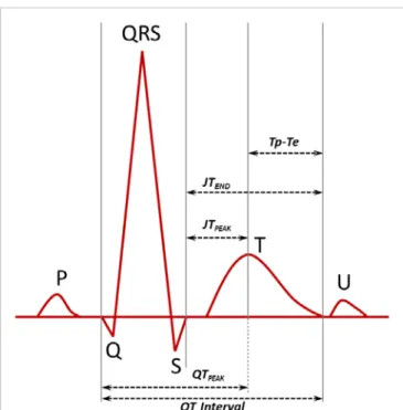
Existenvariasmedicionesposibles(Figura1),¿cuál es la mejor?: QT, QTc, QTPICO, JTPICO, Tp-Tf, JTFINAL,
dispersión del QT, dispersión del Tp-Tf. Todas son útiles pero algunas más confiables, como el Tp-Tf y su dispersión2,3,11-13.
Mecanismos que inducen arritmias en la terapia oncológica
Dorantes Sánchez M. y Bazán Milián M.
CorSalud 2019 Abr-Jun;11(2):146-152 149
dico y endocárdico (por isquemia o inflamación). Estos mecanismos pueden agruparse, como se muestra en el recuadro 2.
Estrategia para valorar trastornos de la repolari-zación ventricular
Desde el punto de vista cardiológico, en un paciente que recibe terapia anticancerígena, deben valorarse a) en la clínica: el síncope, las palpitaciones rápidas, el desmayo y el vértigo; b) en los electrocardiogra-mas: el aumento del IQT (se dice que antes de la terapia, el 6% presenta prolongación del QTc), y c) otros factores como el estado de las vías de elimina-ción (riñón e hígado), empleo de fármacos sinérgi-cos con la terapia que modifica el QTc e inhibidores del metabolismo de medicamentos anticancerígenos que aumentan el QT14-16.
Otros mecanismos de QT prolongado
Como queda dicho, los mecanismos de aumento del QT en esta terapia no están bien conocidos, se han considerado la interacción con la función normal de las proteínas de los canales de potasio en los car-diomiocitos (hERG) y otros. Se produce aumento del QT y de su dispersión, con onda T bifásica, po-tenciales tardíos en la meseta de las células de Pur-kinje, aumento preferencial de la duración del po-tencial de acción mediomiocárdico, aumento de la
dispersión transmural de la repolarización (incluso más importante que el aumento del IQT). Estos ca-sos semejan el síndrome de QTL tipo 2 (Figura 2), con onda T bifásica y posible sustrato de TdP14-16.Al valorar el QTL en la terapia anticancerígena, es ne-cesario tomar en cuenta las comorbilidades, los fac-tores de riesgo y el aumento inicial del QT. Se ha observado que, en general, el riesgo para presentar estas complicaciones se presenta de manera más tardía14-16.
Riesgo del aumento del QT y de las arritmias
En 173 publicaciones relevantes, se considera que el aumento del QTc es un riesgo más común con el tratamiento convencional (antraciclinas), en el no convencional varía del 0-22%. Se considera grave si es mayor de 500 ms (0-5%). Sin embargo, las arrit-mias (TdP, fibrilación y taquicardia ventriculares, y la muerte súbita cardíaca) son extremadamente ra-ras. Si el QTc es mayor de 500 ms o mayor de 60 ms comparado con el basal, debe evaluarse la supre-sión del medicamento14-16.
Medidas a tomar
En casos graves pueden adoptarse las siguientes me-didas: sulfato de magnesio endovenoso, isoprotere-
Recuadro 2. Mecanismos que inducen arritmias en la
terapia oncológica14-16.
1. Afectación directa del corazón
- Cáncer primario - Metástasis al corazón - Amiloidosis cardíaca
2. Trastornos electrolíticos
- Vómitos - Diarrea
- Disbalance inducido por drogas
3. Factores independientes de la terapia anticancerí-gena
- Previo sustrato básico para las arritmias - Arritmias luego de cirugía de cáncer
- Radiación que puede inducir pericarditis, ateros-clerosis
- Medicaciones adyuvantes, antieméticos y otros
4. Efecto sobre los miocitos cardíacos
- Bloqueo hERG (human Ether-a-go-go-Related Gene 2)
- Homeostasis anormal de calcio - Injuria mitocondrial
- Apoptosis cardíaca
Figura 1. Diversas mediciones electrocardiográficas para
nol, choque eléctrico externo, lidocaína, marcapaso auricular o ventricular temporal si existe bradicardia (marcapaso doble cámara), y cardioversor-desfibri-lador automático implantable si la esperanza de vida es mayor de 12 meses y se produce algún episodio de muerte súbita cardíaca reanimada o arritmia gra-ve sin causa corregible (electrolitos). Debe emplear-se monitoreo y decidir si es necesario suspender el
tratamiento o no14-16.
Toxicidad cardiovascular
Existe una clasificación del potencial alargamiento del QT por fármacos anticancerígenos. Según Cop-pola15, Vejpongsa y Yeh (J Am Coll Cardiol. 2014;64: 938-45) refieren que en 1807 sobrevivientes de cán-cer con seguimiento durante 7 años, encontraron
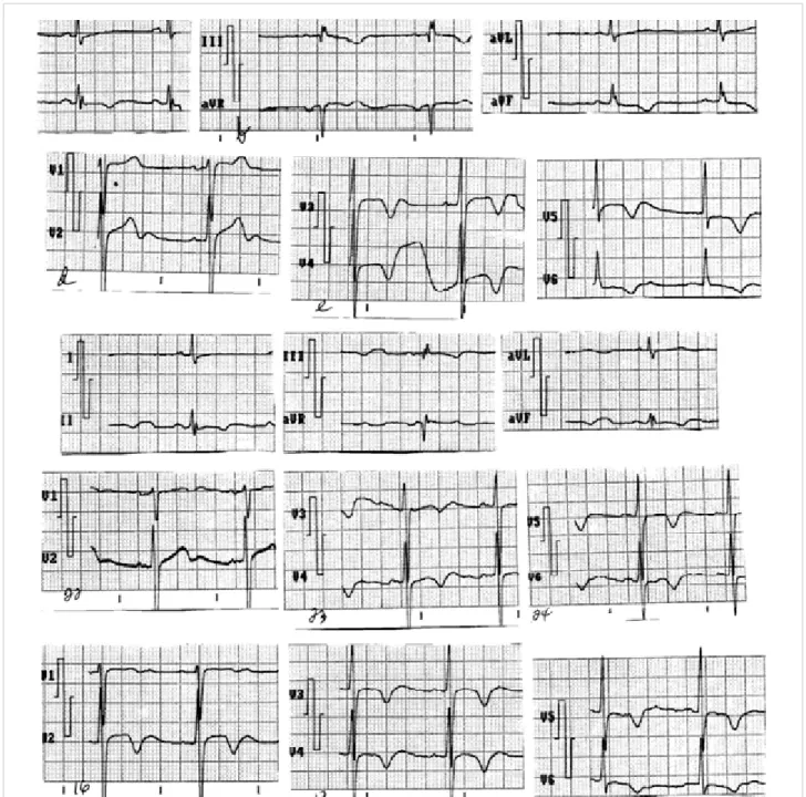
Figura 2. Electrocardiogramas de un mismo paciente en distintos momentos de su evolución: intervalo QT largo por fármacos, alteraciones de la onda T (en magnitud, duración, bimodalismo, onda T1T2). Estas características se
Dorantes Sánchez M. y Bazán Milián M.
CorSalud 2019 Abr-Jun;11(2):146-152 151
que se produjo la muerte en el 51% de los pacientes por el cáncer en sí y en el 33%, se debió a enferme-dad cardiovascular. En estos pacientes habrá de considerarse no sólo la sobrevida sino también la calidad de vida. Los efectos adversos en cuanto a la miocardiopatía se conocen desde 1966 pero el tra-tamiento de cáncer induciendo arritmias se estudió a partir del 2009. La toxicidad cardiovascular es de los conflictos más peligrosos en estos pacientes. Como idea final, el QTL es hallazgo común en ellos, frente a la TdP que es rara pero peligrosa14-16.
Grupos de riesgo
Se han considerado varios grupos según las medi-ciones del IQT: 1) QTc de 450-480 ms, 2) de 481-500 ms, 3) mayor de 500 ms en 2 electrocardiogramas, y 4) mayor de 501 ms o más de 60 ms respecto al ba-sal, TdP, taquicardia ventricular polimórfica o sínto-mas relacionados con estas arritmias. Algunos valo-res aceptados del IQT se refieren en diversos artícu-los2,14-16.
El futuro de los efectos adversos cardiovascula-res por la terapia contra el cáncer, específicamente en relación con las arritmias, apunta hacia la investi-gación de cómo alteran estos fármacos las propie-dades electrofisiológicas del corazón14-16.
Factores coadyuvantes
En estos pacientes son varios los conflictos que pue-den presentarse: el cáncer en sí puede predisponer a las arritmias (en general las series toman en cuen-ta un número inadecuado de pacientes), una enfer-medad preexistente proporcionaría mayor vulnera-bilidad a la terapia anticancerígena induciendo arrit-mias; además, la sinergia, un tratamiento previo con-tra el cáncer, el empleo de polifarmacia, la arritmia preexistente con pobre monitoreo previo y los fac-tores comunes de riesgo14-16.
Estrategias para disminuir riesgos del IQT prolon-gado y de arritmias graves
Como estrategias para minimizar los riesgos del IQT prolongado y de la TdP en pacientes con terapia an-ticancerígena se aconseja14-16:
1. Evitar el empleo de fármacos que prolonguen el QTc en pacientes con intervalo mayor de 450 ms antes de iniciar el tratamiento.
2. Descontinuar estos medicamentos si el QTc se prolonga a más de 500 ms o es mayor de 550 ms, si hay un ensanchamiento del QRS de base (ma-yor de 120 ms, secundario a marcapasos o a blo-queo de rama derecha).
3. Reducir la dosis o descontinuar el tratamiento si el QTc se prolonga más de 60 ms comparado con el valor previo al inicio del tratamiento.
4. Mantener la concentración de electrolitos séricos (K, Mg, Ca), dentro del rango normal.
5. Evitar interacciones medicamentosas.
6. Ajustar las dosis de estos fármacos que se elimi-nen por el riñón en pacientes con disfunción aguda o enfermedad renal establecida.
7. Evitar la administración endovenosa rápida de estos medicamentos.
8. Evitar la administración de un fármaco con peli-gro potencial de prolongación del QT.
9. Evitar el empleo de estos preparados en pacien-tes con historia de TdP por medicamentos o pre-viamente resucitados de episodios de muerte sú-bita cardíaca.
10.Evitar su empleo en pacientes diagnosticados, como síndrome de QTL congénito.
11.Realizar monitoreo electrocardiográfico, concen-tración del fármaco y cambios de dosis de esta terapia.
BIBLIOGRAFÍA
1. Antzelevitch C. Cardiac repolarization. The long and short of it. Europace. 2005;7(Supl 2):3-9. 2. Viskin S. The QT interval: too long, too short or
just right. Heart Rhythm. 2009;6(5):711-5.
3. Parvez B, Darbar D. Novel ECG markers for ven-tricular repolarization: is the QT interval obso-lete? Heart Rhythm. 2011;8(7):1044-5.
4. Morita H, Wu J, Zipes DP. The QT syndromes: long and short. Lancet. 2008;372(9640):750-63. 5. Verrier RL, Huikuri H. Tracking interlead
hetero-geneity of R-and T-wave morphology to disclose latent risk for sudden cardiac death. Heart Rhythm. 2017;14(10):1466-75.
6. Kentta TV, Nearing BD, Porthan K, Tikkanen JT, Vitasalo M, Nieminen MS, et al. Prediction of sud-den cardiac death with automated high-through-put analysis of heterogeneity in standard resting 12-lead electrocardiograms. Heart Rhythm. 2016; 13(3):713-20.
7. Shimizu W. Where does heterogeneity exist in ventricular tachyarrhythmias? Heart Rhythm. 2015;12(6):1304-5.
8. Coronel R, Wilms-Schopman FJ, Opthof T, Janse MJ. Dispersion of repolarization and arrhythmo-genesis. Heart Rhythm. 2009;6(4):537-43.
T-U waves precede torsades de pointes in long QT syndrome. A systematic electrocardiographic analysis in patient with acquired and congenital QT prolongation. J Am Coll Cardiol. 2009;54(2): 143-9.
10.El-Sherif N, Turitto G, Boutjdir M. Acquired long QT syndrome and torsade de pointes. Pacing Clin Electrophysiol. 2018;41(4):414-21.
11.Laksman ZW, Krahn AD. Fast and spurious: cor-recting the QT interval. Heart Rhythm. 2016;13(2): 536-7.
12.Khakpour H, Vaseghi M. Electrocardiographic Tpeak to Tend interval: the short and long of it. Heart Rhythm. 2016;13(4):925-6.
13.Tse G, Gong M, Wong WT, Georgopoulos S, Letsas KP, Vassiliou VS, et al. The Tpeak-Tend interval as an electrocardiographic risk marker of arrhyth-mic and mortality outcomes: a systematic review and meta-analysis. Heart Rhythm. 2017;14(8):1131-7.
14.Porta-Sánchez A, Gilbert C, Spears D, Amir E, Chan J, Nanthakumar K, et al. Incidence, diagno-sis, and management of QT prolongation induced by cancer therapies: a systematic review. J Am Heart Assoc [Internet]. 2017[citado 28 Dic 2018]; 6(12):e007724. Disponible en:
https://www.ahajournals.org/doi/pdf/10.1161/JA HA.117.007724
15.Coppola C, Rienzo A, Piscopo G, Barbieri A, Arra C, Maurea N. Management of QT prolongation in-duced by anti-cancer drugs: target therapy and oldagents.Differentalgorithmsfordifferentdrugs. Cancer Treat Rev. 2018;63:135-43.
16.Buza V, Rajagopalau B, Curtis AB. Cancer treat-ment-induced arrhythmias focus on chemothera-py and targeted therapies. Circ Arrhythm Electro-physiol[Internet]. 2017 [citado 5 Ene 2019];10(8): e005443. Disponible en:
CorSalud 2019 Apr-Jun;11(2):146-152
RNPS 2235-145 © 2009-2019 Cardiocentro Ernesto Che Guevara, Villa Clara, Cuba.
Article licensed under a Creative Commons Attribution – CC BY-NC-ND 4.0
146
Cuban Society of Cardiology
__________________________Special Article
Ventricular repolarization in cancer therapy
Margarita Dorantes Sánchez
1, MD; and Manuel Bazán Milián
2, MD
1 Department of Arrhythmias and Cardiac Pacing, Instituto de Cardiología y Cirugía Cardiovascular. Havana, Cuba. 2 Department of Cardiology, Instituto Nacional de Oncología y Radiobiología. Havana, Cuba.
Este artículo también está disponible en español
ARTICLE INFORMATION
Received: January 11, 2019 Accepted: February 21, 2019
Competing interests
The authors declare no competing interests
Acronyms LQT: Long QT LQTI: Long QT interval QTI: QT interval
TdP: Torsades de pointes Tp-Te: TPEAK-TEND
M Dorantes Sánchez Instituto de Cardiología y Cirugía Cardiovascular.
17 N° 702, Vedado, CP 10400. Plaza de la Revolución. La Habana, Cuba. E-mail address:
ABSTRACT
Ventricular depolarization and repolarization processes are discussed, including their differences and heterogeneity both in patients with a healthy/sick heart, a matter of ranges. Measurements expressing the characteristics of ventricular repo-larization are analyzed: the QT interval and other even more reliable measure-ments such as the TPEAK-TEND interval, its dispersion and others. We emphasize on
the existence of the long QT syndrome (and sign) and the three basic processes of arrhythmogenesis: heterogeneity, alternation and dispersion, with differences in action potentials in the three zones of the ventricular myocardium. The risk factors of long QT (common in this therapy) and ventricular arrhythmias (especially tor-sades de pointes, extremely rare in these cases) are highlighted. The need to as-sess clinical and electrical features, comorbidities, aggregate conflicts, and manage-ment of these patients is also discussed.
Keywords:Ventricular repolarization, Cancer therapy, Antineoplastic agents, Car-diac arrhythmias, QT interval
Repolarización ventricular en la terapia oncológica
RESUMEN
Se discuten los procesos de despolarización y repolarización ventriculares, con su falta de uniformidad y su heterogeneidad, tanto en pacientes con corazón sano como en aquellos enfermos, cuestión de rangos. Se analizan las mediciones que expresan las características de la repolarización ventricular: el intervalo QT y otras mediciones incluso más fidedignas como el intervalo TPICO-TFINAL, su
disper-sión y otras. Se precisa la existencia del signo y del síndrome de QT largo, así co-mo los tres procesos básicos de la arritco-mogenia: la heterogeneidad, la alternancia y la dispersión, con las diferencias de los potenciales de acción en las tres zonas del miocardio ventricular. Se precisan los factores de riesgo del QT largo (común con esta terapia), de las arritmias ventriculares (en especial la torsión de puntas, extremadamente rara en estos casos) y se discute la necesidad de valorar datos clínicos, eléctricos, comorbilidades, conflictos agregados y las medidas a tomar en estos pacientes.
Palabras clave: Repolarización ventricular, Terapia oncológica, Antineoplásicos, Arritmias cardíacas, Intervalo QT
GENERALITIES OF THE VENTRICULAR ARRHYTHMIAS
ous relationship between oncologists and cardiolo-gists is essential when dealing with these conflicts and making the most appropriate decisions. This work is focused in problems related to cancer thera-py and alterations in the ventricular repolarization.
The second author works in the development of the first Cardio-Oncology group at the Instituto
Na-cional de Oncología y Radiobiología, with future
plans to create units in other hospitals throughout the country. This would benefit the patients, by fo-cusing on the stratification of cardiovascular risk, its early detection, prevention and treatment. All of this closely related with oncologists, who would guide and outline the strategies to follow.
A previous basic concept
In the processes of ventricular depolarization and repolarization, there is no uniformity or homogenei-ty, but rather the opposite, heterogeneity and lack of uniformity in the formation and conduction of the impulse at the level of all the structures of the heart’s electrical system, in both, patients with struc-tural heart disease and regular individuals. It is a matter of ranks1.
QT interval and others
The QT interval (QTI) demonstrates the total dura-tion of the depolarizadura-tion and repolarizadura-tion; the TPEAK-TEND (Tp-Te) expresses the dispersion of the
ventricular repolarization, that is, the non-uniformity of recovery. Not only the QTI is important, but also other measures, even better in order to evaluate ventricular repolarization1-3.
Long QT interval
The acquired long QT interval (LQTI) is more fre-quent than the congenital one, with which it shares some similarities. It should be remembered that there may be a sign of long QT (LQT) and LQT syn-drome, the first being the mere presentation of the electrical sign; in the second, syncopal episodes or sudden death events occur. Although during the clinical evolution, the sign can become a syndrome and the behavior to follow, thus, will be different4.
Arrhythmogenesis and M cells
There are three fundamental processes in relation to arrhythmogeny: alternation, heterogeneity, and spa-tial and temporal dispersion. The pathophysiology of these alterations is based on the action potentials and their several characteristics in the three zones of the ventricular myocardium: the epicardium, the
endocardium and the midmyocardium. Therefore, the duration of the epicardial action potential ex-presses the peak QT, as the midmyocardium repre-sents the complete repolarization, until the end of the T wave1,5-8.
The starting order of the action potential is: en-docardial, epicardial and midmyocardial; while the end of the potential follow this order: epicardium, endocardium and midmyocardium. The M cells are a hybrid between the Purkinje cells and the ventric-ular tissue with ionic, electrophysiological and pharmacological differences among the three cell types. The electrical heterogeneity of the ventricular repolarization is present, at the transmural and transseptal level, in both sick and healthy people. The M zone has a longer area than the action poten-tial of the endocardium and epicardium, even more if the heart rate is low or if certain antiarrhythmic drugs are used1,5-7.
The M zone is a subpopulation of cells with unique electrophysiological properties, which im-prove the pump efficiency but increase electrical instability, which is compensated by the epicardial and endocardial zones1.
Risk factors
It is important to recall some risk factors that con-tribute to the increase of the QTI and which can cause torsades de pointes (TdP) due to drugs 9,10:
- Genetic: Genetic susceptibility, mutations.
- Congenital: Subclinical congenital LQTI.
- Sex and age: Females (twice risk), greater age.
- Electrolyte disorders: Hypomagnesemia, hypoka-lemia, hypocalcemia.
- Drugs: Employment of diuretics (regardless of electrolytic alterations) and digitalis, high concen-tration of some drugs (except quinidine), use of more than one antiarrhythmic drug, intravenous bolus administration of a drug.
- Associated heart disease: ventricular hypertro-phy, heart failure with reduced ejection fraction, mitral failure, mitral valve prolapse, cardiomyo-pathies, valvulopathies.
- Comorbidities: Renal or hepatic disease.
- Cardiac arrhythmias: Ventricular arrhythmias (extrasystoles with short coupling interval, non-sustained ventricular tachycardia), atrioventricu-lar block, bradycardia.
mor-Ventricular repolarization in cancer therapy
CorSalud 2019 Apr-Jun;11(2):146-152
148
phology, biphasic T, prominent U, postextrasys-tolic large U wave, aberrant or giant TU, beat-to-beat instability, aberrance of the T wave after a long RR interval.
There are also risk factors for the origin of the TdP, which are shown in Box 1.
Box 1. Risk factors for the origin of the torsade de pointes9,10.
- Start of arrhythmia with giant TU
- Abnormal TU
- Early afterdepolarizations
- Slow rise of the QRS of the ventricular extrasystole
- Short-long-short cycle
- Pauses
- Increased duration of the QRS of the torsades de
pointes’ first beat
- Lower angle of the QRS
- Prominent U
- Alternation of QT-T (in duration, configuration,
po-larity, amplitude
- QRS fragmentation
- Long QT
- Abnormal TPEAK-TEND
Dispersion and heterogeneity
The spatial and temporal dispersion can be present-ed between base and apex, septum and free walls, myocardial wall and between both ventricles (cir-cumferential). In normal hearts, there can be disper-sion until 55 ms and it reflects the non-uniformity of the ventricular repolarization, within certain limits. Heterogeneity occurs in terms of R, J and T waves, if their characteristics are compared in an individual resuscitated from a sudden death event with another who has not had it5-8.
Other measurements
It should be remembered that the LQT is not a per-fect marker for risk of malignant ventricular ar-rhythmias (fibrillation and ventricular tachycardia or death) and the TPEAK-TEND (transmural dispersion of
the repolarization, normal value 100 ms) is more valuable than the cQT and likewise, its dispersion (normal value 20 ms). Furthermore, not all LQT leads to TdP1,3,11-13.
Factors that contribute to the lengthening of the QTI
In patients who receive cancer therapy, there are other conflicts that contribute to the lengthening of the QTI14-16:
1. Coexistent conditions: greater age, female sex, hy-pothyroidism, congenital LQT syndrome, fever, cardiac diseases as: left ventricular dysfunction, myocardial ischemia, bradycardia, high blood pressure, heart failure, pulmonary thromboembo-lism, stroke.
2. Concomitant drugs: antidepressants, antifungals, antihistamines, antiemetics, antibiotics, antipsy-chotics, antianginals, laxatives, antiarrhythmic drugs.
3. Related to the anticancer therapy: poor oral in-take, dehydration, electrolyte disorders, nauseas, vomiting, diarrhea, poorly controlled diabetes, liver dysfunction, renal failure.
Where to measure the QT and some figures
The QT interval must be measured in the lead with the earliest beginning of the QRS and the latest end of the T wave, and always where it is seen more accurately. Some authors indicate as normal cQT that greater than 360 ms and less than 460 ms in the adult woman; and greater than 350 ms and less than 450 ms in the adult man. There are special condi-tions in which these measurements are difficult: branch block, implanted pacemaker, prolonged PR and junctional rhythm with presence of U wave2,4,11,
14,15.
How to consider ventricular repolarization
There are several possible measurements (Figure 1), which is the best?: QT, cQT, QTPEAK, JTPEAK,
Tp-Te, JTEND, QT dispersion, Tp-Te dispersion. They are
all useful but some more reliable, as the Tp-Te inter-val and its dispersion2,3,11-13.
Mechanisms that induce arrhythmias in cancer therapy
Strategy to assess ventricular repolarization disorders
From the cardiological point of view, in a patient re-ceiving anticancer therapy should be considered: a) in clinic: syncope, rapid heartbeat, fainting and ver-tigo; b) in the electrocardiograms: the increase of the QTI (there is referred that before the therapy, 6% present cQT prolongation), and c) other factors such as the condition of the excretion routes (kidney and liver), use of synergistic drugs with therapy that modifies the cQT and metabolism inhibitors of anti-cancer drugs that increase the QT14-16.
Other mechanisms of prolonged QT
As noted above, the mechanisms for increasing the QT in this therapy are not well known, there has been considered the interaction with the average function of the proteins of the potassium channels in cardiomyopathies (hERG) and others. There is an increase in the QT and its dispersion, with biphasic T wave, late potentials in the Purkinje cells’ plateau, preferential increase in the action potential duration of midmyocardium, increase in the transmural dis-persion of repolarization (even more important than the increase in the QTI). These cases mimic the LQT syndrome type 2 (Figure 2), with biphasic T wave and possible TdP substrate14-16. When considering the LQT in the anticancer therapy, it is necessary to
take into account the comorbidities, the risk factors and the initial increase of the QT. It has been ob-served that, in general, the risk of having these com-plications is presented later14-16.
Risk of increased QT and arrhythmias
In 173 relevant publications, it is considered that the increase in the cQT is a more common risk in the conventional treatment (anthracyclines), because in the unconventional one it varies from 0-22%. It is considered serious if it is greater than 500 ms (0-5%). Nonetheless, arrhythmias (TdP, fibrillation and ven-tricular tachycardia, and sudden cardiac death) are extremely rare. If the cQT is greater than 500 ms or greater than 60 ms compared to the basal, the medi-cation suppression should be evaluated14-16.
Decision making
In severe cases, the next measures can be taken: intravenous magnesium sulfate, isoprenaline, exter-nal electric shock, lidocaine, temporary auricular or ventricular pacemaker if there is bradycardia (dual-chamber pacemaker), and implantable cardioverter-defibrillator, if the life expectancy is greater than 12 months and there is an event of resuscitated sudden death or severe arrhythmia without correctable cause (electrolytes). Monitoring is necessary as well
Box 2. Mechanisms that induce arrhythmias in cancer
therapy14-16.
1. Direct affectation of the heart
- Primary cancer
- Metastasis to the heart
- Cardiac amyloidosis
2. Electrolyte disorders
- Vomiting
- Diarrhea
- Drug-induced imbalance
3. Independent factors from the anticancer therapy
- Previous basic substrate for arrhythmias
- Arrhythmias after cancer surgery
- Radiation that can induce pericarditis,
athero-sclerosis
- Adjuvant medications, antiemetic and others
4. Effect on cardiac myocytes
- hERG block ( human Ether-a-go-go-Related Gene
2)
- Abnormal calcium homeostasis
- Mitochondrial damage
- Cardiac apoptosis
Figure 1. Several electrocardiographic measurements to
Ventricular repolarization in cancer therapy
CorSalud 2019 Apr-Jun;11(2):146-152
150
as deciding whether to discontinue the treatment14-16.
Cardiovascular toxicity
There is a classification of the potential QT prolonga-tion by anticancer drugs. According to Coppola15, Vejpongsa and Yeh (J Am Coll Cardiol 2014;64:938-45) report that in 1807, among the survivors of can-cer with follow-up for seven years, 51% of patients
died by cancer itself and 33% due to cardiovascular disease. In these patients, not only their survival must be considered, but also their quality of life. The adverse effects in terms of cardiomyopathy are known since 1966, but the treatment of cancer induc-ing arrhythmias has been studied since 2009. The cardiovascular toxicity is one of the most dangerous conflicts in these patients. As a final thought, the
Figure 2. Electrocardiograms of the same patient at different times in his evolution: long QT interval due to drugs, alterations of the T wave (in magnitude, duration, bimodalism, T1T2 wave). These characteristics are observed in some
LQT is a common finding in them, against the TdP, that is rare but dangerous14-16.
Risk groups
Several groups have been considered according to the QTI measurements: 1) cQT of 450-480 ms, 2) of 481-500 ms, 3) greater than 500 ms in two electrocar-diograms, and 4) greater than 501 ms or more than 60 ms with respect to the basal, TdP, ventricular polymorphic tachycardia or symptoms related to these arrhythmias. Some accepted values of the QTI are referred in several articles2,14-16.
The future of cardiovascular adverse effects in the cancer therapy, specifically in relation to ar-rhythmias, is aimed at researching on how these drugs alter the electrophysiological features of the heart14-16.
Adjuvant factors
In these patients, several conflicts may arise: cancer itself can predispose arrhythmias (generally the series take into account an inadequate number of patients), a preexisting condition would provide greater vulnerability to anticancer therapy inducing arrhythmias; in addition, the synergy, a previous treatment against cancer, the use of polypharmacy, the preexisting arrhythmia with poor previous moni-toring and common risk factors14-16.
Strategies to reduce risks of prolonged QTI and severe arrhythmias
Some recommended strategies to minimize the risks of prolonged QTI and TdP in patients with antican-cer therapy are14-16:
1. To avoid the use of drugs that prolong the cQT in patients with an interval greater than 450 ms be-fore starting treatment.
2. To stop the use of these drugs if the cQT is pro-longed more than 500 ms or greater than 550 ms, if there is a widening of the QRS base (greater than 120 ms, secondary to pacemaker or right bundle branch block).
3. To reduce dosage or discontinuing the treatment if the cQT lasts more than 60 ms compared to the previous value at the start of treatment.
4. To maintain the concentration of serum electro-lytes (K, Mg, Ca), within the standard range. 5. To avoid drug interactions.
6. To adjust the doses of these drugs that are ex-creted by the kidneys in patients with acute dys-function or established renal disease.
7. To avoid intravenous bolus administration of
these drugs.
8. To avoid the administration of a drug with poten-tial danger of QT prolongation.
9. To avoid the employment of these preparations in patients with a history of TdP due to drugs or previously resuscitated from events of sudden death.
10. To avoid its use in diagnosed patients with con-genital LQT syndrome.
11. To carry out an electrocardiographic monitoring, concentration of the drug and dose changes in the therapy.
REFERENCES
1. Antzelevitch C. Cardiac repolarization. The long and short of it. Europace. 2005;7(Supl 2):3-9. 2. Viskin S. The QT interval: too long, too short or
just right. Heart Rhythm. 2009;6(5):711-5.
3. Parvez B, Darbar D. Novel ECG markers for ven-tricular repolarization: is the QT interval obso-lete? Heart Rhythm. 2011;8(7):1044-5.
4. Morita H, Wu J, Zipes DP. The QT syndromes: long and short. Lancet. 2008;372(9640):750-63. 5. Verrier RL, Huikuri H. Tracking interlead
hetero-geneity of R-and T-wave morphology to disclose latent risk for sudden cardiac death. Heart Rhythm. 2017;14(10):1466-75.
6. Kentta TV, Nearing BD, Porthan K, Tikkanen JT, Vitasalo M, Nieminen MS, et al. Prediction of sud-den cardiac death with automated high-through-put analysis of heterogeneity in standard resting 12-lead electrocardiograms. Heart Rhythm. 2016; 13(3):713-20.
7. Shimizu W. Where does heterogeneity exist in ventricular tachyarrhythmias? Heart Rhythm. 2015;12(6):1304-5.
8. Coronel R, Wilms-Schopman FJ, Opthof T, Janse MJ. Dispersion of repolarization and arrhythmo-genesis. Heart Rhythm. 2009;6(4):537-43.
9. Kirchhof P, Franz MR, Bardai A, Wilde AM. Giant T-U waves precede torsades de pointes in long QT syndrome. A systematic electrocardiographic analysis in patient with acquired and congenital QT prolongation. J Am Coll Cardiol. 2009;54(2): 143-9.
10.El-Sherif N, Turitto G, Boutjdir M. Acquired long QT syndrome and torsade de pointes. Pacing Clin Electrophysiol. 2018;41(4):414-21.
Ventricular repolarization in cancer therapy
CorSalud 2019 Apr-Jun;11(2):146-152
152 536-7.
12.Khakpour H, Vaseghi M. Electrocardiographic Tpeak to Tend interval: the short and long of it. Heart Rhythm. 2016;13(4):925-6.
13.Tse G, Gong M, Wong WT, Georgopoulos S, Letsas KP, Vassiliou VS, et al. The Tpeak-Tend interval as an electrocardiographic risk marker of arrhyth-mic and mortality outcomes: a systematic review and meta-analysis. Heart Rhythm. 2017;14(8):1131-7.
14.Porta-Sánchez A, Gilbert C, Spears D, Amir E, Chan J, Nanthakumar K, et al. Incidence, diagno-sis, and management of QT prolongation induced by cancer therapies: a systematic review. J Am Heart Assoc [Internet]. 2017[citado 28 Dic 2018];
6(12):e007724. Disponible en:
https://www.ahajournals.org/doi/pdf/10.1161/JA HA.117.007724
15.Coppola C, Rienzo A, Piscopo G, Barbieri A, Arra C, Maurea N. Management of QT prolongation in-duced by anti-cancer drugs: target therapy and oldagents.Differentalgorithmsfordifferentdrugs. Cancer Treat Rev. 2018;63:135-43.
16.Buza V, Rajagopalau B, Curtis AB. Cancer treat-ment-induced arrhythmias focus on chemothera-py and targeted therapies. Circ Arrhythm Electro-physiol[Internet]. 2017 [citado 5 Ene 2019];10(8): e005443. Disponible en: