Arcuate ligament compression as a cause of early-onset
thrombosis of the hepatic artery after liver transplantation
Mario Vilatobá,* Daniel Zamora-Valdés,** Manuel Guerrero-Hernández,*** Héctor Romero-Talamás,** Rafael P. Leal-Villalpando,**** Miguel A. Mercado**
* Department of Transplantation, ** Department of Surgery, *** Department of Radiology and Imaging, **** Department of Anesthesiology. Instituto Nacional de Ciencias Médicas y Nutrición Salvador Zubirán (INCMNSZ).
ABSTRACT
Background. Early hepatic artery thrombosis (HAT) is a potentially lethal complication after orthotopic li-ver transplantation (OLT) requiring immediate intervention. Aim. To report an infrequent cause of HAT af-ter OLT and by itself a controversial clinical entity, the median arcuate ligament celiac araf-tery compression. Case report. A 59-year-old female with hepatitis C virus-induced cirrhosis, Child B, MELD 15, underwent cadaveric-donor OLT with complete vena cava exclusion. Type 1 hepatic artery anatomy was found both in the donor and the recipient, the gastroduodenal artery was ligated. During the first eight postoperative days, clinical and analytical evolution was satisfactory and Doppler ultrasound showed no ab-normalities. On the ninth postoperative day, the patient developed hypovolemic shock due to bleeding at the hepatic artery anastomosis, surgical reconstruction was performed. Postoperative color Doppler showed absent hepatic artery flow and an angiography suggested celiac artery compression. The patient was explored again the same day, liberating the celiac artery from the median arcuate ligament and per-forming thrombectomy and reconstruction of the hepatic artery anastomosis. The patient made a satisfac-tory recovery and color Doppler showed adequate flow in the hepatic artery. She is alive, free of biliary complications and enjoying a good quality of life 12 months after transplantation. Conclusion. Median ar-cuate ligament celiac artery compression is an infrequent anatomical variant that should be intentionally evaluated in the recipient at the time of arterial reconstruction in OLT and specifically be considered in early HAT to allow recognition and effective correction.
Key words. Liver trasplantation. Arcuate ligament. Hepatic artery thrombosis. Thrombectomy.
Correspondence and reprint request: Mario Vilatobá-Chapa, MD Department of Transplantation, INCMNSZ
Vasco de Quiroga 15, 14000 México, DF Phone and Fax: (525) 5655-9471 E-mail: mvilatoba@hotmail.com
Manuscript received: September 20, 2010. Manuscript accepted: November 03, 2010. INTRODUCTION
Hepatic artery thrombosis is a potentially fatal complication after orthotopic liver transplantation (OLT), representing its more common vascular com-plication, with an incidence of 5% at high-volume centers.1 Hepatic artery thrombosis is a graft and
life-threatening complication in the early postopera-tive course (≤ 30 days) presenting as rapid deteriora-tion of hepatic funcdeteriora-tion tests, ischemic biliary necrosis, primary dysfunction and/or graft loss.2
Hepatic thrombosis can present in the late course (> 30 days) as abnormal liver function tests in an otherwise asymptomatic patient, or as biliary
com-plications including biliary stenosis or necrosis, bile leak, cholangitis and hepatic abscesses.3 Urgent
re-transplantation is considered mainstay therapy for survival after early-onset hepatic artery thrombosis, as only 10% graft salvage with anastomotic revision or thrombolysis has been observed in large series;1
making avoidance of these complications a critical issue, especially with current scarcity of donor li-vers in our country and worldwide.
The aim of this report is to describe a rare cause of hepatic artery thrombosis after OLT and by itself a controversial clinical entity, the median arcuate li-gament celiac artery compression. Intermittent obs-truction of the hepatic artery during expiration caused by the median arcuate ligament may result in hepatic artery thrombosis or as an ischemic da-mage to the bile ducts leading to strictures, cholan-gitis, and eventual loss of the liver graft. Jurim, et al.,4 reported that 10% of their patients undergoing
only the case of Jiang, et al., has been fully descri-bed.5-7
CASE REPORT
A 59-years old female with chronic hepatitis C vi-rus infection genotype 1b and liver cirrhosis Child-Pugh B (8 points) and MELD score 15 with a history of ascites and variceal bleeding in 2002 and 2003 began liver transplantation evaluation in 2006. The patient had a history of hypersplenism, iron-de-ficiency anemia, varicella-zoster infection and positi-ve serology for cytomegalovirus (IgG). For laboratory data, see table 1. She underwent cadave-ric donor orthotopic liver transplantation with total inferior vena cava exclusion in September, 2008. The hepatic artery of the donor had a Carrel’s patch and receptor’s hepatic artery was divided at the point of origin of the gastroduodenal artery, the arterial anastomosis was performed using continuous 7-0 nonabsorbable polypropylene suture at the bifurca-tion of the common hepatic artery and the gastro-duodenal artery. The donor was a previously healthy 18-years-old male with brain death due to head trauma, the graft had a cold ischemia time of 12 hrs and the preoperative biopsy revealed mild steatosis. A total of 2 liters of transoperative blee-ding was quantified and a total of four red blood cell packs were transfused. During the first eight posto-perative days, clinical and analytical evolution was satisfactory and color Doppler ultrasound showed a hepatic artery with flow velocity of 36 cm/sec and a resistance index of 0.84 (Figure 1).
On the early morning of the ninth postoperative day, while preparing her discharge, the patient deve-loped hypovolemic shock. Exploratory laparotomy was immediately done; no pulse was palpated on the hepatic artery and a partial dehiscence was detected
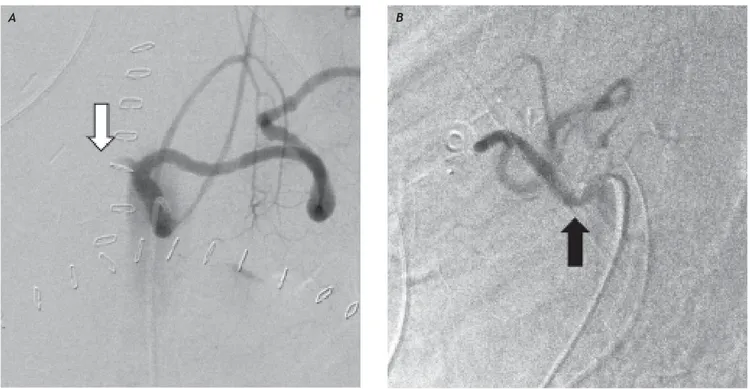
at the arterial anastomosis. Small thrombus were detected and removed distal to the anastomosis, surgical reconstruction was performed using conti-nuous 7-0 nonabsorbable polypropylene suture. In-traoperative color Doppler ultrasound showed a patent intrahepatic HA. A total of four red blood cell packs were transfused. At the ICU, hemodynamic instability persisted and postoperative color Doppler ultrasound showed no flow in the hepatic artery. Di-gital subtraction angiography was performed, showing a characteristic indentation at the origin of the celiac trunk, due to compression by the median arcuate ligament; only the splenic artery was opaci-fied by contrast injection (Figure 2). The patient
Table 1. Laboratory data. Day 0 indicates OLT day prior to surgery. The second and third operations took place during Day 9.
Lab/Day Day 0* Day 0** Day 2 Day 4 Day 7 Day 9* Day 9** Day 10 Day 11 Day 12
Hemoglobin 14.1 13.1 12.0 11.1 11.0 6.0 13.5 14.4 11.4 12.4 Creatinine 0.59 0.82 0.5 0.63 0.71 ND 0.6 0.58 0.37 0.4 T. bilirubin 1.9 5.6 3.4 2.4 3 ND 4.2 3.1 1.5 1.5 D. bilirubin 0.5 1.0 1.3 1.0 1.0 ND 2.8 1.5 0.4 0.5
ALT 57 670 654 463 134 ND 119 124 41 90
AST 77 1,806 413 142 25 ND 103 60 17 28
Alk. Phos. 100 74 55 102 118 ND 40 46 60 53 INR 1.29 1.47 1.3 1.11 1.1 ND 1.46 1.28 2.2 0.8 Albumin 3.5 2.8 3.1 2.9 2.9 ND 1.2 2.3 3.0 1.9
***** Preoperative. ********** Postoperative. ND:ND:ND:ND:ND: No data.
was re-explored later the same day, finding hepatic artery thrombosis. This was corrected with throm-bectomy and arterial anastomosis reconstruction.
Figure 2. A. Digital substraction angiography in anteroposteriorprojection of the celiac artery showing lack of opacification of the hepatic artery (white arrow). B. Lateral projection during end expiration showing an indentation at its origin (black arrow).
A B
Figure 3. Color Doppler ultrasound after surgical median arcuate ligament division, showing a hepatic artery with a flow velocity of 120.5 cm/sec and resistance index of 0.66.
The celiac artery was liberated from the median ar-cuate ligament by dividing its fibers. Immediately, an adequate pulse was palpated in the hepatic ar-tery. The patient made a satisfactory recovery and color Doppler ultrasound showed adequate flow in the hepatic artery (Figure 3). Three days later, an-giography showed normal flow through the hepatic artery (Figure 4) and the patient was discharged ten days after the last intervention. Fortunately, no bi-liary complication overcame and she is alive and well 12 months after transplantation, with normal hepatic artery flow by color Doppler ultrasound.
DISCUSSION
The median arcuate ligament is a fibrous arch that passes superior to the origin of the celiac axis, at the level of the first lumbar vertebral body, uniting the two diaphragmatic crura over the aorta, outlining the anterior margin of the aortic hiatus.8 In about
3.7-10% of the population undergoing OLT,4,9 median
Figure 4.A. Digital substraction angiography in anteroposteriorprojection of the celiac artery performed in day 12, showing a normal hepatic artery (white arrow). B. Lateral projection during end expiration shows a normal celiac artery (black arrow).
A B
A small subset of patients suffer a hemodynami-cally significant compression that cause symptoms, a condition first reported by Harjola in 196311 and
Dunbar, et al., in 1965,12 with radiological
confir-mation by Colapinto, et al., in 1972.13 The original
group of patients described at Columbus experienced intermittent epigastric pain, usually in the pos-tprandial period associated with nausea, vomiting, diarrhea (occasionally malabsorptive) and weight loss.12 An abdominal bruit was found in the
epigas-trium in some patients. The patients improved after surgical division of the median arcuate ligament and the adjacent cords of the celiac plexus, although it was not entirely clear whether this was due to relea-se of compression of the artery or disruption of the neural pathways. More recently, it has been pointed out that not all patient’s symptoms resolves after the operation.14 Some authors have even questioned
the mere relationship of clinical symptoms and iso-lated narrowing of the celiac axis due to the vast co-llateral circulation.15,16
Paulsen, et al.,17 showed that hepatic artery
blood flow is increased after OLT (425.7 ± 25.6 mL/ min). Jurim, et al.,4 found that celiac artery
com-pression decreased the hepatic arterial blood flow af-ter OLT to around 200 mL/min during expiration, which corresponds to two thirds of the respiratory cycle and therefore the majority of the perfusion time. Usually, such a phenomenon would be com-pensated by collateral circulation from the superior
mesenteric and pancreaticoduodenal arteries.18
However, the same scenario in a surgically dissec-ted, newly reconstructed artery could induce decrea-sed flow and predispose recipients to thrombosis after OLT. Probably the thrombosis distal to the anastomosis found in our patient lead to an increa-sed resistance to hepatic artery blood flow and even-tually to the bleeding through the anastomosis that we found in the second operation. Fortunately, our patient belongs to that reduced group of patient whose hepatic artery thrombosis resolves with sur-gical reconstruction without retransplantation, as hepatic artery blood flow was restored after throm-bectomy in the third operation, apparently due to an insignificant intrahepatic arterial thrombosis com-ponent. Lubrano, et al.,7 showed a greater risk of
hepatic artery thrombosis in patients with celiac ar-tery compression undergoing OLT (20% vs. 6.9%; p = 0.17); although not statistical significant, proba-bly due to a small sample size, the fact that the inci-dence of hepatic artery thrombosis increases more than two-fold in this group is clinically significant.
CONCLUSION
patients), by direct examination or through imaging studies, to prevent potentially fatal complications, such as early-onset hepatic artery thrombosis and bleeding, as seen in our patient.
REFERENCES
1. Duffy JP, Hong JC, Farmer DG, et al. Vascular complicatio-ns of orthotopic liver tracomplicatio-nsplantation: experience in more than 4,200 patients. J Am Coll Surg 2009; 208: 895-903. 2. Nikeghbalian S, Kazemi K, Davari HR, et al. Early hepatic
artery thrombosis after liver transplantation: diagnosis and treatment. Transplant Proc 2007; 39: 1195-6. 3. Gunsar F, Rolando N, Pastacaldi S, et al. Late hepatic
ar-tery thrombosis after orthotopic liver transplantation. Li-ver Transpl 2003; 9: 605-11.
4. Jurim O, Shaked A, Kiai K, et al. Celiac compression syn-drome and liver transplantation. Ann Surg 1993; 218: 10-12.
5. Fukuzawa K, Schwartz ME, Katz E, et al. The arcuate liga-ment syndrome in liver transplantation. Transplantation
1993; 56: 223-4.
6. Jiang ZJ, Liang TB, Feng XN, et al. Arcuate ligament syn-drome inducing hepatic artery thrombosis after liver transplantation. Hepatobiliary Pancreat Dis Int 2008; 7: 433-6.
7. Lubrano J, Scatton O, Randone B, et al. Median arcuate ligament in orthotopic liver transplantation: relevance to arterial reconstruction. Transplant Proc 2008; 40: 3532-5.
8. Skandalakis JE, Colborn GL. Skandalakis’ Surgical anatomy. The embryologic and anatomic basis of modern surgery. New York: McGraw-Hill; 2007.
9. Park CM, Chung JW, Kim HB, et al. Celiac axis stenosis: in-cidence and etiologies in asymptomatic individuals. Korean J Radiol 2001; 2: 8-13.
10. Reuter SR. Accentuation of celiac compression by the me-dian arcuate ligament of the diaphragm during deep expi-ration. Radiology 1971; 98: 561-4.
11. Harjola PT. A Rare Obstruction of the Coeliac Artery. Re-port of a Case. Ann Chir Gynaecol Fenn 1963; 52: 547-50. 12. Dunbar JD, Molnar W, Beman FF, et al. Compression of the
celiac trunk and abdominal angina. Am J Roentgenol Ra-dium Ther Nucl Med 1965; 95: 731-44.
13. Colapinto RF, McLoughlin MJ, Weisbrod GL. The routine la-teral aortogram and the celiac compression syndrome. Ra-diology 1972; 103: 557-63.
14. Carey JP, Stemmer EA, Connolly JE. Median arcuate liga-ment syndrome. Experiliga-mental and clinical observations.
Arch Surg 1969; 99: 441-6.
15. Drapanas T, Bron KM. Stenosis of the celiac artery. Ann Surg 1966; 164: 1085-8.
16. Lindner HH, Kemprud E. A clinicoanatomical study of the arcuate ligament of the diaphragm. Arch Surg 1971; 103: 600-5.
17. Paulsen AW, Klintmalm GB. Direct measurement of hepatic blood flow in native and transplanted organs, with accom-panying systemic hemodynamics. Hepatology 1992; 16: 100-11.