http://dvr.sagepub.com/
Diabetes and Vascular Disease Research
http://dvr.sagepub.com/content/10/6/472 The online version of this article can be found at:
DOI: 10.1177/1479164113500680
2013 10: 472 originally published online 3 September 2013 Diabetes and Vascular Disease Research
Anna C Roberts and Karen E Porter
Cellular and molecular mechanisms of endothelial dysfunction in diabetes
Published by:
http://www.sagepublications.com
can be found at:
Diabetes and Vascular Disease Research
Additional services and information for
http://dvr.sagepub.com/cgi/alerts Email Alerts:
http://dvr.sagepub.com/subscriptions Subscriptions:
http://www.sagepub.com/journalsReprints.nav Reprints:
http://www.sagepub.com/journalsPermissions.nav Permissions:
What is This?
- Sep 3, 2013 OnlineFirst Version of Record
- Oct 18, 2013 Version of Record
Diabetes & Vascular Disease Research 10(6) 472 –482
© The Author(s) 2013 Reprints and permissions: sagepub.co.uk/journalsPermissions.nav DOI: 10.1177/1479164113500680 dvr.sagepub.com
Introduction
Largely owing to the vast quantities of energy-dense food consumed in the Western world, coupled with increasingly sedentary lifestyles, diabetes mellitus (DM) is considered to be a public health problem of epidemic proportions.1,2 In the United Kingdom alone, 2.9 million people are diagnosed with the disease, with an estimated 850,000 more undiag-nosed.3 DM is recognised as an independent risk factor for cardiovascular disease (CVD),4 even when under glycaemic control. With up to 75% of mortality in diabetic patients arising from CVD,5 this is an issue of utmost importance. Endothelial cell (EC) dysfunction is associated with both diabetes and the pathogenesis of CVD.6 Establishing the causative mechanisms linking these factors could provide essential insights and inform novel therapeutics.
The aim of this review is to explore the relationship between DM and EC function/dysfunction, focussing pre-dominantly on cellular and molecular mechanisms as opposed to clinical manifestations.
Physiological role of endothelium
Forming a vast interface between the blood and surrounding tissues, the endothelium forms a monolayer comprising the innermost lining of blood vessels. Advances over recent dec-ades have revealed the complexity of this semi-permeable
membrane and its key role in maintaining vascular homeo-stasis.7 While facilitating the passage of substances such as nutrients and leukocytes across the vessel wall, the endothe-lium secretes numerous mediators necessary for normal vas-cular functioning including those that regulate vasvas-cular tone, coagulation, modulate immune responses and control vascu-lar cell growth.8
Arguably, the most significant endothelium-derived mediator is nitric oxide (NO), which plays multiple roles in preventing atheroma formation.9 Produced by the action of endothelial nitric oxide synthase (eNOS), NO diffuses into neighbouring vascular smooth muscle cells (SMC), activat-ing guanylyl cyclase and producactivat-ing cyclic guanosine monophosphate (cGMP) and activating kinases responsible for vascular relaxation.8 Vasodilation via NO synthesis is
Cellular and molecular mechanisms of
endothelial dysfunction in diabetes
Anna C Roberts
1and Karen E Porter
1,2Abstract
In healthy individuals, the vascular endothelium regulates an intricate balance of factors that maintain vascular homeostasis and normal arterial function. Functional disruption of the endothelium is known to be an early event that underlies the development of subsequent cardiovascular disease (CVD) including atherosclerosis and coronary heart disease. In addition, the rising global epidemic of type 2 diabetes is a significant problem conferring a significantly higher risk of CVD to individuals in whom endothelial dysfunction is also notable. This review first summarises the role of endothelium in health and explores and evaluates the impact of diabetes on endothelial function. The characteristic features of insulin resistance and other metabolic disturbances that may underlie long-term changes in vascular endothelial function (metabolic memory) are described along with proposed cellular, molecular and epigenetic mechanisms. Through understanding the underlying mechanisms, novel targets for future therapies to restore endothelial homeostasis and ‘drive’ a reparative cellular phenotype are explored.
Keywords
Diabetes, endothelial function, endothelial dysfunction, metabolic memory, microRNA, novel therapies
1 Division of Cardiovascular and Diabetes Research, Leeds Institute of
Genetics, Health and Therapeutics (LIGHT), University of Leeds, Leeds, UK
2 Multidisciplinary Cardiovascular Research Centre (MCRC), University
of Leeds, Leeds, UK
Corresponding author:
Karen E Porter, Division of Cardiovascular and Diabetes Research, Leeds Institute of Genetics, Health and Therapeutics (LIGHT), University of Leeds, Worsley Building, Clarendon Way, Leeds LS2 9JT, UK.
Email: medkep@leeds.ac.uk
well documented in vitro and in vivo.10–12 Additionally, NO inhibits platelet aggregation, SMC proliferation and nuclear transcription of leukocyte-adhesion molecules including vascular cell adhesion molecule (VCAM) and intercellular adhesion molecule (ICAM). Arterial wall shear stress13 and coupling of agonists such as acetylcholine and bradykinin to endothelial-bound receptors8 are potent stimuli for NO synthesis. Although these stimuli act via different intracel-lular mechanisms, each increases NO production via enhanced expression of eNOS. Other important vasodila-tory factors are endothelium-derived hyperpolarizing factor (EDHF) and prostacyclin (PGI2), the latter also playing a role in platelet inhibition.8
Conversely, the endothelium is able to secrete the potent vasoconstrictor endothelin (ET-1), which through its pro-inflammatory and mitogenic effects augments the pathogenesis of CVD.14 Other endothelial-derived vaso-constrictors include prostaglandin H2 (PGH2), thrombox-ane A2 (TXA2) and reactive oxygen species (ROS). EC-bound angiotensin-converting enzyme (ACE) cataly-ses production of the vasoconstrictive angiotensin II, and ACE inhibitors are commonly used to decrease blood pressure.15 Under physiological conditions, the endothe-lium maintains a fine balance between anti- and pro-thrombotic states. While NO, prostacyclin and thrombomodulin favour blood fluidity and are dominant under basal conditions, ‘activated’ endothelium drives an opposing pro-thrombotic state. Under these conditions, plasminogen activator inhibitor-1 and thromboxane, together with von Willebrand Factor (vWF), a key con-stituent of the coagulation cascade, are deleterious.8,16
Through NO production, the endothelium plays a cru-cial role in preventing leukocyte adhesion; however, when ‘activated’, expression of adhesion molecules aid the pas-sage of leukocytes across the vascular wall promoting a pro-inflammatory environment. Under these conditions, EC produce pro-inflammatory cytokines, including tumour necrosis factor-alpha (TNF-α), which further augment inflammation.17
Finally, the endothelium plays a key role in angiogenesis initiated by tissue growth factors, particularly vascular endothelial growth factor (VEGF). The VEGF receptor is coupled to activation of mitogen-activated protein kinase (MAPK) signalling, promoting angiogenesis by increasing nuclear transcription of relevant genes. This mechanism is counteracted by the anti-angiogenic factors angiostatin and thrombospondin, which suppress angiogenesis unless required.8
Endothelial dysfunction
Endothelial dysfunction refers to inability of the endothe-lium to regulate vascular homeostasis, and essentially describes ‘tipping’ of the physiological balance in favour of
vasoconstrictive, pro-inflammatory and pro-thrombotic effects5 that promote atherosclerosis. As such, abnormalities in endothelial function are detected early in the develop-ment of CVD, often before symptoms are clinically evident.16
Given the vast range of vasoprotective effects of NO, the term endothelial dysfunction generally refers to reduced NO bioavailability, through decreased eNOS expression.16 Procedures employed to evaluate endothelial dysfunction measure blunted vasodilatory responses to certain ago-nists.5 Indeed, by this method, impaired endothelial-dependent vasodilation in patients with confirmed CVD,18–21 and importantly in those who carry risk factors for future CVD events,22 has been demonstrated. This sug-gests that endothelial dysfunction is the commonality by which these risk factors, particularly those associated with the metabolic syndrome, lead to CVD development. This point is underscored by reports that endothelial dysfunction appears to reverse when lifestyle changes and modulatory drug therapies are implemented.23 DM is one of the afore-mentioned risk factors whose association with endothelial dysfunction will now be evaluated.
Endothelial dysfunction in DM
Current evidence links endothelial dysfunction to type 1 DM (T1DM) and type 2 DM (T2DM), through demonstra-tion of impaired endothelial-dependent vasodilademonstra-tion.24–26 Despite many proposed mechanisms for this relationship, the definitive pathogenesis remains unclear, possibly because diabetes patients usually display multiple homeo-static imbalances alongside the typically described hyper-glycaemia. Such disturbances induce endothelial dysfunction independently of the presence of diabetes, indicating a multifactorial aetiology rather than hypergly-caemia per se.27–29 The following sections will evaluate these factors.
Selective insulin resistance
unaffected.34,35 Moreover, this selective resistance pro-motes hyperinsulinaemia, resulting in further stimulation of MAPK signalling.36 The outcome therefore is that of pro-atherogenic signalling that is clearly detrimental to the vas-culature.37 This signalling deficiency has been demonstrated experimentally in obese Zucker rats, in which defective PI3-K/Akt signalling consequently resulted in decreased NO bioavailability.34
Pro-inflammatory signalling
T2DM is associated with a state of chronic systemic inflam-mation; elevated levels of circulating inflammatory mark-ers are observed in patients with diabetes and obesity38,39 and are believed to confer a pathological EC phenotype. A diverse range of harmful stimuli have been shown to con-tribute to vascular complications in diabetes. These include pro-inflammatory cytokines, chemokines, adhesion mole-cules and transcription factors (summarised in Table 1) and have been reviewed recently.40
Animal models of obesity have identified adipose tissue as a site of inflammatory cytokine production, with accom-panying elevated plasma levels of TNF-α.41,42 TNF-α acti-vates nuclear factor κB (NFκB), a transcription factor able to further stimulate expression of inflammatory genes.43 NFκB is also activated by free fatty acids (FFA) and the receptor for advanced glycation end-products (RAGE), both of which are characteristic of the diabetic milieu.16 Evidence suggests that overexpression of either TNF-α or IκB kinase mediates the development of insulin resistance. Obese mice with null mutations in the TNF-α
gene display significantly improved insulin sensitivity.44 Indeed, NFκB that is permanently present in the cell could be viewed as the ‘first responder’ to many inflammatory signals due to rapid activation of its downstream pathways. More recently, new pharmacological roles for the salicylate anti-inflammatory drugs have emerged regarding their abil-ity to decrease insulin resistance via inhibition of IκB kinase. Preliminary results from the TINSAL-T2D (Targeting INflammation Using SALsalate in T2DM) clini-cal trial advocates their use in glucose-lowering, presuma-bly as a consequence of improved insulin sensitivity.45 The association of insulin resistance with inflammation implies that the latter adversely impairs the NO-producing PI3-K/ Akt pathway, ultimately reducing NO bioavailability. Exposure of cultured EC to TNF-α leads to impaired eNOS expression,46 inferring that reduced bioavailability of NO nullifies its otherwise significant anti-inflammatory role, thus augmenting formation of inflammatory atherosclerotic lesions.
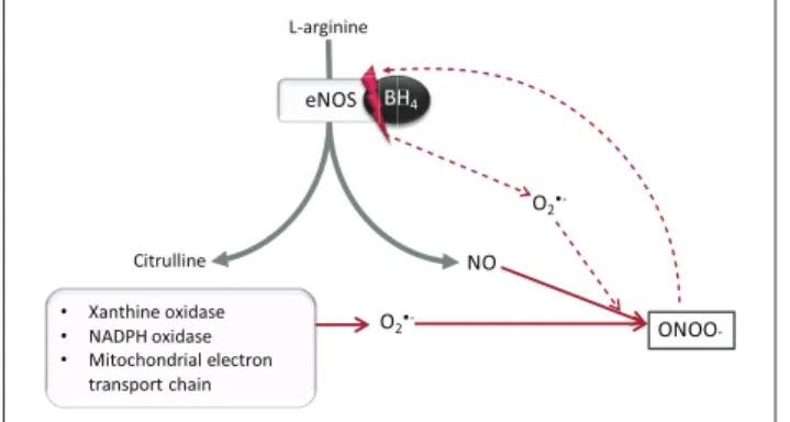
Oxidative stress
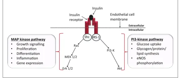
Oxidative stress is commonly implicated as an important unifying mechanism in conferring endothelial injury (see Figure 2). The term refers to the accumulation of ROS such as superoxide anion (O2·−), when production exceeds the capability of the anti-oxidant system to remove them.47 A key adverse effect of oxidative stress is diminished NO bio-availability, either through direct degradation by ROS, or alterations in the functional capacity of eNOS. Direct reac-tion of NO with O2·− yields peroxynitrite (ONOO−), a Shc IRS-1
Ras
MEK 1/2
Erk 1/2
PI3-K
Akt
Intracellular Extracellular
Insulin receptor
Insulin
Endothelial cell membrane
MAP kinase pathway • Growth signalling • Proliferaon • Differenaon • Inflammaon • Gene expression
PI3-kinase pathway • Glucose uptake • Glycogen/protein/
lipid synthesis • eNOS
phosphorylaon
Figure 1. Intracellular insulin signalling pathways: Upon binding to its receptor, insulin initiates two key intracellular signalling pathways. Activation of PI3-Kinase dominates under physiological conditions resulting in a net anti-atherogenic effect. In insulin resistance, selective deficiency in the PI3-kinase pathway leads to a dominant MAP kinase pathway, whose effects are predominantly pro-atherogenic.
highly potent oxidant capable of altering the functions of multiple intracellular molecules, including those related directly to the NO pathway.48 Additionally, ONOO− serves to uncouple eNOS from its critical co-factor tetrahydrobi-opterin (BH4), resulting in preferential formation of O2·− in the place of NO and hence further exacerbation of the prob-lem.49 In the setting of insulin resistance, experimental evi-dence has implicated endogenous ONOO− as a down-regulator of the PI3-K/Akt pathway.50,51
Aside from uncoupled eNOS, significant sources of O2·− include xanthine oxidase, nicotinamide adenine dinucleo-tide phosphate (NADPH) oxidase and mitochondria.47 Mitochondria produce negligible ROS in physiological cir-cumstances, but greatly increase O2·− output when high glu-cose concentrations increase the proton gradient within the electron transport chain.52 Recent evidence has also impli-cated mitochondrial dynamics, namely, increased mito-chondrial fission and fragmentation, as contributing factors to ROS production in DM.53
Anti-oxidant therapy
Anti-oxidant therapies aiming to reduce the detrimental effects of ROS have so far yielded conflicting results.
Studies of diabetic vessels in vivo have demonstrated improvements in NO-mediated vasodilation in response to anti-oxidants such as superoxide dismutase (SOD).54,55 Additionally, the anti-oxidant properties of vitamin C have been shown to effectively augment endothelial-dependent vasodilatation in several studies performed on human fore-arm vessels, in the setting of diabetes and hypertension.56–58 Despite promising experimental data, large-scale clinical trials investigating the effects of anti-oxidant vitamins in patients with diabetes failed to report any benefits.59,60 Although conducting clinical trials is a logical progression following experimental evidence, the discrepancies in these results may indicate that the benefits of anti-oxidant thera-pies are too short term to affect mortality outcomes. The vastly greater sample size and follow-up period in clinical trials indicates that these results should take precedent when interpreting the effects of anti-oxidant therapy. However, further experimental evidence regarding the longer term outcomes of anti-oxidant therapies is required.
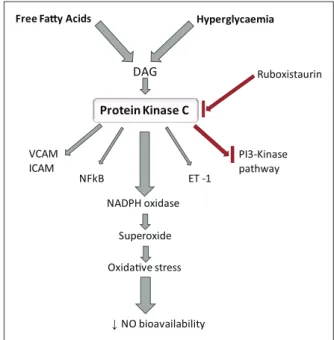
Protein kinase C
Protein kinase C beta (PKCβ) is an endothelial isoform of the serine/threonine kinase family. A host of evidence impli-cated this enzyme as a chief contributor to endothelial dys-function observed in DM.61 PKCβ is activated by diacylglycerol (DAG), which is typically produced subse-quent to ligand-receptor binding.62 However, in conditions of high circulating FFA and hyperglycaemia, a novel route for the chronic activation of DAG has been proposed, via de novo synthesis from glucose.63 Significantly increased levels of PKC and DAG have been identified in animal models of diabetes.63 Upon activation, PKC induces several intracel-lular effects (Figure 3), many of which have been
Table 1. Inflammatory components of diabetes complications.
Inflammatory cytokines
Interleukins IL-1, IL-6, IL-18
Tumour necrosis factor-alpha (TNF-α) C-reactive protein (CRP)
Adhesion molecules
Intercellular adhesion molecule 1 (ICAM-1) Vascular cell adhesion molecule-1 (VCAM1) E-selectin
Chemokines
CCL-2 (MCP-1) CX3CL1 (fractalkine) CCL5 (RANTES)
Transcription factors
NFκB
Toll-like receptors
TLR2 TLR4
Profibrotic cytokines
Transforming growth factor beta (TGF-β) Connective tissue growth factor (CTGF)
Key categories of inflammatory mediators associated with vascular complications of diabetes, indicating the principal components of each category. There is widespread evidence for inflammatory cytokines, adhesion molecules, chemokines and transcription factors. More recent data support roles for profibrotic cytokines and toll-like receptors that appear to mediate responses to a number of ‘diabetogenic’ stimuli (reviewed in Forbes and Cooper40).
L-arginine
eNOS BH4
Citrulline NO
O2•- ONOO
-• Xanthine oxidase
• NADPH oxidase
• Mitochondrial electron transport chain
S B
O2
•-Figure 2. Production of peroxynitrite in endothelial cells: In healthy cells, eNOS catalyses synthesis of NO from l-arginine. The factors listed (bullet-pointed box) produce O2·−, which
reacts with NO to produce ONOO−. This uncouples eNOS from BH4, which enhances O2·− production, and increased
ONOO−.
NO: nitric oxide; ONOO−: peroxynitrite; eNOS: endothelial nitric
oxide synthase; O2·−: superoxide anion; BH4: tetrahydrobiopterin. grey
experimentally identified using PKC inhibitors such as ruboxistaurin.43,64 Such alterations include amplified expres-sion of ET-1, VCAM and ICAM.62 PKC also activates vas-cular NADPH oxidase, a significant source of O2·− and subsequent oxidative stress in the endothelium. Indeed, inhibition of PKC has been shown to diminish O2·− in the diabetic vasculature.65 A trial in healthy humans indicated that PKC inhibition prevented hyperglycaemia-induced impairment of endothelial-dependent vasodilation.66 Disappointingly, a later trial by the same authors showed that this benefit was not apparent in T2DM patients,67 sug-gesting that further work is required to delineate the role of PKC in T2DM. Additional effects of PKC in the endothe-lium include activation of NFκB, alterations in eNOS expres-sion and inhibition of the PI3-K signalling pathway.43
Hyperglycaemia
The hallmark of DM, chronic hyperglycaemia, has been implicated in the development of endothelial dysfunction via four principal mechanisms: PKC activation, activation of the hexosamine and polyol pathways and formation of advanced glycation end-products (AGEs).68 These path-ways are believed to mediate vascular dysfunction through
the unifying mechanism of ROS overproduction, most notably increases in O2·−.69 Briefly, the polyol and hexosa-mine pathways display low affinity for glucose in the phys-iological state; however, intracellular hyperglycaemia leads to shunting of glucose through these pathways, with result-ant increases in PKC activation, changes in gene expression and protein function.68 AGEs initially arise from intra-cellular hyperglycaemia and subsequent non-enzymatic reactions, causing alterations in both intra- and extracellu-lar proteins, as well as modifications to the extracelluextracellu-lar matrix.70 Additionally, activation of RAGE on EC influ-ences ROS production and leads to NFκB activation, which induces atherogenic gene expression.71,72 Collectively, these mechanisms have been shown to increase the proton gradient across the inner mitochondrial membrane, conse-quently increasing O2·− as previously discussed. SOD-enabled inhibition of mitochondrial ROS is capable of inhibiting increases in all these mechanisms.68
Metabolic memory: role of
epigenetics
The mechanisms described suggest routes by which envi-ronmental factors such as diet-induced hyperglycaemia may induce endothelial dysfunction, and it would be easy to believe that correcting these imbalances would promptly normalise endothelial function. This is not the case. Indeed, follow-up trials succeeding the Diabetes Control and Complications Trial (DCCT) and the United Kingdom Prospective Diabetes Study (UKPDS) propose the existence of a so-called metabolic memory, in which the effects of either prolonged or transient changes in glycaemia persist long after these have been re-adjusted.73–75 Not only does this suggest that intensive glycaemic control cannot com-pletely reverse pre-established hyperglycaemia-induced vascular complications, but importantly showed that the benefits of intensive therapy were maintained for many years despite a return to often inferior glycaemic control.73 Such observations have been confirmed in vitro and in vivo, including EC-specific studies.76–78 Together, these data imply that heritable alterations in cell phenotype may be induced by diabetogenic stimuli, particularly hyperglycae-mia. While this was once thought to be due to mutations in mitochondrial DNA,68 more recent evidence has implicated epigenetic mechanisms as the underlying cause of this phe-nomenon–alterations in gene expression without changes in DNA sequence.79 Indeed, epigenetic studies are shedding new light on how environmental factors influence gene expression and the associated susceptibility to T2DM and increased CVD.79 Three highly connected pathways provide the foundation for epigenetic theories, namely, post-translational histone modifications, DNA methylation and non-coding RNA-based mechanisms, detailed studies of which are clearly warranted to investigate endothelial-specific mech-anisms that may inform novel therapeutic targets.
Hyperglycaemia Free Fay Acids
DAG
Protein Kinase C
NADPH oxidase
Superoxide
Oxidave stress
↓NO bioavailability NFkB
Ruboxistaurin
ET -1 VCAM
ICAM PI3-Kinase pathway
Figure 3. Activation and effects of protein kinase C (PKC): High levels of free fatty acids and hyperglycaemia activate DAG, which in turn activates PKC. This increases expression of ET-1, VCAM, ICAM, NFκB and NADPH oxidase. NADPH oxidase activation results in decreased NO bioavailability. PKC may have a role in PI3-kinase pathway inhibition. Ruboxistaurin inhibits PKC activation.
Histone modifications
Post-translational modifications of histones in chromatin alter the conformation of DNA, providing or denying access to specific sites that enhance or retard gene transcription, respectively. Histone modifications (e.g. acetylation/deacet-ylation; methylation/demethylation) are regulated by a fam-ily of enzymes that are responsible for their transfer [histone acetyltransferases (HATs), histone methyltransferases (HMTases)] or removal [histone deacetylases (HDAC), his-tone demethylases (HDM)].79 Several studies have been per-formed specifically in ECs, where changes in the expression and activity of these enzymes have been linked to diabetic complications. For example, specific histone methylations are associated with the chronic inflammatory phenotype in EC exposed to high glucose concentrations.80 Transient exposure of human and bovine endothelium to hyperglycae-mia led to transcriptional activation of inflammatory NFκ B-dependent promoters that were sustained even following restoration of normoglycaemia.80,81
DNA methylation
Studies examining the role of DNA methylation in diabetes and vascular complications are few, although altered pat-terns of DNA methylation have been associated with ath-erosclerosis82 and in the endothelium itself.83 Both global and gene-specific methylation is believed to contribute to the metabolic memory of diabetic complications. Covalent methylation of cytosine residues is a stable DNA modifica-tion that persists throughout experimental processing,84 providing a robust platform on which to base future inves-tigations. Akin to modifications in histones, DNA methyla-tion can confer persistent alteramethyla-tions in gene expression and a clear understanding of the complex mechanisms involved will be of immense value. High fat feeding of obese rats has been reported to induce hypermethylation of particular genes,85 so it is reasonable to consider that this phenome-non may also exist in human diabetic individuals, and spe-cifically at the level of the endothelium.
microRNAs
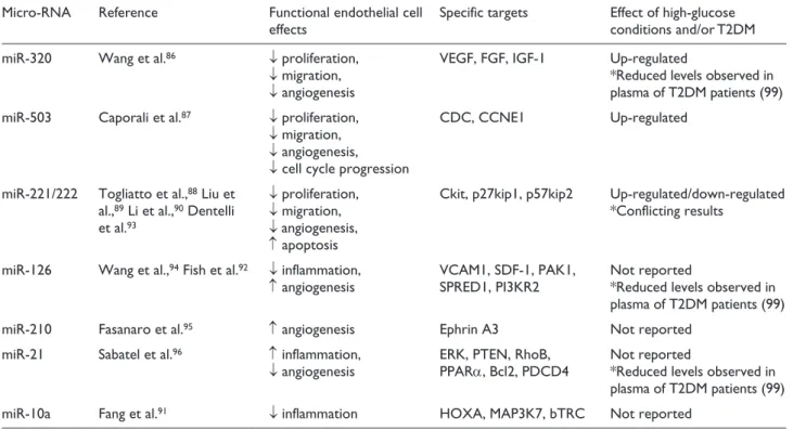
Another area of intense interest in which gene expression can be post-transcriptionally modified is by microRNAs (miRs), the most widely studied of the non-coding RNAs. miRs are short RNAs that inhibit expression of a range of target proteins through mRNA degradation or translational repression. As regards ECs, in vitro studies have exposed a complex network of miRs with regulatory roles in proliferation and migration (miR-320,86 miR-503,87 miR221/22288–90), inflammation (miR-10a,91 miR-126 92) and angiogenesis (miR221/222,93 miR-126,94 miR-210,95 miR-2196). These roles may be divergent in physiological circumstances, for example, specific miRs are capable of exerting protective, pro-angiogenic effects,94,95 while others
confer more unfavourable anti-angiogenic properties.93,96 Furthermore, a distinct signature of miR dysregulation has been proposed to exist in DM,97 whereby high glucose con-ditions may induce over or under expression of detrimental or beneficial miRs, respectively, thereby contributing to the development of vascular complications. In such a way, upregulation of miR-320 was observed in EC exposed to hyperglycaemia, with a resultant reduction in cellular prolif-eration and migration.86 Importantly, inhibition of the over-expressed miRs restored EC regenerative functions.86
The stable and consistent levels of plasma miRs in healthy individuals has identified them as potential bio-markers for diseases such as T2DM,98 not only for evaluat-ing disease progression but also to detect risk of future disease development. In this way, plasma profiling of T2DM patients has revealed altered levels of miRs, which may contribute to a pathophysiological state.99 Highly reported miRs that appear to modulate EC function either beneficially or detrimentally are presented in Table 2.
In summary, while the study of epigenetic mechanisms associated with diabetes and its complications has gained momentum in recent years, there is still much to be learned. Moreover, the complexity of these interactions will pose considerable challenges for their elucidation.
Current therapies for DM
Approach to future therapies
This review has highlighted the critical involvement of endothelial dysfunction in the pathogenesis of vascular disease in DM, in which diabetic mediators inflict a pro-atherogenic, inflammatory phenotype, often with limited or delayed reversibility. Owing to the silent nature of insu-lin resistance and hence an inevitable problem in rapid diagnosis, early intervention is difficult. Manipulation or inhibition of some of the described mechanisms may pro-vide novel avenues for therapeutic intervention. However, the transition from this experimental stage through to pro-duction of safe and effective drugs is not easy, and requires results from valid and reliable large-scale clinical trials. Such clinical trials have already revealed disappointing outcomes regarding the use of anti-oxidants in alleviating oxidative stress;59,60 however, they should not deter further anti-oxidant therapy trials. It is likely that a ‘prevention rather than cure’ approach is preferable in the management of ROS, with the enzymatic sources of superoxide anion a promising target for future therapeutics.16 Other mecha-nisms meriting further investigation include PKC inhibi-tion65 and the previously mentioned use of salicylate anti-inflammatory drugs.45
Rapidly accumulating evidence supports the notion that miRs are cell-type specific and differentially expressed,
and may actually play a causative role in diabetes and its related vascular complications.97 Recent evidence suggests that miRs can be communicated between cells and specifi-cally, EC-derived plasma miRs are reportedly able to regu-late the biological functions of not only other EC, but also vascular SMC and leukocytes (reviewed in Yamakuchi106). In future, understanding and exploiting these mechanisms has potential to be translated into defining clinical biomark-ers and elucidating specific molecular targets for therapeu-tics. In the longer term, these may be of value in reversing the devastating outcomes attributed to endothelial dysfunc-tion in diabetes by erasing persistent metabolic memory by ‘re-setting’ the epigenetic cell signature.
Conclusion
Recent advances in knowledge surrounding metabolic memory and the role of epigenetics have provided exciting new avenues for future research. However, there is still much to learn regarding their roles in diabetic vascular complications, and specifically on the EC. To date, in vitro studies of metabolic memory in cultured EC have largely focused on changes in gene expression in response to hyperglycaemia.80 However, this review has highlighted the contribution of a complex array of factors other than glucose in the pathogenesis of endothelial dysfunction. For
Table 2. Principal endothelial microRNAs and their involvement in cell function.
Micro-RNA Reference Functional endothelial cell
effects Specific targets Effect of high-glucose conditions and/or T2DM
miR-320 Wang et al.86 ↓ proliferation,
↓ migration,
↓ angiogenesis
VEGF, FGF, IGF-1 Up-regulated
*Reduced levels observed in plasma of T2DM patients (99)
miR-503 Caporali et al.87 ↓ proliferation,
↓ migration,
↓ angiogenesis,
↓ cell cycle progression
CDC, CCNE1 Up-regulated
miR-221/222 Togliatto et al.,88 Liu et
al.,89 Li et al.,90 Dentelli
et al.93
↓ proliferation,
↓ migration,
↓ angiogenesis,
↑ apoptosis
Ckit, p27kip1, p57kip2 Up-regulated/down-regulated *Conflicting results
miR-126 Wang et al.,94 Fish et al.92 ↓ inflammation,
↑ angiogenesis VCAM1, SDF-1, PAK1, SPRED1, PI3KR2 Not reported*Reduced levels observed in plasma of T2DM patients (99)
miR-210 Fasanaro et al.95 ↑ angiogenesis Ephrin A3 Not reported
miR-21 Sabatel et al.96 ↑ inflammation,
↓ angiogenesis ERK, PTEN, RhoB, PPARα, Bcl2, PDCD4 Not reported *Reduced levels observed in plasma of T2DM patients (99)
miR-10a Fang et al.91 ↓ inflammation HOXA, MAP3K7, bTRC Not reported
this reason, it is apparent that identifying the presence of a diabetic metabolic memory might be most accurately observed in EC derived from human diabetic vessels. In such a way, we have previously identified inherent func-tional differences between vascular SMC cultured from patients with and without T2DM,107 such data offering an explanation for increased venous bypass graft stenosis after revascularisation surgery in these patients. Identification of any sustained functional differences in T2DM-EC may pro-vide further justification for the aforementioned graft fail-ure. Such information would potentially create a foundation, upon which therapeutics to ameliorate endothelial dysfunc-tion could be developed.
Declaration of conflicting interests
The authors declare no conflict of interest.
Funding
Anna C Roberts undertook this work as part of an Intercalated Bachelor of Science degree in Cardiovascular Medicine, funded by the Alumni Footsteps Fund at the University of Leeds.
References
1. Wang J, Luben R, Khaw K-T, et al. Dietary energy density predicts the risk of clinically incident type 2 diabetes: the
EPIC-Norfolk study. Diabetes Care 2008; 31: 2120–2125.
2. Wilmot E, Edwardson C, Achana F, et al. Sedentary time in adults and the association with diabetes, cardiovascular disease and death: systematic review and meta-analysis.
Diabetologia 2012; 55: 2895–2905.
3. Diabetes UK. What is diabetes? http://www.diabetes.org.uk/ Guide-to-diabetes/Introduction-to-diabetes/What_is_diabetes/ (accessed 17 October 2012).
4. International Diabetes Federation. Diabetes: a major risk fac-tor. http://www.cvd.idf.org (accessed 17 October 2012). 5. Xu J and Zou M-H. Molecular insights and therapeutic
tar-gets for diabetic endothelial dysfunction. Circulation 2009;
120: 1266–1286.
6. Feener EP and King GL. Endothelial dysfunction in
diabe-tes mellitus: role in cardiovascular disease. Heart Fail Monit
2001; 1: 74–82.
7. Michiels C. Endothelial cell functions. J Cell Physiol 2003;
196: 430–443.
8. Levick JR. An introduction to cardiovascular physiology.
4th ed. London: Arnold Publishers, 2003.
9. Yasa M and Turkseven S. Vasoprotective effects of nitric
oxide in atherosclerosis. FABAD J Pharm Sci 2005; 30:
41–53.
10. Tronc F, Wassef M, Esposito B, et al. Role of NO in flow-induced remodeling of the rabbit common carotid artery.
Arterioscler Thromb Vasc Biol 1996; 16: 1256–1262. 11. Gardiner SM, Compton AM, Bennett T, et al. Control of
regional blood flow by endothelium-derived nitric oxide.
Hypertension 1990; 15: 486–492.
12. Rees DD, Palmer RMJ, Schulz R, et al. Characterization of three inhibitors of endothelial nitric oxide synthase in vitro
and in vivo. Br J Pharmacol 1990; 101: 746–752.
13. Buga GM, Gold ME, Fukuto JM, et al. Shear stress-induced release of nitric oxide from endothelial cells grown on beads.
Hypertension 1991; 17: 187–193.
14. Böhm F and Pernow J. The importance of endothelin-1 for
vascular dysfunction in cardiovascular disease. Cardiovasc
Res 2007; 76: 8–18.
15. Varin R, Mulder P, Tamion F, et al. Improvement of endothe-lial function by chronic angiotensin-converting enzyme
inhi-bition in heart failure. Circulation 2000; 102: 351–356.
16. Tabit C, Chung W, Hamburg N, et al. Endothelial dysfunc-tion in diabetes mellitus: molecular mechanisms and clinical
implications. Rev Endocr Metab Disord 2010; 11: 61–74.
17. Sumpio BE, Riley JT and Dardik A. Cells in focus:
endothe-lial cell. Int J Biochem Cell Biol 2002; 34: 1508–1512.
18. Förstermann U, Mügge A, Alheid U, et al. Selective attenua-tion of endothelium-mediated vasodilaattenua-tion in atherosclerotic
human coronary arteries. Circ Res 1988; 62: 185–190.
19. Werns SW, Walton JA, Hsia HH, et al. Evidence of endothe-lial dysfunction in angiographically normal coronary arteries
of patients with coronary artery disease. Circulation 1989;
79: 287–291.
20. Bossaller C, Habib GB, Yamamoto H, et al. Impaired mus-carinic endothelium-dependent relaxation and cyclic guanosine 5’-monophosphate formation in atherosclerotic human
coro-nary artery and rabbit aorta. J Clin Invest 1987; 79: 170–174.
21. Freiman PC, Mitchell GG, Heistad DD, et al. Atherosclerosis impairs endothelium-dependent vascular relaxation to
ace-tylcholine and thrombin in primates. Circ Res 1986; 58:
783–789.
22. Vita JA, Treasure CB, Nabel EG, et al. Coronary vasomotor response to acetylcholine relates to risk factors for coronary
artery disease. Circulation 1990; 81: 491–497.
23. Widlansky ME, Gokce N, Keaney JJF, et al. The clinical
implications of endothelial dysfunction. J Am Coll Cardiol
2003; 42: 1149–1160.
24. McVeigh G, Brennan G, Johnston G, et al. Impaired endothelium-dependent and independent vasodilation in patients with type 2 (non-insulin-dependent) diabetes
mel-litus. Diabetologia 1992; 35: 771–776.
25. Johnstone MT, Creager SJ, Scales KM, et al. Impaired endothelium-dependent vasodilation in patients with
insu-lin-dependent diabetes mellitus. Circulation 1993; 88:
2510–2516.
26. Williams SB, Cusco JA, Roddy M-A, et al. Impaired nitric oxide-mediated vasodilation in patients with
non-insulin-dependent diabetes mellitus. J Am Coll Cardiol 1996; 27:
567–574.
27. Pasimeni G, Ribaudo MC, Capoccia D, et al. Non-invasive evaluation of endothelial dysfunction in uncomplicated
obesity: relationship with insulin resistance. Microvasc Res
2006; 71: 115–120.
28. Steinberg H, Chaker H, Leaming R, et al. Obesity/insu-lin resistance is associated with endothelial dysfunction.
Implications for the syndrome of insulin resistance. J Clin
Invest 1996; 97: 2601–2610.
29. Hamburg NM, Larson MG, Vita JA, et al. Metabolic syn-drome, insulin resistance, and brachial artery vasodilator function in Framingham offspring participants without
clini-cal evidence of cardiovascular disease. Am J Cardiol 2008;
30. Kuboki K, Jiang ZY, Takahara N, et al. Regulation of endothelial constitutive nitric oxide synthase gene
expres-sion in endothelial cells and in vivo. Circulation 2000; 101:
676–681.
31. Montagnani M, Chen H, Barr VA, et al. Insulin-stimulated activation of eNOS is independent of Ca2+ but requires
phosphorylation by Akt at Ser1179. J Biol Chem 2001; 276:
30392–30398.
32. Ferri C, Pittoni V, Piccoli A, et al. Insulin stimulates endothe-lin-1 secretion from human endothelial cells and modulates
its circulating levels in vivo. J Clin Endocrinol Metab 1995;
80: 829–835.
33. Taniguchi CM, Emanuelli B and Kahn CR. Critical nodes
in signalling pathways: insights into insulin action. Nat Rev
Mol Cell Biol 2006; 7: 85–96.
34. Jiang ZY, Lin Y-W, Clemont A, et al. Characterization of selective resistance to insulin signaling in the vasculature of
obese Zucker (fa/fa) rats. J Clin Invest 1999; 104: 447–457.
35. Cusi K, Maezono K, Osman A, et al. Insulin resistance dif-ferentially affects the PI 3-kinase- and MAP
kinase-medi-ated signaling in human muscle. J Clin Invest 2000; 105:
311–320.
36. Montagnani M, Golovchenko I, Kim I, et al. Inhibition of phos-phatidylinositol 3-kinase enhances mitogenic actions of insulin
in endothelial cells. J Biol Chem 2002; 277: 1794–1799.
37. Wheatcroft SB, Williams IL, Shah AM, et al. Pathophysiological implications of insulin resistance on
vas-cular endothelial function. Diabet Med 2003; 20: 255–268.
38. Vozarova B, Weyer C, Hanson K, et al. Circulating inter-leukin-6 in relation to adiposity, insulin action, and insulin
secretion. Obes Res 2001; 9: 414–417.
39. Festa A, D’Agostino R, Howard G, et al. Chronic subclini-cal inflammation as part of the insulin resistance syndrome.
Circulation 2000; 102: 42–47.
40. Forbes JM and Cooper ME. Mechanisms of diabetic
compli-cations. Physiol Rev 2013; 93: 137–188.
41. Hotamisligil G, Shargill N and Spiegelman B. Adipose expression of tumor necrosis factor-alpha: direct role in
obe-sity-linked insulin resistance. Science 1993; 259: 87–91.
42. Sethi JK and Hotamisligil GS. The role of TNFα in
adipo-cyte metabolism. Semin Cell Dev Biol 1999; 10: 19–29.
43. Rask-Madsen C and King GL. Mechanisms of disease: endothelial dysfunction in insulin resistance and diabetes.
Nat Clin Pract Endocrinol Metab 2007; 3: 46–56.
44. Uysal KT, Wiesbrock SM, Marino MW, et al. Protection from obesity-induced insulin resistance in mice lacking
TNF-[alpha] function. Nature 1997; 389: 610–614.
45. Endocrine Today. TINSAL-T2D: salsalate lowered glucose in patients with type 2 diabetes, http://www.endocrinetoday. com/pda.aspx (accessed 12 November 2012).
46. Zhang J, Patel JM, Li YD, et al. Proinflammatory cytokines downregulate gene expression and activity of constitutive nitric oxide synthase in porcine pulmonary artery endothelial
cells. Res Commun Mol Pathol Pharmacol 1997; 96: 71–87.
47. Forstermann U. Oxidative stress in vascular disease: causes,
defense mechanisms and potential therapies. Nat Clin Pract
Cardiovasc Med 2008; 5: 338–349.
48. Pacher P, Beckman JS and Liaudet L. Nitric oxide and
peroxynitrite in health and disease. Physiol Rev 2007; 87:
315–424.
49. Sasaki N, Yamashita T, Takaya T, et al. Augmentation of vascular remodeling by uncoupled endothelial nitric oxide
synthase in a mouse model of diabetes mellitus. Arterioscler
Thromb Vasc Biol 2008; 28: 1068–1076.
50. Song P, Wu Y, Xu J, et al. Reactive nitrogen species induced by hyperglycemia suppresses Akt signaling and trig-gers apoptosis by upregulating phosphatase PTEN (phos-phatase and tensin homologue deleted on chromosome 10)
in an LKB1-dependent manner. Circulation 2007; 116:
1585–1595.
51. Nomiyama T, Igarashi Y, Taka H, et al. Reduction of insulin-stimulated glucose uptake by peroxynitrite is concurrent with
tyrosine nitration of insulin receptor substrate-1. Biochem
Biophys Res Commun 2004; 320: 639–647.
52. Du XL, Edelstein D, Dimmeler S, et al. Hyperglycemia inhibits endothelial nitric oxide synthase activity by
post-translational modification at the Akt site. J Clin Invest 2001;
108: 1341–1348.
53. Shenouda SM, Widlansky ME, Chen K, et al. Altered mito-chondrial dynamics contributes to endothelial dysfunction in
diabetes mellitus. Circulation 2011; 124: 444–453.
54. Voinea M, Georgescu A, Manea A, et al. Superoxide dismutase entrapped-liposomes restore the impaired endothelium-dependent relaxation of resistance arteries
in experimental diabetes. Eur J Pharmacol 2004; 484:
111–118.
55. Diederich D, Skopec J, Diederich A, et al. Endothelial dys-function in mesenteric resistance arteries of diabetic rats:
role of free radicals. Am J Physiol 1994; 266: H1153–H1161.
56. Taddei S, Virdis A, Ghiadoni L, et al. Vitamin C improves endothelium-dependent vasodilation by restoring nitric
oxide activity in essential hypertension. Circulation 1998;
97: 2222–2229.
57. Ting HH, Timimi FK, Haley EA, et al. Vitamin C improves endothelium-dependent vasodilation in forearm resistance
vessels of humans with hypercholesterolemia. Circulation
1997; 95: 2617–2622.
58. Timimi FK, Ting HH, Haley EA, et al. Vitamin C improves endothelium-dependent vasodilation in patients with
insulin-dependent diabetes mellitus. J Am Coll Cardiol 1998; 31:
552–557.
59. Heart Protection Study Collaborative Group. MRC/BHF Heart Protection Study of antioxidant vitamin supplementa-tion in 20 536 high-risk individuals: a randomised
placebo-controlled trial. Lancet 2002; 360: 23–33.
60. Lonn E, Yusuf S, Hoogwerf B, et al. Effects of vitamin E on cardiovascular and microvascular outcomes in high-risk patients with diabetes: results of the HOPE study
and MICRO-HOPE substudy. Diabetes Care 2002; 25:
1919–1927.
61. Das Evcimen N and King GL. The role of protein kinase C activation and the vascular complications of diabetes.
Pharmacol Res 2007; 55: 498–510.
62. Rask-Madsen C and King GL. Proatherosclerotic mechanisms involving protein kinase C in diabetes and insulin resistance.
Arterioscler Thromb Vasc Biol 2005; 25: 487–496.
63. Inoguchi T, Xia P, Kunisaki M, et al. Insulin’s effect on protein kinase C and diacylglycerol induced by diabetes
and glucose in vascular tissues. Am J Physiol 1994; 267:
64. Mehta N, Sheetz M, Price K, et al. Selective PKC beta inhi-bition with ruboxistaurin and endothelial function in type-2
diabetes mellitus. Cardiovasc Drugs Ther 2009; 23: 17–24.
65. Hink U, Li H, Mollnau H, et al. Mechanisms underlying
endothelial dysfunction in diabetes mellitus. Circ Res 2001;
88: e14–e22.
66. Beckman JA, Goldfine AB, Gordon MB, et al. Inhibition of
protein kinase Cβ prevents impaired endothelium-dependent
vasodilation caused by hyperglycemia in humans. Circ Res
2002; 90: 107–111.
67. Beckman JA, Goldfine AB, Goldin A, et al. Inhibition of
protein kinase Cβ does not improve endothelial function in
type 2 diabetes. J Clin Endocrinol Metab 2010; 95: 3783–
3787.
68. Brownlee M. Biochemistry and molecular cell biology of
diabetic complications. Nature 2001; 414: 813–820.
69. Nishikawa T, Edelstein D, Du XL, et al. Normalizing mito-chondrial superoxide production blocks three pathways of
hyperglycaemic damage. Nature 2000; 404: 787–790.
70. Goldin A, Beckman JA, Schmidt AM, et al. Advanced
glyca-tion end products. Circulation 2006; 114: 597–605.
71. Wautier M-P, Chappey O, Corda S, et al. Activation of NADPH oxidase by AGE links oxidant stress to altered gene
expression via RAGE. Am J Physiol Endocrinol Metab 2001;
280: E685–E694.
72. Schmidt AM, Hori O, Brett J, et al. Cellular receptors for advanced glycation end products. Implications for induction of oxidant stress and cellular dysfunction in the pathogenesis of
vascular lesions. Arterioscler Thromb 1994; 14: 1521–1528.
73. DCCT/EDIC Research Group. Retinopathy and nephropa-thy in patients with type 1 diabetes four years after a trial of
intensive therapy. N Engl J Med 2000; 342: 381–389.
74. DCCT/EDIC Study Research Group. Intensive diabetes treatment and cardiovascular disease in patients with type 1
diabetes. N Engl J Med 2005; 353: 2643–2653.
75. The epidemiology of Diabetes Interventions and Complications. Welcome to the EDIC study website, https:// edic.bsc.gwu.edu/en (accessed 21 November 2012). 76. Roy S, Sala R, Cagliero E, et al. Overexpression of
fibronec-tin induced by diabetes or high glucose: phenomenon with a
memory. Proc Natl Acad Sci U S A 1990; 87: 404–408.
77. Engerman RL and Kern TS. Progression of incipient diabetic
retinopathy during good glycemic control. Diabetes 1987;
36: 808–812.
78. Chan P-S, Kanwar M and Kowluru RA. Resistance of reti-nal inflammatory mediators to suppress after reinstitution of good glycemic control: novel mechanism for metabolic
memory. J Diabetes Complications 2010; 24: 55–63.
79. Reddy MA and Natarajan R. Epigenetic mechanisms in
diabetic vascular complications. Cardiovasc Res 2011; 90:
421–429.
80. El-Osta A, Brasacchio D, Yao D, et al. Transient high glu-cose causes persistent epigenetic changes and altered gene
expression during subsequent normoglycemia. J Exp Med
2008; 205: 2409–2417.
81. Brasacchio D, Okabe J, Tikellis C, et al. Hyperglycemia induces a dynamic cooperativity of histone methylase and demethylase enzymes associated with gene-activating
epige-netic marks that coexist on the lysine tail. Diabetes 2009; 58:
1229–1236.
82. Hiltunen MO, Turunen MP, Häkkinen TP, et al. DNA hypo-methylation and methyltransferase expression in
atheroscle-rotic lesions. Vasc Med 2002; 7: 5–11.
83. Chan Y, Fish JE, D’Abreo C, et al. The cell-specific expres-sion of endothelial nitric-oxide synthase: a role for DNA
methylation. J Biol Chem 2004; 279: 35087–35100.
84. Fradin D and Bougnères P. T2DM: why epigenetics? J Nutr
Metab 2011; 2011: 647514 (17 pp.).
85. Jiang M, Zhang Y, Liu M, et al. Hypermethylation of hepatic glucokinase and L-type pyruvate kinase promoters in
high-fat diet–induced obese rats. Endocrinology 2011; 152: 1284–
1289.
86. Wang XH, Qian RZ, Zhang W, et al. MicroRNA-320 expres-sion in myocardial microvascular endothelial cells and its relationship with insulin-like growth factor-1 in type 2
dia-betic rats. Clin Exp Pharmacol Physiol 2009; 36: 181–188.
87. Caporali A, Meloni M, Völlenkle C, et al. Deregulation of microRNA-503 contributes to diabetes mellitus–induced impairment of endothelial function and reparative
angiogen-esis after limb ischemia. Circulation 2011; 123: 282–291.
88. Togliatto G, Trombetta A, Dentelli P, et al. MIR221/ MIR222-driven post-transcriptional regulation of P27KIP1 and P57KIP2 is crucial for high-glucose- and AGE-mediated
vascular cell damage. Diabetologia 2011; 54: 1930–1940.
89. Liu X, Cheng Y, Yang J, et al. Cell-specific effects of miR-221/222 in vessels: molecular mechanism and therapeutic
application. J Mol Cell Cardiol 2012; 52: 245–255.
90. Li Y, Song Y-H, Li F, et al. microRNA-221 regulates high
glucose-induced endothelial dysfunction. Biochem Biophys
Res Commun 2009; 381: 81–83.
91. Fang Y, Shi C, Manduchi E, et al. MicroRNA-10a regula-tion of proinflammatory phenotype in athero-susceptible
endothelium in vivo and in vitro. Proc Natl Acad Sci U S A
2010; 107: 13450–13455.
92. Fish JE, Santoro MM, Morton SU, et al. miR-126 regulates
angiogenic signaling and vascular integrity. Dev Cell 2008;
15: 272–284.
93. Dentelli P, Rosso A, Orso F, et al. microRNA-222 controls neovascularization by regulating signal transducer and
acti-vator of transcription 5A expression. Arterioscler Thromb
Vasc Biol 2010; 30: 1562–1568.
94. Wang S, Aurora AB, Johnson BA, et al. The endothelial-specific microRNA miR-126 governs vascular integrity and
angiogenesis. Dev Cell 2008; 15: 261–271.
95. Fasanaro P, D’Alessandra Y, Di Stefano V, et al. MicroRNA-210 modulates endothelial cell response to hypoxia and inhibits the receptor tyrosine kinase ligand
Ephrin-A3. J Biol Chem 2008; 283: 15878–15883.
96. Sabatel C, Malvaux L, Bovy N, et al. MicroRNA-21 exhib-its antiangiogenic function by targeting RhoB expression in
endothelial cells. PLoS One 2011; 6: e16979.
97. Shantikumar S, Caporali A and Emanueli C. Role of micro-RNAs in diabetes and its cardiovascular complications.
Cardiovasc Res 2012; 93: 583–593.
98. Gilad S, Meiri E, Yogev Y, et al. Serum microRNAs are
promising novel biomarkers. PLoS One 2008; 3: e3148.
99. Zampetaki A, Kiechl S, Drozdov I, et al. Plasma micro-RNA profiling reveals loss of endothelial miR-126 and
other microRNAs in type 2 diabetes. Circ Res 2010; 107:
100. Look AHEAD (Action for Health in Diabetes). Look AHEAD, https://www.lookaheadtrial.org/public/home.cfm (accessed 23 November 2012).
101. US Department of Health and Human Services. Weight loss does not lower heart disease risk from type 2 diabetes, http:// www.nih.gov/news/health/oct2012/niddk-19.htm (accessed 23 November 2012).
102. Cuny T, Guerci B and Cariou B. New avenues for the
phar-macological management of type 2 diabetes: an update. Ann
Endocrinol 2012; 73: 459–468.
103. Selvin E, Bolen S, Yeh HC, et al. Cardiovascular outcomes in trials of oral diabetes medications: a systematic review.
Arch Intern Med 2008; 168: 2070–2080.
104. Desouza CV, Bolli GB and Fonseca V. Hypoglycemia,
dia-betes, and cardiovascular events. Diabetes Care 2010; 33:
1389–1394.
105. Nissen SE and Wolski K. Rosiglitazone revisited: an updated meta-analysis of risk for myocardial infarction and
cardio-vascular mortality. Arch Intern Med 2010; 170: 1191–1201.
106. Yamakuchi M. MicroRNAs in vascular biology. Int J Vasc
Med 2012; 2012: 794898.
107. Madi HA, Riches K, Warburton P, et al. Inherent differences in morphology, proliferation, and migration in saphenous vein smooth muscle cells cultured from nondiabetic and type
2 diabetic patients. Am J Physiol Cell Physiol 2009; 297: