Annals of Hepatology 2(1) 2003: 12-22
12
edigraphic.com
Annals of Hepatology 2003; 2(1): January-March: 12-22
Annals of Hepatology
Abstract
Hepatic encephalopathy and brain edema are impor-tant complications in the course of a patient with acute liver failure. Presumed unrelated for many years, in-creasing evidence suggests that an increase in brain water is seen in all forms of hepatic encephalopathy. Ammonia, traditionally linked to the pathogenesis of hepatic encephalopathy, plays an important role in the increase in brain water. In acute liver failure, an os-motic disturbance in the astrocyte, in combination with an alteration of cerebral blood flow results in overt brain edema and intracranial hypertension. In cirrho-sis, magnetic resonance techniques indicate the pres-ence of a brain osmotic disturbance. Several clinical factors modulate the development of brain swelling.
Key words: Hepatic encephalopathy, brain edema, ammonia, liver disease, cerebral blood flow.
Hepatic encephalopathy (HE) is a frequent clinical com-plication of patients with acute and chronic liver disease, and is defined as the presence of potentially reversible cerebral dysfunction in the absence of other known causes of brain disease.1 Brain edema is defined as the excessive accumula-tion of intracellular and/or extracellular fluid in brain tissue, and is a deadly complication of patients with acute liver
fail-1Section of Hepatology. Department of Medicine.
Lakeside VAMC and Northwestern University Feinberg School of Medicine.
Supported by a Merit Review from the VA Research Administration and the Stephen B. Tips Memorial Fund at Northwestern Memorial Hospital. Dr. Vaquero is supported by Fondo de Investigación Sanitaria (BEFI/FIS), Madrid, Spain.
Abbreviations: Hepatic encephalopathy (HE), Acute liver failure (ALF), Portacaval anastomosis (PCA), Cerebral blood flow (CBF), Glial fibrillary acidic protein (GFAP).
Address for correspondence: Andres T. Blei
Searle 10-573 303 East Chicago Ave Chicago 60611, IL, USA Phone number: 1 312 503 3453 Fax: 1 312 640 2401
E-mail: [email protected]
Concise Review
Brain edema in acute liver failure. A window to
the pathogenesis of hepatic encephalopathy
Javier Vaquero,1 Chuhan Chung,1 Andres T. Blei1
ure (ALF) in deep HE.2 The development of these complica-tions is an important event in the patient with liver disease. In cirrhosis, a first episode of encephalopathy signals a de-creased survival and raises the need to evaluate liver trans-plantation in the appropriate candidate.3 In fulminant hepat-ic failure, the grade of hepathepat-ic encephalopathy is a strong predictor of outcome.4-6 Brain edema and intracranial hyper-tension remain a leading cause of death in ALF,7 despite im-portant advances in intensive medical care.
This review highlights links between HE and brain edema, as well as discuss current concepts of the patho-genesis and pathophysiology of both complications. A clinical perspective is emphasized.
Brain edema and intracranial hypertension in ALF
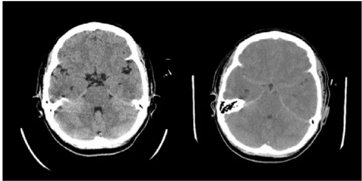
Brain edema and intracranial hypertension are more common in patients with a fulminant presentation of ALF8 (Figure 1), while their frequency decrease in sub-acute cases. Conceptually, it is important to distinguish brain edema from intracranial hypertension. Even though brain edema leads to intracranial hypertension, the latter will have effects per se in the brain that may or may not coincide with those that cause brain edema. For this rea-son, the investigation of the pathogenesis of brain edema has been frequently hampered by the presence of various degrees of intracranial hypertension in different studies.
General anatomical and physiological basis of intracranial hypertension
The cranium is a space with a fixed, non-distensible volume. Three different compartments are traditionally considered, with the brain parenchyma being the largest (approximately 80-90% of intracranial volume in normal subjects), followed by intravascular (approximately 10%) and cerebrospinal fluid (approximately 3-5%) compart-ments. Cells constitute most of the volume of brain pa-renchyma, with the extracellular fluid accounting just for 10-20% of that volume. This distribution is important as small increases in cell volume will lead to large increases in the total volume of the brain.
edigraphic.com
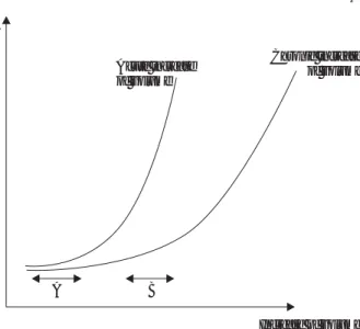
doctrine). The capacity of the brain to compensate ancrease in volume without a corresponding increase of in-tracranial pressure is called Compliance (∆V/∆P). Brain compliance has several important qualities (see figure 2). First, compliance is lower when the increase in volume is acute, which can leave no time for compensating mecha-nisms to develop. Second, when the increase in volume is continuous, compliance decreases progressively with time, leading ultimately to increased intracranial pressure. Third, when compliance is low, a small increase in volume, for example of cerebral blood volume, can lead to large in-creases of intracranial pressure.
In ALF, intracranial hypertension is usually the mani-festation of an increase in water content of brain tissue.9 An increase of cerebral blood flow (CBF) is also a feature seen in many patients and in experimental models of ALF or hyperammonemia, and can contribute to the increase of intracranial pressure via an increase in cerebral blood volume. Cerebral imaging has shown a reduction of cere-brospinal fluid in ALF.9
Neuropathology of brain edema in acute liver failure
Astrocyte swelling is a common finding in neuro-pathological studies of brain autopsy material from pa-tients with ALF10 and from animal models of brain edema due to ALF.11-15 Swelling is more prominent in foot pro-cesses than in cell bodies, and affects predominantly as-trocytes located in gray matter.11,12 In accordance with this observation, the increase in brain water content
de-tected by the gravimetric technique in different animal models of ALF was also selective of cortical gray matter, and was absent from subcortical, mesencephalic or pon-tine white matter or in the cerebellum.11,12 This consistent alteration of astrocytes is in contrast with the ultrastruc-tural preservation of neurons of the same brains.10-14
Tight junctions that characterize endothelial cells of brain capillaries were intact in all ultrastructural studies, excluding a gross disruption of the blood-brain barrier in ALF.10,11.14 However, a significant vacuolization and enlargement of en-dothelial cells, basement membrane and extracellular space was evident in some studies,10,13 suggesting increased perme-ability and pinocytic vesicular transport across the blood-brain barrier. Several functional studies have shown in-creased permeability to inulin,16 sucrose,16 GABA and ana-logues,17,18 Trypan blue15,19 or horseradish peroxidase12 in different animal models of ALF and even in the rat 24 hours after portacaval anastomosis (PCA).12 In contrast, no abnor-malities in the ultrastructure or permeability of the blood-brain barrier are found in other studies.11,20 Differences in methodology, animal species, models used or time of sam-pling (before/after intracranial hypertension has developed) may be some of the reasons for the discrepancies.
Pathogenesis of brain edema in ALF
For many years, several hypotheses have tried to inde-pendently explain the occurrence of brain swelling in ALF. In the last decade, however, we have witnessed a progressive convergence of previously excluding theories to a more rational context, based on solid research obser-Figure 1. Rapid development of brain edema in a patient with fulminant hepatic failure. On the right, the TC scanner made at admission was
edigraphic.com
vations. A critical role for ammonia is now evident. First,hyperammonemia alone appears sufficient as a cause of brain edema, as brain edema is seen in hyperammonemic patients with genetic disorders of urea cycle enzymes who do not have other alterations of liver function.21 Also, exposure to ammonia results in brain edema in vivo22,23 and astrocyte swelling in vitro.24,25 Second, am-monia appears necessary, as there are no reports of brain edema in ALF with normal ammonia levels, and in-creased concentrations of ammonia in blood and brain are consistent findings in experimental ALF.26,27 Further-more, Clemmesen and cols have recently shown that ce-rebral herniation only occurred in their study in those pa-tients with ALF and deep encephalopathy that had plasma ammonia levels above 150 micromols/L.28
An osmotic disturbance
An osmotic disturbance of the astrocyte as a result of ammonia is an observation that finds most support in clinical and experimental data. The main pathway for detoxification of ammonia is through formation of glutamine as the brain lacks a complete urea cycle.29 The formation of glutamine from glutamate is catalyzed by glutamine synthetase, an enzyme located mainly in astro-cytes,30 with the consumption of 1 molecule of ATP. Be-cause glutamine is a compound with osmotic properties, its accumulation within the astrocyte has been proposed as one of the mechanism leading to astrocyte swelling.
The presence of increased brain glutamine levels in ani-mal models and patients with ALF or hyperammonemia has been confirmed in multiple studies.22,23,26,31-37 An in-creased brain efflux of glutamine has been observed in
pa-tients with ALF compared to cirrhotic and healthy controls, which was higher in those who subsequently died of cere-bral herniation.38 The increase of brain glutamine seems to be an early event, as evidenced by the 2-fold increase seen just 24 hours after performance of portacaval anastomosis in the rat.26 Inhibition of glutamine formation results in amelioration of ammonia-induced swelling in the rat brain in vivo22,23 and in isolated astrocytes in vitro.24
Accumulation of glutamine alone, however, does not provide a complete explanation for the development of brain edema. Mild hypothermia32 and indomethacin ad-ministration,39 for example, reduce brain swelling and in-tracranial hypertension in ammonia-infused PCA rats, de-spite similar increases of glutamine in brain. Other ele-ments must be present to account for the development of brain swelling. One possibility would be the involvement of other organic osmoles, such as alanine. Alanine, which can be generated from transamination of glutamine, is also increased in the rat brain of ALF models.26,40 Notably, whereas glutamine increases rapidly in the early stages of HE and remains elevated to the same extent at coma stag-es, alanine continues to progressively rise in parallel with worsening encephalopathy.40 Other organic osmoles, such as myo-inositol or taurine, seem to be unchanged or slightly decreased in experimental models of ALF.31,41-43
The role of the blood-brain barrier
An alteration of the blood-brain barrier could explain a vasogenic theory for brain edema in ALF. Notably, func-tional abnormalities of the blood-brain barrier have been described in various experimental models.12,15-19 However, if an alteration of the blood-brain barrier permeability were the initial and critical event, it could hardly explain the selective swelling of astrocytes and the alterations in aminoacids and organic osmoles described above. Fur-thermore, no beneficial effects of corticosteroid therapy, a measure supposed to improve blood-brain barrier per-meability, were seen in patients with ALF and intracrani-al hypertension.44 Alterations of blood-brain barrier per-meability, if present, appear to play more a secondary and/or facilitating role, than being the central determinant of brain water accumulation in ALF.
Alterations of brain glutamate
Glutamate is the main excitatory neurotransmitter of the brain, participating in more than 80% of synapses. Total brain glutamate levels were found to be decreased in ALF26,36,37 and in hyperammonemia.22,31,32 However, levels of extracellular brain glutamate are increased in patients45,46 and experimental models41,47-50,6-49 of ALF, as measured via brain microdialysis (a technique that allows monitoring of extracellular space composition). The increase of extracel-lular glutamate probably results from the impairment of glutamate re-uptake by astrocytes, given that decreased ex-pression of astrocytic glutamate transporters as well as de-creased astrocytic glutamate re-uptake have been shown in Figure 2. Relation between increase of volume and intracranial
pres-sure (compliance). Continuous increases of volume leads to progressi-vely larger increases of intracranial pressure. Even though the increa-se of volume is the same in A and B, the lower brain compliance resul-ts in higher increases of pressure in B. The curve will be steeper when the insult is acute, due to a lesser capacity of compensation.
ICP
Acute increase of volume
Chronic increase of volume
Increase of volume
edigraphic.com
various experimental models.51-56 An increased release ofglutamate is also possible.57,58
A role for glutamate in the pathogenesis of brain ede-ma in ALF is suggested by 1) glutaede-mate induces astro-cytes swelling when injected into the brain or in isolated preparations59-61 2) brain extracellular glutamate correlates positively with severity of HE and brain water content in some ALF models,41,48 and 3) administration of glutamate antagonists has been reported to increase survival62 and to ameliorate brain edema63 in rodents with acute ammonia intoxication. Despite these observations, the exact role of glutamate in the production of brain edema in ALF re-quires further investigation.
Oxidative and nitrosative stress
A potential role for oxidative and nitrosative stress in the pathogenesis of brain edema in ALF is being increas-ingly explored. In the clinical setting, treatment with the antioxidant N-acetylcysteine was associated with less fre-quent progression to coma and brain edema in patients with ALF.61,62 These effects, however, were thought to arise from an improved brain microcirculation.64 In our lab, treatment with N-acetylcysteine ameliorated ammo-nia-induced brain edema in PCA rats, despite the lack of differences in CBF or haemodynamics compared to pla-cebo-treated controls.65 Ammonia, which has been report-ed to increase the formation of free radicals both in vivo66,67 and in vitro,68 could be one potential source for oxidative stress. An ammonia-induced increase of hemeoxygenase-1 gene expression, which is considered the best gene-marker of oxidative stress, also supports this assumption.69,70 Regarding nitrosative stress, in-creased expression and activity of neuronal nitric oxide synthase71 and increased brain nitric oxide production72 have been shown in experimental models of hyperam-monemia. Evidence for nitrosative stress arises also from cellular studies, where exposure of isolated astrocytes to ammonia resulted in increased nitration of protein ty-rosine residues.73 Astroglial protein tyrosine nitration was also found in brains from rats after acute ammonia intox-ication or after portacaval anastomosis, indicating the possible in vivo relevance of those findings.
Even though of considerable interest, further in vitro and in vivo work is needed in order to clarify the role of oxidative and nitrosative stress in the pathogenesis of brain edema in ALF.
Energy failure
Malfunction of the Na+, K+-ATPase pump by a sub-stance not cleared by the failing liver, which would lead to accumulation of intracellular sodium,was initially sus-pected.74,75 This was not confirmed in subsequent studies. Depletion of brain ATP could not be demonstrated with preserved concentrations of high energy phosphates (phosphocreatine, ATP) in various experimental models of ALF.27,76,77 Thus, brain energy failure is considered to
be an improbable pathogenic event, at least prior to the development of intracranial hypertension.
Cerebral blood flow and the pathogenesis of brain edema and intracranial hypertension in ALF
In patients with ALF, a wide spectrum of values of CBF has been reported, ranging from abnormally low to abnormally high levels.64,78,79 Even though differences in methodology could explain some of the discrepancies, in-tra-individual variations have been noted. Thus, the wide spectrum of CBF in ALF is more likely to reflect a real situation where CBF is subjected to the influence of mul-tiple factors,80 such as disease severity, systemic haemo-dynamics or extrahepatic complications. Despite these variations, it is now well accepted that cerebral oxidative metabolism is preserved in ALF,81,82 with CBF usually higher than the metabolic needs of the brain (the so-called luxury perfusion).79,82
In the following paragraphs, we will examine how the normal coupling between CBF and brain metabolism is altered in ALF, and how changes of CBF can influence the development of intracranial hypertension and brain edema in ALF.
Loss of CBF autoregulation in ALF
In normal conditions, CBF varies according to the metabolic requirements of the brain,83 increasing or de-creasing in parallel with brain activity. This autoregula-tion occurs independently of changes in mean arterial pressure or cardiac output, whenever blood pressure var-ies within the limits of 60 to 160 mmHg. Landmark stud-ies by Larsen and cols have clearly shown that CBF auto-regulation is lost in patients with ALF.84,85 The loss of au-toregulation can be explained by the presence of vasodilatation of cerebral arterioles.86 Maneuvers that in-duce vasoconstriction of cerebral vessels, such as hyper-ventilation leading to hypocapnia, can restore CBF auto-regulation in ALF.87 Restoration of autoregulation can also be achieved by moderate hypothermia88 and liver transplantation.85
Influence of CBF on brain edema in ALF
edigraphic.com
brain edema: 1) there is a striking positive correlationbe-tween CBF and brain water content, 2) abrogation of the increase in CBF by indomethacin or hypothermia results in amelioration of brain edema,32,39 and 3) the increase in CBF does not occur when brain edema is prevented via the inhibition of glutamine formation with methionine-sulfoximine.72 Why CBF increases in this model has not been elucidated, and even though a role of nitric oxide was initially suggested,72 subsequent studies did not con-firm this contention.92
The mechanism by which CBF can influence brain edema deserves further attention. The flux of water/sol-utes across the blood-brain barrier is determined by the terms of the Starling equation, as recently discussed:93
Water flux = Lp x A x [∆P - Σσ (i) x ∆πi]
where Lp is the filtration coefficient of the capillary membrane, A is the capillary membrane area, ∆P is the hy-drostatic pressure gradient between the lumen of the capil-lary and the interstitial tissue, σ (i) is the reflection coeffi-cient of substance i over the capillary membrane, and ∆πi is the osmotic pressure gradient created by the concentration gradient of substance i between blood and tissue.
Changes in CBF and cerebral autoregulation in ALF could favour water flux into the brain by affecting mainly several components of the Starling equation. First, a high-er hydrostatic pressure gradient (∆P) is a normal result of
an increase in CBF if no new capillaries are opened. Also, given that autoregulation is lost in ALF as a consequence of cerebral arteriolar vasodilatation,86 variations in sys-temic arterial pressure will greatly influence the hydro-static pressure in the capillaries. Second, an increase in CBF would result in increased ammonia delivery to the brain. This could worsen ∆πi by inducing further accumu-lation of intracellular glutamine and increase of brain tis-sue osmolarity, which would favour the flux of water into the brain. Finally, an increase in CBF could lead to a larg-er capillary membrane area (A) if new capillaries wlarg-ere opened (capillary recruitment), but the relevance of such mechanism in the brain is controversial.94
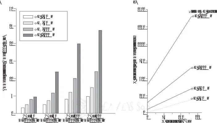
Subtle alterations in the permeability of blood-brain barrier, that would affect Lp and σ (i), have been previ-ously discussed. The net consequence of an increase in blood flow is to raise ammonia delivery to the brain. In-deed, small increases in blood flow will greatly increase ammonia delivery to the brain in a hyperammonemic state (Figure 3).
Clinical pathophysiology of the brain edema and intracranial hypertension
All clinicians recognize that multiple factors can po-tentially influence the development of HE. Thus, it is not surprising that a wide array of factors can affect the ex-pression of brain edema and intracranial hypertension in
NH - 40 M3 m
NH3- 60 Mm
NH3- 100 Mm NH - 200 M3 m
30
25
20
15
10
5
0
CBF-40 mL/100g/min
C F-60 mL/100g/min
B C F-100 mL/100g/min
B C F-120 mL/100g/min
B
Ammonia deli
v
ery (
mol/100 g/min)
m
% Increase in ammonia deli
v
ery
1500
1250
1000
750
500
250
0
0 75 150 225
NH - 40 M3 m
NH - 60 M3 m
NH - 100 M3 m
NH - 200 M3 m
Plasma NH Levels3
a) b)
% Increase in CBF
edigraphic.com
the clinical setting.92 We will examine severalphysiolog-ical variables, applying concepts previously reviewed.
Infection
Infection is a well-known precipitant of hepatic enceph-alopathy in chronic liver disease.95 In ALF, more than 80% of patients present evidence of infection,96 and increased levels of circulating cytokines are a consistent finding.97-99 A correlation between infection or the systemic inflamma-tory response syndrome (SIRS) and presence of severe HE has been reported in ALF.100 Furthermore, infection was an independent predictor of progression from mild to severe HE in a study of the US Acute Liver Failure Study Group.101 The mechanisms by which infection could pre-cipitate or worsen HE are not well understood but are like-ly to be diverse, with effects on the periphery (deterioration of liver function, alteration of haemodynamics) and effects on the brain. Receptors for some cytokines such as IL-1 have been described in brain capillaries, suggesting that cytokines could exerts their effects in the brain even with-out crossing the blood-brain barrier.102
Necrotic liver
Improvement of brain edema and control of intracrani-al pressure after totintracrani-al hepatectomy has been reported in patients with ALF.103,104 An increased release of cytokines from the necrotic liver suggests an inflammatory element in the intracranial hypertension of ALF.105 However, con-founding factors were present in these studies, such as the use of extracorporeal detoxification methods or mild hy-pothermia, clouding the assessment of the effect of hep-actectomy per se.
Temperature
Temperature is a component of the systemic inflam-matory response syndrome. Whereas hypothermia has been shown to exert beneficial effects in decreasing brain edema and intracranial pressure in patients90 and animal models,32,48 of ALF, hyperthermia is a deleterious event. Hyperthermia was shown to precede surges of intracrani-al pressure in patients with ALF,106,107 to increase cerebral blood flow in dogs,108 and to increase blood-brain barrier permeability in some animal models.109 Fever should be vigorously treated in patients with ALF.
Agitation
Episodes of agitation often precede surges of intracra-nial pressure in patients with ALF,106 making the preven-tion and treatment of agitapreven-tion an important aspect in the management of these patients. Agitation may reflect in-crease extra-cellular brain glutamate levels.48,49 Propofol is the preferred approach to handle this situation.
Arterial pressure and cerebral blood flow
Changes in arterial pressure will influence CBF and in-tracranial pressure in the patient with ALF that has a loss of cerebral autoregulation and poor brain compliance. In this setting, even small increases of arterial pressure may lead to increased cerebral blood volume and intracranial hypertension. On the other hand, a decrease of arterial pressure in the patient with intracranial hypertension could lead to brain hypoxia. Thus, monitoring of intracra-nial pressure and of jugular bulb oxygen saturation has been proposed to tailor the treatment with vasoactive drugs in ALF patients.92 Cerebral perfusion pressure (mean arterial pressure minus intracranial pressure) must be maintained above 40 mmHg to avoid tissue hypoxia. Jugular bulb oxygen saturations lower than 55% are in-dicative of cerebral ischemia, while saturations greater than 85% indicate decreased metabolic demands of the brain or, more commonly, cerebral hyperemia.
Glucose and lactate
Even though cerebral energy metabolism seems to be preserved until the last stages of the disease, brain metabo-lism of glucose is altered in ALF. In a preliminary report, hyperglycemia above 12 mM (200 mg/dL) was shown to be associated with intracranial hypertension in patients with ALF.110 Brain edema from other etiologies is known to worsen if hyperglycemia is present.111,112 However, mild hyperglycemia was not associated with worsening of brain edema in the ischemic rat model of ALF.113 Thus, the con-sequences of hyperglycemia in ALF are still unclear.
edigraphic.com
levels of glutamine, which increased in the precoma stagebut did not subsequently rise, the increase in lactate cor-related with the severity of encephalopathy. Based on these findings, the authors suggested that alterations of cellular glucose- and energy metabolism rather than the intracellular (astrocytic) accumulation of glutamine were the major cause of HE and brain edema in that model. Further studies to investigate the role of glucose and lac-tate in HE and brain edema are needed, and could have important repercussions in the clinical management of these patients.
Water and electrolyte abnormalities
Sodium concentration is the main factor determining plasma osmolarity under normal conditions. Hyponatre-mia is common in cirrhosis and water/electrolyte abnor-malities are a common precipitant of HE. The relatively high frequency of central pontyne myelinolysis in hy-ponatremic cirrhotic subjects after liver transplantation116 is probably a reflection of the profound alteration of os-moregulation and osmo-compensation in the brain of these patients.117 Extreme care should be taken in the cor-rection of hyponatremia before, during and after liver transplantation.
The consequences of hyponatremia were studied in ex-perimental models. In the rat with portacaval anastomosis, induction of chronic hyponatremia by 1-desamino-8-D-arginine vasopresin treatment decreased the levels of brain organic osmolytes.118 Infusion of ammonia resulted in the expected rise of glutamine and a reduction of taurine and myo-inositol, the main organic osmolytes in both nor-monatremic and hyponatremic rats. Importantly, infusion of ammonia resulted in a higher degree of brain edema in hyponatremic compared to normonatremic rats, despite an attenuated rise of glutamine in the former. These observa-tions suggest that chronic hyponatremia makes the brain more vulnerable to an ammonia-induced osmotic distur-bance with subsequent brain swelling. In contrast, increas-ing plasma osmolarity by infusion of hypertonic saline has been observed to reduce intracranial hypertension in ALF,93 its use is being evaluated in ongoing clinical trials.
Alterations of potassium could also influence HE by al-tering normal ammonia metabolism. Hypokalemia increas-es renal ammoniagenincreas-esis and seems to rincreas-esult in both in-creased urinary excretion and venous secretion of ammo-nia as seen in humans with chronic potassium depletion.119 Serum potassium and urine flow significantly correlated with kidney ammonia production, with serum potassium accounting for 61.4% of variations in ammonia production.
Ammonia metabolism
A highly orchestrated interorgan metabolism and traf-ficking of ammonia and glutamine is present in physiolog-ical conditions. Chronic and acute liver failure result in hy-perammonemia and leads to an important disturbance of
normal body nitrogen homeostasis (for review see).120 Dif-ferent clinical factors influence per se the plasma levels or the effects of ammonia in the brain in liver failure.
Muscle wasting
Even though glutamine synthetase activity in skeletal muscle is low, its relative increased mass compared to other organs that contain this enzyme makes the muscle one of the main glutamine synthesizing organs. Due to the increased plasma ammonia levels in liver failure, the muscle becomes an important ammonia detoxifying or-gan.121 Avoidance of muscle wasting in cirrhosis and stimulation of ammonia metabolism in the muscle are im-portant potential therapeutic targets in chronic and acute liver failure.
Gastrointestinal bleeding
Gastrointestinal bleeding is a known precipitant of HE. Blood in the digestive tract has been shown to be a potent stimulus for the intestinal production of ammonia.122 In the rat with portacaval anastomosis, simulated gas-trointestinal bleeding led to increased levels of ammonia and glutamine in the brain and to worsening of hepatic encephalopathy,123 indicating a mechanistic association.
Acid-base abnormalities
Ammonia can occur in blood as ammonium ion (NH4+) or as ammonia gas (NH3). The ammonia gas form is lipo-philic and diffuses easily across cell membranes, whereas the ammonium ion is non-diffusible and crosses mem-branes only by carrier-mediated processes.124 At physio-logical pH, most ammonia is in the ion form and only 1% is present as ammonia gas. Circumstances that alter blood pH could, thus, be pathogenically important. Metabolic or respiratory alkalosis, produced for example by diuretics or hyperventilation, could increase the gaseous form of ammonia, facilitating in this way its entry into the brain. However, the global effect of an altered pH in ammonia metabolism is difficult to evaluate, because the conse-quences can be different in different organs. Thus, where-as alkalosis can favour ammonia detoxification by im-proving urea synthesis in the liver,125 it may increase am-monia production in the kidney.126,127 Similarly, whereas metabolic acidosis increases gut release of ammonia, it increases ammonia uptake in the liver and the mus-cle.128,129
Sedatives
edigraphic.com
Sedation is difficult to avoid in the agitated patient withALF, but benzodiazepines should be avoided.
Conclusion
Hepatic encephalopathy of chronic liver disease and brain edema of acute liver failure have been considered two distinct and unrelated clinical entities for many years. Even though their pathogenesis is still not fully resolved, a new paradigm has progressively developed where both entities are different elements of a continuous spectrum. Hepatic encephalopathy and brain edema seem to share common pathogenic roots, with a key role of ammonia and a critical involvement of the astrocyte in both compli-cations. The study of one helps to understand the other: whereas ALF facilitates the study of causal relationships in a firmer manner, chronic liver failure gives the oppor-tunity to study brain compensating mechanisms. This in-tegrated view provides also a better perspective to judge the pathophysiological relevance that other factors may have in the manifestation of the disease.
References
1. Ferenci P, Lockwood A, Mullen K, Tarter R, Weissenborn K, Blei AT. Hepatic encephalopathy—definition, nomenclature, diagnosis, and quantification: final report of the working party at the 11th World Congresses of Gastroenterology, Vienna, 1998. Hepatology 2002;
35: 716-721.
2. Ware AJ, D’Agostino AN, Combes B. Cerebral edema: a major com-plication of massive hepatic necrosis. Gastroenterology 1971; 61:
877-884.
3. Bustamante J, Rimola A, Ventura PJ, Navasa M, Cirera I, Reggiardo V, Rodes J. Prognostic significance of hepatic encephalopathy in patients with cirrhosis. J Hepatol 1999; 30: 890-895.
4. O’Grady JG, Alexander GJ, Hayllar KM, Williams R. Early indica-tors of prognosis in fulminant hepatic failure. Gastroenterology 1989;
97: 439-445.
5. Bernuau J, Goudeau A, Poynard T, Dubois F, Lesage G, Yvonnet B, Degott C, et al. Multivariate analysis of prognostic factors in fulmi-nant hepatitis B. Hepatology 1986; 6: 648-651.
6. Castells A, Salmeron JM, Navasa M, Rimola A, Salo J, Andreu H, Mas A, et al. Liver transplantation for acute liver failure: analysis of applicability. Gastroenterology 1993; 105: 532-538.
7. Hoofnagle JH, Carithers RL, Jr., Shapiro C, Ascher N. Fulminant he-patic failure: summary of a workshop. Hepatology 1995; 21: 240-252.
8. O’Grady JG, Schalm SW, Williams R. Acute liver failure: redefining the syndromes. Lancet 1993; 342: 273-275.
9. Cordoba J, Blei AT. Brain edema and hepatic encephalopathy. Semin
Liver Dis 1996; 16: 271-280.
10. Kato M, Hughes RD, Keays RT, Williams R. Electron microscopic study of brain capillaries in cerebral edema from fulminant hepatic failure. Hepatology 1992; 15: 1060-1066.
11. Traber PG, Dal Canto M, Ganger DR, Blei AT. Electron microscopic evaluation of brain edema in rabbits with galactosamine-induced fulminant hepatic failure: ultrastructure and integrity of the blood-brain barrier. Hepatology 1987; 7: 1272-1277.
12. Traber P, DalCanto M, Ganger D, Blei AT. Effect of body tempera-ture on brain edema and encephalopathy in the rat after hepatic devas-cularization. Gastroenterology 1989; 96: 885-891.
13. Matkowskyj KA, Marrero JA, Carroll RE, Danilkovich AV, Green RM, Benya RV. Azoxymethane-induced fulminant hepatic failure in C57BL/6J mice: characterization of a new animal model. Am J Physiol
1999; 277: G455-462.
14. Gove CD, Hughes RD, Ede RJ, Williams R. Regional cerebral edema and chloride space in galactosamine-induced liver failure in rats.
Hepatology 1997; 25: 295-301.
15. Dixit V, Chang TM. Brain edema and the blood brain barrier in galac-tosamine-induced fulminant hepatic failure rats. An animal model for evaluation of liver support systems. ASAIO Trans 1990; 36: 21-27. 16. Zaki AE, Ede RJ, Davis M, Williams R. Experimental studies of
blood brain barrier permeability in acute hepatic failure. Hepatology
1984; 4: 359-363.
17. Horowitz ME, Schafer DF, Molnar P, Jones EA, Blasberg RG, Patlak CS, Waggoner J, et al. Increased blood-brain transfer in a rabbit model of acute liver failure. Gastroenterology 1983; 84: 1003-1011.
18. Bassett ML, Mullen KD, Scholz B, Fenstermacher JD, Jones EA. Increased brain uptake of gamma-aminobutyric acid in a rabbit model of hepatic encephalopathy. Gastroenterology 1990; 98: 747-757.
19. Livingstone AS, Potvin M, Goresky CA, Finlayson MH, Hinchey EJ. Changes in the blood-brain barrier in hepatic coma after hepate-ctomy in the rat. Gastroenterology 1977; 73: 697-704.
20. Alexander B, Li X, Benjamin IS, Segal MB, Sherwood R, Preston JE. A quantitative evaluation of the permeability of the blood brain bar-rier of portacaval shunted rats. Metab Brain Dis 2000; 15: 93-103. 21. Brusilow SW: Inborn errors of urea synthesis. In: Lloyd J, Scriver C,
eds. Genetic and metabolic diseases in pediatrics. London:
Butterworths, 1985; 140-165.
22. Takahashi H, Koehler RC, Brusilow SW, Traystman RJ. Inhibition of brain glutamine accumulation prevents cerebral edema in hyperammonemic rats. Am J Physiol 1991; 261: H825-829.
23. Blei AT, Olafsson S, Therrien G, Butterworth RF. Ammonia-induced brain edema and intracranial hypertension in rats after portacaval anastomosis. Hepatology 1994; 19: 1437-1444.
24. Norenberg MD, Bender AS. Astrocyte swelling in liver failure: role of glutamine and benzodiazepines. Acta Neurochir Suppl (Wien)
1994; 60: 24-27.
25. Ganz R, Swain M, Traber P, DalCanto M, Butterworth RF, Blei AT. Ammonia-induced swelling of rat cerebral cortical slices: implica-tions for the pathogenesis of brain edema in acute hepatic failure.
Metab Brain Dis 1989; 4: 213-223.
26. Swain M, Butterworth RF, Blei AT. Ammonia and related amino ac-ids in the pathogenesis of brain edema in acute ischemic liver failure in rats. Hepatology 1992; 15: 449-453.
27. Mans AM, DeJoseph MR, Hawkins RA. Metabolic abnormalities and grade of encephalopathy in acute hepatic failure. J Neurochem
1994; 63: 1829-1838.
28. Clemmesen JO, Larsen FS, Kondrup J, Hansen BA, Ott P. Cerebral herniation in patients with acute liver failure is correlated with arte-rial ammonia concentration. Hepatology 1999; 29: 648-653. 29. Felipo V, Butterworth RF. Neurobiology of ammonia. Prog Neurobiol
2002; 67: 259-279.
30. Martinez-Hernandez A, Bell KP, Norenberg MD. Glutamine syn-thetase: glial localization in brain. Science 1977; 195: 1356-1358.
31. Cordoba J, Gottstein J, Blei AT. Glutamine, myo-inositol, and organic brain osmolytes after portocaval anastomosis in the rat: implications for ammonia-induced brain edema. Hepatology 1996; 24: 919-923.
32. Cordoba J, Crespin J, Gottstein J, Blei AT. Mild hypothermia modi-fies ammonia-induced brain edema in rats after portacaval anasto-mosis. Gastroenterology 1999; 116: 686-693.
33. Olafsson S, Gottstein J, Blei AT. Brain edema and intracranial hy-pertension in rats after total hepatectomy. Gastroenterology 1995;
108: 1097-1103.
34. McConnell JR, Antonson DL, Ong CS, Chu WK, Fox IJ, Heffron TG, Langnas AN, et al. Proton spectroscopy of brain glutamine in acute liver failure. Hepatology 1995; 22: 69-74.
35. Haussinger D, Laubenberger J, vom Dahl S, Ernst T, Bayer S, Langer M, Gerok W, et al. Proton magnetic resonance spectroscopy studies on human brain myo-inositol in hypo-osmolarity and hepatic en-cephalopathy. Gastroenterology 1994; 107: 1475-1480.
36. Bates TE, Williams SR, Kauppinen RA, Gadian DG. Observation of cerebral metabolites in an animal model of acute liver failure in vivo: a 1H and 31P nuclear magnetic resonance study. J Neurochem 1989;
edigraphic.com
37. Bosman DK, Deutz NE, De Graaf AA, vd Hulst RW, Van Eijk HM, Bovee WM, Maas MA, et al. Changes in brain metabolism during hyperammonemia and acute liver failure: results of a comparative 1H-NMR spectroscopy and biochemical investigation. Hepatology
1990; 12: 281-290.
38. Strauss GI, Knudsen GM, Kondrup J, Moller K, Larsen FS. Cerebral metabolism of ammonia and amino acids in patients with fulminant hepatic failure. Gastroenterology 2001; 121: 1109-1119.
39. Chung C, Gottstein J, Blei AT. Indomethacin prevents the develop-ment of experidevelop-mental ammonia-induced brain edema in rats after portacaval anastomosis. Hepatology 2001; 34: 249-254.
40. Zwingmann C, Chatauret N, Leibfritz D, Butterworth RF. Selective increase of brain lactate synthesis in experimental acute liver fail-ure: Results of a [H-C] nuclear magnetic resonance study. Hepatology
2003; 37: 420-428.
41. Michalak A, Rose C, Butterworth J, Butterworth RF. Neuroactive amino acids and glutamate (NMDA) receptors in frontal cortex of rats with experimental acute liver failure. Hepatology 1996; 24: 908-913.
42. Peeling J, Shoemaker L, Gauthier T, Benarroch A, Sutherland GR, Minuk GY. Cerebral metabolic and histological effects of thioacetamide-in-duced liver failure. Am J Physiol 1993; 265: G572-578.
43. Zimmermann C, Ferenci P, Pifl C, Yurdaydin C, Ebner J, Lassmann H, Roth E, et al. Hepatic encephalopathy in thioacetamide-induced acute liver failure in rats: characterization of an improved model and study of amino acid-ergic neurotransmission. Hepatology 1989; 9: 594-601.
44. Canalese J, Gimson AE, Davis C, Mellon PJ, Davis M, Williams R. Controlled trial of dexamethasone and mannitol for the cerebral oedema of fulminant hepatic failure. Gut 1982; 23: 625-629.
45. Tofteng F, Jorgensen L, Hansen BA, Ott P, Kondrup J, Larsen FS. Cerebral microdialysis in patients with fulminant hepatic failure.
Hepatology 2002; 36: 1333-1340.
46. Tofteng F, Larsen FS. Monitoring extracellular concentrations of lac-tate, glutamate, and glycerol by in vivo microdialysis in the brain during liver transplantation in acute liver failure. Liver Transpl
2002;8:302-305.
47. Bosman DK, Deutz NE, Maas MA, van Eijk HM, Smit JJ, de Haan JG, Chamuleau RA. Amino acid release from cerebral cortex in ex-perimental acute liver failure, studied by in vivo cerebral cortex microdialysis. J Neurochem 1992; 59: 591-599.
48. Rose C, Michalak A, Pannunzio M, Chatauret N, Rambaldi A, Butterworth RF. Mild hypothermia delays the onset of coma and prevents brain edema and extracellular brain glutamate accumula-tion in rats with acute liver failure. Hepatology 2000; 31: 872-877.
49. de Knegt RJ, Schalm SW, van der Rijt CC, Fekkes D, Dalm E, Hekking-Weyma I. Extracellular brain glutamate during acute liver failure and during acute hyperammonemia simulating acute liver fail-ure: an experimental study based on in vivo brain dialysis. J Hepatol
1994; 20: 19-26.
50. Tossman U, Delin A, Eriksson LS, Ungerstedt U. Brain cortical amino acids measured by intracerebral dialysis in portacaval shunted rats.
Neurochem Res 1987; 12: 265-269.
51. Knecht K, Michalak A, Rose C, Rothstein JD, Butterworth RF. De-creased glutamate transporter (GLT-1) expression in frontal cortex of rats with acute liver failure. Neurosci Lett 1997; 229: 201-203.
52. Chan H, Butterworth RF. Evidence for an astrocytic glutamate trans-porter deficit in hepatic encephalopathy. Neurochem Res 1999; 24: 1397-1401.
53. Chan H, Hazell AS, Desjardins P, Butterworth RF. Effects of ammo-nia on glutamate transporter (GLAST) protein and mRNA in cul-tured rat cortical astrocytes. Neurochem Int 2000; 37: 243-248.
54. Mena EE, Cotman CW. Pathologic concentrations of ammonium ions block L-glutamate uptake. Exp Neurol 1985; 89: 259-263.
55. Bender AS, Norenberg MD. Effects of ammonia on L-glutamate up-take in cultured astrocytes. Neurochem Res 1996; 21: 567-573.
56. Oppong KN, Bartlett K, Record CO, al Mardini H. Synaptosomal glutamate transport in thioacetamide-induced hepatic encephalopa-thy in the rat. Hepatology 1995; 22: 553-558.
57. Moroni F, Lombardi G, Moneti G, Cortesini C. The release and neosynthesis of glutamic acid are increased in experimental models of hepatic encephalopathy. J Neurochem 1983; 40: 850-854.
58. Erecinska M, Pastuszko A, Wilson DF, Nelson D. Ammonia-induced release of neurotransmitters from rat brain synaptosomes: differences between the effects on amines and amino acids. J Neurochem 1987;
49: 1258-1265.
59. Van Harreveld A, Fifkova E. Light- and electron-microscopic changes in central nervous tissue after electrophoretic injection of glutamate.
Exp Mol Pathol 1971; 15: 61-81.
60. Van Harreveld A. Swelling of the Muller fibers in the chicken retina.
J Neurobiol 1982; 13: 519-536.
61. Noble LJ, Hall JJ, Chen S, Chan PH. Morphologic changes in cul-tured astrocytes after exposure to glutamate. J Neurotrauma 1992; 9: 255-267.
62. Hermenegildo C, Marcaida G, Montoliu C, Grisolia S, Minana MD, Felipo V. NMDA receptor antagonists prevent acute ammonia toxic-ity in mice. Neurochem Res 1996; 21: 1237-1244.
63. Vogels BA, Maas MA, Daalhuisen J, Quack G, Chamuleau RA. Memantine, a noncompetitive NMDA receptor antagonist improves hyperammonemia-induced encephalopathy and acute hepatic en-cephalopathy in rats. Hepatology 1997; 25: 820-827.
64. Wendon JA, Harrison PM, Keays R, Williams R. Cerebral blood flow and metabolism in fulminant liver failure. Hepatology 1994; 19: 1407-1413. 65. Vaquero J, Gottstein J, Cahill ME, Blei AT. N-acetylcysteine pre-vents ammonia-induced brain edema in rats after portacaval
anas-tomosis. The 11th International Symposium on Hepatic
Encephal-opathy and Nitrogen Metabolism. Abstract book 2002: p31 [Abstract]. 66. Kosenko E, Kaminsky Y, Kaminsky A, Valencia M, Lee L, Hermenegildo C, Felipo V. Superoxide production and antioxidant enzymes in ammonia intoxication in rats. Free Radic Res 1997; 27:
637-644.
67. Kosenko E, Felipo V, Montoliu C, Grisolia S, Kaminsky Y. Effects of acute hyperammonemia in vivo on oxidative metabolism in nonsynaptic rat brain mitochondria. Metab Brain Dis 1996; 12: 69-82.
68. Murthy CR, Rama Rao KV, Bai G, Norenberg MD. Ammonia-in-duced production of free radicals in primary cultures of rat astro-cytes. J Neurosci Res 2001; 66: 282-288.
69. Warskulat U, Gorg B, Bidmon HJ, Muller HW, Schliess F, Haussinger D. Ammonia-induced heme oxygenase-1 expression in cultured rat astrocytes and rat brain in vivo. Glia 2002; 40: 324-336.
70. Song G, Dhodda VK, Blei AT, Dempsey RJ, Rao VL. GeneChip analy-sis shows altered mRNA expression of transcripts of neurotransmit-ter and signal transduction pathways in the cerebral cortex of porta-caval shunted rats. J Neurosci Res 2002; 68: 730-737.
71. Rao VL, Audet RM, Butterworth RF. Increased neuronal nitric oxide synthase expression in brain following portacaval anastomosis. Brain Res 1997; 765: 169-172.
72. Master S, Gottstein J, Blei AT. Cerebral blood flow and the develop-ment of ammonia-induced brain edema in rats after portacaval anas-tomosis. Hepatology 1999; 30: 876-880.
73. Schliess F, Gorg B, Fischer R, Desjardins P, Bidmon HJ, Herrmann A, Butterworth RF, et al. Ammonia induces MK-801-sensitive nitra-tion and phosphorylanitra-tion of protein tyrosine residues in rat astro-cytes. Faseb J 2002; 16: 739-741.
74. Seda HW, Hughes RD, Gove CD, Williams R. Inhibition of rat brain Na+,K+-ATPase activity by serum from patients with fulminant he-patic failure. Hepatology 1984; 4: 74-79.
75. Ede RJ, Gove CD, Hughes RD, Marshall W, Williams R. Reduced brain Na+, K+-ATPase activity in rats with galactosamine-induced hepatic failure: relationship to encephalopathy and cerebral oedema. Clin Sci (Lond) 1987; 72: 365-371.
76. Holmin T, Agardh CD, Alinder G, Herlin P, Hultberg B. The influ-ence of total hepatectomy on cerebral energy state, ammonia-related amino acids of the brain and plasma amino acids in the rat. Eur J
Clin Invest 1983; 13: 215-220.
77. Bates TE, Williams SR, Busza AL, Gadian DG, Proctor E. A 31P nuclear magnetic resonance study in vivo of metabolic
abnormali-ties in rats with acute liver failure. NMR Biomed 1988; 1: 67-73. 78. Larsen FS, Pott F, Hansen BA, Ejlersen E, Knudsen GM, Clemmesen
JD, Secher NH. Transcranial Doppler sonography may predict brain death in patients with fulminant hepatic failure. Transplant Proc 1995;
edigraphic.com
79. Aggarwal S, Kramer D, Yonas H, Obrist W, Kang Y, Martin M, Policare R. Cerebral hemodynamic and metabolic changes in fulminant he-patic failure: a retrospective study. Hepatology 1994; 19: 80-87.
80. Larsen FS. Cerebral circulation in liver failure: Ohm’s law in force.
Semin Liver Dis 1996; 16: 281-292.
81. Larsen FS, Ejlersen E, Clemmesen JO, Kirkegaard P, Hansen BA. Pres-ervation of cerebral oxidative metabolism in fulminant hepatic fail-ure: an autoregulation study. Liver Transpl Surg 1996; 2: 348-353.
82. Larsen FS, Knudsen GM, Hansen BA. Pathophysiological changes in cerebral circulation, oxidative metabolism and blood-brain bar-rier in patients with acute liver failure. Tailored cerebral oxygen uti-lization. J Hepatol 1997; 27: 231-238.
83. Magistretti PJ, Pellerin L, Rothman DL, Shulman RG. Energy on demand. Science 1999; 283: 496-497.
84. Larsen FS, Ejlersen E, Hansen BA, Knudsen GM, Tygstrup N, Secher NH. Functional loss of cerebral blood flow autoregulation in pa-tients with fulminant hepatic failure. J Hepatol 1995; 23: 212-217.
85. Strauss G, Hansen BA, Kirkegaard P, Rasmussen A, Hjortrup A, Larsen FS. Liver function, cerebral blood flow autoregulation, and hepatic encephalopathy in fulminant hepatic failure. Hepatology
1997; 25: 837-839.
86. Larsen FS, Adel Hansen B, Pott F, Ejlersen E, Secher NH, Paulson OB, Knudsen GM. Dissociated cerebral vasoparalysis in acute liver failure. A hypothesis of gradual cerebral hyperaemia. J Hepatol 1996;
25: 145-151.
87. Strauss G, Hansen BA, Knudsen GM, Larsen FS. Hyperventilation restores cerebral blood flow autoregulation in patients with acute liver failure. J Hepatol 1998; 28: 199-203.
88. Jalan R, Olde Damink SW, Deutz NE, Hayes PC, Lee A. Restoration of cerebral blood flow autoregulation and reactivity to carbon diox-ide in acute liver failure by moderate hypothermia. Hepatology 2001; 34: 50-54.
89. Aggarwal S, Yonas H, Kang Y, Martin M, Kramer D, Obrist WD, Darby J. Relationship of cerebral blood flow and cerebral swelling to outcome in patients with acute fulminant hepatic failure.
Trans-plant Proc 1991; 23: 1978-1979.
90. Jalan R, Damink SW, Deutz NE, Lee A, Hayes PC. Moderate hypo-thermia for uncontrolled intracranial hypertension in acute liver
fail-ure. Lancet 1999; 354: 1164-1168.
91. Clemmesen JO, Hansen BA, Larsen FS. Indomethacin normalizes intracranial pressure in acute liver failure: a twenty-three-year-old woman treated with indomethacin. Hepatology 1997; 26: 1423-1425.
92. Larsen FS, Gottstein J, Blei AT. Cerebral hyperemia and nitric oxide synthase in rats with ammonia-induced brain edema. J Hepatol 2001;
34: 548-554.
93. Larsen FS, Wendon J. Brain edema in liver failure: basic physiologic principles and management. Liver Transpl 2002; 8: 983-989.
94. Hudetz AG. Blood flow in the cerebral capillary network: a review emphasizing observations with intravital microscopy. Microcircula-tion 1997; 4: 233-252.
95. Strauss E, da Costa MF. The importance of bacterial infections as precipating factors of chronic hepatic encephalopathy in cirrhosis.
Hepatogastroenterology 1998; 45: 900-904.
96. Rolando N, Harvey F, Brahm J, Philpott-Howard J, Alexander G, Gimson A, Casewell M, et al. Prospective study of bacterial infec-tion in acute liver failure: an analysis of fifty patients. Hepatology
1990; 11: 49-53.
97. de la Mata M, Meager A, Rolando N, Daniels HM, Nouri-Aria KT, Goka AK, Eddleston AL, et al. Tumour necrosis factor production in fulminant hepatic failure: relation to aetiology and superimposed microbial infection. Clin Exp Immunol 1990; 82: 479-484. 98. Rolando N, Ellis A, de Groote D, Wendon J, Williams R. Correlation
of serial cytokine levels with progression to coma (grade IV) in pa-tients with acute liver failure (ALF). Hepatology 1995; 22: 366A.
99. Muto Y, Nouri-Aria KT, Meager A, Alexander GJ, Eddleston AL, Williams R. Enhanced tumour necrosis factor and interleukin-1 in fulminant hepatic failure. Lancet 1988; 2: 72-74.
100. Rolando N, Wade J, Davalos M, Wendon J, Philpott-Howard J, Wil-liams R. The systemic inflammatory response syndrome in acute liver failure. Hepatology 2000; 32: 734-739.
101.Vaquero J, Schiodt F, Chung C, Rademaker AW, Lee WM, Blei AT. Predictors of progression to deep hepatic encephalopathy in acute liver failure: results of the US Acute Liver Failure Study Group.
Hepatology 2002; 36: 222A.
102.Wong ML, Bongiorno PB, al-Shekhlee A, Esposito A, Khatri P, Licinio J. IL-1 beta, IL-1 receptor type I and iNOS gene expression in rat brain vasculature and perivascular areas. Neuroreport 1996; 7:
2445-2448.
103.Jalan R, Pollok A, Shah S, Madhavan K, Simpson K. Liver derived pro-inflammatory cytokines may be important in producing intrac-ranial hypertension in acute liver failure. J Hepatol 2002; 37: 536. 104.Rozga J, Podesta L, LePage E, Hoffman A, Morsiani E, Sher L, Woolf
GM, et al. Control of cerebral oedema by total hepatectomy and extracorporeal liver support in fulminant hepatic failure. Lancet 1993;
342: 898-899.
105.Jalan R, Williams R. The inflammatory basis of intracranial hyper-tension in acute liver failure. J Hepatol 2001; 34: 940-942.
106.Munoz SJ, Moritz MJ, Bell R, Northrup B, Martin P, Radomski J. Factors associated with severe intracranial hypertension in candi-dates for emergency liver transplantation. Transplantation 1993; 55:
1071-1074.
107.Kodakat SK, Gopal PB, Wendon J. Intracranial pressure is related to body temperature in acute liver failure. Liver Transpl 2001; 7: C87.
108. Katsumura H, Kabuto M, Hosotani K, Handa Y, Kobayashi H, Kubota T. The influence of total body hyperthermia on brain haemodynamics and blood-brain barrier in dogs. Acta Neurochir (Wien) 1995; 135: 62-69. 109. Mueller SM. Increased blood-brain barrier permeability during
hy-perthermia in the awake rat. Trans Am Neurol Assoc 1979; 104: 81-83.
110.Kodakat SK, Gopal PB, Wendon JA. Hyperglycaemia is associated with intracranial hypertension in patients with acute liver failure [ab-stract]. Liver Transpl 2001; 7: C21.
111.Nakakimura K, Fleischer JE, Drummond JC, Scheller MS, Zornow MH, Grafe MR, Shapiro HM. Glucose administration before cardiac arrest worsens neurologic outcome in cats. Anesthesiology 1990; 72:
1005-1011.
112.Lam AM, Winn HR, Cullen BF, Sundling N. Hyperglycemia and neurological outcome in patients with head injury. J Neurosurg 1991;
75: 545-551.
113.Shah V, Webster S, Gottstein J, Blei AT. Reduction of cerebral perfu-sion precedes rise of intracranial pressure in rats with ischemic ful-minant liver failure. Hepatology 1993; 17: 1117-1122.
114.Bihari D, Gimson AE, Lindridge J, Williams R. Lactic acidosis in fulminant hepatic failure. Some aspects of pathogenesis and prog-nosis. J Hepatol 1985; 1: 405-416.
115.Ellis A, Wendon J. Circulatory, respiratory, cerebral, and renal de-rangements in acute liver failure: pathophysiology and management.
Semin Liver Dis 1996; 16: 379-388.
116.Abbasoglu O, Goldstein RM, Vodapally MS, Jennings LW, Levy MF, Husberg BS, Klintmalm GB. Liver transplantation in hyponatremic patients with emphasis on central pontine myelinolysis. Clin
Trans-plant 1998; 12: 263-269.
117.Bluml S, Zuckerman E, Tan J, Ross BD. Proton-decoupled 31P mag-netic resonance spectroscopy reveals osmotic and metabolic distur-bances in human hepatic encephalopathy. J Neurochem 1998; 71:
1564-1576.
118.Cordoba J, Gottstein J, Blei AT. Chronic hyponatremia exacerbates ammonia-induced brain edema in rats after portacaval anastomosis.
J Hepatol 1998; 29: 589-594.
119.Tizianello A, Garibotto G, Robaudo C, Saffioti S, Pontremoli R, Bruzzone M, Deferrari G. Renal ammoniagenesis in humans with chronic potassium depletion. Kidney Int 1991; 40: 772-778. 120.Olde Damink SW, Deutz NE, Dejong CH, Soeters PB, Jalan R.
Interorgan ammonia metabolism in liver failure. Neurochem Int 2002;
41: 177-188.
121.Clemmesen JO, Kondrup J, Ott P. Splanchnic and leg exchange of amino acids and ammonia in acute liver failure. Gastroenterology
2000; 118: 1131-1139.
122. Sugarbaker SP, Revhaug A, Wilmore DW. The role of the small in-testine in ammonia production after gastric blood administration.
edigraphic.com
123.Olde Damink SW, Dejong CH, Deutz NE, Soeters PB. Effects of simulated upper gastrointestinal hemorrhage on ammonia and re-lated amino acids in blood and brain of chronic portacaval-shunted rats. Metab Brain Dis 1997; 12: 121-135.
124.Cooper AJ, Plum F. Biochemistry and physiology of brain ammonia.
Physiol Rev 1987; 67: 440-519.
125.Haussinger D, Steeb R, Gerok W. Metabolic alkalosis as driving force for urea synthesis in liver disease: pathogenetic model and therapeu-tic implications. Clin Investig 1992; 70: 411-415.
126.Tannen RL, Goyal M. Response of ammoniagenesis to acute alkalo-sis. Am J Physiol 1984; 247: F827-836.
127. DuBose TD, Jr., Good DW. Effects of chronic Cl depletion alkalosis on proximal tubule transport and renal production of ammonium.
Am J Physiol 1995; 269: F508-514.
128.Fine A. Effects of acute metabolic acidosis on renal, gut, liver, and muscle metabolism of glutamine and ammonia in the dog. Kidney Int 1982; 21: 439-444.
129.Welbourne TC. Effect of metabolic acidosis on hindquarter glutamine and alanine release. Metabolism 1986; 35: 614-618.
130.Gorg B, Foster N, Reinehr R, Bidmon HJ, Hongen A, Haussinger D, Schliess F. Benzodiazepine-induced protein tyrosine nitration in rat astrocytes. Hepatology 2003; 37: 334-342.
131.Jayakumar AR, Panickar KS, Norenberg MD. Effects on free radical generation by ligands of the peripheral benzodiazepine receptor in cultured neural cells. J Neurochem 2002; 83: 1226-1234. 132.Bender AS, Norenberg MD. Effect of benzodiazepines and
neurosteroids on ammonia-induced swelling in cultured astrocytes.